Note: Descriptions are shown in the official language in which they were submitted.
CA 02345965 2001-03-30
WO 00/24440 1 PCT/SE99/01915
Method and device for measuring access flow
10 AREA OF INVENTION
The present invention relates to a method and device for
measuring blood flow rate in a blood access. Blood is taken out
from the body of a mammal to an extracorporeal blood circuit
through a blood access, via needles or a catheter.
PRIOR ART
There are several types of treatments in which blood is
taken out in an extracorporeal blood circuit. Such treatments
involve, for example, hemodialysis, hemofiltration,
hemodiafiltration, plasmapheresis, blood component separation,
blood oxygenation, etc. Normally, blood is removed from a blood
vessel at an access site and returned to the same blood vessel
or at another location in the body.
In hemodialysis and similar treatments, an access site is
commonly surgically created in the nature of a fistula. Blood
needles are inserted in the area of the fistula. Blood is taken
out from the fistula via an arterial needle and blood is
returned to the fistula via a venous needle.
A common method of generating a permanent access site
having capability of providing a high blood flow and being
operative during several years and even tens of years, is the
provision of an arterio-venous fistula. It is produced by
operatively connecting the radial artery to the cephalic vein
at the level of the forearm. The venous limb of the fistula
thickens during the course of several months, permitting
repeated insertion of dialysis needles.
An alternative to the arterio-venous fistula is the
arterio-venous graft, in which a connection is generated from,
CA 02345965 2001-03-30
WO 00/24440 2 PCT/SE99/01915
for example, the radial artery at the wrist to the basilic
vein. The connection is made with a tube graft made from
autogenous saphenous vein or from polytetrafluorethylene (PTFE,
Teflon). The needles are inserted in the graft.
A third method for blood access is to use a silicon, dual-
lumen catheter surgically implanted into one of the large
veins.
Further methods find use in specific situations, like a
no-needle arterio-venous graft consisting of a T-tube linked to
a standard PTFE graft. The T-tube is implanted in the skin.
Vascular access is obtained either by unscrewing a plastic plug
or by puncturing a septum of said T-tube with a needle. Other
methods are also known.
During hemodialysis, it is desirable to obtain a constant
blood flow rate of 150 - 500 ml/min or even higher, and the
access site must be prepared for delivering such flow rates.
The blood flow in an AV fistula is often 800 ml/min or larger,
permitting delivery of a blood flow rate in the desired range.
In the absence of a sufficient forward blood flow, the
extracorporeal circuit blood pump will take up some of the
already treated blood entering the fistula via the venous
needle, so called access or fistula recirculation, leading to
poor treatment results.
The most common cause of poor flow with AV fistulas is
partial obstruction of the venous limb due to fibrosis
secondary to multiple venipunctures. Moreover, stenosis causes
a reduction of access flow.
When there is a problem with access flow, it has been
found that access flow rate often exhibit a long plateau time
period with reduced but sufficient access flow, followed by a
short period of a few weeks with markedly reduced access flow
leading to recirculation and ultimately access failure. By
constantly monitoring the evolution of the access flow during
consecutive treatment sessions, it is possible to detect
imminent access flow problems.
Several methods have been suggested for monitoring
recirculation and access flow. Many of these methods involve
injection of a marker substance in blood, and the resultant
CA 02345965 2001-03-30
WO 00/24440 3 PCT/SE99/01915
recirculation is detected. The methods normally involve
measurement of a property in the extracorporeal blood circuit.
Examples of such methods can be found in US 5,685,989,
US 5,595,182, US 5,453,576, US 5,510,716, US 5,510,717,
US 5,312,550, etc.
Such methods have the disadvantage that they cannot detect
when the access flow has decreased to such an extent that
recirculation is at risk, but only when recirculation prevails.
Moreover, it is a drawback that injection of a substance is
necessary.
A noninvasive technique that allows imaging of flow
through AV grafts is color Doppler ultrasound. However, this
technique requires expensive equipment.
The measurement of access flow rate necessitates the
reversal of the flows in the extracorporeal circuit. A valve
for such reversal is shown in i.a. US 5605630 and US 5894011.
However, these valve constructions comprises dead ends in which
blood may stand still for a long time and coagulate, which is a
drawback.
DISCLOSURE OF INVENTION
An object of the present invention is to provide a method
and a device for measuring the access flow rate without
interfering with the blood and without injecting a substance in
blood.
Another object of the invention is to provide a method and
a device for measuring access flow rate without measuring on
the blood in the extracorporeal blood circuit or in the access
or blood vessel.
According to the invention, it is required to reverse the
blood flow through the access. Thus, a further object of the
invention is to provide a valve for reversing the blood flow.
A still further object of the invention is to provide a
method for determining when the blood flow rate is so small
that risk for recirculation prevails.
These objects are achieved with a method and an apparatus
for estimating fluid flow rate (Qa) in a fluid flow access,
comprising removing a first fluid flow from said access at a
CA 02345965 2001-03-30
WO 00/24440 4 PCT/SE99/01915
removal position to an external flow circuit comprising a
dialyzer having a semipermeable membrane, said first fluid flow
passing along said membrane at one side thereof and a dialysis
fluid being emitted from the other side thereof, and returning
said first fluid flow from said external flow circuit to said
access at a return position downstream of said removal
position, measuring a first variable which is essentially
proportional to a concentration (Cd norm) of a substance in
said dialysis fluid emitted from the dialyzer, reversing the
removal position with the return position and measuring a
second variable which is essentially proportional to the
concentration (Cd rev) of said substance in said dialysis fluid
in the reversed position; and calculating the fluid flow rate
(Qa) in said flow access from said measured concentrations.
Preferably, the calculation of the fluid flow rate in said
flow access takes place by calculating the ratio between the
first and the second variable and using the formula:
Cd norm / Cd rev = 1 + K/Qa, in which Cd norm and Cd rev are
values proportional to the concentrations of said substance in
the dialysis fluid in the normal and reversed positions,
respectively, and K is the clearance of the dialyzer and Qa is
the access flow rate.
The blood flow access may be in a mammal for obtaining
access to a blood vessel, such as a hemodialysis access in the
nature of an arterio-venous shunt or fistula. In the latter
case, the dialyzer clearance K is replaced by the effective
dialyzer clearance Keff obtained by taking into account a
cardiopulmonary recirculation and in the normal position.
The substance is preferably selected from the group of:
urea, creatinine, vitamin B12, beta-two-microglobuline and
glucose, or may be an ion selected from the group of: Na+, Cl ,
K, Mg , Ca++, HCO3, , acetate ion, or any combination thereof
as measured by conductivity; and wherein said concentration is
measured as the concentration difference between the outlet and
the inlet of the dialyzer, if applicable.
It is possible to measure the actual concentration of the
substance. However, since only the ratio between the
concentrations in the normal and the reversed position,
CA 02345965 2007-07-18
respectively, is needed, it is possible to measure a value
which is proportional to the concentration of said substance,
whereby said value is used in place of said concentration. Said
property may be the blood concentration of said substance in
the external circuit, either before or after the dialyzer.
Alternatively, the relative whole body efficiency (KWh/V) may
be used, as explained in more detail below.
The effective clearance Keff may be obtained by the
equation Keff = Qd * Cd / Cs, where Qd is the flow of dialysis
fluid emitted from the dialyzer, Cd is the concentration of
said substance in said dialysis fluid and Cs is the
concentration of said substance in systemic venous blood.
A method of measuring the concentration (Cs) of said
substance in systemic venous blood comprises the steps of:
stopping the blood flow in the external flow circuit for a time
period sufficient to allow the cardiopulmonary circulation to
equalize; starting the blood flow in the external flow circuit
with a slow speed to fill the arterial line with fresh blood
before the measurement; and measuring the equalized
concentration of said substance in the dialysis fluid at a low
dialysate flow rate or at isolated ultrafiltration. It is
advantageous to make the measurement of the effective clearance
at the initiation of the treatment.
The concentration (Cs) of said substance in systemic
venous blood may be estimated by: calculating a whole body mass
of urea (Murea) in the body of the patient, estimating or
measuring the distribution volume (V) of urea in the body of
the patient; and estimating the concentration (Cs) of said
substance in the blood by dividing the whole body mass of urea
with the distribution volume. In this way, the mean
concentration of urea in the whole body is obtained. However,
the mean concentration in the whole body is slightly higher
than the urea concentration in the systemic blood, except at
the start of the treatment. Thus, this calculation should
preferably be done or be extrapolated to the start of the
treatment.
It is possible to discriminate between the condition when
access or fistula recirculation has developed and not. A method
CA 02345965 2007-07-18
6
for that purpose would be: changing the blood flow rate (Qb);
monitoring the concentration of said substance in the dialysate
emitted from the dialyzer; and detecting a possible fistula
recirculation in the normal position by correlating a change in
said concentration to said change of the blood flow rate.
Preferably, the blood flow rate is decreased and a
corresponding decrease in the urea concentration is monitored,
and the abscence of such a decrease being indicative of fistula
recirculation.
According to an aspect of the present invention there is
provided a method for estimating the flow rate (Qa) of a
liquid flowing in a tube comprising the steps of:
removing a fraction of the liquid from the tube at a
first location;
circulating the fraction of the liquid in the first
compartment of a dialyzer, whereas a dialysis liquid is
circulated in a second compartment of the dialyzer
separated from the first compartment by a semi-permeable
membrane, and whereas the liquid and/or the dialysis liquid
contain a substance that can diffuse through the semi-
permeable membrane;
returning the fraction of the liquid into the tube at a
second location;
measuring a first value (Cd norm) of a property of the
dialysis liquid downstream of the dialyzer, the property
being related to the concentration of the substance;
inverting the first location and the second location so
as to remove the fraction of the liquid from the tube at
the second location and to return the fraction of the
liquid into the tube at the first location;
measuring a second value (Cd rev) of the property of the
dialysis liquid downstream of the dialyzer; and
CA 02345965 2007-07-18
6a
calculating the flow rate (Qa) of the liquid in the tube
from the first and second measured values (Cd norm, Cd rev)
of the property of the dialysis liquid.
According to another aspect of the present invention
there is provided an apparatus for extracorporeal blood
treatment comprising:
a dialyzer having a first and a second compartments
separated by a semi-permeable membrane;
a blood circuit having:
a blood removal line for connecting, in a normal
operative condition, an inlet of the first compartment to a
vascular tube of a patient at a first connecting point, and
a blood return line for connecting, in the normal
operative condition, an outlet of the first compartment to
the vascular tube of the patient at a second connecting
point;
a dialysis liquid circuit having:
a supply line for supplying a dialysis liquid to an
inlet of the second compartment, and
a waste line for draining a waste liquid from an
outlet of the second compartment,
the apparatus further comprising:
a reversal means for switching the connection of the
blood removal line and blood return line to the vascular
tube so that, in a reverse operative condition, the first
connecting point is in fluid communication with the outlet
of the first compartment and the second connecting point is
in fluid communication with the inlet of the first
compartment;
a measuring means for measuring, in the normal
operative condition, a first value (Cd norm) of a property
in the waste liquid, and for measuring, in the reverse
operative condition, a second value (Cd rev) of the
CA 02345965 2007-07-18
6b
property in the waste liquid, the property being related to
the concentration of a substance in the waste liquid; and
a calculating means for calculating a fluid flow rate
(Qa) in the vascular tube from the first and second
measured values (Cd norm, Cd rev) of the property in the
waste liquid.
SHORT DESCRIPTION OF DRAWINGS
Further objects, advantages and features of the invention
appears from the following detailed description of the
invention with reference to specific embodiments of the
invention shown on the drawings, in which
Fig. 1 is a partially schematic view of a forearm of a
patient provided with an AV fistula.
Fig. 2 is a schematic diagram of an extracorporeal
dialysis circuit.
Fig. 3 is a schematic diagram of the blood flow circuit in
a patient and in the attached extracorporeal blood circuit.
Fig. 4 is a schematic diagram similar to Fig. 3, but with
the extracorporeal circuit in an alternative reversed position.
Fig. 5 is a schematic diagram of a blood flow circuit
including a switch valve.
Fig. 6 is a diagram of the dialysis fluid urea
concentration versus time, including a portion with reversed
flow access according to the invention.
Fig. 7 is a schematic diagram similar to the diagram of
Fig. 5 comprising an alternative valve arrangement.
Fig_ 8 is schematic diagram similar to the diagram of Fig-
7 showing the valve arrangement in an idle postion.
Fig. 9 is schematic diagram similar to the diagram of Fig-
7 showing the valve arrangement in a reversed postion.
Fig. 10 is a schematic diagram similar to Fig. 5 with the
pump in an alternative position.
Fig. 11 is a diagram showing calculations with relative
whole body efficiency.
CA 02345965 2001-03-30
WO 00/24440 7 PCT/SE99/01915
Fig. 12 is a cross-sectional view of a valve housing to be
used in the schematic diagram of Figs. 5 and 7 to 10.
Figl 13 is a bottom view of a valve member intended to be
inserted in the valve housing of Fig. 12.
Fig. 14 is a partially schematic plan view of the valve
housing of Fig. 12.
DESCRIPTION OF DETAILED EMBODIMENTS OF THE INVENTION
For the purpose of this description, an access site is a
site in which a fluid in a tube can be accessed and removed
from and/or returned to the tube. The tube may be a blood
vessel of a mammal, or any other tube in which a fluid is
flowing. The access flow rate is the flow rate of the fluid in
the tube or blood vessel immediately upstream of the access
site or removal position.
Fig. 1 discloses a forearm 1 of a human patient. The
forearm 1 comprises an artery 2, in this case the radial
artery, and a vein 3, in this case the cephalic vein. Openings
are surgically created in the artery 2 and the vein 3 and the
openings are connected to form a fistula 4, in which the
arterial blood flow is cross-circuited to the vein. Due to the
fistula, the blood flow through the artery and vein is
increased and the vein forms an thickened area downstream of
the connecting openings. When the fistula has matured after a
few months, the vein is thicker and may be punctured
repeatedly. Normally, the thickened vein area is called a
fistula.
An arterial needle 5 is placed in the fistula, in the
enlarged vein close to the connected openings and a venous
needle 6 is placed downstream of the arterial needle, normally
at least five centimeters downstream thereof.
The needles 5 and 6 are connected to a tube system 7,
shown in Fig. 2, forming an extracorporeal circuit comprising a
blood pump 8, such as a dialysis circuit. The blood pump
propels blood from the blood vessel, through the arterial
needle, the extracorporeal circuit, the venous needle and back
into the blood vessel.
CA 02345965 2007-07-18
8
The extracorporeal blood circuit 7 shown in Fig_ 2 further
comprises an arterial clamp 9 and a venous clamp 10 for
isolating the patient from the extracorporeal circuit should an
error occur.
Downstream of pump 8 is a dialyzer 11, comprising a blood
compartment 12 and a dialysis fluid compartment 13 separated by
a semipermeable membrane 14. Further downstream of the dialyzer
is a drip chamber 15, separating air from the blood therein.
Blood passes from the arterial needle past the arterial
clamp 9 to the blood pump 8. The blood pump drives the blood
through the dialyzer 11 and further via the drip chamber 15 and
past the venous clamp 10 back to the patient via the venous
needle. The drip chamber may comprise an air detector, adapted
to trigger an alarm should the blood emitted from the drip
chamber comprise air or air bubbles. The blood circuit may
comprise further components, such as pressure sensors etc.
The dialysis fluid compartment 13 of the dialyzer 11 is
provided with dialysis fluid via a first pump 16, which obtains
dialysis fluid from a source of pure water, normally RO-water,
and one or several concentrates of ions, metering pumps 17 and
18 being shown for metering such concentrates. The preparation
of dialysis fluid is conventional and is not further described
here.
An exchange of substances between the blood and the
dialysis fluid takes place in the dialyzer through the
semipermeable membrane. Notably, urea is passed from the blood,
through the semipermeable membrane and to the dialysis fluid
present at the other side of the membrane. The exchange may
take place by diffusion under the influence of a concentration
gradient, so called hemodialysis, and/or by convection due to a
flow of liquid from the blood to the dialysis fluid, so called
ultrafiltration, which is an important feature of
hemodiafiltration or hemofiltration.
From the dialysis fluid compartment 13 of the dialyzer is
emitted a fluid called the dialysate, which is driven by a
second pump 19 via a urea monitor 20 to drain. The urea monitor
continuously measures the urea concentration in the dialysate
emitted from the dialyzer, to provide a dialysate urea
CA 02345965 2007-07-18
9
concentration curve during a dialysis treatment. Such urea
concentration curve may be used for several purposes, such as
obtaining a total body urea mass, as described in WO 9855166,
and to obtain a prediction of the whole body dialysis dose Kt/V
as also described in said application.
As described above, the present invention provides a
method of non-invasively measuring the access flow in the
fistula immediately before the arterial needle, using the urea
monitor and the dialysis circuit as shown in Fig. 2.
By measuring the dialysis urea concentration during normal
dialysis and then reversing the positions-of the needles and
measuring the dialysis urea concentration with the needles in
the reversed position, it is possible to calculate the blood
flow in the blood access, without the addition of any substance
to blood or the dialysis fluid.
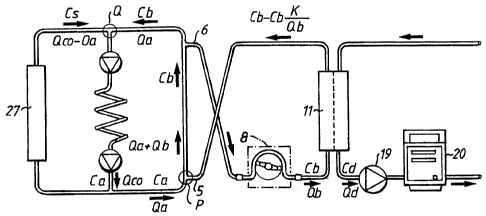
Fig. 3 shows a simplified schematic diagram of the blood
vessel circuit of a patient and a portion of the dialysis
circuit according to Fig. 2. The patient blood circuit
comprises the heart, where the right chamber of the heart is
symbolized by an upper pump 21 and the left chamber of the
heart is symbolized by a lower pump 22. The lungs 23 are
located between the upper and lower pump. From the outlet of
the left chamber pump 22 of the heart, the blood flow divides
into a first branch 24 leading to the access 25, normally in
the left forearm of the patient, and a second branch 26 leading
to the rest of the body, such as organs, other limbs, head,
etc. symbolized by a block 27. Blood returning from the body
from the organs etc., i.e. from block 27, combines with blood
returning from the access and enters the right chamber pump 21.
The cardiac output flow rate is defined as Qco and the
flow rate of the access is defined as Qa, which means that
Qco - Qa enters the block 27. The venous blood returning from
block 27 before being mixed with blood from the access, the
systemic venous blood, has a urea concentration of Cs. The
blood leaving the left chamber pump 22 has a urea concentration
CA 02345965 2001-03-30
WO 00/24440 10 PCT/SE99/01915
of Ca equal to that passing out to the access 25 as well as to
the block 27.
For measuring the access flow rate, it is necessary to
reverse the flow through the arterial and venous needles. One
way of achieving that is to reverse the needles manually.
Alternatively, Fig. 5 shows a valve 28 for performing the
same operation. The arterial needle 5 is connected to an
arterial inlet line 29 of the valve and the venous needle 6 is
connected to a venous inlet line 30 of the valve. The blood
pump is connected to a first outlet line 31 of the valve and
the returning blood from the dialyzer 11 is connected to a
second outlet line 32 of the valve.
The valve comprises a valve housing and a pivotable valve
member 33, which is pivotable from the normal position shown on
the drawing to a reverse position pivoted 90 in relation to
the normal position.
In the normal position shown in Fig. 5, the arterial
needle 5 is connected to the blood pump 8 and the venous needle
6 is connected to the outlet of the dialyzer, via the drip
chamber, see Fig. 2. In the reversed position, the arterial
needle 5 is connected to the outlet of the dialyzer and the
venous needle 6 is connected to the blood pump 8, as required.
An alternative design of the valve arrangement is shown in
Figs. 7, 8 and 9. In the embodiment of Fig. 7, the arterial
line 29 is connected to an enlarged opening 29a and the venous
outlet line 30 is connected to an enlarged opening 30a, the
openings being arranged in the valve housing 28a diametrically
opposite to each other. Two enlarged openings 31a and 32a are
arranged in the valve housing 28a diametrically opposite each
other and displaced 90 in relation to enlarged openings 29a
and 30a. The pivotable valve member 33a is normally arranged as
shown in Fig. 7 and forms a partition dividing the valve
chamber in two semi-circular portions. The valve member has a
width, which is smaller than the peripheral dimension of the
enlarged openings. The valve member is pivotable 90 to a
reverse position, shown in Fig. 9, in which the blood flows
through the arterial and venous needles are reversed.
CA 02345965 2007-07-18
11
During its movement from the normal to the reversed
position, the valve member 33a passes through an idle position
shown in Fig. 8, in which all four enlarged openings are
interconnected, because the width of the valve member is
smaller than the peripheral dimension of the enlarged openings.
By this idle position, harm to blood cells may be avoided. Such
harm may be caused by high shear stresses which may occur if
the inlet line 31 to the blood pump or the outlet line 32 from
the dialyzer are completely occluded. By means of the idle
position, another advantage is obtained, that the blood needles
are not exposed to rapid change of flows, which in some
instances even may result in dislocation of the needles. When
the valve member is moved from the normal position to the idle
position, the flow through the needles change from the normal
flow of, for example, 250 ml/min to essentially zero flow. The
valve member may be placed in the idle position for some
seconds. Then, the valve member is moved to the reversed
position, and the flows through the needles is changed from
essentially zero flow to -250 ml/min. In this way, a more
gentle switch between normal and reversed flows may be
obtained.
It is noted, that the positions of the openings and the
valve member may be different so that the pivotal movement may
be less than or more than 90 . Moreover, the openings need not
be arranged diametrically in order to achieve the desired
operation. Furthermore, the dimensions of the enlarged openings
in relation to the tubes and lines are not in scale, but the
diameter of the enlarged openings is rather of the same
dimension as the tube inner diameter, as appears more clearly
below.
It is noted that the valve is constructed to have as few
dead end portions as possible, in which the blood may stand
still and coagulate. From the drawing, it is appreciated that
no portion of the valve has a dead end construction in any
position of the valve body.
Furthermore, another schematic diagram incorporating a
valve is shown in Fig. 10. Fig. 10 differs from Fig. 5 only in
the placement of the pump 8a, which in the embodiment according
CA 02345965 2001-03-30
WO 00/24440 12 PCT/SE99/01915
to Fig. 10 is placed between the arterial needle 5 and the
valve 28. In this manner, the pressure across the valve body 33
is less compared to the embodiment according to Fig. 5. The
operation is somewhat different. The blood pump is stopped, and
the valve is put in the reversed position. Finally, the pump is
started and pumping the blood in the opposite direction by
reversing the rotational direction of the pump.
In order to ascertain that no air is introduced into the
patient in either position of the valve, it may be advantageous
to add an air detector 34 and 35 immediately before each of the
arterial and venous needle, or at least before the arterial
needle. The air detectors trigger an alarm should they measure
air bubbles in the blood given back to the blood vessel.
Normally, the air detector in the drip chamber is sufficient
for this purpose.
The detailed construction of a valve intended to be used
in the present invention, is disclosed in Figs. 12, 13 and 14.
The valve comprises a valve housing 36 comprising two inlet
connectors and two outlet connectors. All four connectors open
into cylindrical valve chamber 41, the four openings being
displaced 90 in relation to each other.
As shown in Fig. 14, the valve comprises a blood inlet
connector 37 connected to the arterial needle 5 and a blood
outlet connector 38 connected to the venous needle 6. The
connector portions are arranged as male Luer connectors to be
connected to flexible tubes ending with a female Luer
connector.
Furthermore, the valve comprises a circuit outlet
connector 39 connected to the blood pump 8 and a circuit inlet
connector 40 connected to the dialyzer outlet. The connector
portions 39 and 40 are arranged as female Luer connectors to
mate with male Luer connectors of the circuit.
As appears from Fig. 12, the cylindrical valve chamber 41
is closed at the bottom. From the top, a valve member 42 may be
introduced into the cylindrical valve chamber. The valve member
42 comprises a valve partition 43 as appears from Fig. 13.
The valve member also comprises an operating wing 44, by
means of which the valve member may be pivoted 90 between a
CA 02345965 2007-07-18
13
normal position, in which the valve partition 43 is situated as
shown by dotted lines in Fig. 14, and a reversed position. The
pivotal movement is limited by a shoulder 45 of the valve
member 42, which cooperates with a groove 46 in the valve
housing. The shoulder 45 is provided with a protrusion 46a
which cooperates with two recesses 47 and 48 in the normal
position and reverse position, respectively, to maintain the
valve member in either position. The groove 46 may be provided
with a third recess (not shown in the drawing) in order to
define said idle position. Such a third recess is positioned in
the middle between the two recesses 47 and 48.
The valve member and housing are provided with suitable
sealings to ensure safe operation. The operation of the valve
is evident from the above description.
By studying the theoretical dialysate urea concentrations
resulting from a given dialyzer clearance K, a given access
blood flow Qa and a given blood urea concentration Cs in the
systemic venous blood returning from the body, it is found that
the effective urea clearance Keff of the dialyzer, taking the
cardiopulmonary recirculation into account, is needed for the
calculation of access flow. The effective clearance can be
measured, for example as described in EP 658 352,
Alternatively, the effective clearance can be calculated
from simultaneous systemic venous blood Cs and dialysate Cd
measurements of urea concentrations, such as by blood samples.
The systemic blood urea concentration Cs may be measured
by the so called stop flow - slow flow technique, where the
blood flow is substantially stopped for a couple of minutes to
allow the cardiopulmonary recirculation to equalize.
Thereafter, the pump is run slowly to fill the arterial line
with fresh blood before taking the blood sample. The urea
concentration in the so obtained blood sample is equal to the
urea concentration Cs in the systemic venous blood returning
from the body to the heart.
Alternatively to taking a blood sample, the dialysis fluid
flow at the other side of the membrane is stopped and the
CA 02345965 2007-07-18
14
slowly flowing blood is allowed to equalize with the dialysate
at the other side of the membrane, whereupon the urea
concentration of the dialysate is measured to obtain the
systemic venous blood urea concentration Cs.
A further method to obtain effective clearance is
described in WO 9929355. According to the invention described
in WO 9929355, the systemic blood concentration Cs is measured
before or at the initiation of the treatment, for example by
stop flow - slow flow technique with blood sample or
equilisation as described above. After obtaining valid
dialysate urea concentration values Cd from a urea monitor
connected to the dialysator outlet line, the initial dialysate
urea concentration Cdinit at the start of the treatment is
extrapolated by the dialysate urea curve obtained.
A still further method of obtaining systemic blood urea
concentration Cs is to calculate the urea mass Mh in the whole
body and extrapolate the urea mass to the start of the
treatment. By dividing the whole body urea mass MWh with the
distribution volume V, the systemic blood urea concentration Cs
at the start of the treatment is obtained.
By dividing the dialysate urea concentration Cd with the
systemic blood urea concentration Cs and multiplicating with
the dialysate flow rate Qd, the effective clearance Keff is
obtained. It is advantageous to measure the effective clearance
Keff at the initiation of the treatment.
Furthermore, in the method of the invention, the blood
flows in the arterial and venous needles are reversed. The
dialysate urea concentrations in the two cases with normal
position of the needles and with reverse position of the
needles may be calculated as follows, with reference to Figs. 3
and 4.
The blood urea concentration Cs in the venous blood
returning from the body is assumed unchanged when the lines are
reversed, and the dialyzer clearance K is also assumed
unchanged. For simplicity ultrafiltration is assumed to be
zero, but it is also possible to handle a nonzero UF.
The following notations are used:
CA 02345965 2001-03-30
WO 00/24440 15 PCT/SE99/01915
Qco - Cardiac Output
Qa - Access flow
Qb - Blood flow in extracorporeal circuit
Qd - Dialysate flow
K - Dialyzer clearance
Keff - Effective dialyzer clearance
Cs - Blood urea concentration in systemic venous blood
returning from the body
Ca - Blood urea concentration in the access
Cb - Blood urea concentration at the dialyzer inlet
Cd - Dialysate urea concentration
The definition of clearance is:
K = (removed urea) / Cb = Qd * Cd / Cb (1)
Consider first the case in which Qa > Qb and the needles
are in the normal position. In this case Cb = Ca.
Removal from blood must equal appearance in the dialysate
so that
K * Ca = Qd * Cd (2)
A mass balance for urea at the point V, see Fig. 3, when
mixing the venous return blood with the blood from the access
gives:
Ca * Qco = Cs * (Qco - Qa) + Ca * (Qa - K) (3)
Thus, we obtain a relation between Ca and Cs.
By combining equations 2 and 3 we obtain:
Cd = (K/Qd) * Cs / [1 + K/(Qco -Qa)] (4)
The definition of effective clearance Keff implies that Cs
should be used in the denominator instead of Cb as normally
used in dialyzer clearance, which means that
CA 02345965 2001-03-30
WO 00/24440 16 PCT/SE99/01915
Keff = K * (Cb / Cs) = K / [1 + K/(Qco -Qa)] (5)
If we now turn to the case with reversed lines, see Fig.
4, we still have that what is removed from the blood must enter
the dialysate, so that in this case
K * Cb = Qd * Cd (6)
The flow in the fistula between the needles will be
Qa + Qb and we can calculate the blood urea concentration at
the dialyzer inlet from a urea mass balance at the point P
where the dialyzed blood enters the access again
Cb * (Qb - K) + Ca * Qa = Cb * (Qb + Qa) (7)
We also have the mass balance at the point Q where the
venous return blood meets the dialyzed blood in the access
return flow:
Ca * Qco = Cs * (Qco - Qa) + Cb * Qa (8)
By eliminating Ca and Cb we get
Cd = (K/Qd) * Cs / [ 1 + (Qco/Qa) * K / (Qco - Qa)] (9)
Since Cs, K and Qd in the two cases are unchanged, it is
possible to obtain the ratio of dialysate urea concentrations:
Cd norm / Cd rev = 1 + (K/Qa) / [1 + K/(Qco-Qa)] =
= 1 + Keff/Qa (10)
In practice, the two dialysate urea concentrations are
probably best found by a curve fit to the dialysate urea curves
before and after the switch of lines, with an extrapolation to
the time of switching from the respective side, see Fig 6,
which shows the urea concentration Cd of the dialysate during a
normal hemodialysis treatment.
CA 02345965 2007-07-18
17
During a time period of about 10 minutes, marked with a
ring in Fig. 6, the arterial and venous needles are reversed.
After a initial time period for allowing the urea monitor to
measure accurately, the urea concentration with reversed lines
is appr. 0.8 times the original urea concentration, which means
that Cdnorm / Cdrev = 1.25. Thus, if Keff is 200 ml/min, as
measured with the needles in the normal position or estimated
as described above, the access flow is 800 ml/min.
The effective clearance may also be obtained as a rough
estimate from blood and dialyzer flows and dialyzer
characteristics, e.g. from the dialyzer data sheet.
In the present specification, there is used three
different clearances, namely dialyzer clearance, effective
clearance and whole body clearance. If dialyzer clearance is
250 ml/min for a certain blood flow rate and dialysate flow
rate, the effective clearance is normally 5 to 10% lower, such
as 230 ml/min. The whole body clearance is still 5 to 15%
lower, such as 200 ml/min. The dialyzer clearance is the
clearance as measured directly on the dialyzer. The effective
clearance is the clearance also taking into account the cardio-
pumonary recirculation. Finally, the whole body clearance is.
the effective clearance further taking into account other
membranes in the body restricting the flow of urea from any
part of the body to the dialyasate. The concept of whole body
clearance is described in WO 9855166,
The effective clearance used in the formula may also be
obtained from a measurement according to the method described
in EP 658 352 mentioned above, with the needles in the normal
position. This will give a measure of the effective plasma
water urea clearance, which then has to be converted to whole
blood clearance. The method of EP 658 352 essentially comprises
that the conductivity of the dialysis fluid upstream of the
dialyzer is increased by for example 10% and then returned to
the original value. The result at the outlet side of the
dialyzer is measured and reults in a measure of the effective
clearance Keff of the dialyzer.
CA 02345965 2001-03-30
WO 00/24440 18 PCT/SE99/01915
Alternatively, the effective clearance may be calculated
according to equation Keff = Qd * Cd / Cs. The systemic venous
urea concentration may be measured at the same time as the
dialysate urea concentration Cd, or by the methods described
above.
Another method would be to use the value of total body
urea mass Murea obtained by the method according to WO 9855166,
mentioned above. By obtaining the urea distribution volume V by
Watson's formula or any other method, the venous urea
concentration would be approximately:
Cs = Murea / V (11)
In the method of WO 9855166, the relative whole body
efficiency of the dialyzing process Kwb/V is obtained. Note,
that whole body clearance is used, as indicated by the
subscript wb. According to said WO 9855166, urea concentration
is proportional to the relative whole body efficiency according
to the formula:
Kwb/V = (Qd = Cd) / m (12)
Thus, if (Kwb/V) is used instead of Cd in the above
equation (10), a similar result is obtained, if it is presumed
that m is constant, i.e. the measurement must be extrapolated
to the same time instance:
(Kwb/V) norm / (Kwb/V) rev = 1 + Keff/Qa (13)
As is mentioned in said WO 9855166, it is possible to
calculate the relative whole body efficiency only from
dialysate urea measurement. Since we are interested only in the
ratio in the normal and reversed position, we do not need to
calculate the actual Kwh.
Fig. 11 shows a plot of the relative whole body efficiency
K/V (min-1). The period with reversed lines is shown inside a
circle. In all other respects, the same discussion applies as
is given above.
CA 02345965 2001-03-30
WO 00/24440 19 PCT/SE99/01915
The calculations above assume that the extracorporeal
blood flow rate Qb does not exceed the access flow rate Qa. If
this is the case there will be access recirculation and the
flow in the access will be reversed when the needles are in the
normal position. The calculation of dialysate urea
concentration is unchanged for the needles in reversed
position, but has to be modified for the needles in normal
position. Calculations corresponding to those above show that
the ratio above between dialysate urea concentrations for
normal and reversed needle positions will be:
Cd norm / Cd rev = 1 + Keff / Qb (14)
where Keff is the effective clearance with the effect of
recirculation included, that is with the needles in the normal
position.
The only difference is that the calculation will now give
the extracorporeal blood flow Qb instead of the access flow.
This blood flow is known, so in practice this means that when
the result is an access flow rate Qa close to the blood flow
rate Qb, recirculation should be suspected, and this always
means that the access has to be improved.
Keff/Qb is a figure lower than one, normally for example
0.6 - 0.9. Keff/ Qa should be considerably lower, for example
0.1 - 0.4. Thus, when Cd norm/Cd rev approaches or is lower
than a predetermined number, such as 1.2 or 1.5, further
calculations should be done for determining if access
recirculation is present.
A simple procedure is to decrease the blood flow Qb
somewhat. If the dialysate concentration then decreases, this
means that there is no access or fistula recirculation at least
at the lower blood flow.
The above calculations can also be made for the situation
where ultrafiltration is present. However, it is a simple
measure to reduce the ultrafiltration to zero during the
measurement interval. Moreover, the error induced by
ultrafiltration is small and may be neglected.
CA 02345965 2001-03-30
WO 00/24440 20 PCT/SE99/01915
The measurement should be performed during a time
interval, which is considerably larger than 30 seconds so that
cardio-pulmonary recirculation has been developed. The
measurement time for obtaining valid results may be 5 minutes
with the needles reversed, while measurements with the needles
in correct position may be done in 5 minutes or continuously
during the treatment.
The method is also applicable to the methods of treatment
comprising infusion of a dialysis solution to the blood before
or after the dialyzer, called hemofiltration and
hemodiafiltration. The result is the same as given above.
If the access is a venous catheter, there is no cardio-
pulmonary recirculation and the calculations becomes simpler.
The result is the same, except that the effective clearance
Keff is replaced by the dialyzer clearance K, since the
systemic venous urea concentration Cs becomes the same as the
dialyzer inlet urea concentration Cb.
It should be noted that all flow rates, clearances and
urea concentrations in the calculations relate to whole blood.
Approximately 93% of plasma is water, depending on the protein
concentration, and about 72% of erythrocytes is water.
Depending on the hematocrit value, the blood water volume is
10 - 13 % lower than the volume of whole blood, see for example
Handbook of Dialysis, Second Edition, John T. Daugirdas and
Todd. S Ing, 1994, page 18.
The effective urea clearance obtained according to
EP 658 352 relates to blood water, and must therefore be
increased by 10 - 13 % before being used in the present
formulas. Blood urea concentration values obtained from a
laboratory relate in general to plasma, and must therefore be
decreased by about 7% in order to relate to whole blood.
Alternatively, all urea concentrations, flow rates and
clearances may be used as relating to blood water. The
effective clearance is then used unchanged, but the calculated
access flow will relate to blood water, and has to be increased
by 10 - 13 % to relate to whole blood.
The invention has been described above with reference to
use in the human body and using urea as a marker for measuring
CA 02345965 2001-03-30
WO 00/24440 21 PCT/SE99/01915
access flow. However, any other substance present in blood and
which can be measured at the dialysate side of the dialyzer may
be used according to the invention, such as creatinine, vitamin
B12, beta-two-microglobuline, NaCl or any combination of ions.
Another alternative is to measure conductivity.
It is also possible to measure a property proportional to
the concentration, since it is the ratio that is involved in
the equations. Thus, urea concentration may be measured by
measuring conductivity differences after passing the urea
containing fluid through a urease column, and such conductivity
difference can be used directly in place of the concentration
values in the equations.
Other indirect methods of measuring any of the above-
mentioned substances concentrations may be used as long as the
measurements are made at the dialysate side of the dialyzer.
Another alternative is to measure the blood urea concentrations
by any known method, either before or after the dialyzer, since
these concentrations are proportional to the concentrations in
the formulas.
The invention has been described above with reference to
use in the human body. However, the invention can be used in
any tube system where a fluid is passed and a portion thereof
is taken out for dialysis, such as in beer or wine production.