Note: Descriptions are shown in the official language in which they were submitted.
CA 02346769 2003-12-03
IMPLANTS COMPRISING COMBINATIONS OF
ALLOGENEIC CELLS FOR USE IN CANCER TREATMENT
10
FIELD OF THE INVENTION
The present invention relates generally to the frelds of cellular immunology
and cancer
therapy. More specifically, it relates to the treatment of tumors or the
generation of an anti-tumor
immune response by implanting a mixture of alloactivated allogeneic cells in
or around the tumor
site.
BACKGROUND
Cancer continues to be a leading cause of mortality around the globe.
Tradiflona! regimens
of cancer management have been successful in the management of a selective
group of circulating
and slow-growing solid cancers. However, many solid tumors are resistant to
traditional
approaches, and the prognosis in such cases is correspondingfy-grave.
One example is brain cancer. Each year, approximately 15,000 cases of high
grade
astrocytomas are diagnosed in the United States. The number is growing in both
pediatric and adult
populations. Standard treatments include cytoreductive surgery followed by
radiation therapy or
chemotherapy. There is no cure, and virtually all patients ultimately succumb
to recurrent or
progressive disease. The overall survival for grade IV astrocytomas
(glioblastoma multiforme) is
poor, with ~50% of patients dying in the first year after diagnosis. Because
these tumors are
aggressive and highly resistant to standard treatments, new therapies are
needed.
Another example is pancreatic cancer, the fifth leading cause of cancer-
related deaths in the
United States. The disease is associated with a high mortality rate, with a
medium survival far
untreated patients after diagnosis of about 4 months. Ninety percent of
pancreatic cancer patients
initially -present with locally advanced, surgically unresectable disease.
Current therapy for these
patients is strictly palliative and does not significantly impact on overall
patient survival. Most
recently, the chemotherapeutic agent, Gemcitabine (GEMZARTM) was shown to
improve overall
median survival to 5.7 months compared to that of 5-fluorouracyl (4.2 months)
and had a better
-1-
CA 02346769 2001-04-09
WO 99/18981 PCT/US98/21413
clinical benefit index. However, it is clear that even with these newer
agents, palliation of the
disease is highly temporary.
An emerging area of cancer treatment is immunotherapy. There are a number of
immunological strategies under development, including: 1. Adopfrve
immunotherapy using
stimulated autologous cells of various kinds; 2. Systemic transfer of
allogeneic lymphocytes; 3.
Vaccination at a distant site to generate a systemic tumor-specific immune
response; 4. Implantation
of immune cells directly into the tumor.
The first of these strategies, adoptive immunotherapy, is directed towards
providing the
patient with a level of enhanced immunity by stimulating cells ex vivo, and
then readministering them
to the patient. The cells are histocompatible with the subject, and are
generally obtained from a
previous autologous donation.
One version is to stimulate autologous lymphocytes ex vivo with tumor-
associated antigen to
make them tumor-specific. Zarling et al. (1978) Nature 274:269-71 generated
cytotoxic lymphocytes
in vitro against autologous human leukemia cells. In U.S. Patent No.
5,192,537, Osband suggests
activating a tumor patient's mononuclear cells by culturing them ex vivo in
the presence of tumor cell
extract and a non-specific activator like phytohemagglutinin or IL-1, and then
treating the culture to
deplete suppresser cell activity. Despite these experimental observations,
systemic administration
of ex vivo-stimulated autologous tumor-specific lymphocytes has not become
part of standard
cancer therapy.
Autologous lymphocytes and killer cells may also be stimulated non-
specifically. In one
example, Fc receptor expressing leukocytes that can mediate an antibody-
dependent cell-mediated
cytotoxicity reaction are generated by culturing with a combination of IL-2
and IFN-y (U.S. Patent No.
5,308,626). In another example, peripheral blood-derived lymphocytes cultured
in IL-2 form
lymphokine-activated killer (LAK) cells, which are cytolytic towards a wide
range of neoplastic cells,
but not normal cells. In combination with high dose IL-2, LAK cells have had
some success in the
treatment of metastatic human melanoma and renal cell carcinoma. Rosenberg
(1987) New EngL J.
Med. 316:889-897. For examples of trials conducted using LAK in the treatment
of brain tumors, see
Merchant et al. (1988) Cancer 62:665-671 8~ (1990) J. Neuro-Oncol. 8:173-198.
While not
associated with serious clinical complications, efficacy is typically only
anecdotal or transient.
Another form of adoptive therapy using autologous cells has been proposed
based on
observations with tumor-infiltrating lymphocytes (TIL). TILs are obtained by
collecting lymphocyte
populations infiltrating into tumors, and culturing them ex vivo with IL-2.
TILs have activity and tumor
specificity superior to LAK cells, and have been experimentally administered,
for example, to
humans with advanced melanoma. Rosenberg et al. (1990) New Engl. J. Med.
323:570-578.
Unfortunately, TILs can only be prepared in sufficient quantity to be
clinically relevant in a limited
number of tumor types, and remain experimental.
The second of the strategies for cancer immunotherapy listed earlier is
adoptive transfer of
allogeneic lymphocytes. The rationale of this experimental strategy is to
create a general level of
-2-
CA 02346769 2001-04-09
WO 99/18981 PCT/US98121413
immune stimulation, and thereby overcome the energy that prevents the host's
immune system from
rejecting the tumor. Strausser et al. (1981) J. lmmunol. Vol. 127, No. 1
describe the lysis of human
solid tumors by autologous cells sensitized in vitro to alloantigens. Zarling
et al. (1978) Nature
274:269-71 demonstrated human anti-lymphoma responses in vivo following
sensitization with
allogeneic leukocytes. Kondo et al. (i984) Med Hypotheses 15:241-77 observed
objective .
responses of this strategy in 20-30% of patients, and attributed the effect to
depletion of suppresser
T cells. The studies were performed on patients with disseminated or
circulating disease. Even
though these initial experiments were conducted over a decade ago, the
strategy has not gained
general axeptance, especially for the treatment of solid tumors.
The third of the immunotherapy strategies listed earlier is the generation of
an active
systemic tumor-specific immune response of host origin by administering a
vaccine composition at a
site distant from the tumor.
Various types of vaccines have been proposed, including isolated tumor-antigen
vaccines
and anti-idiotype vaccines. Another approach is to use tumor cells from the
subject to be treated, or
a derivative of such cells. For review see, Schirrmacher et al. (1995) J.
Cancer Res. Clin. Oncol.
121:487-489. In U.S. Patent No. 5,484,596, Hanna Jr. et al. claim a method for
treating a resectable
carcinoma to prevent recurrence or metastases, comprising surgically removing
the tumor,
dispersing the cells with collagenase, irradiating the cells, and vaccinating
the patient with at least
three consecutive doses of about 10' cells.
In yet another approach, autologous or syngeneic tumor cells are genetically
altered to
produce a costimulatory molecule. For reviews see, Pardoll et al. (1992) Curr.
Opin. Immunol.
4:619-23; Saito et al. (1994) Cancer Res. 54:3516-3520; Vieweg et al.(1994)
Cancer Res. 54:1760
1765; Gastl et a1. (1992) Cancer Res. 52:6229-6236; and WO 96107433. Tumor
cells have been
genetically altered to produce TNF-a, IL-1, IL-2, IL-3, !L-4, iL-6, IL-7, IL-
10, IFN-a, IFN-y and GM
CSF.
International patent application WO 98/16238 describes cancer immunotherapy
using
autologous tumor cells combined with allogeneic cytokine-secreting cells. The
vaccines comprise a
source of tumor-associated antigen, particularly tumor cells from the patient
to be treated, combined
with an allogeneic cytokine-secreting cell line. Exemplary cytokines are IL-4,
GM-CSF, IL-2, TNF-a,,
and M-CSF in the secreted or membrane-bound form. The cytokine-producing cells
provide
immunostimulation in traps to generate a specific immune response against the
tumor antigen.
Vaccines can be tailored for each type of cancer or for each subject by mixing
tumor antigen with an
appropriate number of cytokine-producing cells, or with a cocktail of such
cells producing a plurality
of cytokines at a favorable ratio.
The fourth of the immunotherapy strategies listed earlier is infra-tumor
implantation, directed
at delivering effector cells directly to the site of action. The proximity of
the effector cells to the
target is supposed to promote the ability of the transplanted cells to react
with the tumor, generating
a graft versus tumor response.
-3-
CA 02346769 2001-04-09
- WO 99/18981 PCT/US98I21413
Kruse et al. (Proc. NatY. Acad. Sci. USA, 87:9377-9381, 1990) analyzed various
effector cell
populations in adoptive immunotherapy of the 9L rat gliosarcoma cell line.
Different cell populations
were prepared that were designed to have a direct effector function against
the cancer cells.
Included were syngeneic lymphocytes, nonadherent lymphocyte-activated killer
(LAK) cells,
adherent LAK cells, syngenelc cytotoxic T lymphocytes (CTL) raised against
tumor antigens, and
allogeneic CTL raised against alloantigens. The allogeneic cytotoxic T
lymphocytes were claimed to
prevent tumor take. The CTL were prepared by coculturing thoracic duct
lymphocytes from one
inbred rat strain with spleen cells from rats syngeneic to the challenged
animals, under conditions
and for a period designed to enrich for cytotoxic effector cells. Treatment
was effected by
coinjecting the CTL with the tumor cells into the brains of rats in
conjunction with recombinant IL-2,
and then readministering the CTL on two subsequent occasions. The regimen was
claimed to
forestall tumor take by 17 days. The authors state that the tumor is
successful in the brain, because
the brain is an immunologicaily privileged site which prevents the
administered cells from being
eliminated before they perform their function. A corollary of this is that the
treatment would not be
effective at other sites (such as the pancreas and the breast) that are not
immunologically privileged.
In a subsequent study, Kruse et al. (J. Neuro-Oncol., 19:161-168, 1994)
performed
intracranial administrations of single or multiple source allogeneic cytotoxic
T lymphocytes. In this
study, the 9L cancer cell line was injected into rats only 6 days before the
initiation of treatment. A
series of four injections of allogeneic T lymphocytes within the next 17 days
was performed, and had
the effect of extending the median life span of the rats by 19 days (about the
same interval as the
treatment protocol). There is no evidence for any lasting effect, despite the
fact that four doses of
the effector cells are given. This is consistent with the author's hypothesis
that the tumoricidal effect
is generated by the CTL themselves, and disappears once the administered cells
are eliminated.
Two other publications by the same group demonstrates the natural progression
of this CTL
implantation technology in a direction towards greater enrichment for cells
with a direct effector
action against the tumor.
J.M. Redd, et al. Cancer lmmunol. Immunother., 34:349, 1992 describe a method
of
generating allogeneic tumor specific cytotoxic T lymphocytes. CTL were
generated in culture from
an inbred rat strain allogeneic to the tumor cell line. The cells were found
to lyse both tumor cells
and Con A stimulated lymphoblasts of the same tissue type. The tumor-speck
subset was
deliberately selected and enriched as being specific for a determinant
expressed only by the tumor.
The article concludes by stating that the ultimate goal of the authors is to
transfer the technology to
humans using allogeneic CTL lacking specficity for normal brain antigens
(i.e., depleted of
atloreactive cells). This is a significant elucidation of the previous article
by Kruse et al. in Proc.
Nad. Acad. Sci. (supra, p. 9579 coi. 1), in which they refer to two types of
allogeneic CTL, one of
which is tumor specific and one of which is allospecific. The yield of tumor
specific cells was
substantially lower. The article by Redd et al. teaches that the tumor
specific cells are preferred, and
provides a way of enriching for them when using cultured rat cells.
-4-
CA 02346769 2001-04-09
WO 99/1»981 PGT/US98/21413
More recently, Kruse et al. (Pnx. Am. Assoc. Cancer Res. 36:474, 1995; FASEB
J.
10:A1413, 1996) briefly outline a clinical study of human brain cancer
patients. The pa5ent's
lymphocytes were expanded with OKT3 and IL-2, then co-cultured with allogeneic
donor cells for 18-
21 days in the presence of IL-2. Such culture conditions would result in a
population highly enriched
for terminally differentiated effector cells. Patients enrolled in the Phase I
study received CTL into
the tumor bed and were placed with a catheter for subsequent infusions.
Ongoing treatment
involved ! to 5 treatment cycles every other month, with each cycle consisting
of 2-3 CTL infusates
within a 1 to 2 week period. Again, the ongoing necessity to readminister the
cells is consistent with
the author's stated objective of providing cells with a direct cytolytic
effect on the tumor.
The necessity of ongoing repeated administration of the effector cells to the
tumor through a
cannula severely limits the practical utility of this technology, both in
terms of expense and the
inconvenience to the patient.
In view of the limitations of may of these strategies, new approaches to the
treatment of
cancer are needed.
Considerable progress was made towards a simpler and more effective
immunotherapeutic
strategy by the development of cytoimplants. See International patent
application WO 96/29394, a
"Method for Treating Tumors. Potent cellular compositions are placed directly
into the tumor bed,
leading to beneficial effects for patients with different types of cancers.
The method can be
conducted as follows: The tumor patient's leukocytes are co-cultured in a
mixed lymphocyte cell
reaction with healthy lymphocytes derived from an allogeneic donor. The
alloactivated cells are
surgically implanted at the tumor site, and produce a mixture of cytokines
which induce a primary
immune response. During this reaction, the host lymphoid cells identify both
the graft lymphoid cells
and tumor tissue as foreign.
SUMMARY OF THE INVENTION
The present invention provides compositions and methods for treating a tumor
or eliciting an
anti-tumor immunological response in a human patient. The compositions contain
a combination of
cells that are allogeneic to the subject being treated, at least one of which
has been alloactivated in
culture. The compositions are designed for implantation into the tumor bed of
the patient, where
they evoke a local reaction with a long-term beneficial effect on the tumor.
Certain embodiments of the invention relate to methods for preparing a
pharmaceutical
composition containing alloactivated human donor lymphocytes for treating a
tumor in a human
patient, comprising the steps of (a) coculturing lymphocytes from a first
human donor allogeneic to
the patient, and leukocytes from a second human donor allogeneic to both the
first human donor and
the patient, so as to alloactivate the lymphocytes; (b) harvesting the cells
and preparing them for
human administration at a time when they are effective in the treatment of the
tumor. The
-5-
CA 02346769 2003-12-03
alloactivated cells are typically harvested from culture near the time of peak
cytokine
secretion, and are typically effective when given as a single dose.
Other embodiments of this invention relate to a method for preparing a
cultured cell
population containing alloactivated human donor lymphocytes effective in
treating a
tumor in a human patient, comprising the steps of: a) obtaining lymphocytes
from a first
human donor allogeneic to the patient, and b) obtaining leukocytes from a
second human
donor allogeneic to both the first human donor and the patient; c) coculturing
the
lymphocytes ex vivo with the leukocytes so as to alloactivate the lymphocytes;
d)
harvesting the cocultured cells from culture at a time when the harvested
cells, upon
implantation in the bed of a solid tumor in the patient, are effective in
treating the tumor
or eliciting an anti-tumor immunological response; e) washing culture medium
from the
harvested cells; and f) verifying that the washed cells are sufficiently
sterile for human
administration.
Also embodied are cellular compositions prepared according to methods of this
invention, which, upon implantation at or around the site of a solid tumor in
a human
patient with or without partial resection of the tumor, is effective in the
treatment of the
tumor including eliciting an anti-tumor immunological response.
Also embodied is use of a cell population containing lymphocytes from a first
human that are alloactivated against leukocytes from a second human, for the
manufacture of a medicament for the treatment of a tumor in a third human,
including for
eliciting an anti-tumor immunological response in the third human.
Also embodied are pharmaceutical compositions comprising a cell population
containing lymphocytes from a first human that are alloactivated against
leukocytes from
a second human, for treatment of a tumor in a third human, including for
eliciting an anti-
tumor immunological response in the third human.
-6-
CA 02346769 2003-12-03
Also embodied are pharmaceutical compositions prepared according to the
aforementioned methods, in some forms containing approximately 2 x 10⁹ to 2 x
10' °
alloactivated cells. The pharmaceutical compositions are suitable for human
use after
washing substantially free of substances like growth factors and serum
inappropriate for
administration, and in substantially sterile condition.
Also embodied is a cell population containing lymphocytes from a first human
that
are alloactivated against leukocytes from a second human, for use in a method
of
treatment of a third human by surgery or therapy. Optionally, the cell
population contains
leukocytes from at least three different humans. The cell populations can be
used in a
medicament for treatment of a tumor or raising an anti-tumor immune response
in a
human patient. The medicament is implanted at the site of a solid tumor, with
or without
prior resection or partial resection.
Also embodied are methods for treating a tumor in a human patient or raising
an
anti-tumor immune response, comprising implanting in or around the bed of a
solid tumor
in the patient a cell population comprising alloactivated human lymphocytes,
the cell
population having been produced by coculturing lymphocytes from a first human
donor
ex vivo with leukocytes from a second human donor allogeneic to both the first
human
donor and the patient. The effect can optionally be boosted by implanting a
second
alloactivated cell population or administering a cellular vaccine.
Potential benefits of administering the compositions of this invention include
limiting the extent of tumor growth, improving quality of life, or extending
the median
life expectancy.
BRIEF DESCRIPTION OF THE FIGURES
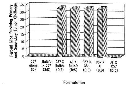
Figure 1 is a bar graph showing the effect of different alloactivated
lymphocyte
preparations on providing resistance to a secondary challenge with J588L
lymphoma cells
in Balb/c mice. Allogeneic cells stimulated either with syngeneic splenocytes
or certain
third-party splenocytes are both effective,
Figure 2 is a bar graph showing the effect of different cell culture ratios on
survival time in the mouse lymphoma model.
-6a-
CA 02346769 2003-12-03
Figure 3 is a bar graph showing the degree of functional activity in different
human alloactivated cell preparations, as determined in four different assays.
Figure 4 is a bar graph showing the level of secretion of the cytokines IL-2
and
IFN-y by human alloactivated cell preparations.
Figure 5 is a bar graph showing the enhancement of alloactivation of human
lymphocytes by using a plurality of different stimulator cells.
Figure 6 is a bar graph showing the degree of functional activity of different
human alloactivated cell preparations, depending on the ratio of responder:
stimulator
cells.
-6b-
CA 02346769 2001-04-09
WO 99/18981 PCT/US98/21413
Figure 7 is a bar graph showing the effect of including 20 pglmL of histidine
(dark shading)
or cimetidine (light shading) into cultures of human cells; either the
responder alone, the stimulator
alone, or mixed cultures at a responderstimulator ratio of 10:1.
DETAILED DESCRIPTION
This invention provides therapeutic compositions for use in cancer treatment.
The
compositions contain live cells, and confer a long-term benefit to human
cancer patients when
administered into a solid tumor mass.
The results of an instructive experiment are shown in Figure 1. Balblc mice
were treated
with a histocompatible lymphoma and an alloactivated cell population. When the
lymphocytes in the
population have been against host alloantigens (C57 x Balb/c or Aj x Balb/c),
a proportion of the
mice clear the first dose of lymphoma cells. Some surviving mice are
sufficiently well protected to
survive a second challenge with the lymphoma cells, consistent with ongoing
specific immunity
against tumor antigens. It has now been discovered that lymphocytes activated
against alloantigens
unrelated to those of the treated subject (C57 x Aj) are also effective in
conferring survival from
tumor challenge.
Thus, cells from one donor can be alloactivated against alloantigens a second
donor, and
still be effective when administered into the tumor bed in a subject who is
unrelated to either donor.
The invention shares several features with the method for treating tumors
described in
International Patent Application WO 96129394. The live cells in the
composition of the present
invention include lymphocytes that are allogeneic to the subject being
treated, and which have been
alloactivated before use in treatment. The cells are implanted directly in or
around a solid tumor
mass in the patient, with or without resection or partial resection of the
tumor.
A key difference is the alloantigens that the lymphocytes in the composition
have been
activated against. In the WO 96/29394 application, the lymphocytes are
activated using leukocytes
of the patient to be treated, and are therefore primed specifically against
the alloantigens of the
patient. In the present invention, the lymphocytes are activated against
alloaniigens of a second
unrelated donor. The donor is invariably allogeneic to the patient at a number
of loci for both class I
and class II histocompatibility antigens. As a consequence, the lymphocytes
are typically not primed
specifically against alloantigens of the intended recipient.
Not all third party responderstimulator cell combinations are equally
effective in generating
a strong alloreaction. This disclosure provides a number of strategies for
overcoming relatively less
active combinations.
One strategy is a number of screening assays to measure the extent of
alloactivation early
in culture. These are detailed in Example 3. Preferred cell populations for
use in this invention are
those that show a high degree of alloactivation within the first three days,
as measured by one or
--7-
CA 02346769 2001-04-09
- .WO 99/18981 PCT/US98/21413
more of the screening assays. This permits various donor:donor cell
populations to be screened in
advance of use in therapy.
A second strategy is to use a plurality of third party donors as a source of
responder cells,
stimulator cells, or both. This is illustrated in Example 5. Using a plurality
of donors helps ensure
that at least some histoincompatibilities will lead to sufficient
alloacGvation, as measured in the
screening assays. In addition, it has been found that cultures prepared with
leukocytes from three or
more donors can achieve higher overall levels of alloactivation.
A third strategy is to include in the alloactivation culture an H2 receptor
antagonist such as
cimetidine. This is also illustrated in Example 5. The use of H2 receptor
antagonists brings certain
relatively inacctivve cell combinations over the threshold to measurable
alloactivation, and increases
the extent of alloacctivation in others.
The present invention confers a number of advantages in comparison with
previously known
technology. For example, it is sometimes is difficult to get enough patient
leukocytes to use as
stimulators for preparing alloactivated cells. This invention provides that
leukocytes from unrelated
healthy donors can be used instead, thereby providing an almost limitless
supply. In addition,
particular donor combinations that generate high levels of alloactivation can
be identified in advance,
and used to provide a reliable source of effective material. Alloactivated
cells may be stored or
produced on an ongoing basis, eliminating the necessity of withholding
treatment for the two to three
days necessary to alloactivate lymphocytes using leukocytes of the patient.
A further description of preferred methods to prepare and use the compositions
of this
invention are provided in the sections that follow.
DEFINITIONS
"Mixed lymphocyte reactior", "mixed lymphocyte culture", "MLR", and "MLC" are
used
interohangeably to refer to a mixture comprising a minimum of two different
cell populations that are
allotypically different. At least one of the allotypically different cells is
a lymphocyte. The cells are
cultured together for a time and under suitable conditions to result in the
stimulation of the lymphocytes.
A frequent objective of an MLC is to provide allogeneic stimulation such as
may initiate proliferation of
the lymphocytes; but unless indicated, proliferation during the culture is not
required. In the proper
context, these terms may alternatively refer to a mixture of Celts derived
from such a culture. When
cells from an MLC are administered as a bolus to a human, especially in a
tumor bed, it is referred to as
a "cytoimplant".
The terms "vaccine", "immunogen", or "immunogenic composition" are used herein
to refier
to a compound or composition, as appropriate, that is capable of either: a)
generating an immune
response against an antigen (such as a tumor antigen) in a naive individual;
or b) reconstituting,
boosting, or maintaining an immune response in an individual. The
immunological response may
comprise antibodies, immunoreaetive cells (such as helper/inducer or cytotoxic
cells), or any
-g-
CA 02346769 2001-04-09
WO 99/18981 PCT/US98/21413
combination thereof, and is preferably directed towards an antigen that is
present on a tumor
towards which the treatment is directed.
A "cell line" or "cell culture" denotes higher eukaryotic cells grown or
maintained in vitro. It is
understood that the descendants of a cell may not be completely identical
(either morphologically,
genotypicafly, or phenotypically) to the parent cell.
"Inactivation" of a cell is used herein to indicate that the cell has been
rendered incapable of
cell division to form progeny. The cell may nonetheless be capable of response
to stimulus, or
biosynthesis and/or secretion of cell products such as cytokines. Methods of
inactivation are known in
the art. Preferred methods of inactivation are treatment with toxins such as
mitomycin C, or irradiation.
Cells that have been fixed or permeabilized and are incapable of division are
also examples of
inactivated cells.
The term "cancer cell", used either in the singular or plural form, refer to
cells that have
undergone a malignant transformation that makes them pathological to the host
organism. Primary
cancer cells (that is, cells obtained from near the site of malignant
transformation) can be readily
distinguished from non-cancerous cells by well-established techniques,
particularly histoiogical
examination. The definition of a cancer cell, as used herein, includes not
only a primary cancer cell, but
any cell derived from a cancer cell ancestor. This includes metastasized
cancer cells, and in vitro
cultures and cell lines derived from cancer cells.
The term "tumor-associated antigen" or "TAA" refers to a molecule, complex, or
epitope that
is detected at a higher frequency or density by tumor cells than by non-tumor
cells of the same
tissue type. Knowledge of the existence or characteristics of a particular
tumor-associated antigen
target is not necessary for the practice of the invention.
As used herein, "treatment" refers to clinical intervention in an attempt to
alter the natural
course of the individual or cell being treated, and may be performed either
for prophylaxis or during
the course of clinical pathology. Desirable effects include preventing
occurrence or recurrence of
disease, alleviation of symptoms, diminishment of any direct or indirect
pathological consequences
of the disease, preventing metastasis, lowering the rate of disease
progression, amelioration or
palliation of the disease state, and remission or improved prognosis.
The "pathology" associated with a disease condition is anything that
compromises the well
being, normal physiology, or quality of life of the affected individual. This
may involve (but is not
limited ta) destructive invasion of affected tissues into previously
unaffected areas, growth at the
expense of normal tissue function, irregular or suppressed biological
activity, aggravation or
suppression of an inflammatory or immunological response, increased
susceptibility to other
pathogenic organisms or agents, and undesirable clinical symptoms such as
pain, fever, nausea,
fatigue, mood alterations, and such other features as may be determined by an
attending physician.
An "effective amount" is an amount sufficient to effect a beneficial or
desired clinical result,
particularly the generation of an immune response, or noticeable improvement
in clinical condition.
An immunogenic amount is an amount sufficient in the subject group being
treated (either diseased
-g-
CA 02346769 2001-04-09
_ WO 99/18981 PCT/US98/21413
or not) sufficient to elicit an immunok~gicaf response, which may comprise
either a humoral
response, a cellular response, or both. In terms of clinical response for
subjects bearing a
neoplastic disease, an effective amount is amount sufficient to palliate,
ameliorate, stabilize, reverse
or slow progression of the disease, or otherwise reduce pathological
consequences of the disease.
An effective amount may be given in single or divided doses. Preferred
quantities and cell ratios for
use in an effective amount are given elsewhere in this disclosure.
An "individual" or "subject" is a vertebrate, preferably a mammal, more
preferably a human.
Non-human mammals include, but are not limited to, farm animals, sport
animals, and pets.
GENERAL TECHNIQUES
The practice of the present invention will employ, unless otherwise indicated,
conventional
techniques of molecular biology, microbiology, cell biology, biochemistry and
immunology, which are
within the skill of the art. Such techniques are explained fully in the
literature, such as, "Molecular
Cloning: A Laboratory Manual", second edition (Sambrook et al., 1989);
"Oligonucleotide Synthesis"
(M.J. Gait, ed., 1984); "Animal Cell Culture" (R.I. Freshney, ed., 1987);
"Methods in Enzymoiogy"
(Academic Press, Inc.); "Handbook of Experimental Immunology" (D.M. Weir &
C.C. Blackwell,
eds.); "Gene Transfer Vectors for Mammalian Cells" (J.M. Miller & M.P. Calos,
eds., 1987); "Current
Protocols in Molecular Biology" (F.M. Ausubel et al., eds., 1987); "PCR: The
Polymerase Chain
Reaction", (Mullis et al., eds., 1994); "Current Protocols in Immunology"
(J.E. Coligan et al., eds.,
1991 ). See also Gately et al., Lee et al., and Zarling et al. (infra) for
examples of techniques in
mixed lymphocyte cultures.
General procedures for the preparation and administration of pharmaceutical
compositions
are outlined in Remington's Pharmaceutical Sciences 18th Edition (1990), E.W.
Martin ed., Mack
Publishing Co., PA.
There are a number of animal models for cancer that can be used to test and
adjust the
compositions and methods of this invention, if desired. Certain models involve
injecting in-bred
animals with established syngeneic tumor lines. The tumors can be co-injected
with a potentially
therapeutic composition, allowed to establish before therapy is commenced, or
administered as a
challenge at some time following vaccination of a naive animal. Illustrations
are provided in the
Example section. Also useful are chimeric animal models, described in U.S.
Patent Nos. 5,663,481,
5,602,305 and 5,476,993; EP application 379,554; and International application
WO 91/01760.
All patents, patent applications, articles and publications mentioned herein,
both supra and
infra, are hereby incorporated herein by reference.
-10-
CA 02346769 2001-04-09
WO 99/18981 PCT/US98/21413
PREPARATION OF ALLOACTIVATED CELL POPULATIONS
The cellular compositions of this invention are prepared by afloactivating one
or more
responder cell populations containing lymphocytes with one or more stimulator
cell populations
expressing alloantigens. The source of the responder and stimulator cells are
allogeneic both to
each other, and to the patient to be treated with the resultant composition.
Source of donor cells: The cells that are used to prepare the composition are
typically taken from
healthy unrelated human donors allogeneic to the subject to be treated.
Cells are generally described as allogeneic if they are from the same species
but bear a
phenotypic difference sufficient to stimulate an alloreaction. In the context
of this disclosure, use of
the term "allogeneic° is restricted to a difference in phenotype of
major histocompatibility complex
(MHC) antigens. Any qualitative difference in the identity of MHC allotypes
between cells of the
same species means they are allogeneic cells. In humans, differences at any of
the H1.A-A, B, C, D,
DP, DQ, and DR loci constitute allotypic differences relevant for this
invention. Identity of HLA A, B,
C, DP, DQ, and DR are typically determined using allotype-specific antibodies
in a cytotoxicity or
immunofluorescence technique.
Preferred allotypic differences for the purposes of the present invention
relate to HLA class II
antigens. Comparing the class II antigens of the DP, DQ, and DR loci between
the putative
allogeneic cells and cells of the subject to be treated, preferably at least
1, and increasingly more
preferably 2, 3, 4, 5, or even 6 loci are different between allogeneic cells.
Class II antigens may also
be determined at the D locus by mixed lymphocyte reaction using typed cells.
Donors of allogeneic
cells are generally unrelated to the subject being treated, to maximize the
number of MHC
mismatches. In a normal outbred population, unrelated individuals will almost
invariably differ at a
number of different loci.
The number of class II region mismatches is related but secondary to a
functional
determination of allogenicity. Allogeneic cells are particularly suitable for
use in the present
invention if they demonstrate a strong proliferative response when tested in
alloreactive cultures.
Donors of cells previously known or empirically shown to produce a
particularly strong response are
especia8y suitable for use in therapy. As described elsewhere in this
disclosure, a panel of different
allogeneic cells can be tested in combinations to determine those that elicit
the strongest degree of
alloactivation.
The "responder" cells are capable of specifically reacting to an allogeneic
stimulus. The cell
population generally contains lymphocyte cells or cells of the lymphocyte
lineage, particularly T
cells. lymphocytes expressing CD4 antigen (CD4+ cells), and cells expressing
CD8 antigen (CD8+
cells) are both included in the definition of T lymphocytes, and either or
both may be included in the
composition. Generally, the responder cells are leukocytes obtained from
peripheral blood, typically
enriched for mononuclear cells (PBMC), and optionally further enriched for
cells of the lymphocyte
-11-
CA 02346769 2001-04-09
WO 99/18981 PCT/US98/21413
lineage. Particular enriched populations contain at least 10% CD4+ cells or
10% helperrnducer
cells; more preferably they are at least about 20% of CD4+ or helper/inducer
cells; even more
preferably the portion is at least about 30% of CD4+ or helpeNinducer cells.
CD4+ cells may be
conveniently quantified with commercially available specific antibody such as
OKT4 in conjunction
with fluorescence-activated counting. However, standard peripheral blood
mononuclear cell
preparations are suitably enriched for many applications of this invention.
Assays for determining
the extent of alloactivation are described in the next section.
The "stimulator" cells are allogeneic to the responder cells and capable of
eliciting an
alloreaction in the responders. Suitable cell types for use as stimulator
cells are those that bear a
high density of allogeneic histocompatibility antigens, particularly class il
antigens. Any type of cell
(not limited to blood cells) bearing sufficient alloantigens can be used. A
particularly suitable source
is peripheral blood leukocytes or white cells. It is desirable to enrich for,
or at least not to deplete
cells expressing class II histocompatibility antigens from the population,
such as B cells and
monocytes. Extensive subfractionation of the cells is not usually required,
and a simple peripheral
blood mononuclear cell population (PBMC) is adequate for most purposes.
The combined cell population is not necessarily restricted to one source for
the responder
cells and one source for the stimulator cells. Two, three, for, or a higher
plurality donors may
optionally be used to facilitate collection of the allogeneic cells, to
increase stimulation of the
allogeneic cells, to minimize the elicitation of an anti-allotype response, or
to otherwise enhance the
therapeutic efficacy.
Collection and preparation of donor cells: Donors are typically prescreened to
identify those with
sufficient leukocyte count, and exclude those with neoplastic conditions or
transmissible infections.
Collection may be performed by whole blood donation followed by separation of
blood cell
populations, or by leukapheresis. Leukapheresis is especially appropriate for
collecting the
responder cell population, because the number of cells required is
substantial. Sufficient blood is
processed to obtain about 100-500 mL leukapheresis suspension, preferably at
least about 200 mL.
For example, leukapheresis may be performed using a Cobe 2997 (CORE SPECTRA~,
Lakewood
CO); Fenwail CS 300 (Fenwall, Deerflield IL); or Haemonetrics (Braintree MA)
blood cell separator.
Flow rates of ~40-50 mUmin for 2-4 h yield -200-250 mL leukapheresis
suspension having < 1 mL
red cells, with variations between individual donors and the equipment used.
The collected leukocytes are generally washed to remove platelets, and
resuspended in a
suitable medium, such as AIM V supplemented with 2% inactivated fetal calf
serum. Separation of
PBMC and other enrichment procedures include centrifugation over a suitable
medium such as
FICOLLT"" or HISTOPAQUE~, passage over a nylon-wool column, affinity
separation methods such
as panning, or sorting in a fluorescent cell sorter using an antibody against
a relevant cell-surface
marker. Where possible, it is generally preferable to decrease the number of
manipulation steps.
-12-
CA 02346769 2001-04-09
.WO 99/18981 1PGT/US98/21413
For example, better leukapheresis separation may obviate the need for
subsequent separation on
FICOLLT"".
Mixed lymphocyte cultures: Responder and stimulator cells are combined in a
suitable culture
medium, typically supplemented with fetal calf serum or a serum substitute,
and optionally including
other growth factors. The ratio of respondecatimulator cells is preferably
between about 100:1 to
1:10; more preferably about 50:1 to 1:1; still more preferably about 20:1 to
5:1, and even more
preferably about 10:1. Where there are a plurality of stimulator or responder
cells in a one-way
MLC, the same approximate ratio of respondersatimulators is maintained. Thus,
when using 2
inactivated stimulators, the ratio may be approximately 9:(1:1); when using 3
inactivated stimulators,
the ratio may be approximately 8:(1:1:1). Simila~y, when using multiple
responders, the ratio may
be (5:5):1 or (3:3:3):1. If cultured together, the multiple responder
composition becomes a multi-way
MLC. One-way activation of multiple responders can be achieved by conducting a
separate culture
for each responder population at a 10:1 ratio, and then combining the
alloactivated cells just before
use.
This invention encompasses the use of two-way or multi-way mixed lymphocyte
cultures,
wherein a plurality of cell populations act as both responders and
stimulators. In certain
embodiments of the invention, one-way MLCs are performed by inactivating the
stimulator cells, for
example, by treating ~10' cellslmL with 50 uglmL mitomycin C or sublethal
irradiation, followed by
washing.
Once combined in the desired ratio, the cells cultured at an appropriate
density in a suitable
atmosphere (such as 95% Oz, 5% C02 at about 37°C). The culture period
is preferably at least
about 12 h, more preferably between about 24 h and 72 h. Additional
stimulation may be obtained
by culturing for 3-5 days, although this is generally not preferred, since
cytokine levels are normally
higher during the first 48 to 72 h of culture.
The recitation within this disclosure of preferred cell sources, cell ratios,
culture conditions,
timing, and other features, is intended as an aid to the practitioner and is
not meant to limit the scope
of the invention, unless explicitly required. No limitation is implied with
respect to any of the
individual parameters, since various other parameter combinations will
generate a cell population
with the desired functional effect.
Measuring functional criteria of the alloactivated cell population: Once the
culture is initiated
but before use in therapy, the functional activity of the culture can be
determined using one or more
functional assays.
Since cytokine secretion is believed to play an important role in eliciting
the response in the
treated subject, cytokines can be tested in a standard immunoassay. Particular
cytokines of interest
are IL-2, IL-4, IL-6, TNF-a, LT, IFN-y, G-CSF, M-CSF (both membrane and
secreted form), and GM-
-13-
CA 02346769 2001-04-09
- . WO 99/18981 PCT/US98/21413
CSF. For example, particular degrees of stimulation is indicated by levels of
biological activity of
TNF-a or LT at 50-150 UImL, or 500-3500 pglmL.
Proxies for functional activity of the alloactivated cells include: I: MTT
Formazan Reduction
Assay; II: XTT Formazan Reduction Assay; II1: Flow Cytometry for CD3ICD69 or
CD3/FDA; IV:
FDA Plate Assay; V: Acid Production Assay, VI: Acridine Orange Assay. These
assays are detailed
in Example 3. More traditionally, alloactivation can be determined by cell
proliferation, measured by
culturing a test sample for 5 days and conducting a standard (~Hj-thymidine
uptake assay, or by
counting blast cells. The predictive value of functional assays can be
determined by comparing
results of the assays on cultured cells with the effect of the cells in a
suitable animal model. See
Example 4. _
Preferred cultures are those that show a level of activation ~ 10% above
unstimulated donor
control value within one of the first 3 days of culture, as measured by the
Tetrazolium Reduction
Assay (XTT), the Acridine Orange Assay (AO) or by Flow Cytometry (C069), more
preferably
attaining the threshold in several of these assays in combination.
Optimizing the functional effect: Experience in animal model experiments shows
that not all third-
party donors provide the same degree of alloactivation when third party donor
are used for both the
stimulator and responder cells.
To the extent that variability is donor-cell dependent, donors can be chosen
according to
experience, both in terms of the degree of aifoactivation observed in culture,
and the clinical result.
Functional criteria indicating a particular level of activation, such as the
Tetrazolium Reduction
Assay (XTT), Flow Cytometry Assay, or the level of secretion of certain
lymphokines determined by
ELISA, may be sufficiently predictive of outcome, depending on clinical
experience. Once
successful donors are identified, they can be constituted in a panel of
regular donors sourced by the
service lab providing the immunogenic compositions.
To the extent that the variability depends on the match between donors and
patient, several
other selection criteria can also be used. Since the efficacy of certain donor-
patient combinations
may migrate according to histocompatibility, donors can be selected, if
desired, on the basis of
tissue match. Donors of particular human histocompatibitity types can be
tested for efficacy with
particular tumors, if desired, using one of the chimeric animal models listed
earlier.
A more immediate donor identification test can be conducted using PBLs from
the patient
and PBL from a selection of potential donors in an in vitro assay. One such
assay is a reverse
functional test. In this assay, patients cells are set up in a mixed
lymphocyte culture as the
responder, using the potential donor of the alloactivated cells as the
inactivated stimulator.
Since the response is thought to involve cytokine secretion by the
alloactivated cells, an
alternative predictor may be a two-stage culture. In this approach, a
responderstimulator culture is
set up using the same responder and stimulator cells being tested for use in
the preparative culture.
At 3 days, the culture is inactivated with mitomycin or sub-lethal
irradiation, so that cells can still
-14-
CA 02346769 2001-04-09
_ WO 99/18981 PCT/US98/21413
produce cytokines but not replicate. Leukocytes from the patient are then
added, and their response
is followed by a functional assay, cytokine secretion, or T cell
proliferation. In a variation of this
approach, inactivated tumor cells are also provided in the second stage of the
culture, and read-out
is determined at the end of the second stage by measuring cytolysis of s'Cr
labeled tumor cells.
These assays are described for the benefit of the reader who may wish to
optimize the .
compositions of this invention in various ways, particularly in setting up a
donor pane! enriched for
high responders. It should be emphasized that the invention can be practiced
without employing all
of these screening procedures.
As an alternative or in addition to pretesting the
responderatimulator:recipient combination,
the degree of alloactivation or the potential therapeutic outcome can be
enhanced by employing
either of the following strategies: a) using a plurality of donor cells as the
responder or stimulator in
the MLC; and/or b) adding an H2 receptor antagonist to the culture medium of
the MLC.
Using a plurality of donors for the responder or stimulator cell population
confers a number
of advantages. It is predicted that there will be a normalizing effect - when
there is a variety of
alloincompatibilities present, there is a stronger possibility that at least
one stimulator cell will
stimulate at least one responder cell, and in turn, that at least one
responder cell will stimulate the
treated subject. It is also more convenient, in that the same mixed population
will be suitable for a
variety of patients. Thus, a large batch of mixed alloactivated cells can be
prepared and stored
frozen, for dispensation on demand. It has also been discovered that having a
plurality of different
stimulators can achieve levels of alloactivation higher than one of the
stimulators alone. This is
illustrated in Example 4.
Adding an H2 receptor antagonist to the culture medium also has an enhancing
effect on
alloact'rvation during the first three days of culture. This is illustrated in
Example 5. Without
intending to be bound by theory, it is hypothesized that the H2 receptor
antagonist inhibits the
activity of suppressor T cells in the culture. Thus, it is especially
effective in restoring alloactivation
to cell combinations that are clearly incompatible, but show little reactivity
in a standard MLC. A
preferred H2 receptor antagonist is cimetidine, added to the culture medium at
between 5 IIgImL and
100 ~glmL, typically 20 ~g/mL.
USE OF CELLULAR COMPOSITIONS IN CANCER TREATMENT
The composifions of this invention can be administered to subjects, especially
human
subjects. They are particularly useful for eliciting an immune response
against a tumor-associated
antigen, or for treating cancer.
Objectives of treatment: One purpose of implanting the cellular compositions
of this invention is to
elicit an immune response. The immune response may include either humoral or
cellular
-15--
CA 02346769 2001-04-09
WO 99/18981 PCT/US98/21413
components, or both. Humoral immunity can be determined by a standard
immunoassay for
antibody levels in a serum sample from the treated individual.
Since cellular immunity is thought to play an important role in immune
surveillance of
cancer, generating a cellular immune response is frequently a particular
objective of treatment. As
used herein, a "cellular immune response" is a response that involves T cells,
and can be observed
in vitro or in vivo.
A general cellular immune response can be measured as the T cell proliferative
activity in
cells (particularly PBL) sampled from the subject after administration.
Inactivated tumor cells,
preferably derived from the subject, are used as stimulators A non-specific
mitogen such as PHA
serves as a positive control; incubation with an unrelated stimulator cell
serves as a negative control.
After incubation of the PBMCs with the stimulators for an appropriate period
(typically 5 days),
['HJthymidine incorporation is measured. If desired, determination of which
subset of T cells is
proliferating can be performed using flow cytometry. T cell cytotoxicity (CTL)
can also be measured.
In this test, an enriched T cell population from the subject are used as
effectors in a standard 5'Cr
release assay. Tumor cells are radiolabeled as targets with about 200 ~Ci of
Naz 5'CrO, for 60
minutes at 37° C, followed by washing. T cells and target cells (~1 x
10'Iwell) are then combined at
various effector-to-target ratios in 96-well, U-bottom plates. The plates are
centrifuged at 100 x g for
5 minutes to initiate cell contact, and are incubated for 4-16 hours at
37°C with 5% C02. Release of
5'Cr is determined in the supernatant, and compared with targets incubated in
the absence of T cells
(negative control) or with 0.1% TRITONTM X-100 (positive control).
Another purpose of implanting the cellular compositions of this invention is
for treatment of a
neoplastic disease, particularly cancer. Beneficial effects are typically
immunologically mediated or
the result of an inflammatory infiltrate into the injection site and
collateral tumors. Evidence of a host
response can be shown inter alia by infiltration of host leukocytes (such as
lymphocytes, histiocytes,
and other leukocytes) into the tumor site by standard histomorphology
analysis. The response is
preferably an immunological response, which may have humoral or cellular
components, and
preferably includes cytotoxic T cell activity. Immunological activity can be
measured systemically in
standard antibody binding immunoassays or cytotoxicity assays on peripheral
blood components
taken from the treated subject, using tumor cells as targets. Monitoring the
effect according to these
methods is optional, and the recited features need not be positively
demonstrated in order for the
compositions and treatment methods to fall within the scope of this invention,
except where required.
Suitable subjects: The compositions of this invention may be used for
administration to both
human and non-human vertebrates.
Typically, the subject will either have cancer, or be at substantial risk of
developing cancer.
Typical human subjects for therapy comprise two groups, which may be
distinguished by clinical
criteria. Patients with "advanced disease" or "high tumor burden" are those
who bear a clinically
measurable tumor. A clinically measurable tumor is one that can be detected on
the basis of tumor
-16-
CA 02346769 2001-04-09
_ . WO 99/1$981 pCT/US98/21413
mass (e.g., by palpation, MRI, CAT scan, X-ray, or radioscintigraphy; positive
biochemical or
histopathological markers on their own are insufficient to identify this
population).
A cellular composition for use in this invention is administered to patients
with advanced
disease with the objecctivve of palliating their condition. Ideally, reduction
in tumor mass occurs as a
result, but any clinical improvement constitutes a benefit. Clinical
improvement includes decreased
risk or rate of progression or reduction in pathological consequences of.the
tumor.
A second group of suitable subjects is known in the art as the "adjuvant
group". These are
individuals who have had a history of cancer, but have been responsive to
another mode of therapy.
The prior therapy can have included (but is not restricted to) surgical
resection, radiotherapy,
traditional chemotherapy, and other modes of immunotherapy. As a result, these
individuals have
no clinically measurable tumor by the definition given above. However, they
are suspected of being
at risk for recurrence or progression of the disease, either near the original
tumor site, or by
metastases. The adjuvant group may be further subdivided into high-risk and
low-risk individuals.
The subdivision is made on the basis of features observed before or after the
initial treatment.
These features are known in the clinical arts, and are suitably defined for
each different cancer.
Features typical of high risk subgroups are those in which the tumor has
invaded neighboring
tissues, or which show involvement of lymph nodes.
A cellular compositson for use in this invention is administered to patients
in the adjuvant
group in order to elicit an anti-cancer response primarily as a prophylactic
measure against
recurrence. Ideally, the composition delays recurrence of the cancer, or more
preferably, reduces
the risk of recurrence (i.e., improves the cure rate). Such parameters may be
determined in
comparison with other patient populations and other modes of therapy.
Of course, crossovers between these two patient groups occur, and the cellular
compositions can be administered at any time that is appropriate. For example,
therapy can be
conducted before or during traditional therapy of a patient with high tumor
burden, and continued
after the tumor becomes clinically undetectable. Therapy may be continued in a
patient who initially
fell in the adjuvant group, but is showing signs of recurrence.
Examples of tumors that can be treated according to this invention include but
are not
limited to those on the following list. The list includes sites that are
thought to be immune privileged,
such as the brain, and sites that are not immune privileged, such as the
pancreas, colon, breast, and
prostate.
~ Brain tumors, such as astrocytoma, oligodendroglioma, ependymoma,
medulloblastomas, and PNET {Primitive Neural Ectodermal Tumor);
~ Pancreatic tumors, such as pancreatic ductal adenocarcinomas.
~ Lung tumors, such as small and large cell adenocarcinomas, squamous cell
carcinoma,
and bronchoalveolarcarcinoma;
~ Colon tumors, such as epithelial adenocarcinoma, and liver metastases of
these tumors;
~ Liver tumors, such as hepatoma, and cholangiocaroinoma;
-17-
CA 02346769 2001-04-09
_ . WO 99/18981 PC1YUS98/21413
~ Breast tumors, such as duct"ai and lobular adenocarcinoma;
~ Gynecologic tumors, such as squamous and adenocaroinoma of the uterine
cervix, and
uterine and ovarian epithelial adenocarcinoma;
~ Prostate tumors, such as prostatic adenocaroinoma;
~ Bladder tumors, such as transfional, squamous cell carcinoma;
~ Tumors of the RES System, such as B and T cell lymphoma (nodular and
diffuse),
plasmacytoma and acute and chronic leukemia;
~ Skin tumors, such as malignant melanoma; and
~ Soft tissue tumors, such as soft tissue sarcoma and le'romyosarcoma.
The immune status of the individual may be any of the following: The
individual may be
immunologically naive with respect to certain tumor-associated antigens
present in the composition,
in which case the compositions may be given to initiate or promote the
maturation of an anti-tumor
response. The individual may not currently be expressing anti-tumor immunity,
but may have
immunological memory, particularly T cell memory relating to a tumor-
associated antigen, in which
case the compositions may be given to stimulate a memory response. The
individual may also have
active immunity (either humoral or cellular immunity, or both) to a tumor-
associated antigen, in which
case the compositions may be given to maintain, boost, or maturate the
response, or recruit other
arms of the immune system. The subject should be at least partly
immunocompetent, so as to
minimize a graft versus host reaction of pathological scope. However, it is
recognized that cancer
patients often show a degree of immunosuppression, and this does not
necessarily prevent the use
of the compositions of the invention, as long as the compositions may be given
safely and
effectively.
Modes of administration and dose: The compositions of this invention can be
administered to the
subject at the site of any solid tumor. Circulating cancers are treatable so
long as there is at least
one solid tumor mass. Metastatic sites, affected nodes, and other sites away
from the primary
neoplasm are suitable, so long as they are accessible and contain sufficient
tumor antigen.
If the solid tumor mass is resectable or partly resectable, then the
composition can be
administered at or near the site or in a cavity created by the resection. If
the tumor is completely
removed, however, then it may be preferable to administer the alloactivated
cells to a metastatic site
to increase the local amount of bystander tumor antigen. The most convenient
time to administer
the alloactivated cells to a resectable site is during the time of surgery. To
keep the cells at the site
until completion of the surgical procedure, it is convenient to administer the
cells in a
pharmaceutically compatible artificial gel, or in clotted plasma.
When the solid tumor mass is not resectable, or where less invasive procedures
are
desired, then the composition can be injected at or near the tumor site
through a needle. For deeper
sites, the needle can be positioned using ultrasound, radioscintigraphy, or
some other imaging
technique, alone or in combination with the use of an appropriate scope or
cannula. Pancreatic
-18.
CA 02346769 2001-04-09
WO 99/18981 PCT/US98121413
tumors are preferably implanted using an injection needle positioned by an
endoscopic ultrasound
guided technique, as described by Chang et al., Gastroenterology 112:A346, i
996 (abstract). For
this application, the cell population is conveniently administered when
suspended in isotonic saline
or a neutral buffer to a volume of about 10 ml.
The dose given is an amount "effective" in bringing about a desired
therapeutic response, be .
it the stimulation of an immune response, or the treatment of cancer as
defined elsewhere in this
disclosure. For the pharmaceutical compositions of this invention, effective
doses typically fall within
the range of about 10° to 10" cells, including aUogeneic stimulators
and responders. Preferably,
between about 1 x 10⁹ to 5 x 10'° cells are used; more preferably
between about 2 x 10⁹ to 2 x 10'°.
Multiple doses when used in combination to achieve a desired effect each fall
within the definition of
an effective amount.
The various components of the implant composition are present in an "effective
combination", which means that there are sufficient amounts of each of the
components for the
composition to be effective. Preferably, at least about 108, more preferably
between about 1 x 10s to
5 x 10'° and; more preferably between about 2 x 10⁹ to 2 x 10'°
responder cells are present.
Preferably, at least about 10', more preferably between about 5 x 10' to 5 x
109 and; more
preferably between about 1 X 10° to 2 x 10⁹ stimulator cells are
present. Ratios of allogeneic
lymphocytes to stimulator leukocytes is generally between 1:1 and 100:1,
usually between about 5:1
and about 25:1, and typically about 10:1. However, any number of component
cells or other
constituents may be used, as long as the composition is effective as a whole.
This will also depend
culture conditions and other factors during preparation.
The pharmaceutical compositions of this invention may be given following,
preceding, in lieu
of, or in combination with, other therapies relating to generating an immune
response or treating
cancer in the subject. For example, the subject may previously or concurrently
be treated by
chemotherapy, radiation therapy, and other forms of immunotherapy and adoptive
transfer. Where
such modalities are used, they are preferably employed in a way or at a time
that does not interfere
with the immunogenicity of the compositions of this invention. The subject may
also have been
administered another vaccine or other composition in order to stimulate an
immune response. Such
alternative compositions may include tumor antigen vaccines, nucleic acid
vaccines encoding tumor
antigens, anti-idiotype vaccines, and other types of cellular vaccines,
including cytokine-expressing
tumor cell lines.
Certain embodiments of this invention relate to combination therapies. In one
preferred
combination therapy, the subject is given an infra-tumor implant of stimulated
allogeneic
lymphocytes, either before, during, or after treatment at a site distant from
the tumor with a
composition comprising stimulated allogeneic lymphocytes and autologous tumor
cells. The
preparation and use of vaccines of this nature is described in detail in
International application V110
98116238, which is hereby incorporated herein by reference in its entirety. An
illustrative protocol for
this combination therapy is provided in Example 6. In the illustration, the
vaccine is given weekly for
-19-
CA 02346769 2001-04-09
WO 99/18981 PCT/US98/21413
four weeks following the cytoimplant, to enhance the extent of the anti-tumor
response in the host or
the therapeutic effectiveness. The vaccine can also be given after intervals
of several months in
order to replenish the response. Accordingly, certain embodiments of this
invention relate to
administering a cytoimplant, and subsequently boosting the therapeutic effect
or immunological
response by administering to the patient a composition comprising
alloactivated human lymphocytes
allogeneic to the patient and an inactivated cel! population consisting of
tumor cells from the patient
or progeny thereof.
While the methods and compositions of this invention are generally effective
when given at a
single dose, it may be desirable to readminister the composition at intervals
of 3-6 months,
especially for fast-growing tumors that can be injected through a positioned
needle. Accordingly,
certain embodiments of this invention relate to administering a cytoimplant,
and subsequently
boosting the therapeutic effect or immunological response by implanting in or
around the bed of a
solid tumor in the patient a second cell population comprising alloactivated
human lymphocytes
allogeneic to the patient.
Timing of administration of compositions of this invention is within the
judgment of the
managing physician, and depends on the clinical condition of the patient, the
objectives of treatment,
and concurrent therapies also being administered. Suitable means of
immunological monitoring
include a one-way MLR using patient's PBL as responders and primary tumor
cells as stimulators.
An immunological reaction may also be manifest by a delayed inflammatory
response at the injection
site. Suitable means of monitoring of the tumor are selected depending on the
tumor type and
characteristics, and may include CT scan, magnetic resonance imaging (MRI),
radioscintigraphy
with a suitable imaging agent, monitoring of circulating tumor marker
antigens, and the subject's
clinical response. Additional doses may be given, such as on a monthly or
weekly basis, until the
desired effect is achieved. Thereafter, and particularly when the
immunological or clinical benefit
appears to subside, additional booster or maintenance doses may be given as
required.
When multiple cytoimplants or combinations of implants and cellular vaccines
are given to
the same patient, some attention should be paid to the possibility that the
allogeneic lymphocytes in
the vaccine may generate an anti-allotype response. The use of a mixture of
allogeneic cells from a
plurality of donors, and the use of different allogeneic cell populations in
each dose, are both
strategies that can help minimize the occurrence of an anti-allotype response.
During the course of therapy, the subject is evaluated on a regular basis for
general side
effects such as a febrile response. Side effects are managed with appropriate
supportive clinical
care.
The examples presented below are provided as a further guide to a practitioner
of ordinary
skill in the art, and are not meant to be limiting in any way.
-20-
CA 02346769 2001-04-09
WO 99/18981 PGT/US98I21413
EXAMPLES
EXAMPLE 1: MIXED LYMPHOCYTE CULTURE PROCEDURR.
Collection of responder PBMC from unrelated donor: Peripheral blood mononuGear
cells (PBMCs) were collected by leukapheresis from normal healthy donors
unrelated to the patient
to be treated. Donors were pre-screened to test for complete blood count (CBC)
with differential,
Hepatitis A, B, and C, VDRL, and HIV-I.
Approximately 150 to 300 ml of leukapheresis suspension containing PBMC was
collected
from each donor, using standard blood donation procedures for supportive
apheresis according to
the manufacturers' instructions. The ieukapheresis was pertormed using a
Fenwall CS 3000
(Deerfield, EL) blood cell separator. A flow rate of 40 to 50 mUmin for 2 to 4
hours with lymphocyte
yield of 2-4 x 10s processed a total donor blood volume of 7,000 to 12,000 ml
to yield 200 to 250 ml
of leukapheresis suspension having less than 1 ml of red cells. If a Cobe 2997
blood cell separator
was used, the centrifuge rate was 5 x g, the flow rate was up to 45 ml/min,
and the collection rate
was no more than or equal to 2.5 mUmin.
However, if donor pre-absolute lymphocyte counts were in the 0.6 x 10⁸ to 1.0
x 109 range,
as little as 150 ml of leukapheresis product was drawn. Hematocrit for the
final product was 3.5%.
At least one total blood volume was processed for 80% efficiency of lymphocyte
collection.
The anticoagulant used was either 2% citrate or a citratelanticoagulant ratio
of ACDA -
15 mUcitrate-100 ml; ACDB - 25 mUcitrate - 100 ml; or CPD - 14 mU citrate -
100 ml. To obtain the
utmost product purity, the actual and final product from the cell separator
was transported as a pure
concentrate of cells in autologous plasma. The cells were not washed, and no
albumin was added.
Preparation of donor cells: The leukapheresis product was transported to the
MC
Oncology Research Laboratory for the production of allogeneic mixed lymphocyte
cells (MLCs) for
immunotherapy.
Cells were drained from the leukapheresis pack into two or three 250 ml
centrifuge tubes;
removing and setting aside 3 ml for sterility tests to be done during
centrifugation. Cell concentrate
was diluted with phosphate buffered saline (PBS) and centrifuged for 7 minutes
at 2,000 rpm.
Centrifugation was repeated twice for a total of three times to wash the cells
free of the clotting factor
in the donor's serum.
Three 1 ml aliquots from the 3 ml removed from the leukocyte suspension were
placed into
sterile capped tubes for sterility testing. The 5rst 1 ml aliquot was added to
thioglycollate medium
(Difco, Detroit, MI) (30-35°C, 48 hr.); a second 1 ml was added to
tryptic soy broth (Difco, Detroit, MI)
(25-30°C, 48 hr.); and the third 1 ml was added to RPMI 1640 (GIBCO,
Gaithersburg, MD) with 10%
heat inactivated FBS (RPMI-10%) and 1 % L-glutamine, but without antibiotics.
- 21 -
CA 02346769 2001-04-09
WO 99/18981 PCT/US98/Z1413 _
Cells were spin washed twice at 150g for 10 minutes in PBS to remove
platelets. The
supernatan! was very carefully discarded as cells were in a slurry and not a
pellet. Cells were
resuspended in AIM V (GIBCO, Gaithersburg, MD) supplemented with 2% heat
inactivated FBS (2%
AIM V) to 420 ml, and placed into a T-175 CM2 flask.
Patient or donor blood was diluted 1:1 with sterile saline. For cell
separation, 35 ml of cell
suspension was carefully layered onto 15 ml Histopaque~ 1.077 suspension
medium (Sigma; St.
Louis, MO) in each 50 ml tube and centMuged at 250g for 45 minutes.
Centrifugation was started
slowly and gradually increased to full speed. After centrifugation, the
intertace containing
mononuclear cells between the Histopaque~ suspension medium and the plasma
layer was
carefully collected with a 25 ml sterile pipet deposited into clean 50 ml
centrifuge tubes, diluted with
2% AIM V Media 1:1, and centrifuged at 5508 for 7 to 10 minutes to form a cell
pellet. Cells
remained a minimum of time in the Histopaque~ suspension medium, because it is
toxic to the cells.
The supernatant was discarded, the pellet was resuspended in 2% AIM V and
divided into
two 50 ml centrifuge tubes to a total volume 40 ml, and centrifuged at 5508
for 5 minutes. After
washing, the supernatant was discarded. The washing step was repeated twice
for a total of three
times. After the last wash, cells in each tube were resuspended in 50 ml of 2%
AIM V. Aliquots of 1
ml of the resuspended cells were diluted to a ratio of 1:10 in 2% AIM V per
tube, then further diluted
1:1 in Trypan Blue (Sigma, St. Louis, MO) to distinguish dead from live cells,
and the live cells were
counted in a hemocytometer. Cells were set at 2 x 10⁸lm1 with 2% AIM V.
Collection of stimulator PBMC from tumor patients: From 200 to 400 ml of
peripheral
blood cells were drawn from glioblastoma patients by vena puncture and placed
into 250 ml
centrifuge tubes, removing and setting aside 3 ml for sterility tests to be
done during spinning. Blood
cells in the centrifuge tubes were diluted with saline and centrifuged for 7
minutes at 550g.
Centrifugation was repeated twice for a total of three times to wash the cells
free of the clotting factor
in the patient's serum. Sterility testing was conducted as described above.
Cells were washed twice by centrifugation at 150g for 10 minutes in saline to
remove
platelets, the supernatant was very carefully discarded, and 420 mi of cells
were resuspended in a
T-175 CMZ flask in saline.
15 ml of Histopaque~ 1.077 cell separation medium was added to twelve 50 ml
centrifuge
tubes, and 35 ml of cells suspended in saline were layered onto the
Histopaque~ 1.077 in each
50 ml tube. The cell suspensions were spun at 2508 for 45 minutes, starting
centrifugation slowly
and gradually increasing speed.
After centrifugation, the mononuclear cells at the intertace between the
Histopaque~ cell
separation medium and the plasma layer were carefully collected with a 25
sterile pipet into 2 sterile
250 ml centrifuge tubes and diluted with 2% AIM V to a final volume of 250 m1.
The diluted
mononuclear cells were centrifuged at 5508 for 7 to 10 minutes. For washing,
the supernatant was
CA 02346769 2001-04-09
WO 99/18981 pCT/US98/21413
discarded, then the cell pellet was re-suspended with 2% AIM V and centrifuged
at 550g for 5
minutes. The washing step was repeated for a total of three times.
After the last washing step, cells were re-suspended in 50 ml of 2% AIM V, 1
ml of the cell
suspension was diluted 1:10 in 2% AIM V per tube, and the number of viable
cells was determined
by enumeration in a 1:1 in Trypan Blue as described above.
It is readily appreciated that this procedure is equally suitable for
obtaining stimulator cells
from healthy third-party donors.
Alloactivation: The isolated patient PBMCs were re-suspended at 10' cellslml
in AIM V,
50 N,g Mitomycin C (Bristol-Mayer Squibb, Princeton, NJ) were added per ml of
patient cell
suspension, and the suspension of PBMCs was incubated at 37°C for one
hour to block response of
the stimulator cells to the responder cells. After one hour of incubation, the
excess mitomycin C was
washed from the cells by alternate centrifugation (250g for 5 min), and the
cells were resuspended in
AIM-V. After mitomycin treatment of the patient's PBMCs, the cells were added
at a 20:1 to 10:1
donor cell:patient cell ratio to the donor culture).
For co-culture, the donor and mitomycin C-treated patient PBMC suspension was
placed in
a sealed sterile Fenwal tissue culture system especially designed for culture
of PBMC for
reimpiantation into patients. Cells were passed in sealed systems via Fenwal
cell transfer units and
pumps according to the manufacturers instructions, and cultured in a
37°C incubator for 48 hours.
Sterility testing of alloactivated cells: Two days prior to implantation of
the cell
suspension, the following three sterility tests were performed. 10 ml sterile
aliquots were removed
from each tissue culture bag, placed into sterile capped 15 ml centrifuge
tubes, and centrifuged for
10 minutes at 450g. In each tube, the pellet was resuspended in 3.0 ml of PBS.
A 1 ml aliquot of
the cell suspension was added to each of three sterile capped tubes containing
2 ml of thioglycollate
broth, tryptic soy broth, or RPMI-10% and incubated for 48 hours. Each cell
suspension was
examined microscopically prior to implant to detect signs of microbial growth.
On the day of surgery, the cells were centrifuged out of their medium, washed
two times with
saline and re-suspended in platelet free, decalcfied plasma obtained from the
patient the previous
day. The cells were transported to the operating room in plasma, then the
plasma was re-calcified
by the addition of calcium gluconate so that it clots just before implantation
into the tumor bed.
The day of surgery a drop of collected cell pellet was again examined for
sterility under the
microscope. Just prior to clotting, a 100 pl aliquot of the cell suspension
was added to 2 ml each of
RPMI-10% without antibiotics, thioglycollate and tryptic soy broth in a
sterile capped tube. The
samples were then incubated for four days after surgery, and a running log was
kept of this last
sterility test.
-23-
CA 02346769 2001-04-09
WO 99!18981 PCT/US98/21413
EXAMPLE 2: CONICAL TRIAL USING ALLOACTIVATED CELLS IMPLANTED AT THE TUMOR
SITES
This experiment confirms that allogeneic cells alloactivated using patient
leukocytes are
effective in cancer treatment.
A Phase I/11 clinical trial was conducted to examine the feasibility,
tolerability, toxicities and
clinical effects associated with a single intratumoral injection of allogeneic
lymphocytes sensitized
against patient alloantigens. This trial is conducted under the auspices of
the appropriate ethical
approval committee, and in accordance with a protocol under the U.S. Food &
Drug Administration.
Eligible patients were men and women between 18 and 85 years of age. A total
of ten
patients were studied. Eight patients were enrolled in the trial, and iwo
additional patients were
treated off study on a compassionate basis. Nine of ten patients had locally
advanced, surgically
unresectable pancreatic tumors; 40% of the patients had Stage II disease, 30%
had Stage 111
disease and 20% had Stage IV disease. One patient with Stage I disease was 89
years old,
declined surgery and was treated on a compassionate basis. Seven of ten
patients had received no
prior therapy, one patient had received prior radiation therapy and two
patients had received prior
radiation and chemotherapy.
Preparation of cells: The procedure for preparing the cytoimplant cells was
generally in
accordance with the main features of Example 1. Typically, a volunteer third-
party donor for
responder cells is screened by normal blood bank criteria for suitability. No
special matching or
identification of HLA type is performed. Whole blood or leukapheresis is
collected from the patient to
be treated; and leukapheresis is collected from the donor on the same day.
Mononuclear cells are
prepared from both patient and donor by centrifugation on FicoIlT"", and
counted to ensure that
enough cells are present to prepare the intended dose. Patient cells are
inactivated by treating for 1
hour with mitomycin C, and then washed.
The cells are combined at a donor:patient ratio of 10:1 to 20:1, depending on
the number of
patient cells available. The cells are suspended at 3 x 10⁶ per mL in AIM 5
medium containing 2%
fetal calf serum and antibiotics in a gas-permeable plastic bag, and incubated
at 37°C in an
atmosphere of 5% CO~I95% 02. No cytokines or other growth factors are added.
After three days,
the cells are collected by centrifugation and washed. The cells are then
transferred to the clinic in a
medium suitable for administration. For the treatment of pancreatic cancer,
the cells were
suspended in a volume of about 10 mL isotonic saline.
Features of the cells are shown on the following table:
-24-
CA 02346769 2001-04-09
WO 99/18981 PGT/US98/Z1413
TABLE 1: Alloactivated Cells Administered to Humans with Pancreatic Cancer
~!::!!:7P y..~. ~ ~...:. ..n..?F>.
f! .' !:: Y ~' F
.S I fi ~ ~ iN~ ~
fiW _~ i;~::i . fi f C C.:: :IIIfLCf
. . .: .MLC
ft ~ >.:n::
v-:
:4'.Y'.!u!y
.
..::.~:.~ nlv. " y ..$ ,. .. .:.:.r..,.
.... Y..; c,:. h . ~: ,......,..:.
:, .,::: ,:.!~::.: . .
i::,:::;.~,.n:. :.. ...:::..w.,
::N.~.n..F.. ~ :.n ...~.:,.:.1
...4....: : ! ~ s..$... C . f
.?:.3iyN.,.: :al
.,.. .: D39P" '1L- P
f..;:..::v. ''v~l~i~a!.' ''
: ::. : :: ~ :
/iS ~~ ~.
. c.5........:.:',
:; :.
./v!
r
> r
..,
<..::
.. ..
:.. .. .. : ... . , .. ..... ...
.. . .: .. ..:... .... : ::
. . ::. ..: .. .::: :::. ....:
! .:......::. . :.: CD89 ::~t f...~.rv
...: .,. r:'. v ditty . . :.: n
.$:.;$.~R :: n ~: .. .n (
':fifi ~ ~. gEmL)
F~tt~s ~?osage ~~Ilt ~'
g m~.
..~.:... ;: : ,;.: _ .. ,.- _ ,
!i. .. . . : ... .
,, , ,
,~,
.fill 10:1 1.76 91% Sterile NA 2779 202
' x 10s
'
:y.
'
.'
~. i<..
s: 10:1 3.5 x 98% Sterile 11.8% 10852 438
.;::k:y:;i.. 108
?y~;ri:,;.
3;1' 10:1 2.8 x 88% Sterile NA 2334 383
109
4"'~3;~'...:~20:1 5.9 x 92% Sterile NA 1927 0
. ;;i.:r,~:: 108
~5, 10:1 6.0 x 95% Sterile NA 3941 298
d~l~ 109
""
'006, 20:1 6.04 96% Sterile NA 118 0
0$ x 109
r
p0.7;.:8'~10:1 5.8 x 93% Sterile NA 7307 981
108
, 008; 10: 8.9 x 91 % Sterile 18.6% 1137 308
LM i 10s
'
009; 13:1 10.5 95% Sterile NA 433 0
GS x 109
~
010;. 15:1 9.6 x 90% Sterile NA 11858 291
JH 109
Administration: The treatment was conducted as follows: A sufficient amount of
whole
blood or leukapheresis was collected from each patient to prepare the cultured
cells used in
treatment. The sample was forwarded to the Immunotherapy Lab, and used to
prepare stimulator
cells for allogeneic stimulation of third-party lymphocytes.
Three days later the cytoimplant cells were administered to the subject on an
out-patient
basis. Under light anesthesia, an injection needle was positioned into the
tumor using an
endoscopic ultrasound guided technique. The implant cells were rescued from
culture, washed,
suspended in about 10 mL of injectable isotonic saline, and delivered to the
diagnostic service
center. The cells were injected into the tumor mass, the device was removed,
and the patient was
allowed to recover.
Three patients were administered with a single dose of 3 x 10⁹ implant cells.
Four patients
were administered with a single dose of 6 x 10⁹ implant cells. Three patients
were administered with
a single dose of 9 x 10⁹ implant cells.
Follow-up was done one day, one week, one month, and every three months after
implantation. Criteria assessed included evidence of toxicity, survival, tumor
response (endoscopic
~ ultrasound and/or CT-scan), tumor markers (CEAICA19-9) and Kamofsky
performance score.
Results: Patients treated with 3 x 10⁹ cells: Patient 001 was a 78 year old
male with an
unresectable clinical Stage IV tumor. The patient received treatment on a
compassionate basis, and
survived 6.5 months. Patient 002 was a 53 year old female with an unresectable
Stage III tumor.
-25-
CA 02346769 2001-04-09
.WO 99/18981 PCT/US98/21413
The patient later presented with elevated total bilirubin and died at 4.2
months after developing liver
metastasis. Patient 003 was a 60 year old male with unresectable Stage III
tumor. There was a
hospital admission for synocopal episode. The patient survived 20.8 months.
Patients treated with 6 x 10g cells: Patient 004 was a 52 year old male with
an unresectable
Stage II tumor. The patient was later admitted to hospital with biliary
obstruction, cholangitis, and
dehydration. This patient died 20.7 months after treatment. Patient 005 was an
89 year old female
with a Stage 1 tumor, who was not a candidate for resection due to her age.
She received treatment
on a compassionate basis, and died 11.3 months later due to myocardial
infarction. Patient 006 is a
54 year old female with an unresectable Stage III tumor. She was later
admitted to hospital for
intractable nausea, vomiting and dehydration, and subsequently for
gastrointestinal hemorrhage.
There was increased tumor size and liver metastasis. The patient died 4.3
months after treatment.
Patient 007 is a 61 year old female with an unresectable Stage II tumor. On
follow-up, there was
elevated total bilirubin, and the patient was admitted with intractable nausea
and vomiting, diarrhea,
and dehydration, possibly related to colitis flare-up. The patient is still
alive >13 months after
treatment.
Patients treated with 9 x 10⁹ cells: Patient 008 is a 55 year old female with
an unresectable
Stage IV tumor. No serious adverse events were observed, and the patient is
still alive >13 months
after treatment. Patient 009 is a 54 year old male with an unresectable Stage
II tumor. The patient
was later admitted for two days for pain and nausea and vomiting. The patient
died 11.7 months
after treatment. Patient 070 is a 68 year old male with an unresectable Stage
II tumor. No serious
adverse events were observed, and the patient died 8.5 months after treatment.
Clinical Interpretation: Elevated bilirubin, liver enzymes and nauseaivomiting
with
dehydration were the most common serious adverse events documented which were
considered to
be due to obstruction of biliary stents. These adverse events were considered
to be associated with
the disease rather than the therapy. Because of its relationship to the timing
of the administration of
the cytoimplant, one adverse event (elevated total bilirubin, Grade 4) was
considered possibly
related to therapy. No other serious adverse effects were observed that were
considered to be
associated with therapy.
The median survival for all patients treated in this study was 11.5 months
(range 4.2 to >21)
with a mean survival of greater than 10 months. The 6 month, 9 month, and 12
month probability of
survival was 80% (n=8), 60% (n=6), and 50% (n=5), respectively. The
probability of greater than
eight month survival by dose was 33% for 3 x 10a cells, 75% for 6 x 10s cells,
and 100°~ for 9 x 10⁹
cells. Comparison of median survivals of patients treated with cytoimplant to
those treated with 5-
fluorouracyl (median = 4.2 months) or GEMZARTM (median = 5.7 months) was
significant at p <
0.006 and p < 0.004, respectively.
-26-
CA 02346769 2001-04-09
WO 99/18981 PCT/US98/Z1413 _
Hlstomorphology: Histology slides were prepared from tissue samples from an 89
year
old female diagnosed with pancreatic adenocarcinoma. At the time of diagnosis,
the tumor was
advanced and surgically unresectable. The only treatment performed on this
patient was the
injection of the tumor with 6 x 10⁹ cytoimplant cells. The patient died of a
myocardial infarct 11.5
months later.
One photomicrograph showed flbrovascular tissue with scattered individualized
tumor cells.
There is a dense lymphocytic and plasma cell infiltrate. Another field showed
lymphocytes rosetting
the separated tumor cells. The tumor cells were dark and shrunken, which is
evidence of apoptosis.
Another field showed scattered islands of necrotic tumor cells. There was a
very dense infiltrate of
lymphocytes, and lymphocytes appear to be trafficking into the site from
adjacent venules. In a high,
magnification view there was clear evidence of direct contact between
lymphocytes and necrotic
tumor cells.
The histomorphology analysis provide clear evidence of a local response by
cells of the
patient after implantation of the alloactivated cells. The data are consistent
with the cell response in
the patient having a direct role in the beneficial effects of the treatment,
as shown by direct contact
between lymphocytes and necrotic tumor cells. To the limited extent that
infiltrating cells are present
in untreated pancreatic cancer, this type of direct contact is not observed.
EXAMPLE 3: MEASUREMENT OF THE DEGREE OF ALLOACTIVATTON
In order to ensure the production of high quality effective MLC cells, a
method of measuring
the potency of the altoactivated cells can be employed. Only cell cultures
with activity over and
above unstimulated control cells should be used clinically. It is beneficial
to compare the activity to
the unstimulated control, since baseline activity of mononuclear cells from
different individuals varies
widely.
Several methods are available for measuring lymphocyte activation. Compared
with
unstimulated mononuclear cells, alloactivated cells reduce more Formazan dye
and have more
esterase activity. Turnover of XTT (a Formazan dye) can be easily demonstrated
in a 96-well plate
by colorimetric spectrophotometry at 470 nm (reference fi50 nm). Activated
cells typically show
higher absorbance than controls. Lymphocyte activation can also be
demonstrated by flow
cytometric determination of esterase activity using the esterase substrate,
fluorescein diacetate
(FDA). T cells with high esterase are not determined using FDA and a
Phycoerythrin-labeled CD3
antibody. Esterase activity can be accurately measured in a plate assay by
using higher
concentrations of FDA and determination of esterase activity by
spectrophotometry at 494 nm
(reference 650 nm) in a 9&well plate format. Background esterase activity
inherent to serum-
containing media is inhibited by addition of a competitive esterase inhibitor
(~10 mM), arginine
methyl ester. For the most part, these measures show good correlation with
each other and with
blastogenesis.
-27-
CA 02346769 2001-04-09
WO 99/18981 PCT/US98/21413
I: MTT Formazan Reduction Assay
This assay is used to enumerate live cells by ability for culture sample to
reduce MTT to
blue-green Formazan dye, and is also helpful for the distinguishing activated
from inactive cells. It
can be used for practically any cell in practically any media. The useful cell
range is between 105
and 5 x 10° per mL.
Reagents:
~ 96 well plates, flat bottom (not EL1SA plates)
~ 5 mglml MTT (Sigma) in PBS (frozen)
~ 20% SDS in 45°~ DMF, 0.2 N HCI (pre-warmed to 37°C)
Procedure:
Place 100 pL of culture media with cells in 96 well plate in duplicate or
triplicate. Use 100 ~L
of media alone for controls. Leave first column blank.
Add 10 pL of MTT to each well. Tap plate to mix. Cover plate and incubate
37°C for 4
hours.
Add 50 pL of SDS solution , avoiding bubbles. Tap to mix. If bubbles are
present, blow on
surface. Count plate at 570 nm (reference 650 nm).
II: XTT Formazan Reduction Assay
This assay is used to enumerate live cells by ability for culture to sample to
reduce XTT to
red-orange Formazan dye, and is also helpful for the distinguishing activated
from inactive cells. It
can be used for practically any cell in practically any media. The useful cell
range is between 105
and 5 x 10⁶ per mL.
Reagents:
~ 96 well plates, flat bottom (not ELISA plates)
~ 1 mglmL MTT (2,3-bis (2-methoxy-4-vitro-5-sulfo-phenyl-2H-tetrasolium-5-
carboxanilinide salt, Sigma) in PBS (fresh)
1.53 mglmL PMS (phenylmethanesulfonyl fluoride, Sigma) in PBS (frozen,
protected
from light)
Procedure:
Place 100 N,L of culture media with cells in 96 well plate in duplicate or
triplicate. Use 100 pL
of media alone for controls. Leave first column blank.
Pre-mix PMS with XTT immediately before use (5 ~L. per mL XTT). Add 50 ~L of
XTT to
each well. Tap plate to mix.
-28-
CA 02346769 2001-04-09
WO 99/18981 PCT/ITS98/21413
Cover plate and incubate 37°C for 4 hours. Count plate at 470 nm
(reference 650 nm).
III: Flow Cytometry for CD31CD68 or CD3IFDA
This is a measurement of T lymphocyte activation after mixed lymphocyte
alloactivation.
Activities such as CD69 expression or esterase activity correlate with
cytokine secretion and can be
used as surrogate measures of lymphocyte acctivvity. Unstimulated lymphocytes
do not express
surface CD69 and have only low levels of non-specific esterases. Once
activated by alloantigens or
non-specific mitogens, the expression of CD69 appears within 48 hours (peak at
24). Esterase
activity increases shortly after stimulation, and continues for several days.
Not all allostimulated
lymphocyte reactions proceed with the same kinetics, and it is preferable to
measure activation on
day 1, 2 and 3 of the culture.
Sample:
Test samples of donor and patient cells are mixed in small cultures at 0.5 x
10g cellslmL in
2% FCS-RPMI. These cultures are maintained at 37°C in 5% COZ incubator
until testing.
Reagents:
~ Monoclonal antibodies:
~ CD3-PE (Coulter)
~ CD69-FITC (Becton-Dickinson). Keep refrigerated when not in use and protect
from
light.
~ Fluorescein Diacetate (Sigma): Stock solution is prepared at 10 mglmL DMSO,
protected from light, and stored in frozen lot tested aliquots. Make working
solution
weekly by diluting stock 1:100 in DMSO, keep working solution refrigerated and
protected from light.
~ D-PBS, 0.5% paraformaldehyde-0.05% TRITONTM X-100 in PBS
Procedure:
Internal control unstimulated and activated mononuclear cells samples are
produced on an
as-needed basis. Large lot-tested batches will be frozen in 250 ~I aliquots in
10% DMSO freezing
media.
Mononuclear cells from a normal donors can be used to produce activated
control
specimens. These cells are placed in 2°~ FCS-RPMI at 0.5 x 10⁸ cellslmt
up to 100 mL. Cells are
cultured for 2 days at 37°C in the presence or absence of 2 Ng/mL PHA
lectin, or admixed at a redo
of 10:1 with a second donor populai~on. The cells are collected by
centrifugation at 350 X g for 5
minutes. The media is removed and replaced by 1/l0th the volume of DMSO
Freezing media, and
frozen. When needed, control unstimulated and stimulated cells can be thawed
quickly and
resuspended at the original volume by adding 9 volumes of PBS.
-29-
CA 02346769 2001-04-09
WO 99/18981 PCT/US98/Z1413
Control cells are analyzed according to the protocol below along with samples
from the test
culture. The duplicate use of control specimens is an addition quality
assurance measure. The
percentage of CD69 or esterase posifrve lymphocytes should be within a 5%
variance.
Dilute 5 pL of CD3-PE antibody (per sample) in 0.5 mL PBS (per sample). Add
either 10 pL
CD69 (per sample) or 1 ~L of working solution of FDA (per sample).
To 12 x 75 mm labeled polystyrene tubes, deliver 0.5 mL of diluted antibody.
Add 100 pl of
well mixed sample to each tube, including reference controls, unstimulated
donor cells and the alto-
activated cells. Gently vortex and incubate 30 minutes at room temperature.
Add 0.5 mL of 0.5%
paraformaldehyde-0.05% TRITONT"" X-100 PBS and mix.
Counting is performed on an appropriately equipped flow cytometer, such as the
EPICS XL
Coulter Flow Cytometer. Histogram 1 (forward scatter vs. CD3) of either
protocol should have a
generous gate around the CD3+ mononuclear cells. Region A should approximate %
T-
Lymphocytes and should be passed to Histogram 2. In Histogram 2, the use of
side scatter versus
CD3 permits discrimination of lymphocytes (low side scatter level) from
unlysed RBSs, RBC ghosts,
platelet aggregates, residual granulocytes andlor other debris. A gate is
drawn around the
lymphocytes (see Histogram 2 for example). This second gate is passed to
Histogram 3, where the
CD3+ CD69+ cells or CD3+ FDA+ cells are displayed. Run the control values
first to set gates
(unstimulated controls). Place the quad stat cursor of Histogram 3 so that the
CD69 or FDA high
values (Quad 2) are 2%. Leave this gate set when analyzing stimulated samples.
Count at least 5,000 gated cells for each sample to obtain a 97% confidence
interval.
IV: FDA Plate Assay
This assay is used to enumerate live cells by ability for culture sample to
turnover the
esterase substrate, fluorescein diacetate, and is also helpful for the
distinguishing activated from
inactivated cells. This assay can be used for practically any media. The
useful cell range is
between 105 and 5 x 10⁶ per mL.
Reagents:
~ 96 well plates, flat bottom (not ELISA plates)
~ 10 mglmL FDA (Sigma) in DMSO (stock, protect from light)
~ 10 mglmL Arginine methyl ester (Sigma) in DMSO
Pnxeduna:
Place 100 pl. of culture media with cells in 96 well plate in duplicate or
triplicate. Use 100 ~L
of media alone for conVols.
Make a fresh working solution of FDA by adding 10 uL per ml of PBS of stock
FDA plus 50
uL AME stock per mL. Add 20 pL of FDA working solution to each well. Tap plate
to mix.
Cover plate and incubate 37°C for 1 hour. Count plate at 494 nm
(reference 650 nm).
-30-
CA 02346769 2001-04-09
WO 99/18981 PCT/US98/21413
V: Acid Production Assay
This assay is used to quantitative relative organic acid production in
cultures. This
correlates with the state of activation of cells. This assay requires the use
of medium containing no
more than 2% serum. Practical cell range is 1-5 x 10⁸ ceIIsJmL incubated from
24-48 hours.
Reagents:
~ 96 well plates, flat bottom (not EL1SA plates)
~ Acid Analysis Reagent. This is made in bulk and stored at 4°C. Add
0.1 mglmL
i3romophenol Blue in distilled water. Add sufficient concentrated HCI until
the
appropriate titration point is met. Titration is pertormed until yellow-green
color is
obtained after adding 75 ~L of reagent to 100 pL RPMI 2% FCS in a well of a 96
well
plate.
Procedure:
Place 100 uL of culture media with cells in 96 well plate in duplicate or
triplicate. Use 100 wl.
of media alone for controls.
Add 75 ~L of Reagent to each well. Tap plate to mix. Count plate at 470 nm
(reference
650 nm).
VI: Blastogenesis Quantitation
This assay is used to quantitate the absolute number of lymphoblasts produced
in cultures
after 7 days. The useful cell range is between 1 x 10⁵ and 5 x 10⁸ per mL.
Place 1-2 drops of a 7
day culture in a Cytospin chamber and perform Cytospin. Stain dried glass
slide with either WrighYs
Stain or Diff Quick Stain. Count number of lymphoblasts and other cells under
oil immersion 100X
lens of microscope. Count over 300 total cells.
In an alternative procedure, spleen cells are cultured in 5 x 75 mm
polypropylene tubes
identical to the AO test After 7 days at 37 C, the cells are mixed by
vortexing and a cytospin
preparation is made (Shandon cytocentrifuge, ). The slides are stained with
Wright/Giemsa stain
using an automated slide stainer and the blasts enumerated manually by
counting at least 300
cellslslide. The percent blasts is calculated by dividing the number of blasts
by the total number of
nucleated cells.
VII: Cell Proliferation Assay
[H']-Thymidine incorporation into DNA is measured as follows: Responder spleen
cells are
suspended at 1 million celis/ml in RPMI-1640 containing 10% fetal bovine
serum, antibiotics
(streptomycin/penicillin) and 5 x 10~s M 2-Mercaptoethanol. One hundred ul of
these cells are
seeded in triplicate wells of a u-bottom microtiter plate (Costar). Stimulator
spleen cells are then
-31-
CA 02346769 2001-04-09
WO 99/18981 PCT/US98/21413
prepared identical to responder spleen cells but are irradiated with 3000 R
{Cs"' source) prior to
use. One hundred ul of the stimulator cells are added and the mixed lymphocyte
culture is
incubated at 37 C for 7 days in a 95% air/5% CO= atmosphere. After 7 days 10
ul of H'-thymidine (.5
mCilml, ICN Pharmaceuticals, Costa Mesa, CA) is added to each well for 6
hours. The microtiter
plate is then harvested used a MASH harvestor and the amount of incorporated
thymidine
determined by counting the harvested wells in a liquid scintillation counter.
The stimulation index
{SI) is then determined by calculating the ratio of the CPM of H'-Thymidine
incorporated into the
MLC culture divided by the CPM of H'-thymidine incorporated into the control
{unstimulated) culture.
VIII: Acridine Orange Incorporation
Potency determination is conducted by incorporation of Acridine Orange {AO):
Spleen cells
are cultured at 1 millionlml in the same media as the cell proliferation assay
but in 5 x 75 mm
polypropylene tubes. Each tube receives 1 ml of reaction mixture. After 3 to 7
days of incubation at
37 C, the tubes are mixed by vortexing, and 200 ul removed and placed in a
fresh 5 x 75 mm
polypropylene tube. 50 ul of acridine orange (50 mglml in PBS) is then added
for 15 minutes at
room temperature. The tubes are again mixed by vortexing and the cells
analyzed for the
incorporation of acridine orange by flow cytometry. Results are expressed as
the ratio of
fluorescence intensity of samples of MLC activated cells versus samples of
control (inactivated)
cells.
DCAMPLE 4: ANIMAL MODELING OF IMPLANT THERAPY
Efficacy of alloactivated cells prepared using third-party stimulators
Cell compositions were prepared, composed of either unstimulated allogeneic
cells alone,
allo-activated syngeneic cells, syn-activated allogeneic cells or
alloactivated allogeneic cells (two
separate allogeneic cells), or all-activated allogeneic cells {two separate
allogeneic donors).
Splenocytes form the mice were used to produce the alloactivated cells by
culturing at a ratio of 10:1
responderatimulator cells. Splenocyte combinations were cultured in RPMI plus
10% fetal calf
serum (FCS) supplemented with penicillin-streptomycin at 3 x 1061mL at
37°C for 3 days.
1 x 10⁶ live J588L lymphoma cells were admixed with 10 x 108 cultured mouse
splenocytes,
and then injected into the subcutaneous tissue over the right flank of Balblc
mice. Treated mice
were watched for tumor growth for 3 weeks.
Mice without tumor were rechallenged 1 month later with 1 x 10⁶ live lymphoma
cells alone
by left flank subcutaneous injections, and watched for tumor growth.
Figure 1 shows the results of these experiments. The presence of activated
allogeneic cells
correlates with a subsequent in vivo antitumor host response. Cell populations
prepared using two
donors allogeneic to the treated animal could be used in place of syngeneic or
autologous cells in
-32-
CA 02346769 2001-04-09
WO 99/18981 PCT/US98/21413
order to induce an antitumor response. However, not all combinations of
acctivvated allogeneic
Donor:Donor cell populations were equally effective.
Effect of Ratio of Responder:Stlmulator Cells on Efficacy
Cell populations were prepared composed of allogeneic cells actirvated by a
variable number
of syngeneic stimulator cells, using C57 splenocytes as the responder and
Balblc splenocytes as
the stimulator. The cells were admixed with live lymphoma cells (J588L cells)
and injected into the
flanks of Balblc mice. Treated mice were watched for tumor growth for 3 weeks.
Figure 2 shows the percentage of mice without tumors after primary tumor
challenge (6
mice per group). A lower cell ratio may on some occasions be better at
inducing an antitumor
response in mice.
Impact of Using Splenocytes from Tumor Bearing Mice on the Antitumor Effect
Splenocytes were taken from naive C57 or Balb/c mice or from a mouse bearing a
1 cm
lymphoma in the right flank. The cells were cultured for 3 days either alone
or after admixture with
Balb/c cells at a 10:1 ratio at a concentration of 0.5 x 10⁶ cellslmL in RPMI-
10% FCS. Lymphocyte
activation was judged by analyzing the percentage of CD3+/Esterase high
population by Flow
Cytometry. The percent FDA positive cells was ~3.5% using stimulators from
healthy Balblc donors,
but only ~2.5% using stimulators from tumor-bearing donors.
The cell populations alloactivated with stimulators either from naive Balb/c
mice or from
mice bearing J588L tumors were admixed with live lymphoma cells (J588L cells)
and injected into
the flanks of naive Balb/c mice. The mice were monitored for tumor growth for
3 weeks. Mice
without tumors were next rechallenged with 1 x 10⁶ live lymphoma cells alone
in the left flank, and
watched for tumor growth. Percent mice without tumors after secondary tumor
challenge was
between 30 and 40% in both groups, being slightly higher in the group treated
with cells stimulated
using naive 8alb/c donors.
This indicates that cells obtained from healthy donors are equally effective
as stimulators as
cells obtained from animals bearing the target tumor.
Resistance of Mice Immunized with Alloactivated Lymphocytes and Irradiated
Tumor Cells to
Subsequent Tumor Challenge
This experiment tested the immunogenic effect of a cell vaccine containing
alloactivated
lymphocytes mixed with inactivated tumor cells.
C57/BL6 mice (3 per group) were injected subcutaneously with 106 irradiated
B16
melanoma cells alone, mixed with 10' Balb/c x C57 alloactivated lymphocytes,
or mixed with 106 IL-4
secreting J588L lymphoma cells (allogeneic to C57). The alloactivated cells
were prepared by
culturing Balblc splenocytes with C57 splenocytes at a ration of 10:1 at 3 x
1061mL in RPMI 10%
FCS for 3 days.
-33-
CA 02346769 2001-04-09
WO 99/18981 PCTIUS98/21413
Cells were washed in PBS, and injected subcutaneously in the flanks of naive
C57 mice.
After 3 weeks, the mice were rechallenged with 5 x 10b B16 live melanoma cells
subcutaneously in
the opposite flank. Mice were observed for tumor formation and sacrificed
after tumors reached 1
cm in diameter.
The mice treated with the alloactivated cells survived significantly longer
than the other
groups. The two longest surviving mice finally developed cone-shaped tumors,
both of which
ulcerated. No other mice developed ulcers. Two days after the ulcers appeared,
both mice expired.
Necropsy of these mice revealed the presence of extremely necrotic tumor
cells, with evidence of
recent tumor cell lysis in the form of massive DNA deposition. This necrosis
was accompanied by
an inflammatory infiltrate, consisting mostly of lymphocytes. No other form of
infection was observed
anywhere in the body. No lung metastases were seen. This is in contrast to the
large number of
lung metastases in control mice harboring B16 melanoma tumors in the flank.
Bilateral kidneys in
both mice showed extensive glomerulonephritis, suggesting death from tumor
lysis syndrome. No
other mice demonstrated these changes.
These results are consistent with the mice treated with the alloactivated cell
vaccine
developing a specific response that caused massive iysis of the live cancer
cells given in the
subsequent challenge.
In another experiment using a different tumor model, C571BL6 mice (3 per
group) were
injected subcutaneously with 106 Lewis Lung carcinoma cells alone, mixed with
10' Balb/c x C57
alloactivated lymphocytes cells, or mixed with 108 IL-4 secreting J588L
lymphoma cells (allogeneic
to C57). The alloactivated cells were prepared by culturing Balblc splenocytes
with C57 splenocytes
at a ratio of 10:1 at 3 x lOgImL in RPMI 10% FCS for 3 days. All cells were
washed in PBS and
injected subcutaneously in the flanks of naive C57 mice. Mice were observed
for tumor formation,
and sacrificed after tumors reached 1 cm in diameter. Mice treated with IL-4
secreting cells survived
significantly longer than the other groups with 2 out of 3 long term
survivors. The group treated with
alloactivated cells alone had no long term survivors.
Correlation of Functional Markers with Antitumor Effect
To determine the correlation between in vitro functional assay results and
potential
therapeutic benefit, cultures showing various degrees of activation are tested
in the mouse
lymphoma treatment model. Mixed lymphocyte cultures are set up using
splenocytes from a variety
of inbred mouse strains at a 10:1 responderatimulator cell ratio.
Alternatively, cultures are set up
using a particular responderatimulator strain combination, but at different
cell ratios. After three
days of culture, the activity is measured in XTT Formazan assay and esterase
assay.
Just before injection, the cultured cells are supplemented with additional
splenocytes, as
necessary, to normalize the cell ratio, and admixed with 1 x 10B live or
irradiated J588L lymphoma
cells. The preparation is the injected into Balb/c mice, and the effect on
survival is monitored. The
mice can be rechallenged with a subsequent dose of live lymphoma cells to test
for a persisting
-34-
CA 02346769 2001-04-09
WO 99/18981 PCT/US98/21413
immunologicai response. The survival data is then correlated with the
functional activity measured
during the culture period.
Effect of Alloactivated Cell Composition on Antitumor Effect
As described elsewhere in this disclosure, histamine impairs alloactivation
during the
lymphocyte culture, as measured in the functional assays. Cimetidine, which is
an H2 receptor
antagonist, promotes alloactivation. In this study, alloacctivation cultures
are prepared in the
presence or absence of 20 ~glmL histidine or cimetidine, tested in the XTT
Formazan and esterase
assays, and then injected into Baiblc mice with J588L lymphoma cells to
correlate with efficacy.
In another study, the effect of having a plurality of different stimulator or
responder cells is
tested. Standard cultures containing C57:Balblc splenocytes (10:1) are
compared for efficacy in the
mouse lymphoma model with cultures containing: a) C57:Aj:Balb/c splenocytes
(9:1:1 or 5:5:1); b)
C57:Aj:C3H splenocytes {9:1:1 or 5:5:1); c) C57:Aj:C3H:Balb/c splenocytes
(8:1:1:1 or 3:3:3:1).
EXAMPLE 5: EXPERIMENTS H?TH CULTURED HUMAN CELLS
Criteria for functionality of alloactivated cells
The degree of alloactivation (a potential reflection of potency in therapy)
can be measured
according to the functional assays detailed in Example 3. This example
illustrates the degree of
activation revealed by the assays.
Human peripheral blood monocytes were isolated from samples taken from a
number of
unrelated human volunteers, and set up in one-way mixed lymphocyte cultures at
a 10:1
responderatimulator ratio as described elsewhere in this disclosure. The
assays were run after 2-3
days in culture.
The results are shown in Figures 3 and 4. Each of the individuals is indicated
by a unique
letter, with the responder cells being indicated before the stimulator cells.
Thus, the designation A x
B means that cells from individual A were cultured with inactivated cells from
individual B.
Compared with unstimulated mononuclear cells, alloactivated cells have more
esterase
activity and reduce more XTT (a Formazan dye). Esterase activity can also be
measured by flow
cytometry using the esterase substrate, fluorescein diacetate (FDA). T cells
with high esterase
activity can be identified by Phycoerythrin-labeled CD3 antibody in
conjunction with FDA. These
measures correlate well with blastogenesis (determined after culturing for one
week), or the level of
IL-2 or IFN-y in the supernatant
Impact of Using Multiple Allogeneic Stimulator Cells
Allo-activated human lymphocyte cultures were produced using cells from either
one, two,
three or four unrelated donors. 3 x 10⁸ ceIIs/mL were cultured in 2% FCS-RPMI
at 37°C for 2 days.
Two-donor populations were produced by admixing responder cells with
stimulator cells at a 10:1
-35-
CA 02346769 2001-04-09
WO 99/18981 PCT/US98/Z1413
ratio. Populations containing three or four donor cells were produced by
mixing responder cells with
two or three different stimulator cells at ratios of 9:1:1 or 8:1:1:1.
Figure 5 shows the characteristics of the cells measured using flow cytometry.
All values
represent percentage of brighHy fluorescent cells after counting 4000 cells on
a Coutter EPICS XL
Cytometer.
The results show that cultures prepared with stimulators from a plurality of
donors in certain
conditions reach higher levels of activation.
Impact of Altering the Ratio of Responder:Stimulator Cells
Mixed lymphocyte cultures composed of alto-activated human peripheral blood
mononuclear
cells were produced using cells from the same two unrelated donors at ratios
of 10:1, 5:1, or 1:1.
Cells were cultured at 0.5 x 10⁸ cells/mL in 2% FCS-RPMi for 3 days. The
strength of these cultures
was measured using the XTT Formazan reduction assay.
The results are shown in Figure 6
Impact of Histamine or Cimetidine on alloactivation
Histamine is known to induce the activity of T suppressor cells. Since T
suppressor cells
can play a role in controlling the activity of the MLR, the effect of
histamine and of a potent histamine
type 2 (H2) receptor blocking drug, Cimetidine, was tested in alloreacting
cell cultures. Cell
populations composed of alloactivated human peripheral blood mononuclear cells
were produced
using cells from unrelated donors. All cultures contain a 10:1 ratio of
responderatimulator
mononuclear cells at 0.5 x 10⁸ ceIIsImL. In some cultures, 20 ~g/mL histamine
or 20~.gImL
Cimetidine were added on day 0.
Figure 7 shows the results measured using a Formazan reduction (XTT) assay.
Histamine
induced suppression and decreased strength of the alto-activation. Cimetidine
enhanced activity,
possibly by blocking the development of suppression.
EXAMPLE 6: TUMOR REGRESSION ACHIEVED USING STIMULATOR-RESPONDER CELL
COMBINATIONS FROM
TWO THIRD-PARTY DONORS
This example describes animal experiments in which immunological treatment of
established malignant tumors leads to tumor regression and induction of
permanent, long lasting
tumor specific immunity.
The tumor used in this study is a non-immunogenic glioma histocompatibte with
the Fischer
344 (F344) rat, designated RT-2 (also known as D74). D74 is an extremely
aggressive,
transplantable tumor in the F344 rat with histologic and clinical
characteristics of Glioblastoma
Multiforme. It is essentially incurable by standard therapeutic protocols.
Intracranial implantation of
as few as 10 cells results in fatal brain tumors in about 40 days. When
injected subcutaneously, as
- 3g _
CA 02346769 2001-04-09
WO 99/18981 PCT/US98/21413
few as 500,000 cells form progressively growing tumors, first palpable in
about 5 days, then
progressing to large, 2 to 3 cm tumors in 3 to 4 weeks. D74 is not immunogenic
on its own, as
inoculation of naive rats with multiple doses of large numbers of lethally
irradiated {10,000 reds) D74
tumor cells does not confer immunity. Surgical removal of well established
growing tumors also
does not result in subsequent immunity of the host.
Previous studies had established that cytoimplants are effective both in
stimulating an anti-
cancer immunological response in the treated subjects, and providing a
significant clinical
improvement. Intratumor implantation of alloactivated cells 10 days after
administration of D74 cells
resulted in a significant slowing of tumor growth, increasing median survival
from 21 to 31 days.
Animals receiving a single implant ultimately died of progressive tumor
growth. Injection of two
successive cytoimptants on Day 10 and Day 17 slowed tumor growth, with 3 out
of 5 animals
showing essentially complete tumor regression. The response in these animals
was found
histologically to be accompanied by infiltration of lymphocytes leading to
tumor cell apoptosis and
necrosis. Animals showing regression of the primary tumors also rejected a D74
parental challenge,
indicating that systemic immunity to D74 had been established. The reaction
was specific, because
challenge with the breast adenocarcinoma line MADB106 led to progressively
growing tumors. In
another study, animals were treated with two successive cytoimplants and those
not showing
complete tumor regression had their tumors removed. These animals were also
found to be immune
to rechallenge with D74 cells in a cell-specific manner.
The current study was designed to test the effect of different donor cell
populations in the
preparation of the ailoactivated cells.
Allogeneic cells were sensitized by in vitro mixed lymphocyte culture (MLC) in
the following
manner. Spleen cells for use as a source of responder or stimulator
lymphocytes were aseptically
removed and minced into single cell suspensions in phosphate-buffered saline
(PBS). The cells
were passed through fine mesh gauze to remove small particulate debris, and
washed twice by
centrifugation (1500 rpm). The stimulator cells were inactivated by
irradiation with 3000 Rads using
a Cs"' source. Responder cells were cultured at 3 million/mL in RPMI-1640
containing 10% fetal
calf serum, antibiotics (streptomycinlpenicillin) and 5 x 10~ M a-
mercaptoethanol; then stimulated
with irradiated spleen cells at a 5:1 responderstimulator cell ratio. After 3
days at 37°C, the cells
were harvested by centrifugation, washed twice in PBS, and suspended in PBS at
500 millioNmL.
This preparation is referred to in this example as a cytoimplant.
Cytoimpiants were administered to F344 rats bearing established (4 to 7 mm)
D74 tumors
growing in the left thigh. The tumors were initiated approximately 10 days
earlier by injecting naive
F344 rats subcutaneously with 0.5 million D74 cells suspended in 100 NL PBS.
Cytoimplants were
suspended in a tuberculin syringe fitted with a 25 gauge needle, and were
injected directly into the
tumor nodule in a volume of about 100 to 250 NL. Tumor sizes were measun:d
bidirectionally using
calipers 2 to 3 timeslweek, until the tumors reached 3.0 cm, at which time the
animals were
sacrificed.
-37-
CA 02346769 2001-04-09
WO 99/18981 PC1'/US98/21413
The animals received one of several treatment regimens: Group 1 received
intratumor
injections of 250 NL PBS alone on Days 10 and 17 (control, n=4); Group 2
received intratumor
injections of 150 million Wstar anti-F344 cytoimplant cells in 250 NL PBS on
both Day 10 and Day
17 (n=5); Group 3 received intratumor injections of Wistar anti-ACt
cytoimplant cells on both Day 10
and Day 17 (n=5); Group 4 received an intratumor injection of Wstar anti-ACI
cytoimplant cells on
Day 10 followed by PVG anti-ACI cytoimplant cells on Day 17 (n=5); Group 5
received an intratumor
injection of Lewis anti-ACI cytoimplant cells on both Day 10 and Day 17 (n=5).
Thus, Groups 3, 4,
and 5 all were treated with cytoimplants made with two donor cell populations
from different strains
than the F344 tumor-bearing subjects. The Wistar is an outbred rat strain; the
others are inbred. All
strains are allogeneic at the MHC in comparison with F344 rats, except for the
Lewis strain which is
syngeneic.
At Day 23 (six days after the second implant), average tumor diameters were as
follows:
Group 1 (saline control): 20 mm (representing growth of ~ 2 mm per day); Group
2: 10 mm (down
from 12.5 mm on Day 20, statistically significant with respect to Group 1 );
Group 3: 14 mm
(statistically significant with respect to Group 1 ); Group 4: 15 mm (down
from 17 mM on Day 21,
with at least one animal showing signs of regression and three showing signs
of stabilization);
Group 5: 15 mm (with several animals still showing some evidence of tumor
progression).
These results show that lymphocytes from a first subject that are
alloactivated against
leukocytes from a second subject can be used to treat established tumors of a
third subject by
implantation into the tumor bed. Results in individual subjects treated by the
same protocol are
heterogeneous. Sequential implants have a synergistic effect, and can lead to
not only extended
survival, but also tumor regression. Implant cells work well when they are
cultured with stimulator
cells that are MHC incompatible, and certain donor combinations seem to work
better than others.
EXAMPLE 7: CLINICAL TRIALS
This example outlines the testing of implant compositions in conjunction with
a peripherally
administered cellular vaccine composition. The preparation and use of MLC
tumor vaccines is
described in more detail in International application WO 98116238, which is
hereby incorporated
herein by reference in its entirety.
All patients are enrolled with informed consent, and randomized into the
various treatment
groups. Tumor cells are obtained during surgical resection of the primary
neoplasm, and
cryopreserve at the time of, surgery. The tumor cells are proliferated ex vivo
if necessary to obtain
sufficient cells for the anticipated course of therapy. Thawed or cultured
tumor cells are subjected to
10,000 rods of gamma irradiation. Preparative-scale mixed lymphocyte cultures
using inactivated
patient stimulator cells and donor leukocytes are conducted generally as
described in Example 1.
The mononuclear cells used to prepare each cellular vaccine are obtained from
healthy,
unrelated donors. Donors are prescreened to minimize risk for infectious
diseases as described in
- 38 -
CA 02346769 2001-04-09
WO 99/18981 PGT/US98/21413
Example 7, and those that test positive are eliminated. By using genetically
disparate donors, the
likelihood of hyperacute rejection of the second administration is decreased.
The mixed lymphocyte
culture is conducted by mixing donor and inactivated patient peripheral blood
mononuclear cells at a
ratio of 10:1, and culturing at 3 x 10⁸ cellslmL in AIMV supplemented with 2%
fetal calf serum for 3
days at 37°C. The total number of mononuclear cells required for a
single inoculum is no more than .
1 x 10⁹. The stimulated cells are collected and washed by centrifugation, then
suspended in sterile,
injectable saline. Quality control of the production of activated cells
includes monitoring cell counts
and viability, testing for mycoplasma and endotoxin, and monitoring for
lymphocyte activation using
early activation markers, as described in Example 7.
Before use in treatment, the alloactivated cell preparation is also evaluated
according to
functional release criteria. The Tetrazolium Reduction Assay (XTT) described
in Example 3 is
conducted on a cell sample. Flow Cytometry is conducted to measure cell
surface expression of
CD69 using fluorescent antibody; or increased intracellular esterase activity
using fluorescein
diacetate. Cultured cells are considered to be sufficiently activated if the
level measured in either
one (but preferably both) of these assays is z 10% above unstimulated donor
control value on any
day of the culture period (day 1, day 2, or day 3). Once the culture passes
the criteria, testing on
subsequent days is not needed. The cells are harvested on day 3, mixed with
the requisite number
of primary or cultured tumor cells, and prepared for human administration.
The study is conducted on patients with Stage IV (metastatic) colon cancer.
Patents are
enrolled in the study under terms of informed consent, and undergo a standard
coiectomy. About 1
week later (around the time they are discharged from the hospital), they begin
a course of four
vaccine injections.
The vaccine composition consists essentially of an alloactivated cell
population mixed with
tumor cells. Patients receive one of three different doses: 1 x 10⁸ MLC cells;
3 x 10⁸ MLC cells; or
1 x 10⁹ MLC, mixed with up to 1 x 10' inactivated tumor cells, depending on
availability. The same
dose is given four times on a weekly schedule.
Initial studies are conducted primarily to determine the maximum tolerated
dose (MTD).
Undesirable clinical side effects at the injection site include an
unacceptable level of induration,
inflammation, or ulceration.
Once the MTD is determined, . a comparison is made between the 4-week
vaccination
schedule alone, and a vaccination course initiated by direct implantation into
a tumor mass. The
implant group is treated two days to a week after colectomy, using ultrasound
to guide an injection
needle into a sizeable metastatic tumor mass in the liver. The metastatic site
is injected with a
preparation of 10 x 10° MLC alloactivated cells alone, suspended in a
minimum volume of saline.
Beginning one week later, the patients in this group also receive the 4-week
course of the MLC-
tumor cell vaccine.
-39-
CA 02346769 2001-04-09
WO 99/18981 PCT/US98/21413
Safety of the compositions is monitored by several criteria, including local
induration,
pruritus, or necrosis at the injection site; systemic effects such as fever,
malaise, headache, and
altered hematological or renal parameters.
The presence of a cellular immune response in the heated patient can be
monitored by
several criteria. Paflent lymphocytes obtained before and after each
inoculation are cultured with
irradiated allogeneic cells of donor origin or from a third party (for anti-
allotype response}, or
irradiated patient tumor cells, or third-party tumor cells (for specific anti-
tumor response). The
response of patient lymphocytes in culture is determined by measuring
proliferation using reduction
of MTT or one of the other functional assays as a surrogate marker for
cellular division. Expression
of CD69 is determined by immunofluorocytometry using PE-labeled antibody.
Optionally, the responding T cells are costained for CD4, CDB, or C031 to
identify helper or
suppressor subsets, or for CD45RF to distinguish TH, from T"z cells. Cytokines
IL-2, IL-4, IFN~y and
TNF-a. secreted into the culture media are quantified by ELISA. IL-2 and IFN-y
correlate with T",
activity, IL-4 correlates with T"2 activity, and TNF-a correlates with the
activity of both. Patients' PBL
are also optionally tested for their ability to respond to autologous tumor
cells in culture. General T
cell activation can be measured by the functional assays described in Example
3, ['HJ thymidine
incorporation, or blastogenesis. Cytotoxic T cell activity can be measured as
cytolysis of 5'Cr
labeled tumor cells. The effective delayed type hypersensitivity (DTH) anti-
tumor response in the
treated patient is measured by comparing the 48-hour response of the
intradermal administration of
5 x 10⁵ autologous tumor cells, mumps, tricophyton, or PPD antigens with that
observed for the
same series before treatment.
The patients are monitored for the extent of the clinical and immunological
response for at
least three months following therapy. Clinical criteria is monitored, in part,
by tracking the volume of
tumor metastasis present in the liver. A CT scan is pertormed at regular
intervals, the volume of
each metastatic site is calculated, and the volumes are compared with the
measurements obtained
before treatment. Progression of disease is indicated by an increase in volume
of the metastasis, or
an increase in the number of metastatic sites. A successful outcome is
indicated by reversal of the
disease, or slower progression in comparison with the typical outcome for
patents with colon cancer
of the same grade.
EXAMPLE 8: COMMERCIAL PRODUCTION OF ALLOACTIVATED CELL COMPOSITIONS
This protocol describes the overall approach to production of tt~e mixed
lymphocyte culture.
The design of this methodology takes into account Good Manufacturing (GMP) and
Good Laboratory
(GLP) Practices, and complies with requirements of Code 21 of U.S. Federal
Regulations.
Patient peripheral blood mononuclear cells, at least 2 x 10⁹ cells are
collected by modified
leukapheresis from the patient to be treated. Isolation of cells is pertormed
on a Baxter Fenwall
apheresis machine or equivalent machine using the Stem Cell Collection
Procedure. Cells are
-40-
CA 02346769 2001-04-09
WO 99/1$981 PGT/US98/21413
shipped in a Baxter-type component bag on ice (4-10°C). Transit
temperature is monitored using
MONITOR-MARKT"' TimeITemperature Tags.
Donor peripheral blood mononuclear cells, at least 10 x 10° cells, are
collected by modiiled
leukapheresis from a healthy individual. Isolation of cells is performed on a
Baxter Fenwall
apheresis machine or equivalent ,using the Stem Cell Collection Procedure.
Donors are unrelated,
anonymous, and random individuals, picked from a list of prescreened potential
donors.
Prescreening of the donors should indicate negative risk factors for HIV,
Hepatftis,
Spongioform Encephalitides, or Tuberculosis. Each cell component is tested
negafrve for HIV 1/2
Ab, HIV Ag, CMV Ab, HTLV IIII Ab, HCV Ab, HBcAb, HBsAg and RPR. Cells are
shipped in a
Baxter-type component bag on ice (4-10°C).
Upon receipt each component is tested for sterility, appropriate cell counts,
and viability.
Components are maintained at 4-10°C until use, and used or frozen
within 72 hours of collection.
Thawed frozen material are used within 2 hours and not re-frozen. Pre-clinical
studies indicate that
components stored at 4° C in ACD anticoagulated plasma or material
frozen in DMSO-containing
media are suitable for the production of effective cell compositions.
Plasma is removed form both the donor and patient components by
centrifugation. Donor
plasma may be collected and heat-inactivated for use as a medium supplement.
Component cells
are suspended in small volumes of PBS and appropriate volumes of each
suspension is mixed to
produce a culture that contains 3 x 10⁸ mononuclear cellslml in AIMV medium at
a ratio of 10:1 to
20:1 (donor:patient cells). Heat-inactivated donor plasma is added to a final
concentration of 2%.
Mixed cells are pumped into Fenwall 3 liter gas permeable culture bags through
the use of the
Fenwall solution pump and sterile set-up. Samples of the component cells may
also be set up in
small culture tubes for testing of lymphocyte activation. Testing of
functional activity is compared
with control cultures containing unstimulated donor cells alone.
Cell mixtures are cultured in a ISO °9000 Forma 37°C incubator
with 5% humidified and
HEPA filtered CO2 for 3 days, and closely monitored. Cells are harvested after
culture by
centrifugation. Samples are taken for quality assurance assays. Each
preparation is tested for final
sterility, adequate cell counts, adequate viability and functional activity.
The cell preparation is suspended in sterile 25% human albumin, and placed in
sterile
injectable vials for transport. Each preparation is labeled with an expiration
date and time, whicfi is
30 hours after packaging, and accompanied by appropriate instructions, release
specification
results, and a MONITOR-MARKT"" Time/Temperature Tag. CeA preparations are
packaged and
shipped via overnight courier service. If not used immediately, the cells are
stored in a refrigerator
at 4-10°C. Any preparation not implanted before the expiration date is
discarded.
-41 -
CA 02346769 2001-04-09
WO 99/18981 PCT/US98/Z1413
In process tests that measure product consistency include:
~ pre-screen infectious disease tests;
~ in process and final product sterilit)r tests;
~ final product mycoplasma and endotoxin;
~ in process and final product cell counts; in process and final product
viability
(Z 85%).
Cells must also meet satisfactory functional criteria. Preparations not
meeting any of these
criteria are not used for treating patients.
TABLE 2: Donor and Patient Screening
(At Time Of Leukapheresis Procedure)
;:TEST; ,: METHOtI~ : ~ SPECIFICIefrT.l~ill
(AS PER.HOSP1TAL:'BLOOD
.
BANK SOPS) ,... -. ' ,
Pre-screen for risk HIV Report Only
factors
Hepatitis
Spongioform encephalitis
Tuberculosis
Adventitious agent HIV 1 and 2 Ab All negative"
screening
HN Ag
HBs-Ag
HBc Ab"
HCV Ab
HTLV 1 and 2 Ab
CMV Ab"
RPR
" Patient may be positive for HBcAb or CMV Ab, and components are labeled as
such.
If CMV negative donor components are not available, a CMV Ab positive donor
component may be used,
even for CMV negative patients.
TABLE 3: Pre-Process Testing Of Donor And Patient Mononuclear Cells
(At Time Of Receipt At Facility, Prior To irradiation)
::: :.._. ::::::.... .::.:...,..:.......:..:.:.
.::: ~~: :. --~~~-,-, : :.:.: .
. .::.::,:;..:..:..:.::.:::..::.::.... .: .::. ...:
- >ff ~~ ~ ~~x,~,~y ~y~~-r ~, rY~' ~ r.a~Cil!
r t~ y5 , n~.a< .. ....q'
..:CS6. n~~5.,.x ~ k~.aYe~ . '..Fei.....
.i >f
Sterility Sterile
Ce0 Count
Patient: z 2 x 10⁹ cells
Donor. Z 10 x 109 cells
-42-
CA 02346769 2001-04-09
WO 99/18981 PCT/US98/21413
TABLE 4: In Process Testing Of Alloactivated Cells
3(.;::; ~:;, lrx. Mr .y/: ,i, .n/Jm;W : ..41,:~~:.. :ijpc
- Mqj. ~ 4!. ::. .. 4 '~:N ~. .
A,H ::3t:'
.~~bj:n( .i. Ji$::y f ' ~i~' y , : 9u :: :.
uG h:. ;~1.."... _ :::::Si:"~ . rW .; .. t ~<
.; ' - '''~y~,~~/ .:8P Ct C '~
ii:.j r".,~..n .,/ .e ..4>.:':; n ~ FI A
~ M .m r.
: n
h ~ ,
~~'~
~ ~
'~ T
. , ::
...:: _ . .
: ... . . .,.;.;:.:... .:.
:..<.:,.:: . . ~ . ......:... . ..
.':4.s},:.,.:::fv:...... . .. . . ....... .....~.y.a
s;.,u. . ...CN. .. ..."::. .
vi:.7~:dG...... . .:.. :.<.?:?
. ...
~ ::. . ::..... . x r
. . ......... '~ fx. . %4. :.. . v ::......... ....
.. . r . .. :. :............... .... ........ .... .s . . ...r:. ....:.4.
......... .. .. ..,. ... .. ... .. ..... - -.
...... .:....:.. . :f~Ay. .. . .
....::.:.. . ......
. M............r......~.......r.'~'......~'A.........::....- ..i... .,....~:,
.. . . . :.d:.,..k....:...,.:.~:.
BioacSvity of lymphocytesTetrazolium Reductionz 10~ above unstimulated
Assay
activation (Tests (X1~ donor control value
on days 1, 2, on any day
and/or 3 of culture) of test
Flow Cytometry z 10~ above unstimulated
(cell surface expressiondonor control value
of on any day
CD69 by fluorescent of test
antibody;
or increased intracellular
esterase activity
by fluorescein
diacetate)
TABLE 5: Final Product Testing
.: v - gPECIFICAT10N
Sterility Sterile
Cell Count 9 x 10⁸ cells (t 10%)
Viability z 85% viable cells
Mycoplasma Negative (results not available
until after the
implantation)
Endotoxin s 350 EUltotal body
Although the foregoing description includes details of some preferred
embodiments to
facilitate understanding, the skilled practitioner will readily appreciate
that substitutions and
modifications may be implemented without departing from the invention.
Examples in the disclosure
should not be construed as limiting the scope of the invention, which is
delineated by the appended
claims.
-43-