Note: Descriptions are shown in the official language in which they were submitted.
CA 02346857 2001-04-10
Implant With Cavities Containing Therapeutic Agents
The present invention relates to an implant according to the preamble of
claim 1 and to a method for producing an implant according to the preamble
of claim 25, 29 or 30.
Here, the term "implant" is first of all, to be understood in a narrow sense,
as
referring to an element at least temporarily insertable into the body of an
animal or a human, which may perform e.g. therapeutic, support and/or joint
functions like temporary implants, for example the so-called "seeds", or
stents
for tumor treatment or therapy, tracheal stents and the like. However, in a
broader sense, this term is also to be understood as referring to elements or
the like being able to be brought, preferably temporarily, into contact with
the body on the outside.
Implants in the form of stents are applied e.g. for supporting widened
vessels.
After having widened constricted vessels, these tube-shaped inserts are
inserted and then radially widened so that the stents support the vessel walls
from the inside.
The stents grow into the vessel walls within about one to three months. A
local radioactive irradiation of the vessel walls has proved to be effective
in
preventing an overgrowth of the vessel walls towards the inside which may
lead to a re-stenosis, i.e. a re-constriction. The following possibilities
present
2 5 themselves in this respect.
Firstly, a balloon catheter filled with a radioactive liquid is applied. Since
the
balloon catheter at least partly closes the vessel in its expanded condition,
contact with the vessel wall and thus application of the balloon catheter is
3 0 very strongly limited in time. In order to locally obtain an effective
dose, very
large activity amounts must thus be applied which leads to technical
problems in protection against radiation. In addition, there is a very high
risk
for the patient in the event of a mechanical failure of the balloon.
CA 02346857 2001-04-10
-2-
Secondly, a sealed radiation source may be inserted via a catheter. Here,
because of the limited dwell time of the catheter in the vessel, great amounts
of activity must also be applied which demands a great technological effort
with regard to protection against radiation. Furthermore, there is the problem
of centering the radiation sources.
Thirdly, radioactive stents may be applied. As a result, the aforementioned
problems and risks are avoided and the desired or effective dose may be
achieved with low amounts of radioactivity over an extended exposure time.
In the last case, i.e. the radioactive embodiment of the stents, it is already
known to provide ion implantation. Here, radioactive phosphorus (32P) is
implanted in existing stent surfaces by means of an ion beam. Further, it is
known that a nickel-titanium stent may be bombarded with protons in a
cyclotron or the like, in order to activate the titanium contained in ordinary
nickel/titanium alloys into radioactive vanadium (4gV).
Both ion implantation and proton activation are marked by a great
technological effort, i.e. the stents can only be produced on a "custom-made
basis". Moreover, both methods are hitherto limited to a few manufacturing
sites and a few radionuclides.
A further method for producing radioactive stents is provided by
electrochemically precipitating radioactive rhenium on stent surfaces and
2 5 then by covering them with a gold layer as a protective layer. Here, as in
all
mufti-layer structures, there is the risk of segmentation, i.e. detachment,
which
is even very high for stents because of the deformation during the radial
widening on the inside of the vessels. Even if only the protection layer is
dissolved or in the event that it was applied incompletely, there is the risk
3 0 that radioactive rhenium lying freely on a large surface area may then be
partly dissolved in the blood and may be transported to other locations in the
body with undesirable consequences.
CA 02346857 2001-04-10
-3-
Moreover, having drugs act as locally as possible may be meaningful in order
to prevent e.g. an expulsion of the implant or to perform local tumor
treatment, for example.
A stent is already known from CA - A - 2,235,031 corresponding to EP - A
- 0 875 218 which forms the starting point of the present invention; a stent
which comprises a non-porous support with a porous covering layer in one
embodiment. The porous covering layer is formed of sintered metal particles.
A drug or a therapeutic agent is absorbed in the pores of the porous covering
layer and it may be re-released from the stent in the implanted state if the
porous covering layer is covered with a dissolvable or permeable covering
layer for example. A radioactive material may also possibly be applied as a
drug.
In the known stmt, it is detrimental that the sintered metal particles of the
porous covering layer form very irregular, indefinite pores. Accordingly, in
the case of a drug to be released, only a relatively indefinite release
behavior
is achieved.
When a radioactive material is absorbed in the pores of the covering layer,
there is the risk that the radioactive material uncontrollably and undesirably
escapes because of irregular pores with indefinite openings. The optionally
provided coating of the covering layer does not provide sufficient protection
in this respect.
The mechanical strength and rigidity of the covering layer formed from the
combined sintered metal particles is not very good, especially when
deforming the stem. In particular, there is the risk that at least some
individual
metal particles break away from the covering layer. In addition, there is the
3 0 risk of segmentation of the covering layer, especially in the radial
widening of
the stent. Here, there is the risk that, for example, blood circulation will
transport portions of the covering layer to other locations in the body with
undesirable consequences. This risk is particularly high in the application of
radioactive material which, as a drug or a therapeutic agent, should remain
3 5 fixed in the porous covering layer.
CA 02346857 2001-04-10
-4-
In addition, nickel, in particular, is suspected in metal implants of at least
favouring excess cell growth, in particular in the area around an inserted
implant. Moreover, other metals from metal surfaces - even when only in
small amounts - which may also be dissolved by body fluids, such as blood,
are increasingly made responsible for undesirable consequences or at least for
unpredictable reactions in the body. In this respect, the large surface area
of
the metal particles from the known stent's porous covering layer which may
come into contact with body fluids or with the body tissue growing into the
porous covering layer, is particularly detrimental. However, e.g. the
application of ceramic covering layers or the coating of metal surfaces with
implants is already known, for example from DE - A - 43 11 772, DE - A -
40 40 850, DE - A - 32 41 589 or EP - A - 0 520 721.
The object of the present invention is to provide an implant and a method for
producing an implant so that an implant, in particular formed as a stent, may
be produced relatively simply, wherein in particular the aforementioned
drawbacks of the prior art may be avoided or at least minimized and wherein
a therapeutic agent may be absorbed by the implant and - if desired - is
locally re-releasable in the implanted condition, and in particular so that
the
implant, in particular a stent, enables radionuclides to be fixed securely on
or
in the surface.
The above object is achieved by an implant according to claim 1 or by a
2 S method according to claims 25, 29 or 30. Advantageous enhancements are
subject of the subclaims.
In particular, the covering layer comprises a plurality of defined cavities
with
separate openings to the surface of the covering layer for absorbing at least
3 0 one therapeutic agent. The term "cavities" should also be understood here
as
defined vacancies in crystal structures or the like which are suitable for
absorbing a therapeutic agent.
Unlike the prior art, the structure of defined and preferably separate
cavities
3 5 in the covering layer allows very precise amounts of a therapeutic agent
to
CA 02346857 2001-04-10
- 5 - ,.
be stored in the cavities, to be fixed in the cavities if necessary and to be
re-
released - if desired - in the implanted condition under definite conditions,
such as with a desired release rate.
The term "therapeutic agent" should be understood in the present patent
application as drugs in the broadest sense, optionally also radioactive
material
or other therapeutic substances. In particular, all therapeutic agents quoted
in
EP - A - 0 875 218 designated as "medication" or receptor-agonists,
receptor-antagonist, enzyme inhibitors, neurotransmitters, cytostatics,
antibiotics, hormones, vitamins, metabolic substrates, anti-metabolites,
diuretics, and the like are also taken into consideration as therapeutic
agents.
In addition, an implant as proposed is provided with a support and a covering
layer, wherein preferably the covering layer at least essentially consists of
a
metal oxide and/or ceramic material. In particular, the covering layer
essentially comprises aluminium oxide, magnesium oxide, tantalum oxide, iron,
oxide and/or titanium oxide. Such a covering layer is relatively easy to
produce, for example via electrolytic precipitation and oxidization, and it
forms a highly chemically and mechanically stable, in particular, very dense
coating of the support. This coating may prevent, at least to a large extent,
(ionic) dissolution of nickel or other metals from the support. Excess cell
growth induced by the dissolved metals may thus at least be minimized in the
surroundings and in the contact area of the implant, respectively.
2 5 A simple structure of the cavities in the covering layer is preferably
achieved
by anodic oxidization of a surface layer which may be part of the support or
of a coating deposited thereon.
Similarly-shaped cavities of defined dimensions may thus be formed in a
3 0 simple way. Preferably, highly similarly-shaped cavities may be produced
very simply by electrolytically forming an aluminium oxide layer as a
covering layer on the surface of the support. In such an artificial
oxidization
of aluminium (anodization), defined cavities may be formed in dependence
with the applied voltage. Apart from aluminium oxide, all the so-called valve
CA 02346857 2001-04-10
-6-
metal oxides, e.g. titanium and tungsten oxides, are particularly suited for
this
purpose. Furthermore, magnesium oxide is also taken into consideration.
By varying the electrical voltage during the anodization, the diameter of the
S cavities and the surface density of the cavities, i.e. the number of
cavities per
unit surface, may be varied. The length of the cavities depends on the
duration of the anodization. As a result, the shape of the cavities may be
controlled in large ranges, so that e.g. in view of a desired release behavior
(release rate, release amount), an optimized shape of the cavities may be
produced in a simple way. For example, the cavities are formed at least
essentially as tubes and extend from the surface of the covering layer,
essentially perpendicularly into the inside of the covering layer, wherein the
portion of the cavities and/or their openings are reduced in diameter or
portionally in area in order to obtain desired properties.
When needed and depending on the application, several therapeutic agents
which, for example, are re-released in succession and/or with an irregular
release rate in the implanted state, may be absorbed by the cavities. For
example, therapeutic agents of different molecular size may thus be absorbed
in different cavities of suitable dimensions of the covering layer of the
implant. If necessary, the cavities or their openings to the surface of the
covering layer, may also be formed small as compared with the components
normally contained in body fluids, like blood, in particular proteins, with
the
result that an otherwise occurring dissolution or wash-out of the therapeutic
2 S agent situated in the cavities does not occur through blood macro-
molecular
components or the like, as the latter cannot penetrate into the cavities.
The integration of the cavities in the covering layer of the support makes a
relatively thin structure possible with a correspondingly low tendency to
3 0 segmentation, i.e. a structure with favorable mechanical properties.
The forming of cavities in certain locations with a relatively low superficial
extent with respect to the superficial extent of the covering layer leads to
the
advantage that the mechanical properties of the covering layer essentially
3 S only depend on the material of the covering layer and not on the
therapeutic
CA 02346857 2001-04-10
_ 7 _ ..
agent or the like in the cavities. Accordingly, an optimized covering layer
with regards to the large mechanical stress in stents can be applied on the
one hand and on the other hand optimally suitable therapeutic agents with
regards to the treatment can be used.
Basically, the cavities may be linked with one another. But, preferably, the
cavities are formed separated from one another preferably with respect to low
height or thickness of the covering layer.
In particular, in the case of separately formed cavities, it is possible to
arrange
a therapeutic agent or several therapeutic agents in the cavities in a
different
concentration or amount or with different release behavior in order to
achieve, for example, a desired inhomogeneous dose distribution in time
and/or in space, with e.g. a higher dose at the ends of a stmt.
The introduction of the therapeutic agent and/or the complexing agents or
binding partners in the cavities is performed preferably by evacuation of the
cavities of the covering layer and by subsequent addition of the therapeutic
agent or the complexing agents or binding partners, so that it (they) is (are)
absorbed by the cavities or sucked into them. If necessary, this is repeated
e.g. for cavities in certain surface areas, in particular end areas of the
implant,
in order to achieve a local increase in the amount of absorbed therapeutic
agent.
2 5 Alternatively or additionally, introduction of the therapeutic agent or of
the
binding partners in the cavities may be achieved or assisted by means of
ultrasound which may purge air or other gases present in the cavities upon
dipping the implant into the agent to be introduced.
3 0 A further aspect of the present invention consists in fixing or binding
the
therapeutic agent or the therapeutic agents in the cavities according to
needs, for example ionically via hydrogen bridges, via complexing agents, via
Van der Waal forces, or the like in order to achieve the desired release or
liberation of the therapeutic agent or of the therapeutic agents. Bonds are
3 5 also possible which are chemically or enzymatically cleaved or broken up
in
CA 02346857 2001-04-10
-8- ..
biological systems and thereby cause a release. Desired properties of the
cavities may be obtained relatively easily by chemically altering the walls of
the cavities, in particular by chemically fixing suitable binding partners for
the relevant therapeutic agent on the wall surfaces.
Finally, it should be pointed out that the implant as proposed may also be
provided with cavities open to the outside in the covering layer, wherein the
size of the cavities may be selected so that cells or portions of cells from
the
body tissue adjacent to the implant may grow into the cavities and thus, for
example, a very secure anchoring of the implant in the body may be
achieved.
In addition, there is the possibility of covering the covering layer or the
openings of the cavities with a cover layer as protective layer. This cover
layer may be made very thin, as essentially it is only used for obtaining the
desired surface properties or a covering up of the material of the covering
layer. For example, depending on the application, the cover layer may be
formed so that it dissolves or loosens from the surface of the covering layer
in
the body, for example due to the body's temperature, to artificial heating,
chemical or enzymatic effects from liquids or body-specific substances, or so
that it is permeable for a therapeutic agent to be absorbed in the cavities.
In
particular, the cover layer may be formed like the one in the coating of
porous material disclosed in EP - A - 0 875 218.
2 5 In the specially provided application of radioactive material as
therapeutic
agent, an essential aspect of the present invention is that the radioactive
material is not localized or arranged over the entire surface, but only in
individual locations and in the covering layer of a support, respectively. The
covering layer may basically be formed by a surface layer, i.e. an upper
3 0 portion, of the support or in particular by a layer or coating applied on
the
surface of the support. Thus, it is possible to form the cavities or their
openings to the surface of the covering layer, small as compared with the
components normally found in blood, particularly proteins, so that in the case
of exposure to radioactive material over a large area, no normally occurring
_ 7 _ ..
agent or the like in the
CA 02346857 2001-04-10
-9-
dissolution or removal of the radioactive material by macromolecular blood
components occurs, as the latter cannot penetrate into the cavities.
A further advantage provided by the cavities lies in that the walls of the
cavities create a very large inside surface area. This internal surface
represents
an essentially larger surface than the outside surface of the covering layer
and accordingly it allows a particularly tighter or stronger binding of more
radioactive material as compared with standard mufti-layer structures.
Another advantage provided by the arrangement of the radioactive material
in the cavities lies in the different concentration of radioactive material
according to need in order to achieve a desired spatial inhomogeneous dose
distribution with, for example, a higher dose at the ends of a stmt, by
"filling"
the cavities with different amounts of radioactive material in some areas of
the surface.
Preferably, the cavities are formed at least essentially as tubes and extend
from the surface of the covering layer, essentially perpendicularly into the
inside of the covering layer, wherein the cross-sections of the cavities
and/or
2 0 their openings are preferably dimensioned so small that at least most of
the
proteins normally present in blood cannot penetrate into the cavities because
of their molecular size, especially when they are only partly filled.
Accordingly, the radioactive material provided in the cavities cannot be
carried away by blood.
The use of an oxide layer, in particular of aluminium oxide, as a covering
layer
results in the additional advantage that the oxide layer in a liquid is
subject to
a sort of swelling which results in closure or further reduction of the
opening
area of the openings of the cavities in the covering layer, thereby providing
3 0 an obstacle or impediment to the penetration of the relatively large
proteins
in blood. Of course, this swelling should be taken into account, when for
example in a desired release of some therapeutic agents, the openings should
not be closed.
CA 02346857 2001-04-10
- 10-
Preferably, the introduction of the radioactive material and/or of the
complexing agents in the cavities may be achieved by evacuating the
cavities and then adding the radioactive material or the complexing agents
which are then absorbed by the cavities or sucked into them, so to speak.
When needed, this may be repeated e.g. for cavities in certain areas of the
surface, in particular the end areas of the implant in order to achieve a
local
increase in radioactivity.
A further, independent aspect of the present invention lies in that the
radioactive material, i.e., a particularly predetermined amount of a
radionuclide or of different radionuclides, is to be fixed preferably in the
cavities via complexing agents, such as amines, phosphines, carboxylates,
and/or thiols. In particular, thiols are provided as complexing agents and for
example, technetium and rhenium as radioactive material, since technetium(V)
and rhenium(V)-compounds form metal complexes with sulphur-containing
ligands which exhibit an extremely high stability in vivo. On the other hand,
as another example, it is better to bind radioactive copper via carboxylates.
With the help of complexing agents, in particular radioactive cations (metals)
may thus be very tightly bound chemically, in particular in the cavities or
pores of the covering layer. Preferably the complexing agents themselves are
fixed or formed on the walls of the cavities, in particular by silanization,
so
that the complex is entirely fixed on the surface or in the covering layer of
the support.
2 5 Alternatively, a binding of radioactive (non-metal) anions, for example
iodine,
may also be provided by forming a complex with appropriate complexing
agents or with appropriate binding partners, for example in metals fixed in
the
cavities, such as noble metals, in particular silver.
3 0 A further, independent, essential aspect of the present invention lies in
that
different radionuclides with correspondingly different half life times and
emission energies, such as 186Re (T"~ = 90 hrs, E(3m~ = 1.071 MeV) and 188Re
(T 1,2 = 16.7 hrs, E~maX = 2,116 MeV), are used together in predetermined
amounts and ratios as a blend or mixture, respectively. An optimal dose
3 S distribution may thus be obtained for the relevant application both with
CA 02346857 2001-04-10
- 11 -
respect to space and time considerations. The fixing of different
radionuclides
is especially enabled by the provision of cavities for absorbing the
radionuclides, since the mechanical properties of the radionuclides or of the
compounds formed with the radionuclides in the cavities play a minor role for
the mechanical properties of the covering layer anyway because of the
relatively small expansion of the cavities, so that radionuclides or
radionuclide compounds which may not normally be used for large surface
coatings may be absorbed in the cavities and fixed therein.
Moreover, there is the possibility of covering the covering layer or the
openings of the cavities with a cover layer, for example in gold, as a
protective layer. This cover layer may be made very thin as essentially it is
only used for achieving the desired surface properties or a covering of the
material of the covering layer, wherein, unlike the prior art, preventing
contact between radioactive material and blood is of secondary importance,
as the radioactive material is fixed in the cavities chemically and so it is
already protected by the cavities anyhow. Furthermore, an essentially better
adhesion of the cover layer on the covering layer may be achieved because
of the free choice of materials, as essentially the mechanical and chemical
properties of the covering layer are not influenced by the radioactive
material
used.
In the following, the present invention will be explained in more detail with
reference to the drawings of preferred embodiments. It shows:
2~
Fig. 1 a schematic illustration of a proposed implant formed as stent in
the non-widened condition;
Fig. 2 a schematic illustration of the stent according to Fig. 1 in radially
3 0 widened condition;
Fig. 3 a schematic cross-section of the stent inserted in a vessel and
radially widened according to Fig. 2;
CA 02346857 2001-04-10
- 12-
Fig.4 a sectional enlargement of a support with an associated
covering layer with several cavities of the implant;
Fig. S a, b, c sectional enlargements of the cavities of the covering layer
S according to Fig. 4 and of an associated cover layer; and
Fig. 6, 7 electron micrographs of an aluminium oxide layer with cavities,
in different enlargements.
A proposed implant 1 is schematically shown in Figs. 1 - 3. The implant 1
comprises the shape of a stent in the present exemplary embodiment, i.e. an
essentially tube-shaped insert for vessels, as may be inferred from Figs. 1
and
2.
The implant 1 or the stent comprises a preferably metal or metallized support
2. The support 2 is deformable here, so that the stent may be widened
radially. Fig. 1 shows the stent in the non-widened state, Fig. 2 in the
radially
widened state.
Fig. 3 shows the stmt in the radially widened state in a vessel 3, wherein the
stent or the implant 1 sits close with its outer side on the inner side of the
vessel wall and thus it supports the e.g. expanded vessel 3 from inside. The
vessel 3 forms therefore body tissue which is in contact with the support 2.
Furthermore, the support 2 or implant 1 is in contact with body fluids, like
2 S blood 4 which e.g. flow through the vessel 3 and the stmt.
At least one therapeutic agent or drug 5, respectively, is assigned to the
support 2 which is fixed on or in the support 2 as inferred from the schematic
partial enlargement of a surface area of the support 2 with an associated
3 0 covering layer 6, partly cut away, according to Fig. 4. With regard to the
therapeutic agent 5, reference is in particular made to the above definition.
Here, the covering layer 6 is applied preferably on the whole surface 7 of the
support 2, for example by means of electrolytic precipitation and oxidization
3 5 or a plasma deposition process. Alternatively, the covering layer 6 may
CA 02346857 2001-04-10
-13-
however also be formed by a surface layer of the support 2, depending on
the material of the support 2 and on the desired composition and structure of
the covering layer 6.
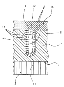
The covering layer 6 comprises a plurality of distributed openings 9 spaced
from one another and attached cavities 10 on its surface 8 facing away from
the support 2. The therapeutic agent 5, which will be dealt with later on in
more detail, is absorbed and optionally chemically fixed in the cavities 10,
as
will be explained later on in more detail with reference to Fig. Sa.
Here, the cavities 10 are formed essentially tube-like and each is closed on
its
ends. They extend from the surface 8 of the covering layer 6, essentially
perpendicularly to the support 2.
1 S In particular, the cavities 10 extend neither up to the surface 7 of
support 2
nor into support 2, but they each terminate blind in the covering layer 6 and
are separated from the surface 7 of the support 2 by a barrier layer 11 of the
covering layer 6. The whole surface 7 of the support 2 is thereby at least
extensively sealed off against body tissues and fluids. High chemical
stability
of the covering layer 6 in the body is of importance here.
Here, the cavities 10 are formed essentially as a circular cylinder. However,
they may also comprise a polygonal cross-section or an irregular cross-
sectional shape. Here, the cavities 10 extend essentially parallel to one
2 5 another and are separated from each other without having the cavities 10
linked to one another. However, this is not absolutely necessary; possibly,
links may also exist between the cavities 10 in the covering layer 6.
The covering layer 6 preferably comprises aluminium oxide which is
3 0 precipitated or formed electrolytically on the surface 7 of the support 2.
During electrolytic oxidization, the diameter of the openings 9 or of the
cavities 10 may be readily changed by appropriate adjustment of the applied
voltage. Here a diameter of about 1.2 to 1.4 nm is obtained per 1 V of anodic
voltage.
CA 02346857 2001-04-10
- 14-
Alternatively, the covering layer 6 or the non-oxidized covering layer
material, like aluminium, may be applied e.g. via a plasma deposition process
on the surface 7 of the support 2 and may possibly be subsequently
oxidized. This is, in particular, advantageous when only an external coating
is
desired; however, an extra inner coating is also possible in this way.
However, production of the covering layer 6 is not limited to the above
examples, for example an oxidization of an appropriate surface layer of the
support 2 may also be taken into consideration. Furthermore, the material for
the covering layer 6 is not limited to aluminium oxide, but e.g. magnesium
oxide and/or titanium oxide may also be applied. Moreover, in addition to
oxides, in particular, ceramic materials may also be applied for forming the
covering layer 6; the mechanical properties of the resulting covering layer 6
and preferably the structure of the cavities 10 for absorbing the therapeutic
agent 5 are essential.
The schematic, enlarged sectional diagram of a cavity 10 according to Fig. Sa
illustrates the possible fixing of the therapeutic agent 5 in the cavities 10
of
the covering layer 6. For example, the wall 12 of the cavity 10 is provided
2 0 with reaction partners such as complexing agents 13 which, for example,
are
bound by silanization in the cavities 10 or on their walls 12.
Instead of the exemplary complexing agents 13 in Fig. Sa, the walls 12 of the
cavities 10 may also be provided with other binding partners, if needed,
2 S causing a desired binding of the therapeutic agent S. Alternatively, at
least
one therapeutic agent 5 is preferably absorbed by the cavities 10 without it
being bound or fixed therein. In particular, in this case, if necessary, but
also
normally, a cover layer 14 is preferably provided on the surface 8 of the
covering layer 6 which also covers the cavities 10 and their openings 9,
3 0 respectively.
In particular, the cover layer 14 is further used for preventing a premature
escape or release of the therapeutic agent 5 from the cavities 10, e.g. before
implantation of the implant 1. However, the cover layer 14 may also be used
3 S for preventing a direct contact between body tissue and/or fluids and the
CA 02346857 2001-04-10
- 15 - ,.
therapeutic agent 5, especially when the therapeutic agent S is radioactive
material. As the total surface area of the openings 9 is preferably smaller,
in
particular substantially smaller than the surface area of the surface 8 of the
covering layer 6 in contact with the cover layer 14, the cover layer 14 may
adhere to the covering layer 6 very well, regardless of the therapeutic agent
5, depending on the selected material for the covering layer 6 and the cover
layer 14. Preferably, the walls 12 of the cavities 10 form an essentially
larger
internal surface as compared with the surface 8 of the covering layer 6, in
particular when fixing of the therapeutic agent in the cavities 10 is desired.
It is essential that the covering layer 6 and the optionally provided cover
layer 14 be dimensioned and formed so as to safely exclude any
segmentation, for example when widening the stent radially. In this respect,
here, the thickness of the covering layer 6 is preferably less than 1.5 pm,
preferably not more than 200 nm and in particular from 30 nm to 150 nm.
However, the thickness of the covering layer may also be e.g. up to 150 pm,
in particular for absorbing larger volumes in the cavities 10.
Fig. Sb shows an alternative embodiment with altered cavities 10 in a cut-out
sectional view corresponding to Fig. Sa. Here, cavities 10 are approximately
bottle-shaped in a section perpendicular to the main extension plane of the
covering layer 6 and/or respectively comprise a reduced portion 15 in the
area of the opening 9, a transition portion 16 adjacent to the portion 15 on
the side opposite the opening 9 with an increasing cross-section and finally
2 5 an adj acent end portion 17 with the largest cross-section or diameter. In
this
exemplary embodiment, the portion 15 reduced in cross-section or diameter
limits the release rate or rapidity with which the therapeutic agent S is
released from the cavities 10 in the implanted state with removed or
permeable cover layer 14. Depending on the dimensioning of the cavities 10
3 0 - by varying the voltage in an electrolytic anodization - it is thereby
possible
to achieve a desired release rate.
If necessary, the sequence of portions 15 - 17 of the cavities 10 illustrated
as
an example in Fig. Sb may also be reversed, so that portion 17 comprising the
3 S largest diameter or cross-section opens to the surface 8, so as to first
achieve
CA 02346857 2001-04-10
- 16-
a very strong or high release rate and then a reduced release rate. In each
case, a desired timely distribution and possibly also a spatial distribution
of
the liberated or released dose of therapeutic agent 5 may be set by the shape
or dimensioning of the cavities 10. The definite structure of the cavities 10
is
essential here.
For example, in Fig. Sb, it is indicated that a single therapeutic agent S is
absorbed by the cavities 10. If necessary, various therapeutic agents S, for
example stacked, may also be absorbed by the cavities 10 in order to achieve
successive release of the various therapeutic agents 5. Alternatively or
additionally, various therapeutic agents 5 may also be absorbed, for example,
in differently structured cavities 10 of the covering layer 6 and/or with
different binding partners, in order to be able to achieve a possibly
simultaneous release of various therapeutic agents 5 in a desired dose.
Fig. Sc, in an illustration corresponding to Figs. Sa and Sb, shows a further
exemplary embodiment of the implant 1 with altered cavities 10 once again
for explaining the different implementation possibilities. In this case, the
cavities 10 each comprise a first portion 18 opening to the surface 8 of the
covering layer 6 and several portions 19 adjacent to the portion 18 on the
end opposite the opening 9 significantly reduced in their diameter or cross-
section with respect to portion 18. Due to their reduced diameter or cross-
section, the root-like or projection-like portions 19 attached to portion 18
of
the cavity cause an e.g. slower release or liberation of an absorbed
2 S therapeutic agent 5 than portion 18 when compared with the release or
liberation from portion 18. If necessary, portions 18 and portions 19 of the
cavities 10 may also be provided or filled with different therapeutic agents
5,
wherein the length of portions 18 and 19, i.e., their perpendicular extent
with
respect to the main plane or surface 8 of the covering layer 6, may be adapted
3 0 individually and commonly to a desired release behavior.
In order to be able to obtain a sufficiently high dose, a certain amount of
the
therapeutic agents) 5 is required which is absorbed by the cavities 10.
Preferably, about 10g to 10' ' cavities per cm2 of the surface 8 of the
covering
3 S layer 6 are provided.
CA 02346857 2001-04-10
- 17 -
Figs. 6 and 7 show electron micrographs of a surface of an aluminium oxide
layer with different magnifications. It clearly shows how the light appearing,
tube-shaped cavities are homogeneously distributed and formed in the
S aluminium oxide layer.
According to a more preferred exemplary embodiment, radioactive material as
a therapeutic agent 5 is absorbed in the cavities 10 and in particular is
fixed
therein.
The schematic, enlarged sectional view of a cavity 10 according to Fig. Sa
illustrates the fixing of the radioactive material in the cavities 10 of the
covering layer 6. The wall 12 of the cavity 10 is provided with reaction
partners or complexing agents 13, preferably thiols or carboxylates which, for
example, are bound via silanization in the cavities 10 or on their walls 12,
and
bind or fix the radioactive material in the cavities 10 via mercapto groups,
for
example.
For example, the radioactive material contains radioactive technetium and/or
rhenium, wherein technetium(V)- and/or rhenium(V)-compounds are in
particular formed with sulphur-containing ligands which exhibit extremely
high stability in vivo. According to an another example, radioactive material
in the form of 86Y Soy a9sr ~s3Sm 64Cu 6'Cu and/or '°SRh is fixed in
the
> > ~ > >
cavities 10 via (poly)carboxylates, wherein the carboxylates in turn are
preferably bound via silanization in the cavities 10.
However, other radionuclides may also be fixed, for example also anions, like
iodine, as a radioactive material in the cavities 10 and in particular may be
bound chemically by means of appropriate reaction partners such as noble
3 0 metals, especially silver. As a further example, the binding of, in
particular,
liquid-introduced radioactive material 5 in the form of '2°I, '23h 'Zah
'2sI, ~3'I
and/or 2' 'At and its binding via silver in the cavities 10 should be
mentioned,
wherein the silver in turn is bound, for example, via (poly)carboxylates which
are in turn preferably bound via silanization in the cavities 10.
CA 02346857 2001-04-10
-18-
Preferably, the radioactive material contains various radionuclides in a
desired
ratio, so that an optimal dose is achieved with respect to space and/or time
considerations because of the different properties of the various
radionuclides. This is possible in a relatively simple way by the proposed
S introduction of radioactive material into the cavities 10 as, e.g., various
radio-
isotopes and/or various radionuclides with different half life values,
energies
and/or types of radiation (a, (3+, (3-, 'y) may be mixed or blended in the
cavities
and may be fixed therein, e.g. via appropriately selected complexing
agents 13.
Alternatively, various radionuclides may also be introduced in succession,
i.e.
for example stacked, in the cavities 10 and may be fixed therein by means of
appropriate or e.g. selective complexing agents 13.
Alternatively or additionally, it is possible not to completely fill up the
cavities 10 with radioactive material, but to add, for example, extra filler
material for stabilization and/or closure of the openings 9 when only
partially
filled with radioactive material S.
The further possible different filling of the cavities 10 with radioactive
material for changing the dose distribution has been already mentioned.
In particular, the diameter of the cavities 10 and/or of the openings 9 is
selected so that the blood components or the molecules normally present in
2 5 blood 4, which are relatively large, may not penetrate into the cavities
10
through the openings 9 because of their size. This can be ensured by a
diameter of the openings 9 from about S to a maximum of 100 nm.
In order to be able to achieve a sufficiently high dose, a certain amount of
3 0 radioactive material is required which is absorbed by the cavities 10.
Preferably about 108 to 10" cavities 10 per cm2 of the surface 8 of the
covering layer 6 are provided.
Finally it should be pointed out that the proposed localization of radioactive
3 5 material in the cavities 10 of a proposed covering layer 6 is not limited
to
CA 02346857 2001-04-10
- 19-
implants, but it may also be applied in other components or radioactive
emitters with desired radioactive properties.