Note: Descriptions are shown in the official language in which they were submitted.
CA 02346879 2001-04-10
WO 00/23052 PCTNS99/24228
Liposome-Entrapped To~oisomerase Inhibitors
Field of the Invention
The present invention relates to a liposome composition having an entrapped
topoisomerase inhibitor.
Background of the Invention
Next to heart disease, cancer is the major cause of death in the United
States,
causing over 500,000 fatalities annually (Katzung, B., "Basic and Clinical
to Pharmacology", 7'" Edition, Appleton & Lange, Stamford CT, 1998, p. 882).
With
present methods of treatment, one-third of patients are cured with local
measures, such as
surgery or radiation therapy, which are quite effective when the tumor has not
metastasized by the time of treatment. Earlier diagnosis might lead to
increased cure of
patients undergoing such local treatments. However, in many cases, early
micrometastasis is a characteristic feature of the neoplasm, indicating that a
systemic
approach such as chemotherapy may be required, often along with a local
treatment
method, for effective cancer management.
Cancer chemotherapy can be curative in certain disseminated neoplasms that
have
undergone either gross or microscopic spread by the time of diagnosis. These
include
2o testicular cancer, diffuse large cell lymphoma, Hodgkin's disease and
choriocarcinoma as
well as childhood tumors such as acute lymphoblastic leukemia. For other forms
of
disseminated cancer, chemotherapy provides a palliative rather than curative
therapy.
Effective palliative therapy results in temporary clearing of the symptoms and
signs of
cancer and prolongation of useful life. Advances in cancer chemotherapy have
recently
provided evidence that chemical control of neoplasia is possible for a number
of cancers.
One category of drugs used for cancer therapy is topoisomerase inhibitors.
These
compounds inhibit the action of topoisomerase enzymes which play a role in the
replication, repair, genetic recombination and transcription of DNA. An
example of a
topoisomerase inhibitor is camptothecin, a natural compound that interferes
with the
3o activity of topoisomerase I, an enzyme involved in DNA replication and RNA
transcription. Camptothecin and the camptothecin analogues topotecan and
irinotecan are
approved for clinical use.
CA 02346879 2001-04-10
WO 00/23052 PCT/US99/24228
Camptothecin and its analogues are effective in cancer chemotherapy by
interfering
with the breakage/reunion actions of topoisomerase I. The compounds stabilize
and form
a reversible enzyme-camptothecin-DNA ternary complex which prevents the
reunion step
of the breakage/union cycle of the topoisomerase reaction.
One problem with camptothecin is its water insolubility, which hinders the
delivery
of the drug. Numerous analogues of camptothecin have been prepared to improve
the
compound's water solubility. Another problem with camptothecin and its
analogues is
that the compounds are susceptible in aqueous environments to hydrolysis at
the a-
hydroxy lactone ring. The lactone ring opens to the carboxylate form of the
drug, a form
t o that exhibits little activity against topoisomerase I.
Various approaches to improving the stability of camptothecin and its
analogues
have been described. One approach has been to entrap the compounds in
liposomes.
Burke (U.S. Patent No. 5,552,156) describes a liposome composition intended to
overcome the instability of camptothecin and its analogues by entrapping the
compounds
~ 5 in liposomes having a lipid bilayer membrane which allows the compound to
penetrate,
or intercalate, into the lipid bilayer. With the compound intercalated into
the bilayer
membrane, it is removed from the aqueous environment in the core of the
liposome and
thereby protected from hydrolysis.
One problem with this approach is that the liposomes are quickly removed from
the
2o bloodstream by the reticuloendothelial system (RES), preventing delivery,
and preferably
accumulation, at the tumor site.
Subramanian and Muller (Oncology Research, 7(9):461-469 (1995)) describe a
liposome formulation of topotecan and report that in liposome-entrapped form,
topotecan
is stabilized from inactivation by hydrolysis of the lactone ring. However,
the biological
25 activity of the liposome-entrapped drug in vitro has only 60 % of the
activity of the free
drug.
Lundberg (Anti-Cancer Drug Design, 13:453 (1998)) describes two lipophilic,
oleic
acid ester derivatives of camptothecin analogues which are entrapped in
liposomes and
intercalated into the bilayer for stabilization of the lactone ring. Daoud
(Anti-Cancer
3o Drugs, 6:83-93 (1995)) describes a liposome composition including
camptothecin, where
the drug is also intercalated into the lipid bilayer. The liposomes in both of
these
references are prepared conventionally, where the drug is passively entrapped
in the
liposomes to sequester the drug in the lipid bilayer membrane for
stabilization. Using
2
CA 02346879 2001-04-10
WO 00/23052 PCTNS99/24228
this method of preparation it is difficult to achieve a sufficient drug load
in the liposomes
for clinical efficacy.
Accordingly, there is still a need in the art for a liposome formulation which
(i)
includes a topoisomerase inhibitor, such as camptothecin and its analogues;
(ii) remains
in the bloodstream for a prolonged period of time; (iii) retains antitumor
activity; and (iv)
includes a sufficient drug load for clinical relevance.
Summary of the Invention
Accordingly, it is an object of the invention to provide a topoisomerase
inhibitor
composition for improved cancer therapy.
It is another object of the invention to provide a liposome composition for
administration of a topoisomerase inhibitor for antitumor therapy.
In one aspect, the invention includes a composition for treating a tumor in a
subject,
comprising liposomes composed of a vesicle-forming lipid and between about 1-
20 mole
t s percent of a vesicle-forming lipid derivatized with a hydrophilic polymer.
The liposomes
are formed under conditions that distribute the polymer on both sides of the
liposomes'
bilayer membranes. Entrapped in the liposomes is a topoisomerase I inhibitor
or a
topoisomerase I/II inhibitor at a concentration of at least about 0.10 pmole
drug per
pmole lipid. The liposomes have an inside/outside ion gradient sufficient to
retain the
2o topoisomerase I inhibitor or topoisomerase I/II inhibitor within the
liposomes at the
specified concentration.
In one embodiment, the topoisomerase inhibitor is a topoisomerase I inhibitor
selected from the group consisting of camptothecin and camptothecin
derivatives. For
example, the camptothecin derivative can be 9-aminocamptothecin, 7-
ethylcamptothecin,
2s 10-hydroxycamptothecin, 9-nitrocamptothecin, 10,11-
methlyenedioxycamptothecin, 9-
amino-10,11-methylenedioxycamptothecin or 9-chloro-10,11-
methylenedioxycamptothecin.
In other embodiments, the camptothecin derivative is irinotecan, topotecan, (7-
(4-
methylpiperazinomethylene)-10,11-ethylenedioxy-20(S)-camptothecin, 7-(4-
methylpiperazinomethylene)-10,11-methylenedioxy-20(S)-camptothecin or 7-(2-(N-
3o isopropylamino)ethyl)-(20S)-camptothecin.
In another embodiment, the topoisomerase inhibitor is a topoisomerase I/II
inhibitor, such as 6-[[2-{dimethylamino)-ethyl]amino]-3-hydroxy-7H-indeno[2,1-
3
CA 02346879 2001-04-10
WO 00/23052 PCTNS99/24228
c]quinolin-7-one dihydrochloride, azotoxin or 3-methoxy-11H-pyrido[3',4'-
4,SJpyrrolo(3,2-cJquinoline-1,4-dione.
The hydrophilic polymer included in the liposome composition can be
polyvinylpyrrolidone, polyvinylmethylether, polymethyloxazoline,
polyethyloxazoline,
polyhydroxypropyloxazoline, polyhydroxypropylmethacrylamide,
polymethacrylamide,
polydimethylacrylamide, polyhydroxypropyimethacrylate,
polyhydroxyethylacryiate,
hydroxymethylcellulose, hydroxyethylcellulose, polyethyleneglycol and
polyaspartamide.
In a preferred embodiment, the hydrophilic polymer is polyethyleneglycol
having a
molecular weight between 500-5,000 daltons.
In still another embodiment, the liposomes further include a vesicle-forming
lipid
having a phase transition temperature above 37°C.
In yet another embodiment, the vesicle-forming lipid is hydrogenated soy
phosphatidylcholine, distearoyl phosphatidylcholine or sphingomyelin. One
preferred
liposome composition is composed of 20-94 mole percent hydrogenated soy
is phosphatidylcholine, 1-20 mole percent distearoyl phosphatidylcholine
derivatized with
polyethyleneglycol and 5-60 mole percent cholesterol.
Another preferred composition is 30-65 mole percent hydrogenated soy
phosphatidylcholine, 5-20 mole percent distearoyl phosphatidylcholine
derivatized with
polyethyleneglycol and 30-50 mole percent cholesterol.
2o In another aspect, the invention includes a composition for administration
of a
topoisomerase I inhibitor or a topoisomerase I/II inhibitor, comprising
liposomes composed
of vesicle-forming lipids and having an inside/outside ion gradient effective
to retain the
drug within the liposomes. Entrapped in the liposomes is the topoisomerase I
inhibitor or
the topoisomerase I/II inhibitor at a concentration of at least about 0.20
mole drug per
2s p,mole lipid.
In another aspect, the invention includes a method of treating a tumor in a
subject,
comprising preparing liposomes composed of vesicle-forming lipids including
between 1-20
mole percent of a vesicle-forming lipid derivatized with a hydrophilic polymer
chain, the
liposomes being formed under conditions that distribute the polymer on both
sides of the
30 liposomes' bilayer membrane. The liposomes contain a topoisomerase I
inhibitor or a
topoisomerase I/II inhibitor entrapped in the liposomes at a concentration of
at least about
0.10 mole per ,mole lipid, the liposomes having an inside/outside ion gradient
sufficient to
4
CA 02346879 2001-04-10
WO 00/23052 PGT/US99124228
retain the topoisomerase I inhibitor or topoisomerase I/II inhibitor within
the liposome at
the specified concentration. The liposomes are then administered to the
subject.
In one embodiment of this aspect, the method further includes entrapping the
topoisomerase I inhibitor or topoisomerase I/II inhibitor in the liposomes by
remote
loading, for example, via an ammonium sulfate gradient.
These and other objects and features of the invention will be more fully
appreciated
when the following detailed description of the invention is read in
conjunction with the
accompanying drawings.
t o Brief Description of the Drawings
Figs. lA is a plot of the blood circulation lifetime of liposome-entrapped MPE-
camptothecin (solid circles), taken as the percent of injected dose as a
function of time,
compared to the free form of the drug (solid squares);
Fig. 1B shows the blood concentration of MPE-camptothecin, as a function of
time,
~5 in hours, after administration of liposome-entrapped MPE-camptothecin
(solid circles)
and of free (non-liposomal) MPE-camptothecin (solid squares) to rats;
Fig. 2A is a plot showing the body weight of mice, in grams, as a function of
days
after tumor inoculation with an HT29 colon tumor. The animals were treated on
days
10, 16 and 23 after tumor inoculation with liposome-entrapped MPE-camptothecin
at
2o dosages of 24 mg/kg (closed circles), 15 mg/kg (closed triangles) and 6
mg/kg (closed
squares) and with free MPE-camptothecin at doses of 24 mg/kg (open circles),
15 mg/kg
(open triangles) and 6 mg/kg (open squares);
Fig. 2B is a plot showing tumor volume, in mm3, as a function of days after
inoculation with an HT29 colon tumor. The animals were treated on days 10, 16
and 23
25 after tumor inoculation with liposome-entrapped MPE-camptothecin at dosages
of 24
mg/kg (closed circles), 15 mg/kg (closed triangles) and 6 mg/kg (closed
squares) and
with free drug at doses of 24 mg/kg (open circles), 15 mg/kg (open triangles)
and 6
mg/kg (open squares);
Fig. 3A is a plot showing the body weight of mice, in grams, as a function of
days
3o after inoculation with an HT29 colon tumor. The animals were treated on
days 9, 16 and
23 after tumor inoculation with liposome-entrapped MPE-camptothecin at dosages
of 5
mg/kg (open triangles), 3 mg/kg (open inverted triangles), 1 mg/kg (open
diamonds), 0.5
mg/kg (open circles) and 0.1 mg/kg (open squares) and with free MPE-
camptothecin at a
S
CA 02346879 2001-04-10
WO 00/23052 PCT/US99/24228
dose of 20 mg/kg (closed squares);
Fig. 3B is a plot showing tumor volume, in mm3, as a function of days after
inoculation with an HT29 colon tumor. The animals were treated on days 9, 16
and 23
s after tumor inoculation with liposome-entrapped MPE-camptothecin at dosages
of 5
mg/kg (open triangles), 3 mg/kg (open inverted triangles), 1 mg/kg (open
diamonds), 0.5
mg/kg (open circles) and 0.1 mg/kg (open squares) and with free MPE-
camptothecin at a
dose of 20 mg/kg (closed squares);
Figs. 4A-4B are plots showing the plasma concentration of topotecan as a
function
of time, in hours, after administration of liposome-entrapped topotecan (solid
triangles)
and of free (non-liposomal) topotecan (solid squares) to rats at dosages of 2
mg/kg (Fig.
4A) and 5 mg/kg (Fig. 4B);
Fig. SA is a plot showing the body weight of mice, in grams, as a function of
days
after inoculation with an HT29 colon tumor. The animals were treated on days
9, 16 and
23 after tumor inoculation with liposome-entrapped topotecan at dosages of 2
mg/kg
(diamonds), 5 mg/kg (circles), 8 mg/kg (open squares); liposome-entrapped MPE-
camptothecin at 4 mg/kg (triangles); free topotecan at a dose of 25 mg/kg
(inverted
triangles) and saline (closed squares);
Fig. SB is a plot showing tumor volume, in mm3, as a function of days after
2o inoculation with an HT29 colon tumor. The animals were treated on days 9,
16 and 23
after tumor inoculation with liposome-entrapped topotecan at dosages of 2
mg/kg
(diamonds), 5 mg/kg (circles), 8 mg/kg (open squares); liposome-entrapped MPE-
camptothecin at 4 mg/kg (triangles); free topotecan at a dose of 25 mg/kg
(inverted
triangles) and saline (closed squares);
Fig. 6 is a plot of plasma concentration of CKD602 as a function of time, in
hours,
after administration of liposome-entrapped CKD602 (solid circles) and of free
(non-
liposomal) topotecan (solid squares) to rats at a dosage of 1 mg/kg;
Fig. 7A is a plot showing the body weight of mice, in grams, as a function of
days
after inoculation with an HT29 colon tumor. The animals were treated on days
9, 16 and
23 after tumor inoculation with liposome-entrapped CKD602 at dosages of 4
mg/kg
(diamonds), 2 mg/kg (circles), 1 mg/kg (open squares); liposome-entrapped MPE-
camptothecin at 4 mg/kg (triangles); free CKD602 at a dose of 20 mg/kg
(inverted
triangles) and saline (closed squares); and
6
CA 02346879 2001-04-10
WO 00123052 PCT/US99/24228
Fig. 7B is a plot showing tumor volume, in mm3, as a function of days after
inoculation with an HT29 colon tumor. The animals were treated on days 9, 16
and 23
after tumor inoculation with liposome-entrapped CKD602 at dosages of 4 mg/kg
(diamonds), 2 mg/kg (circles), 1 mg/kg (open squares); liposome-entrapped MPE-
camptothecin at 4 mg/kg (triangles); free CKD602 at a dose of 20 mg/kg
(inverted
triangles) and saline (closed squares).
Detailed Description of the Invendan
I. Definitions
0 Unless otherwise indicated, the terms below have the following meaning:
"Effective amount" or "effective dose" refers to the amount necessary or
sufficient to
inhibit undesirable cell growth, e. g. , prevent undesirable cell growth or
reduce existing cell
growth, such as tumor cell growth. The effective amount can vary depending on
factors
known to those of skill in the art, such as the type of cell growth, the mode
and regimen of
~ 5 administration, the size of the subject, the severity of the cell growth,
etc. One of skill in
the art would be able to consider such factors and make the determination
regarding the
effective amount.
"Therapeutically effective antitumor therapy" refers to a therapy which is
effective
to maintain or decrease the size, e. g. , volume, of a primary tumor or
metastatic tumor.
20 "Topoisomerase I inhibitor" refers to any compound that inhibits or reduces
the
action of topoisomerase I enzyme.
"Topoisomerase I/II inhibitor" refers to any compound that inhibits or reduces
the
action of both topoisomerase I enzyme and topoisomerase II enzyme.
"Topoisomerase inhibitor" refers to a topoisomerase I inhibitor or a
topoisomerase
2s I/11 inhibitor.
"MPE-camptothecin" refers to 7-(4-methyl-piperazino-methylene)-10,11-
ethylenedioxy-20(S)-camptothecin.
"Topotecan" refers to 9-dimethyl-aminomethyl-10-hydroxycamptothecin.
"CKD-602" refers to 7-(2-(N-isopropylamino)ethyl)-(20S)-camptothecin.
II. Liposome Composition
The present invention is directed to a liposome composition for administration
of a
topoisomerase I inhibitor or a topoisomerase I/II inhibitor. In studies
performed in
7
CA 02346879 2001-04-10
WO 00/23052 PCT/US99/24228
support of the invention, three topoisomerase inhibitors were entrapped in
liposomes and
characterized in vivo: topotecan, 7-(4-methyl-piperazino-methylene)-10,11-
ethyIenedioxy-20(S)-camptothecin (referred to hereing as "MPE-camptothecin")
and 7-(2-
(N-isopropylamino)ethyl)-{20S)-camptothecin (referred to herein as "CKD-602").
The
drugs were entrapped in liposomes by remote loading to achieve a high drug
load stably
retained in the liposomes, as will be described. In vivo studies with the
formulations
demonstrated that the liposome composition achieves a surprising and
unexpected degree
of improvement in therapeutic activity when compared to therapy with the
topoisomerase
inhibitor in free form. More specifically, and as will be described below, the
dose of the
to liposome-entrapped topoisomerase I inhibitor MPE-camptothecin required to
achieve
therapeutic antitumor therapy is about 20 times lower than the dose required
when the
drug is administered in free form.
In this section, the liposome composition will be described, including methods
for
preparing the liposomes.
A. Liposome Components
Liposomes suitable for use in the composition of the present invention include
those
composed primarily of vesicle-forming lipids. Vesicle-forming lipids can form
spontaneously into bilayer vesicles in water, as exemplified by the
phospholipids. The
liposomes can also include other lipids incorporated into the lipid bilayers,
with the
hydrophobic moiety in contact with the interior, hydrophobic region of the
bilayer
membrane, and the head group moiety oriented toward the exterior, polar
surface of the
bilayer membrane.
The vesicle-forming lipids are preferably ones having two hydrocarbon chains,
typically acyl chains, and a head group, either polar or nonpolar. There are a
variety of
synthetic vesicle-forming lipids and naturally-occurring vesicle-forming
lipids, including
the phospholipids, such as phosphatidylcholine, phosphatidylethanolamine,
phosphatidic
acid, phosphatidylinositol, and sphingomyelin, where the two hydrocarbon
chains are
typically between about 14-22 carbon atoms in length, and have varying degrees
of
3o unsaturation. The above-described lipids and phospholipids whose acyl
chains have
varying degrees of saturation can be obtained commercially or prepared
according to
published methods. Other suitable lipids include glycolipids and sterols such
as
cholesterol.
CA 02346879 2001-04-10
WO 00/23052 PCT/US99/24228
Cationic lipids are also suitable for use in the liposomes of the invention,
where the
cationic lipid can be included as a minor component of the lipid composition
or as a major
or sole component. Such cationic lipids typically have a lipophilic moiety,
such as a
sterol, an acyl or diacyl chain, and where the lipid has an overall net
positive charge.
s Preferably, the head group of the lipid carries the positive charge.
Exemplary cationic
lipids include 1,2-dioleyloxy-3-(trimethylamino) propane (DOTAP); N-[1-(2,3,-
ditetradecyloxy)propyl]-N,N-dimethyl-N-hydroxyethylammonium bromide (DMRIE); N-
[1-(2,3,-dioleyloxy)propyl]-N,N-dimethyl-N-hydroxy ethylammonium bromide
(DORIE);
N-[1-(2,3-dioleyloxy) propyl]-N,N,N-trimethylammonium chloride (DOTMA); 3 [N-
to (N',N'-dimethylaminoethane) carbamoly] cholesterol (DC-Chol); and
dimethyldioctadecylammonium (DDAB).
The cationic vesicle-forming lipid may also be a neutral lipid, such as
dioleoylphosphatidyl ethanolamine (DOPE) or an amphipathic lipid, such as a
phospholipid, derivatized with a cationic lipid, such as polylysine or other
polyamine
~5 lipids. For example, the neutral lipid (DOPE) can be derivatized with
polylysine to form a
cationic lipid.
In another embodiment, the vesicle-forming lipid is selected to achieve a
specified
degree of fluidity or rigidity, to control the stability of the liposome in
serum and to control
the rate of release of the entrapped agent in the liposome.
2o Liposomes having a more rigid lipid bilayer, or a liquid crystalline
bilayer, are
achieved by incorporation of a relatively rigid lipid, e.g., a lipid having a
relatively high
phase transition temperature, e. g. , above room temperature, more preferably
above body
temperature and up to 80°C. Rigid, i.e., saturated, lipids contribute
to greater membrane
rigidity in the lipid bilayer. Other lipid components, such as cholesterol,
are also laiown to
25 contribute to membrane rigidity in lipid bilayer structures.
On the other hand, lipid fluidity is achieved by incorporation of a relatively
fluid
lipid, typically one having a lipid phase with a relatively low liquid to
liquid-crystalline
phase transition temperature, e. g. , at or below room temperature, more
preferably, at or
below body temperature.
3o Vesicle-forming lipids having a main phase transition temperatures from
approximately 2°C-80°C are suitable for use as the primary
liposome component of the
present composition. In a preferred embodiment of the invention, a vesicle-
forming lipid
having a main phase transition temperature above about 37°C is used as
the primary lipid
9
CA 02346879 2001-04-10
WO 00/23052 PCT/US99/24228
component of the liposomes. In another preferred embodiment, a lipid having a
phase
transition temperature between about 37-70°C is used. By way of
example, the lipid
distearoyl phosphatidylcholine (DSPC) has a main phase transition temperature
of 55.1 °C
and the lipid hydrogenated soy phosphatidylcholine (HSPC) has a phase
transition
temperature of 58°C. Phase transition temperatures of many lipids are
tabulated in a
variety of sources, such as Avanti Polar Lipids catalogue and Lipid
Thermotropic Phase
Transition Database (LIPIDAT, NIST Standard Reference Database 34).
The liposomes also include a vesicle-forming lipid derivatized with a
hydrophilic
polymer. As has been described, for example in U.S. Patent No. 5,013,556 and
in WO
t o 98/07409, which are hereby incorporated by reference, such a hydrophilic
polymer
provides a surface coating of hydrophilic polymer chains on both the inner and
outer
surfaces of the liposome lipid bilayer membranes. The outermost surface
coating of
hydrophilic polymer chains is effective to provide a liposome with a long
blood
circulation lifetime in vivo. The inner coating of hydrophilic polymer chains
extends into
1s the aqueous compartments in the liposomes, i.e., between the lipid bilayers
and into the
central core compartment, and is in contact with the entrapped compound after
the
compound is loaded via remote loading. As will be illustrated below, the
liposome
formulation having a surface coating of hydrophilic polymer chains distributed
on the
inner and outer liposome surfaces provides for a topoisomerase I inhibitor or
2o topoisomerase I/II inhibitor composition where the compound is retained in
the liposomes
for improved therapeutic activity.
Vesicle-forming lipids suitable for derivatization with a hydrophilic polymer
include any of those lipids listed above, and, in particular phospholipids,
such as
distearoyl phosphatidylethanolamine (DSPE).
2s Hydrophilic polymers suitable for derivatization with a vesicle-forming
lipid
include polyvinyipyrrolidone, polyvinyimethylether, polymethyloxazoline,
polyethyloxazoline, polyhydroxypropyloxazoline,
polyhydroxypropylmethacrylamide,
polymethacrylamide, polydimethylacrylamide, polyhydroxypropylmethacrylate,
polyhydroxyethylacrylate, hydroxymethylcellulose, hydroxyethylcellulose,
3o polyethyleneglycol, and polyaspartamide. The polymers may be employed as
homopoiymers or as block or random copolymers.
A preferred hydrophilic polymer chain is polyethyleneglycol (PEG), preferably
as a
PEG chain having a molecular weight between 500-10,000 daltons, more
preferably
CA 02346879 2001-04-10
WO 00/23052 PCT/US99/24228
between 500-5,000 daltons, most preferably between 1,000-2,000 daltons.
Methoxy or
ethoxy-capped analogues of PEG are also preferred hydrophilic polymers,
commercially
available in a variety of polymer sizes, e. g. , 120-20,000 daltons.
Preparation of vesicle-forming lipids derivatized with hydrophilic polymers
has
s been described, for example in U.S. Patent No. 5,395,619. Preparation of
liposomes
including such derivatized lipids has also been described, where typically,
between 1-20
mole percent of such a derivatized lipid is included in the liposome
formulation. It will
be appreciated that the hydrophilic polymer may be stably coupled to the
lipid, or
coupled through an unstable linkage which allows the coated liposomes to shed
the
1o coating of polymer chains as they circulate in the bloodstream or in
response to a
stimulus.
B. Topoisomerase Inhibitor
The liposomes of the invention include a topoisomerase inhibitor entrapped in
the
15 liposome. Entrapped is intended to include encapsulation of an agent in the
aqueous core
and aqueous spaces of liposomes. It will be appreciated that for compounds
having some
hydrophobicity, entrapment in the lipid bilayer(s) of the liposomes may also
occur.
Topoisomerases catalyze the introduction and relaxation of superhelicity in
DNA.
Several types of enzymes with varying specifities are known to be important in
the
2o replication of DNA, as well as in the repair, genetic recombination and
transcription of
DNA. The simplest topoisomerases, designated topoisomerase I, relax
superhelical
DNA, a process that is energetically spontaneous. The gyrases, which are known
as
topoisomerase II, catalyze the energy-requiring and ATP-dependent introduction
of
negative superhelical twists into DNA. In DNA replication, topoisomerases I
and II have
25 the function of relaxing the positive superhelicity that is introduced
ahead of the
replicating forks by the action of helicases. In addition, gyrases introduce
negative twists
into segments of DNA that allow single-strand regions to appear.
Topoisomerase inhibitors, then, are compounds that inhibit topoiosmerase
activity.
Compounds known as topoisomerase I inhibitors have activity against
topoisomerase I,
3o and the topoiosmerase II inhibitors have activity against topoisomerase II.
Some
compounds have activity against both topoisomerase I and topoisomerase II and
are
known as topoisomerase I/II inhibitors.
Preferred topoisomerase I inhibitors for use in the present invention are
camptothecin
11
CA 02346879 2001-04-10
WO 00/23052 PCT/US99/24228
and analogs of camptothecin. Camptothecin is a pentacyclic alkaloid initially
isolated from
the wood and bark of Camptotheca acuminata, a tree indigenous to China (Wall,
M.E. et
al., J. Am. Chem. Soc., 94:388 (1966)). Camptothecin exerts its
pharmacological effects
by irreversibly inhibiting topoisomerase I. Methods for the synthesis of
camptothecin and
camptothecin analogs or derivatives are known, and are summarized and set
forth in U.S.
Patent No. 5,244,903, which is herein incorporated by reference in its
entirety.
Analogues of camptothecin include SN-38 ((+)-(4S)-4,11-diethyl-4,9-dihydroxy-
1H-
pyrano[3',4':6,7]-indolizino[1,2-b]quinoline-3,14(4H,12H)-dione); 9-
aminocamptothecin;
topotecan (hycamtin; 9-dimethyl-aminomethyl-10-hydroxycamptothecin);
irinotecan (CPT-
to 11; 7-ethyl-10-[4-(1-piperidino)-1-piperidino]-carbonyloxy-camptothecin),
which is
hydrolyzed in vivo to SN-38); 7-ethylcamptothecin and its derivatives (Sawada,
S. et al.,
Chem. Pharm. Bull., 41(2):310-313 (1993)); 7-chloromethyl-10,11-methylene-
dioxy-
camptothecin; and others (SN-22, Kunimoto, T. et al., J. Pharmacobiodyn.,
10(3):148-151
(1987); N-formylamino-12,13,dihydro-1,11-dihydroxy-13-(beta-D-glucopyranosyl)-
SH-
indolo[2,3-a]pyrrolo[3,4-c]carbazole-5,7(6H)-dione (NB-506, Kanzawa, F et al.,
Cancer
Res., 55(13):2806-2813 (1995); DX-8951f and lurtotecan (GG-211 or 7-(4-
methylpiperazino-methylene)-10,11-ethylenedioxy-20(S)-camptothecin)
(Rothenberg,
M.L., Ann. Oncol., 8(9):837-855 (1997)) and 7-(2-(N-isopropylamino)ethyl)-
(20S)-
camptothecin (CKD602, Chong Kun Dang Corporation, Seoul Korea).
2o Topoisomerase inhibitors having activity against both topoisomerase I and
topoisomerase II include 6-[[2-(dimethylamino)-ethyl]amino]-3-hydroxy-7H-
indeno[2,1-
c]quinolin-7-one dihydrochloride, (TAS-103, Utsugi, T., et al., Jpn. J. Cancer
Res.,
88(10):992-1002 (1997)) and 3-methoxy-11H-pyrido[3',4'-4,5]pyrrolo[3,2-
c]quinoline-1,4-
dione (AzaIQD, Riou, J.F., et al., Mol. Pharmacol., 40(5):699-706 (1991)).
In one embodiment of the invention, the topoisomerase I inhibitor administered
is the
pharmacologically active enantiomer of a camptothecin analogue having a chiral
center.
The enantiomer can be resolved from the racemic mixture using techniques known
to those
of skill in the art.
3o C. Method of Preparing ~e Liposome Composition
The liposomes may be prepared by a variety of techniques, such as those
detailed in
Szoka, F., Jr., et al., Ann. Rev. Biophys. Bioeng. 9:467 (1980), and specific
examples of
liposomes prepared in support of the present invention will be described
below. Typically,
12
CA 02346879 2001-04-10
WO 00/23052 PCT/US99/24228
the liposomes are multilamellar vesicles (MLVs), which can be formed by simple
lipid-film
hydration techniques. In this procedure, a mixture of liposome-forming lipids
and
including a vesicle-forming lipid derivatized with a hydrophilic polymer are
dissolved in a
suitable organic solvent which is evaporated in a vessel to form a dried thin
film. The film
is then covered by an aqueous medium to form MLVs, typically with sizes
between about
0.1 to 10 microns. Exemplary methods of preparing derivatized lipids and of
forming
polymer-coated liposomes have been described in co-owned U.S. Patents Nos.
5,013,556, 5,631,018 and 5,395,619, which are incorporated herein by
reference.
The therapeutic agent of choice can be incorporated into liposomes by standard
~ o methods, including (i) passive entrapment of a water-soluble compound by
hydrating a lipid
film with an aqueous solution of the agent, (ii) passive entrapment of a
lipophilic compound
by hydrating a lipid film containing the agent, and (iii) loading an ionizable
drug against an
inside/outside liposome ion gradient, termed remote loading. Other methods,
such as
reverse evaporation phase liposome preparation, are also suitable.
~ 5 In the present invention, a preferred method of preparing the liposomes is
by remote
loading. In the studies performed in support of the invention, three exemplary
topoisomerase I inhibitors were loaded into pre-formed liposomes by remote
loading
against an ion concentration gradient, as has been described in the art (U.S.
Patent No.
5,192,549) and as described in Example 1. In a remote loading procedure, a
drug is
2o accumulated in the liposomes' central compartment at concentration levels
much greater
than can be achieved with other loading methods. In a preferred embodiment of
the
invention, the topoisomerase I inhibitor or topoisomerase I/II inhibitor is
loaded into the
liposomes to a concentration of at least about 0.10 ~cmole drug per ,mole
lipid, more
preferably of at least about 0.15 ~cmole drug per .mole lipid, most preferably
of at least
25 about 0.20 .mole drug per ,mole lipid. The liposomes prepared in support of
the
invention contained MPE-camptothecin, topotecan or CKD602. As set forth in
Example
1, these compounds were loaded into the liposomes by remote loading, discussed
below, to
a drug concentration level of greater than 0.20 ,mole drug per ,mole lipid
(see the table in
Example 1).
3o Liposomes having an ion gradient across the liposome bilayer for use in
remote
loading can be prepared by a variety of techniques. A typical procedure is as
described
above, where a mixture of liposome-forming lipids is dissolved in a suitable
organic solvent
and evaporated in a vessel to form a thin film. The film is then covered with
an aqueous
13
CA 02346879 2001-04-10
WO 00/23052 PCT/US99/24228
medium containing the solute species that will form the aqueous phase in the
liposome
interior spaces.
After liposome formation, the vesicles may be sized to achieve a size
distribution of
liposomes within a selected range, according to known methods. The liposomes
are
s preferably uniformly sized to a selected size range between 0.04 to 0.25
~,m. Small
unilamellar vesicles (SiJVs), typically in the 0.04 to 0.08 ~cm range, can be
prepared by
extensive sonication or homogenization of the liposomes. Homogeneously sized
liposomes
having sizes in a selected range between about 0.08 to 0.4 microns can be
produced, e, g. ,
by extrusion through polycarbonate membranes or other defined pore size
membranes
having selected uniform pore sizes ranging from 0.03 to 0.5 microns,
typically, 0.05, 0.08,
0.1, or 0.2 microns. The pore size of the membrane corresponds roughly to the
largest
size of liposomes produced by extrusion through that membrane, particularly
where the
preparation is extruded two or more times through the same membrane. The
sizing is
preferably carned out in the original lipid-hydrating buffer, so that the
liposome interior
15 spaces retain this medium throughout the initial liposome processing steps.
After sizing, the external medium of the liposomes is treated to produce an
ion
gradient across the liposome membrane, which is typically a lower
inside/higher outside
concentration gradient. This may be done in a variety of ways, e.g., by {i)
diluting the
external medium, (ii) dialysis against the desired final medium, (iii)
molecular-sieve
2o chromatography, e.g., using Sephadex G-50, against the desired medium, or
(iv) high-
speed centrifugation and resuspension of pelleted liposomes in the desired
final medium.
The external medium which is selected will depend on the mechanism of gradient
formation
and the external pH desired, as will now be considered.
In the simplest approach for generating an ion gradient, the hydrated, sized
liposomes
25 have a selected internal-medium pH. The suspension of the liposomes is
titrated until a
desired final pH is reached, or treated as above to exchange the external
phase buffer with
one having the desired external pH. For example, the original medium may have
a pH of
5.5, in a selected buffer, e. g. , glutamate or phosphate buffer, and the
final external medium
may have a pH of 8.5 in the same or different buffer. The internal and
external media are
3o preferably selected to contain about the same osmolarity, e. g. , by
suitable adjustment of the
concentration of buffer, salt, or low molecular weight solute, such as
sucrose.
In another general approach, the gradient is produced by including in the
liposomes,
a selected ionophore. To illustrate, liposomes prepared to contain valinomycin
in the
14
CA 02346879 2001-04-10
WO 00/23052 PCTNS99/24228
liposome bilayer are prepared in a potassium buffer, sized, then exchanged
with a sodium
buffer, creating a potassium inside/sodium outside gradient. Movement of
potassium ions
in an inside-to-outside direction in turn generates a lower inside/higher
outside pH gradient,
presumably due to movement of protons into the liposomes in response to the
net
electronegative charge across the liposome membranes (Deamer, et al., 1972).
In another more preferred approach, the proton gradient used for drug loading
is
produced by creating an ammonium ion gradient across the liposome membrane, as
described, for example, in U.S. Patent No. 5,192,549. Here the liposomes are
prepared in
an aqueous buffer containing an ammonium salt, typically 0.1 to 0.3 M ammonium
salt,
t o such as ammonium sulfate, at a suitable pH, e. g. , 5.5 to 7.5. The
gradient can also be
produced by using sulfated polymers, such as dextran ammonium sulfate or
heparin sulfate.
After liposome formation and sizing, the external medium is exchanged for one
lacking
ammonium ions, e. g. , the same buffer but one in which ammonium sulfate is
replaced by
NaCI or a sugar that gives the same osmolarity inside and outside of the
liposomes.
1 s After liposome formation, the ammonium ions inside the liposomes are in
equilibrium
with ammonia and protons. Ammonia is able to penetrate the liposome bilayer
and escape
from the liposome interior. Escape of ammonia continuously shifts the
equilibrium within
the liposome toward the right, to production of protons.
The topoisomerase inhibitor is loaded into the liposomes by adding the drug to
a
2o suspension of the ion gradient liposomes, and the suspension is treated
under conditions
effective to allow passage of the compound from the external medium into the
liposomes.
Incubation conditions suitable for drug loading are those which (i) allow
diffusion of the
derivatized compound, with such in an uncharged form, into the liposomes, and
(ii)
preferably lead to high drug loading concentration, e.g., 5-500 mM drug
encapsulated,
25 more preferably between 20-200 mM, most preferably between 50-300 mM.
The loading is preferably carried out at a temperature above the phase
transition
temperature of the liposome lipids. Thus, for liposomes formed predominantly
of
saturated phospholipids, the loading temperature may be as high as 60 C or
more. The
loading period is typically between 15-120 minutes, depending on permeability
of the drug
3o to the liposome bilayer membrane, temperature, and the relative
concentrations of liposome
lipid and drug.
With proper selection of liposome concentration, external concentration of
added
compound, and the ion gradient, essentially all of the compound may be loaded
into the
CA 02346879 2001-04-10
WO 00/23052 PCT/US99/24228
liposomes. For example, with a pH gradient of 3 units (or the potential of
such a gradient
employing an ammonium ion gradient), the final internal:external concentration
of drug
will be about 1000:1. Knowing the calculated internal liposome volume, and the
maximum
concentration of loaded drug, one can then select an amount of drug in the
external medium
which leads to substantially complete loading into the liposomes.
Alternatively, if drug loading is not effective to substantially deplete the
external
medium of free drug, the liposome suspension may be treated, following drug
loading, to
remove non-encapsulated drug. Free drug can be removed, for example, by
molecular
sieve chromatography, dialysis, or centrifugation.
to In another embodiment of the invention, the topoisomerase inhibitor is
loaded into
preformed liposomes which include in the liposome interior a trapping agent
effective to
complex with the topoisomerase inhibitor and enhance retention of the
compound. In a
preferred embodiment, the trapping agent is a polyanionic polymer, e. g. , a
molecule
consisting of repetitive units of preferably similar chemical structure and
having ionizable
15 groups, that is, chemical functional groups capable of electrolytic
dissociation resulting in
the formation of ionic charge, and preferably an anionic charge. Polymers
having a
molecular weight over a broad range are suitable, from 400-2,000,000 Daltons.
The polyanionic polymer is entrapped in the liposomes during lipid vesicle
formation.
Upon loading of a drug into the pre-formed liposomes, the polymer serves to
trap or
2o retain the drug within the liposomes. In the studies described herein,
dextran sulfate was
used as an exemplary polyanionic polymer. Dextran sulfate is a polymer of
anhydroglucose with approximately 2.3 sulfate groups per glucosoyl residue. It
is
composed of approximately 9S% alpha-D-(1-6) linkages and the remaining (1-3)
linkages
account for the branching of dextran. The polymer is readily available in
molecular
2s weights ranging from 5,000 to 500,000 Daltons. However, other polymer are
suitable
including sulfated, sulfonated, carboxylated or phosphated hydrophilic
polymers. For
example, sulfated proteoglycans, such as sulfated heparin, sulfated
polysaccharids, such as
sulfated cellulose or cellulose derivatives, carrageenin, mucin, sulfated
polypeptides, such
as polylysine with sulfated amine groups, glycopeptides with sulfonate-
derivatized
3o saccharide or peptide subunits, and hyaluronic acid. Chondroitin sulfates
A, B and C,
keratin sulfates, dermatan sulfates are also contemplated. The polymer can
also be a
neutral polymer modified to include an anionic functional group. For example,
amylose,
pectin, amylopectin, celluloses, and dextran can be modified to include an
anionic subunit.
16
CA 02346879 2001-04-10
WO 00/23052 PCTNS99/24228
Polymers bearing a sulfo group such as polyvinylsulfate, polyvinylsulfonate
polystyrenesulfonate and sulfated rosin gum are also suitable.
Preparation of liposomes which include such a trapping agent is described with
respect to Example 4. In this example, the polyanionic polymer dexuan sulfate
is
entrapped in the liposomes by adding the liposome lipids, which are first
dissolved in
ethanol, to a solution of dextran sulfate ammonium salt and mixed to form
liposomes
having dextran sulfate ammonium salt entrapped within the liposomes. The
external media
was exchanged to establish an ammonium ion gradient across the liposomes for
remote
loading of drug.
III. In vivo Administration of the Composition
Liposomes were prepared in support of the invention as described in Example 1.
The topoisomerase I inhibitors {7-(4-methylpiperazino)-methylene)-10,11-
ethylenedioxy-
20(S)-camptothecin), referred to herein as "MPE-camptothecin"; topotecan; and
7-(2-(N-
~s isopropylamino)ethyl)-(20S)-camptothecin, referred to herein as "CKD-602",
were
loaded into liposomes under an ammonium sulfate ion concentration gradient.
The
liposomes were composed of hydrogenated soy phosphatidylcholine, cholesterol
and
polyethylene glycol derivatized to distearoyl phosphatidylethanolamine (PEG-
DSPE) in a
molar ratio 55.4:39:5.6. The table in Example 1 summarizes the drug to lipid
ratios for
2o the liposome formulations prepared. The calculated liposomal drug
concentrations for the
three compounds, based on an extruded liposome captured volume of 0.9
~.1/~,mole lipid,
are 284 mM for MPE-camptothecin, 264 mM for topotecan and 298 for CKD-602.
Based
on an extruded liposome captured volume of 1.5 ~,l/~mole lipid, the calculated
liposomal
drug concentrations are 189 mM for MPE-camptothecin, 174 mM for topotecan and
198
25 for CKD-602. The in vivo studies performed with each drug will now be
described.
1. In vivo Administration of MPE-Camptothecin
The long-circulating, PEG-coated liposomes containing MPE-camptothecin were
administered to rats to determine the blood circulation lifetime of the drugs
in liposome-
3o entrapped form. The pharmacokinetic profile of the liposome-entrapped drug
and of the
free drug are shown in Fig. lA as the percent of injected dose as a function
of time. As
can be seen, the blood circulation time of the topoisomerase I inhibitor in
iiposome-
entrapped form (solid circles) is significantly longer than the free form of
the drug (solid
17
CA 02346879 2001-04-10
WO 00/23052 PCTNS99/24228
squares). For MPE-camptothecin, the blood circulation half life of the
liposome-entrapped
drug was 14 hours, compared to about 50 minutes for the free drug. The blood
clearance
of the liposome-entrapped drug in rats was approximately 35-fold lower and the
area under
the curve was approximately 1250-fold higher than that of the free drug.
Analytical results
indicate that essentially all the drug remains entrapped in the liposomes in
the
bloodstream.
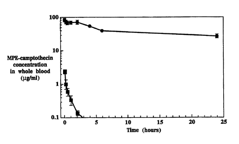
Fig. 1B shows the concentration of MPE-camptothecin in whole blood after
administration of the liposome formulation (solid circles) and of the free
drug to rats.
The longer circulation lifetime results in a higher concentration of the drug
in the blood.
1 o The anti-tumor efficacy of the MPE-camptothecin liposome formulation was
determined in xenograft tumor models, where homozygous nude mice were
inoculated
with human tumor cells of colon, HT29 origin. Surprisingly, these toxicity and
antitumor
efficacy studies showed that liposomal MPE-camptothecin was significantly more
toxic
than the free form of the drug at equivalent doses. These studies and the
results will now
be described.
Liposomes were prepared as set forth in Example 1 to include entrapped MPE-
camptothecin. Nude mice with HT-29 colon xenografts were treated with liposome-
entrapped MPE-camptothecin at dosages of 24 mg/kg, 15 mg/kg and 6 mg/kg or
with free
MPE-camptothecin at the same dosages. Treatment began 10 days after tumor
inoculation
2o and doses were administered at days 10, 16 and 23. The tumor volume in each
animal was
assessed during and following treatment as described in Example 2.
The body weight of each test animal and the tumor volume of each animal are
shown, respectively in Figs. 2A and 2B, where animals were treated with
liposomal
entrapped MPE-camptothecin at dosages of 24 mg/kg (closed circles), 15 mg/kg
(closed
triangles) and 6 mg/kg (closed squares) and with free MPE-camptothecin at
doses of 24
mg/kg (open circles), 15 mg/kg (open triangles) and 6 mg/kg (open squares).
With respect to the animals treated with the liposome-entrapped MPE-
camptothecin, all of the animals dosed with 15 mg/kg and 24 mg/kg died after
two doses
due to drug-related toxicity, with most deaths on day five after the first
dose. All of the
3o animals treated with 6 mg/kg liposome-entrapped MPE-camptothecin survived
until
administration of the third dose on day 23, after which five of the ten
animals died within
a few days. The toxicity of the liposome-entrapped MPE-camptothecin is
reflected in the
greater body weight losses, as seen in Fig. 2A.
18
CA 02346879 2001-04-10
WO 00/23052 PCT/US99/24228
In contrast, all of the animals treated with the free form of the drug
survived the
study, with the exception of one animal in the 24 mg/kg dosing group that died
a few
days after the third dose on day 23.
Table 1
Number
of Surviving
Animals
Treatment Dose Number after doseafter doseafter dose
mg/kg of 1 2 3
Test (day 9) (day 16) (day 23)
Animals
Saline na 20 20 20 20
free MPE-camptothecin24 10 10 10 9
free MPE-camptothecin15 10 10 10 10
free MPE-camptothecin6 10 10 10 10
liposome-entrapped24 10 1 0 0
liposome-enuapped15 10 5 0 0
liposome-entrapped6 10 10 10 5
With respect to antitumor activity of the formulations, the liposome-entrapped
MPE-camptothecin was more effective than the free form of the drug in
inhibiting tumor
growth, despite its greater toxicity. This can be seen in Fig. 2B, where the 6
mg/kg dose
of liposome-entrapped MPE-camptothecin was significantly more effective in
inhibiting
to tumor growth (log growth rate of -0.026) than even the highest dose level
of free MPE-
camptothecin {24 mg/kg, log growth rate 0.004$).
The complete and partial remission of the tumors in the test animals was
monitored
and is presented in Table 2. Complete remission of a tumor is defined as the
elimination
of tumor mass until the end of the experiment. A partial remission is defined
as a tumor
volume of less than 50 % of the peak tumor volume for an individual animal.
19
CA 02346879 2001-04-10
WO 00/23052 PCT/U599/24228
Table 2
Treatment Dose mg/kgComplete Remission'Partial Remission2
Saline 0/20 0/20
free MPE-cam tothecin24 3/10 1/10
free MPE-cam tothecin15 2/10 0/10
free MPE-cam tothecin6 O/10 0/10
li some-entry d 24 _-' -'
li osome-entry 15 --' --'
ed
li osome-entry 6 10/10 na
ed
' complete remission defined as elimination of tumor mass until experiment
termination.
2 partial remission defined as a tumor volume of less than 50 % of the peak
tumor vohtme for an
individual animal.
' all 10 animals in test groups died after the second dose on day 16.
° na = not applicable
As can be seen in Table 2, the liposome-entrapped MPE-camptothecin at a dose
of
6 mg/kg was effective to cause a complete remission of tumors in all 10 test
animals.
This effect was observed within five days after the second treatment on day
16. As noted
above, five of the test animals in the 6 mg/kg liposome-entrapped test group
died shortly
after the third dose. In the surviving five animals, the tumors did not recur
by the end of
the study, approximately 30 days after the final treatment on day 23. Data is
unavailable
for the animals treated with 15 mg/kg and 24 mg/kg liposome-entrapped MPE-
to camptothecin, since all of the animals in these test groups died due to
drug-related
toxicity, as noted above.
Administration of MPE-camptothecin in free form at a dose of 24 mg/kg resulted
in
3 animals with complete tumor remission and 1 animal with partial tumor
remission, as
seen in Tabte 2.
15 Comparison of the results observed for the drug administered in free form
and in
liposome-entrapped form indicate that the drug is more potent when
administered in
liposome-entrapped form. In fact, the liposome-entrapped drug is at least four
times
more potent than the free form of the drug, as can be seen by comparing the
results
obtained for a 6 mg/kg of liposome-entrapped MPE-camptothecin dosage to a 24
mg/kg
2o free MPE-camptothecin dosage (Fig. 2B, Table 2). It is clear from these
results that the
dose of liposome-entrapped MPE-camptothecin required for therapeutically
effective anti-
tumor therapy is four times lower than the dose required when the drug is
administered
CA 02346879 2001-04-10
WO 00/23052 PCT/US99/24228
in free form.
Example 2 describes the details of a second study to determine the maximum
tolerated dose and the lowest effective dose of the liposome-entrapped MPE-
camptothecin. In this study, liposomes were prepared as described in Example 1
and the
liposome formulation was administered to test animals at drug dosages of 0.1
mg/kg, 0.5
mg/kg, 1 mg/kg, 3 mg/kg and 5 mg/kg. The free drug was administered at 20
mg/kg as
a comparison.
Table 3 summarizes the number of test animals in each group, specifying the
number of animals surviving at each dosing phase of the study. As seen in the
table, all
of the control, saline treated animals and all of the animals treated with
free MPE-
camptothecin survived for the duration of the study. Of the ten animals
treated with 5
mg/kg liposome-entrapped MPE-camptothecin, four of the animals died of drug-
related
toxicity and one additional animal died of apparently nonspecific causes after
the third
dose. One of the ten animals in the test group receiving 3 mg/kg liposome-
entrapped
MPE-camptothecin died after the second dose, but the death was not considered
due to
drug treatment because of the absence of any correlating signs of toxicity.
All other
animals treated with liposome-entrapped MPE-camptothecin survived the entire
study
duration.
2o Table 3
Number
of Surviving
Animals
Treatment Dose Number after after after
m k of dose dose dose
Test Animals1 2 3
(da 9) (da 16) da 23)
Saline 20 20 20 20
free MPE-cam tothecin20 10 10 10 10
li some-entry d 5 10 10 10 5
li some-entry d 3 10 10 9 9
li osome-entry ed 1 10 x0 10 10
li some-entry ed 0.5 10 10 10 10
li some-entry d 0.1 10 10 10 10
The results of the study are shown in Figs. 3A-3B, where Fig. 3A shows the
body
weight of mice, in grams, as a function of days after inoculation with the HT-
29 colon
tumor. The animals were treated on days 9, 16 and 23 after tumor inoculation
with
21
CA 02346879 2001-04-10
WO 00/23052 PCT/US99/24228
liposomal entrapped topoisomerase I inhibitor at dosages of S mg/kg (open
triangles), 3
mg/kg (open inverted triangles), 1 mg/kg (open diamonds), 0.5 mg/kg (open
circles) and
0.1 mg/kg (open squares) and with free drug at a dose of 20 mg/kg (closed
squares). As
can be seen in Fig. 3A, body weight changes were dose-related and, these
changes were
correlated with other observations of toxicity.
Fig. 3B is a similar plot showing tumor volume, in mm', as a function of days
after
tumor inoculation, where the dosages are represented by the same symbols as in
Fig. 3A.
Fig. 3B shows that both the 5 mg/kg and 3 mg/kg dose levels of liposome-
entrapped
MPE-camptothecin were more therapeutically effective in inhibiting tumor
growth than
to the 20 mg/kg dvse of the free drug. Treatment with 20 mg/kg of free MPE-
camptothecin
(log growth rate of 0.011) was approximately equivalent in antitumor activity
to the 1
mg/kg dosage level of the drug in liposome-entrapped form (log growth rate of
0.017).
Table 4 summarizes the complete and partial tumor remission in the test
animals.
Table 4
Treaunent Dose Complete Partial
m /k Remission'Remission'-
Saline 0/20 0/20
free MPE-camptothecin 20 0/10 1/10
liposome-entrapped MPE-camptothecin5 10/10 na'
liposome-entrapped MPE-camptothecin3 7/10 1/10
liposome-entrapped MPE-camptothecin1 0/10 O/10
liposome-entrapped MPE-camptothecin0.5 0/10 1/10
liposome-entrapped MPE-camptothecin0.1 O/10 0/10
'Complete remission defined as elimination of tumor mass until experiment
termination.
iPartial remission defined as a tumor volume of less than 50% of the peak
tumor volume for an individual
animal.
'na = not applicable
There were no complete tumor remissions in the animals treated with 20 mg/kg
of
free MPE-camptothecin. In contrast, all ten of the animals treated with
liposome-
entrapped MPE-camptothecin at the 5 mg/kg dosage level had complete
remissions. At
2o the 3 mg/kg dosage, seven of the animals had complete remission of their
tumor.
The results from the study of Example 3 shows that antitumor activity of the
liposome-entrapped topoisomerase inhibitor MPE-camptothecin is significantly
better
22
CA 02346879 2001-04-10
WO 00/23052 PCTNS99/24ZZ8
when compared to the free form of the drug, indicating that the liposome-
entrapped form
was about 20-fold more potent since the antitumor activity of the free drug at
a dose of
20 mg/kg was most comparable to the activity of a 1 mg/kg dose of the liposome-
entrapped form of the drug. That the 3 mg/kg and S mg/kg liposome-entrapped
MPE-
camptothecin dosages were significantly more effective in antitumor therapy
than the 20
mg/kg dose of the drug in free form indicates that the therapeutic index of
the drug
entrapped in liposomes is approximately four-fold to five-fold higher than the
drug in
free form.
to 2. In vivo Adminstration of topotecan
In another study performed in support of the invention, topotecan was
entrapped in
liposomes composed of DSPC and mPEG-DSPE in a 95:5 molar ratio, as described
in
Example 4. Early studies, not reported here, indicated that topotecan was not
readily
retained in the liposomes. The lipid bilayer was selected to use a single
component
~s phospholipid having an acyl chain length close to DSPE in the mPEG-DSPE
component.
Such a bilayer has minimal packing defects which arise from imperfections in
nearest
neighbor interactions in a solid phase bilayer, which have reduced lateral and
rotational
mobility relative to fluid bilayers. In addition, a dextran-sulfate loading
battery was used
in order to achieve precipitation of the topotecan in the liposome interior.
Other
2o polymers, in particular polyanionic polymers, are suitable for this
purpose, such as
chondroitin sulfate A, polyvinylsulfuric acid, and polyphosphoric acid.
The pre-formed liposomes containing dextran atnmounim sulfate in the central
compartment were loaded with topotecan as described in Example 4. After
loading,
unentrapped drug was removed by diafiltration and the liposomes were
characterized.
25 The liposomes were loaded to a drug:lipid ratio of 0.238 and the liposomes
had an
average particle diameter of 87 nm.
The liposomes containing topotecan were administered intraveneously to rats to
determine the blood circulation lifetime. Figs. 4A-4B show the plasma
concentration of
topotecan as a function of time after administration to rats. Fig. 4A compares
the
3o concentration of liposome-entrapped topotecan administered at 2 mg/kg
(solid triangles)
to the concentration of free topotecan administered at the same dosage (solid
squares).
Fig. 4B compares the two forms of the drug at a dosage of 5 mg/kg. The
calculated
pharmacokinetic parameters are given in Table 5.
23
CA 02346879 2001-04-10
WO 00/23052 PCT/US99/24228
Table 5
Parameter milk '' Dc~a a
Dosage = ~.iin
= 2 .k
F ~ " Free L,ap~some
To otecan ~Posome- 'to otecanEritra
Entra ed ed
lasma Cmax /mL) 2.89 54.5 8.23 119.3
AUC ( /mL h) 0.57 523 1.57 1140
T tk (h) 0.20 7.2 0.30 9.8
CL (mL/h) 887 0.96 820 1.10
Vol. Dist. (mL) 173 9.2 278 17.5
elimination rate 3.45 0.096 2.33 0.071
constant
(1/h)
The data in Table 5 shows that the liposome-entrapped drug has a significantly
longer circulation time than the free form of the drug.
The efficacy of the liposomes was determined in another study. As described in
Example 4, the liposomes were administered to mice bearing a subcutaneous
xenograft
tumor. Tumor-bearing mice were randomized into six treatment groups of 12 mice
for
treatment with one of the following: saline, liposome-entrapped MPE-
camptothecin 4
mg/kg; free topotecan 25 mg/kg; liposome entrapped topotecan at drug dosages
of 2
to mg/kg, 5 mglkg or 8 mg/kg. All treatments were administered as intravenous
bolus
injections given weekly for 3 treatments, specifically on days 9, 16 and 23.
The tumor size in each animal was measured twice weekly during the study to
evaluate therapeutic efficacy. Body weight of each animal was monitored twice
weekly
to assess toxicity of the formulations. The results are shown in Tables 6 and
7 and in
Figs. SA-SB.
Table 6
Treatment Dose Complete Partial Non-
mglk~ ,__ Remission'Remission2Res nsive'
Saline 0 0 12
liposome-entrapped MPE-camptothecin4 8 4 0
free topotecan 25 0 1 11
liposome-enuapped topotecan2 1 2 9
liposome-enuapped topotecanS 2 8 2
liposome-entrapped topotecan8 7 3 2
'Complete remission defined as elimination of tumor mass until experiment
termination.
2Partial remission defined as a tumor volume of less than 50% of the peak
tumor volume for an individual animal.
'Non-responsive defined as a tumor volume equal to or greater than initial
tumor volume.
24
CA 02346879 2001-04-10
WO 00123052 PCTNS99/24228
As can be seen from Figs. SA and Table 6, left untreated the tumors grew at a
rate
of 17.8 mm' per day for the duration of the study. The animals treated with
iiposome-
entrapped MPE-camptothecin (positive control animals) experienced a tumor
growth rate
-1.2 mm' per day for the duration of the study. Animals treated with
nonencapsulated
topotecan, which was administered at 25 mg/kg somewhat below the maximum
tolerated
dosage (MTD) of 40 mg/kg, had tumor growth of 14.1 mm3 per day. Animals
treated
with liposome-entrapped topotecan had tumor growth of 0.9 mm3 per day for a
dosage of
2 mg/kg, -1.9 mm' per day for a dosage of 5 mg/kg and -0.8 mm' per day for a
dosage
of 8 mg/kg. The negative growth rate indicates regression of tumor size below
the
1 o starting tumor volume.
The size of treated tumors as a function of the size of control tumors (%T/C)
was
examined for all treatment groups and is summarized in Table 6. The National
Cancer
Institute defines significant anti-tumor activity as a %T/C less than 42.
Table 7
Treaunent Dose %T/C' %T/C ~T/C
m lk Da 29 Da 33 Da 36
liposome-entrapped MPE-camptothecin4 1.8 0.6 1.9
free topotecan 25 82.8 79.0 85.9
liposome-entrapped topotecan2 19.5 12.9 16.3
liposome-entrapped topotecan5 10.5 5.6 5.6
liposome-entrapped topotecan$ 2.0 2.2 2.2
%T/C defined as the average tumor volume at day indicated over the average
tumor vowme of me conrrm, Senne
treated animals.
3. In vivo Adminstration of CKD-602
Example 5 describes another study conducted in support of the invention using
the
topoisomerase inhibitor CKD-602. The drug was remotely loaded into liposomes
against
2o an ammonium-sulfate gradient with dextran as a trapping agent. The liposome
lipid
composition was identical to that used for the study using topotecan - HSPC
and mPEG-
DSPE in a 9515 mole ratio.
Fig. 6 is a plot showing the plasma concentration of CKD-602 as a function of
time
after administration to rats at a dosage of 1 mg/kg. The liposome-entrapped
form of the
drug (solid circles) had a calculated half life of 9.8 hours and an AUC of 274
p,g/mL/hr.
CA 02346879 2001-04-10
WO 00/23052 PCT/US99/24228
The free form of the drug had a calculated half life of 0.2 hours and an AUC
of 0.37
~.g/mL/hr.
Therapeutic efficacy of the CKD-602 formulation was evaulated using mice
hearing
a HT-20 colon cancer xenograft. Seventy-two mice were inoculated with HT-29
tumor
s cells and nine days later were randomized into six treatment groups. The
animals in each
group were treated with one of the following formulations: saline, liposome-
entrapped
MPE-camptothecin 4 mg/kg; free CKD-602 20 mg/kg; liposome entrapped CKD-602 at
drug dosages of 1 mg/kg, 2 mg/kg or 4 mg/kg. All treatments were administered
as
intravenous bolus injections given weekly for 3 treatments, specifically on
days 11, 18
1 o and 25 .
The tumor size in each animal was measured twice weekly during the study to
evaluate therapeutic efficacy. Body weight of each animal was monitored twice
weekly
to assess toxicity of the formulations. The results are shown in Tables 8 and
9 and in
Figs. 7A-7B.
Table 8
Treatment Dose Complete Partial Non-
m /k Remission'Remission2Res nsive'
Saline 0/10 0/10 10/10
liposome-entrapped MPE-camptothecin4 6/10 0/10 4/10
free CKD602 20 0/6 0/6 616
liposome-entrapped CKD6021 2/10 7/10 1/10
liposome-entrapped CKD6022 6/10 2/10 2110
liposome-entrapped CKDb024 4/4 0/4 0/4
'Complete remission defined as elimination of tumor mass until experiment
termination.
=Partial remission defined as a tumor volume of less than 50% of the peak
tumor volume for an individual animal.
'Non-responsive defined as a tumor volume equal to or greater than initial
tumor volume.
As can be seen in Table 8 and in Fig. 7B, the animals treated with saline
experienced continuous tumor growth, at a rate of 15.45 mm' per day for the
duration of
2o the study. The animals treated with the liposome-entrapped MPE-camptothecin
(positive
control animals)had a tumor growth rate of -0.63 mm' per day for the duration
of the
study. Animals treated with free, unentrapped CKDb02 had tumor growth of 15.21
mm3
per day. Animals treated with liposomai CKD602 had tumor growth of -2.21 mm'
per
26
CA 02346879 2001-04-10
WO 00/23052 PCT/US99/24228
day for animals treated with a dose of 1 mg/kg, -0.96 mm' per day for a dose
of 2 mg/kg
and -2.37 mm' per day for a dose of 4 mg/kg. The negative growth rate
indicates
regression of tumor size below the starting tumor volume.
The size of treated tumors as a function of the size of control tumors (%T/C)
was
examined for all treatment groups and is summarized in Table 9. The National
Cancer
Institute defines significant anti-tumor activity as a %T/C less than 42.
Table 9
Treatment Dose 9~T/C' ~T/C 96T/C
m /k Da 29 Da 33 Da 36
liposome-entrapped MPE-camptothecin4 2.9 2.3 1.6
free CKD602 20 129.1 120.1 99.9
liposome-entrapped CKD6021 11.4 7.7 4.4
liposome-entrapped CKD6022 4.8 2.8 1.6
liposome-entrapped CKD6024 1.0 1.3 0.9
T/C defined as the average tumor volume at day indicated over the average
tumor volume of the control, saline
treated animals.
to IV. EXAMPLES
The following examples illustrate methods of preparing, characterizing, and
using
the composition of the present invention. The examples are in no way intended
to limit
the scope of the invention.
Materials
The topoisomerase inhibitor (7-(4-methyl-piperazino-methylene)-10,11-
ethylenedioxy-20(S)-camptothecin trifluoroacetate (GI147211 ) (MPE-
camptothecin), was
provided by Glaxo Research Institute, Research Triangle Park, NC. CKD602 (7-(2-
(N-
isopropylamino)ethyl)-{20S)-camptothecin) was provided by Chong Kun Dang
2o Corporation, Seoul Korea. Topotecan (Hycamtiri ) was purchased
commercially.
Materials for preparation of the liposomes and all other reagents were from
commercially available sources.
27
CA 02346879 2001-04-10
WO 00/23052 PCTNS99/2422$
Methods
Animal Studies: Homozygous nude mice were obtained from Taconic Farms
{Germantown, NY) and allowed to acclimate for 7 days prior to initiation of
the
experiment. Animals were housed in appropriate isolated caging with ad lib
sterile
rodent food and acidified water and a 12:12 light:dark cycle. Animals were
randomized
into treatment groups prior to tumor inoculation based on body weight.
Randomization
was confirmed based on tumor size immediately prior to initiation of
treatment.
Tumors: Tumors were inoculated by trochar placement of fragments from rapidly
growing tumors on donor animals. The human colon cancer cell line, HT-29, was
used
1o to initiate subcutaneous xenograft tumors. Cultured cells were trypsinized,
washed,
counted and resuspended at 50 million cells per mL normal growth media. Tumors
were
inoculated by injection of O.lmL (5 million cells) at the back of the neck.
Tumors were
allowed to grow to an average size of 100 mm' prior to initiation of
treatment.
Monitoring: All animals were observed daily for general well-being throughout
the experiments. Animals were weighed prior to tumor inoculation and weekly
thereafter. Tumors were measured twice weekly throughout the experiment,
beginning
5-10 days after tumor inoculation. Any animal observed to have 15 % or greater
weight
loss from the initial starting weight and any animal observed to have greater
than 4,000
mm' tumor volume were excluded from the study.
Example 1
Preparation of Liposomes with Entrapped Topoisomerase Inhibitor
Liposomes were prepared and loaded with a selected topoisomerase inhibitor as
follows.
A. Liposome Preparation
The lipids hydrogenated soy phosphaticylcholine (HSPC), cholesterol (Chol) and
mPEG-DSPE (at a ratio of 564:38.3:5.3 mol/mol) were dissolved in ethanol at
65°C in
a 250 mL round bottom. The lipids were agitated continuously for at least 30
minutes at
65°C. The total lipid concentration in ethanol solution was 3.7 g total
lipid per 10 mL
ethanol.
The dissolved lipid solution was transferred to another 250 mL round bottom
flask
containing 100 mL of 250 mM ammonium sulfate solution equilibrated to
65°C. The
28
CA 02346879 2001-04-10
WO 00/23052 PCT/US99/24228
ethanol:lipid:ammonium sulfate hydration mixture was mixed continuously for at
least
one hour while maintaining the temperature using a 65 °C water bath to
form
oligolamellar ethanol hydration liposomes.
The oligolameliar liposomes were size reduced using a Lipex thermobarrel
extruder
s to pass the hydration mixture through polycarbonate membranes with known
pore size
dimensions. The mixture was passed 5 times through a 0.20 pm pore diameter
membrane, followed by 10 passes through a 0.10 ~.m pore diameter membrane. The
extruded liposomes contained ammonium sulfate within the interior aqueous
compartments) of the liposomes, as well as in the exterior aqueous bulk phase
medium
1o in which they are suspended. The sized liposomes were stored in the
refrigerator until
diafiltration preceding the remote loading procedure.
100 mg of a selected topoisomerase inhibitor, MPE-camptothecin, CKD-602 or
topotecan, was dissolved in 40 mL 10% sucrose solution to yield a
concentration of 2.5
mg/mL. After dissolution, the solution was passed through a 0.20 ~,m filter to
remove
t s insoluble particulates.
B. Remote Loading of Liposomes
Ammonium sulfate and ethanol were removed from the external bulk aqueous phase
immediately prior to remote loading by hollow fiber tangential flow
diafiltration with a
20 100KDa nominal molecular weight cutoff cartridge. Constant feed volume was
maintained, and at least seven exchange volumes were used resulting in
liposomes
suspended in an exterior aqueous phase comprised of IO% sucrose.
After diafiltration, the liposomes were mixed with a selected drug solution at
a ratio
(drug solution:liposomes) of 1:4 (vollvol) and rapidly warmed to 65°C
using a pre-
2s equilibrated jacketed vessel containing water. The temperature of the
mixture was
maintained at 65 °C for 40 to 60 minutes, after which the mixture was
rapidly cooled in
an ice-water bath. After remote loading, a sample of the liposomes was taken
to check
for the presence of crystals, to determine percent encapsulation and to
measure the mean
particle diameter.
30 Unencapsulated drug was removed from the bulk phase medium by hollow fiber
tangential flow diafiltration using a 100 kDa nominal molecular weight cutoff
cartridge.
At least eight exchange volumes were used, resulting in liposomally
encapsulated drug
suspended in an external aqueous phase comprised of 10 % sucrose 10 millimolar
29
CA 02346879 2001-04-10
WO 00/23052 PCTNS99/24228
Histidine pH 6.5.
The final liposome preparation was sterile filtered using a 0.22 ~m cellulose
acetate
syringe filter and stored refrigerated and protected from light until use.
C. Characterization of Liposomes
Percent encapsulation was determined using size exclusion chromatography to
compare the percent drug in the void volume (liposomal encapsulated) to the
total drug
(void volume plus included volume). Drug concentration in the column fractions
was
determined by absorbance. Mean particle diameter was determined using
quasielectric
to laser light scattering (QELS). The total lipid concentration was assayed at
the post-
sterile filtration stage in order to determine the drug to lipid ratio.
Liposomes loaded
with MPE-camptothecin, topotecan and 7-(2-(N-isopropylamino)ethyl)-(20S)-
camptothecin (CKD-602) were prepared and characterized. The results are shown
in the
table below.
t
s
Parameter liposome-entrappedliposome-entrappedliposome-entrapped
MPE- To tecan CKD-
cam tothecin 602
lot no. 221AZ43A 221AZ43B 221A253
Total Li id 17.81 mol/mL I5.97 moI/mL 14.079 mol/mL
concentration
Drug concentration2.69 mg/mL 1.72 mg/mL 1.77 mg/mL
(4.55 mol/mL) (3.76 mol/mL (3.77 moI/mL
drug:lipid ratio0.26 0.24 0.27
{mol/mol) ( 1 : 3.92 ( 1 : 4.25) 1 : 3.73
Mean Particle 99 ttm 95.4 nm 9ti.7 nm
diameter
Percent Encapsulation_ T99.9% I 95.39'0
96.4%
Examule 2
In vivo Efficacy of Liposome-entrapped MPE-Camptothecin
Liposomes containing entrapped MPE-camptothecin were prepared as described in
2o Example 1. The liposome entrapped drug and the free drug were diluted in 5
% dextrose
in water as required to achieve the desired concentrations.
Nude mice were inoculated with the human colon cancer cell line HT-29 as
described above in the methods section. Seventy mice were randomized to one of
seven
treatment groups as follows: free drug at 24 mg/kg, 15 mg/kg or 6 mglkg;
liposome
2s entrapped drug at 24 mg/kg, 15 mg/kg or 6 mg/kg; saline. Treatment was
initiated when
average tumor volume was approximately 75 mm3 on day 10 post-tumor
inoculation. All
treatments were administered as intravenous bolus injections given weekly for
3
CA 02346879 2001-04-10
WO 00/23052 PCTNS99/24228
treatments, specifically on days 10, 16 and 23.
Tumor size during and following each experiment was used as the primary
evaluation of therapeutic efficacy. Body weight was evaluated to assess
toxicity. All
tumor bearing animals were observed following cessation of treatment, until
euthanized
s based on criteria above. Experiments were concluded when a majority of
control tumors
achieved the maximal allowed volume (4,000 mm').
Tumor size in each animal was measured repeatedly at various time points, thus
these measurements were regarded as correlated information. Since the tumor
sizes over
time after treatment were of interest, repeated measurement analyses was done
for each
to data set. By examining the data, a log transformation seemed reasonable.
Let Y denote
the original tumor measurement, let Z = log(Y+1). After transforming data,
repeated
measurement analyses was done for the transformed data Z. The SAS procedure
PROC
MIXED was used. The log growth rate for each treatment group was calculated
and
used to compare the different treatment groups. Statistical significance was
declared at
15 the 0.05 level, but due to multiple comparisons, adjustment to the type I
error were done
and a P-value of < 0.0033 indicated a statistically significant difference in
any designated
comparison.
The results are summarized in Tables 1 and 2 and in Figs. 2A-2B.
2o Example 3
Dose Finding Stud~r for Lposome-entrapped MPE-Camntothecin
Liposomes containing entrapped MPE-camptothecin were prepared as described in
Example 1. The liposome entrapped drug and the free drug were diluted in S %
dextrose
in water as required to achieve the desired concentrations.
25 Nude mice were inoculated with the human colon cancer cell line HT-29 as
described above in the methods section. Seventy mice were randomized to one of
seven
treatment groups as follows: liposome entrapped drug at 0.1 mg/kg, 0.5 mglkg,
1
mg/kg, 3 mg/kg, 5 mg/kg or 20 mg/kg; and saline. Treatment was initiated when
average tumor volume was approximately 75 mm' on day 9 post-tumor inoculation.
All
3o treatments were administered as intravenous bolus injections given weekly
for 3
treatments, specifically on days 9, 16 and 23.
The tumor size was evaluated and analyzed as described in Example 2, and the
results are shown in Tables 3 and 4 and in Figs. 3A-3B.
31
CA 02346879 2001-04-10
WO 00/23052 PCT/US99/24228
Examgle 4
In vivo Efficacy of Liposome-Entrapped Topotecan
A. Liposome Preparation
Liposomes containing topotecan were prepared as follows.
s The lipids distearoylphospatidylcholine (DSPC) and (N-(carbonyl-
methoxypolyethylene glycol 2000)-1,2-distearoyl-sn-glycero-3-
phosphoethanolamine)
(mPEG-DSPE) were combined at a molar ratio of 95:5 and dissolved in ethanol at
70°C
using continuous agitation. The lipid concentration in the ethanol solution
was 8.9 grams
per 10 mL ethanol.
Dextran sulfate-ammonium salt was prepared by ion exchange chromatography
using dextran sulfate sodium salt as the starting material. A 100 mg/mL
solution of
dextran sulfate ammonium salt was prepared by dissolving dextran sulfate
sodium salt in
water and adjusting the solution pH to 5 using ammonium hydroxide.
100 mL of dextran sulfate solution was heated to 70°C and combined with
the
~ 5 ethanol solution of lipid while mixing to form oligolamellar liposomes.
The temperature
of the oligolamellar ethanol hydration liposome dispersion was maintained at
70°C for
one hour with continuous mixing.
The post hydration mixture was heated to 70 degrees and size reduced using a
Lipex thermobarrel extruder through a series of polycarbonate membranes to
arrive at a
2o particle size near 100 nm mean particle diameter. Typically, the sequence
involved 5
passes through an 0.2 pm pore diameter membrane, followed by 10 passes through
an
0.1 pm pore diameter membrane.
Unentrapped dextran sulfate polymer and remaining ethanol were removed from
the
external bulk aqueous phase immediately prior to the active drug loading step
with eight
25 volume exchanges using 350 mM sodium chloride solution, followed by eight
volume
exchanges using a 10% sucrose solution. The diafiltration cartridge employed
had a
specified nominal molecular weight cutoff of 100,000 Daltons.
A solution of topotecan was prepared at a concentration of 2.5 mg/mL in 10
sucrose. The drug solution and diafiltered liposomes were combined at a volume
ratio of
30 4:1, and the temperature of the resulting mixture was raised to 70°C
and maintained with
constant stirring for one hour. Active drug loading was terminated by rapidly
cooling
the post-loading mixture using an ice water bath.
32
CA 02346879 2001-04-10
WO 00/23052 PCT/US99/24228
Unentrapped drug was removed by diafiltration employing a cartridge having
nominal molecular weight cutoff of 100,000 Daltons. Typically, 8-10 volume
exchanges
were employed using 10% sucrose 10 mM Histidine pH 6.5 as the exchange buffer.
Drug concentration was adjusted to the final value by assaying for potency
with a
s uv-vis absorbance measurement and diluting accordingly.
The final process step involved sterile grade filtration employing a 0.22 ~tm
filter
prior to filling vials.
B. Liposome Characterization
1o Percent encapsulation was determined using size exclusion chromatography to
determine the percent drug in the void volume ("liposomal drug") to the total
amount
recovered in both the included and void volume fractions. Drug concentration
was
monitored using uv-vis absorbance spectrophotometry. Mean particle diameter
was
determined using quasielastic laser light scattering. Total lipid was
determined using
is phosphorous assay. The results are summarized in the table below.
ameter Liposome-entrapped
.:
Topotet~ , :..
'.. . .;
total lipid 17.2 mg/mL
total drug 2.1 mg/mL
drug:lipid ratio 0.238
(mol:mol)
mean particle diameter 87.3 nm
percent encapsulation 98.8
C. In vivo Pharmacokinetics and Efficacy
Seventy two mice were inoculated with HT-29 cancer cells as described above in
2o the methods section. Nine days after tumor inoculation, the animals were
treated weekly
with one of the following intravenous treatments: saline; liposome-entrappea
m~r~-
camptothecin 4 mg/kg; free topotecan 25 mg/kg; liposome entrapped topotecan at
drug
dosages of 2 mg/kg, 5 mg/kg or 8 mg/kg. All treatments were administered as
intravenous bolus injections given weekly for 3 treatments, specifically on
days 9, 16 and
2s 23.
33
CA 02346879 2001-04-10
WO 00/23052 PCT/US99/24228
The tumor size was evaluated and analyzed as described in Example 2, and the
results are shown in Tables 6 and 7 and in Figs. SA-SB.
Example 5
s In vivo Efficacy of Liposome-Entrapped CKD-602
A. Li~osome Preparation and Characterization
Liposomes containing CKD-602 were prepared as described in Example 4, except
using a drug solution of CKD-602. The liposomes were characterized as
described in
Example 4 and the results are summarized below.
to
Parameter Liposom,e entrapped
T
CYCD-602 ' .
total lipid 12.5 mg/mL
total drug 2.07 mg/mL
drug:lipid ratio 0.315
(mol:mol)
mean particle diameter 92.8 nm
percent encapsulation 94.7
B. In vivo Pharmacokinetics and Efficacy
Seventy two mice were inoculated with HT-29 cancer cells as described above in
the methods section. Eleven days after tumor inoculation, the animals were
treated
15 weekly with one of the following intravenous treatments: saline, liposome-
entrapped
MPE-camptothecin 4 mg/kg; free CKD602 20 mg/kg; liposome entrapped CKD602 at
drug dosages of 1 mg/kg, 2 mg/kg or 4 mg/kg. All treatments were administered
as
intravenous bolus injections given weekly for 3 treatments, specifically on
days 11, 18
and 25.
2o The tumor size in each animal was measured twice weekly during the study to
evaluate therapeutic efficacy. Body weight of each animal was monitored twice
weekly
to assess toxicity of the formulations. The results are shown in Tables 8 and
9 and in
Figs. 7A-7B.
Although the invention has been described with respect to particular
embodiments,
2s it will be apparent to those skilled in the art that various changes and
modifications can
be made without departing from the invention.
34