Note: Descriptions are shown in the official language in which they were submitted.
CA 02347028 2001-04-17
WO OOI24452 PCT/US99/25068
SYSTEMS AND COMPOUNDS FOR DRUG DELIVERY TO INTERSTITIAL REGIONS
OF THE MYOCARDIUM
Field of the Invention
The present invention relates to the interstitial delivery
of particulate drug delivery systems for large and small
molecule therapeutic agents within the heart.
Background of the Invention
Local drug delivery provides many advantages. Approaches
for local controlled~release of agents at a depth within a
tissue such as the heart, pancreas, esophagus, stomach, colon,
large intestine, or other tissue structure to be accessed via a
controllable catheter will deliver drugs to the sites where they
are most needed, reduce the amount of drug required, increase
the therapeutic index, and control the time course of agent
delivery. These, in turn, improve the viability of the drugs,
lower the amount (and cost) of agents, reduce systemic effects,
reduce the chance of drug-drug interactions, lower the risk to
patients, and allow the physician to more precisely control the
effects induced. Such local delivery may mimic endogenous modes
of release, and address the issues of agent toxicity and short
half lives.
Local drug delivery to the heart is known. In U.S. Pat.
No. 5,551,427, issued to Altman, implantable substrates for
local drug delivery at a depth within the heart are described.
The patent shows an implantable helically coiled injection
needle which can be screwed into the heart wall and connected to
an implanted drug reservoir outside the heart. This system
allows injection of drugs directly into the wall of the heart
acutely by injection from the proximal end, or on an ongoing
basis by a proximally located implantable subcutaneous port
reservoir, or pumping mechanism. The patent also describes
implantable structures coated with coating which releases
bioactive agents into the myocardium. This drug delivery may be
performed by a number of techniques, among them infusion through
CA 02347028 2001-04-17
WO 00/24452 PCT/US99/25068
a fluid pathway, and delivery from controlled release matrices
at a depth within the heart. Controlled release matrices are
drug polymer composites in which a pharmacological agent is
dispersed throughout a pharmacologically inert polymer
substrate. Sustained drug release takes place via particle
dissolution and slowed diffusion through the pores of the base
polymer. Pending applications 08/8816850 by Altman and Altman,
and 09/057,060 by Altman describes some additional techniques
for delivering pharmacological agents locally to the heart.
Implantable drug delivery systems, such as controlled release
matrices, have been well described in the literature, as has the
use of delivering particulate delivery systems or particulate
drug carriers such as microcapsules, lipid emulsions,
microspheres, nanocapsules, liposomes, and lipoproteins into the
circulating blood. However, local delivery of such micro drug
delivery systems to a depth within the myocardium using
endocardial catheter delivery and epicardial injection systems
have not been described, and have many advantages that have not
been foreseen.
Recently, local delivery to the heart has been reported of
therapeutic macromolecular biological agents by Lazarous
[Circulation, 1996, 94:1074-1082.], plasmids by Lin
[Circulation, 1990; 82:2217-2221], and viral vectors by French
[Circulation, Vol. 90, No 5, November 1994, 2414-2424] and
Muhlhauser [Gene Therapy (1996) 3, 145-153]. March
[Circulation, Vol. 89, No 5, May 1994, 1929-1933.] describes the
potential for microsphere delivery to the vessels of the heart,
such as to limit restenosis, and this approach has also been
used for the delivery of bFGF by Arras [Margarete Arras et. al.,
The delivery of angiogenic factors to the heart by microsphere
therapy, Nature Biotechnology, Volume 16, February 1998.] These
approaches for microsphere delivery obstruct flow, and will be
delivered preferentially to capillary beds which are well
perfused. Further, these approaches do not deliver therapeutic
agents to the interstitial spaces. None of this work recognizes
the potential to use particulate drug delivery system to
optimize local drug delivery at a depth within the myocardium.
This art also does not recognize the potential such delivery
2
CA 02347028 2001-04-17
WO 00/24452 PCT/US99/25068
systems have in treating disease substrates in the myocardium if
delivered to an appropriate region of the myocardial
interstitium.
Problems exist for delivering small molecules or lipophilic
molecules which rapidly transport through the capillary wall, to
well-perfused tissues such as the myocardium. These problems
are due to the convective losses of the agents to the systemic
circulation. By going rapidly across the capillary wall, the
small molecules are rapidly carried away by the bloodstream.
Local delivery of an easily transported molecule is difficult
because local delivery concentrations are rapidly reduced at
very small distances from the delivery site due to convective
losses. Such easily transported agents cannot treat an
effective area of tissue locally without raising the systemic
concentrations of the agents to a therapeutic level.
Summary
The therapeutic compounds described below comprise very
small capsules which can be injected into body tissue,
particularly the heart. The capsules include an encapsulating
layer which surrounds a therapeutic agent. After injection, the
encapsulating layer degrades or dissolves, and the therapeutic
agent is released within the heart. The therapeutic agent may
be one of any number of known agents such as anti-arrhythmic
drugs, gene therapy solutions, and macromolecules intended to
have either acute or long-term effects on the heart. While some
of these therapeutic agents are used to treat the heart by
injecting them into the heart, they are of such small size that
they readily enter the cardiac capillary system and the cardiac
lymphatic system, and are quickly transported away from the
injection site. Thus, in prior treatment methods, relatively
large doses and repeated dosed are required to provide
therapeutic effect at the injection site. To provide a solution
to this problem, the capsules described below are provided in
sizes that are too large to permit capillary transport or
lymphatic transport. Thus, injected capsules are immobile within
the heart tissue, and upon degradation they will release a
3
CA 02347028 2001-04-17
WO 00124452 PC'T/US99/25068
therapeutic agent very near the site of injection. The capsules
may also be provided in sizes that are too large to permit
capillary transport, but small enough to enter the lymphatic
system and be transported away from the injection site in the
cardiac lymphatic system, so that the therapeutic effect is
provided at some distance from the injection site. The
encapsulating layer may be made from various materials including
biodegradable polymers in the form of microspheres, or from
standard vesicle forming lipids which form liposomes and
micelles.
Brief Description of The Drawings
Figure 1 illustrates an encapsulated therapeutic agent
designed for injection into the heart.
Figure 1a illustrates a microsphere encapsulated
therapeutic agent designed for injection into the heart.
Figure 2 illustrates a method for injection of therapeutic
agents into the heart.
Figure 3 illustrates the expected transportation of
molecules released from degrading microspheres injected within
the myocardium.
Figures 4a through 4d illustrate the progression of
injected liposome encapsulated small molecules within the heart
tissue after injection.
Figure 5 illustrates a method of delivering therapeutic
agents to the coronary arteries through the lymphatic vessels.
Detailed Description of the Invention
Figure 1 illustrates a microdrug delivery system which is
comprised of a compound or substance for use in delivering a
therapeutic agent to the heart. The compound is comprised of
numerous capsules 1 which are made up of an encapsulating layer
2 which may form a microsphere formulated from ProleaseTM or
other biodegradable microsphere material, or from vesicle
forming lipids which may form a liposome or micelle, and a
4
CA 02347028 2001-04-17
WO 00/24452 PCT/US99/25068
therapeutic agent 3 within the encapsulating layer. Therapeutic
agent may be embedded in a biodegradable polymer, or in a
carrier fluid 4. The encapsulating layer is typically
pharmacologically inactive, although techniques to make it
active to promote cellular uptake and / or receptor binding are
known in the art. The therapeutic agent may be any of a wide
variety of drugs and other compounds used for treatment of
various ailments of the heart. The capsules are carried within a
solution such as pH controlled saline to create a slurry which
can be injected into the heart of a patient. Prior to
injection, the encapsulating layer will protect the
macromolecule from mechanical and chemical degradation within
the catheter or needle used for injection. Once injected into
the heart tissue, the size of the encapsulating layer will
inhibit transport of the compound away from the injection site,
either through the cardiac capillary system and/or the cardiac
lymphatic system. Also once injected, the encapsulating layer
will degrade, either due to chemical conditions, biological
conditions, or temperature conditions within the heart wall, and
release the encapsulated molecule. The time period over which
the encapsulating layer degrades is variable, depending upon its
formulation, such formulations being available in the art. The
half-life for degradation may be selected from several minutes
to several days, depending on the therapy intended. Thus a
sustained reservoir of therapeutic agent is created within the
heart tissue near the injection site, and therapeutic agents are
slowly released near the injection site to treat nearby tissue.
The need to flood the entire heart and/or the entire blood
system of the patient is eliminated, so that very small doses of
therapeutic agents are enabled. This reduces the cost of
treatment, and minimizes the otherwise harsh side effects
associated with many effective therapeutic agents.
Figure 1a illustrates the formulation of the microdrug
delivery system from a microsphere formulated from ProleaseTM,
biodegradable polymers, or particulate controlled release matrix
with molecules of therapeutic agent dispersed throughout the
microsphere. The microsphere 5 in Figure 1a includes numerous
molecules or particles of therapeutic agents 3 dispersed
5
CA 02347028 2001-04-17
WO 00/24452 PCT/US99/25068
throughout the solid biodegradable microsphere or particulate
controlled release matrix 6. As the microsphere material
degrades, therapeutic agents are slowly released from the
microsphere. This formulation differs from the capsule
formulation, but may be employed to achieve similar results. In
one preferred embodiment, the core 7 of the solid biodegradable
microsphere contains no therapeutic drug at a radius less then
approximately 20 um, preferably about 15 um. Thus the core of
the microsphere, to a radius of up to 20 um, preferably 15 um,
may be devoid of therapeutic agent. Alternatively, the core of
the microsphere, to a radius of up to 10 um, preferably 7.5 um,
may be devoid of therapeutic agent. This prevents problems
associated with migration of the potentially potent depot within
the lymphatic system. The core of the microsphere may also be
designed to have a longer degradation half-life so that
essentially all of the drug will be delivered before the
microsphere can substantially migrate through the lymphatic
networks. Thus, the particulate micro delivery systems includes
millispheres, microspheres, nanospheres, nanoparticles,
liposomes and micelles, cellular material and other small
particulate controlled release structures which can be advanced
in a fluid suspension or slurry and be delivered to a depth
within the heart muscle. These small drug delivery systems may
deliver therapeutic agents as diverse as small molecule
antiarrhythmics, agents that promote angiogenesis, and agents
that inhibit restenosis. They may also be combined in cocktails
with steroid agents such as dexamethasome sodium phosphate to
prevent inflammatory response to the implanted materials.
Separate particulate drug delivery systems for delivering
different agents to the same region of the heart may also be
used. The release kinetics of separate micro delivery systems
may also be different.
Delivery of small drug delivery systems reduce the
likelihood of causing embolic events in the brain, kidneys, or
other organs should these drug delivery systems escape into the
left chambers of the heart. Because the systems are small only
very small arterioles would be occluded should one of them
escape into the blood within the left chambers of the heart.
6
CA 02347028 2001-04-17
WO 00/24452 PCTNS99/25068
This is not a problem in the right side of the heart, as the
lungs act as a filter of potentially embolic materials.
Figure 2 shows a catheter system 9 with centrally located
drug delivery catheter 20 implanted at a depth within the left
ventricular apex 15 of the heart 10. Hollow penetrating
structure 30 has penetrated the heart muscle, and has
transported particulate encapsulated agents 35 such as VEGF,
bFGF, or other therapeutic agent to a depth within the heart
muscle. The encapsulated agents are injected into the heart
muscle (the myocardium) in an intact portion of the heart muscle
(that is, not into a vessel such as the ventricle chamber, a
coronary artery or a TMR channel which are subject to blood flow
and immediate transport of the injected particles from the
area). The capsules or microspheres are suspended within a
fluid inside the catheter to facilitate injection. The use of
small drug delivery systems in slurry or suspension delivered by
a fluidic pathway (a needle or catheter) to a depth within the
myocardium can solve different problems in pharmacokinetics of
local cardiovascular drug delivery. Such an approach can
provide for well controlled and easily administered sustained
dosage of therapeutic macromolecules, eliminate the issue of
connective losses of small molecules for local delivery, and
increase the ability of gene therapy preparations to gain access
through the cell membrane.
Problems exist for macromolecular therapies in the heart
such as short half-Lives and the presence of endogenous
inhibitors. Many macromolecular therapies may be improved by
providing a sustained dosage over time to overcome endogenous
inhibitors, as well as encapsulation to protect the
macromolecule from degradation.
The interstitial (intramuscular or intra-myocardial)
delivery of particulate drug delivery systems for sustained
release such as biodegradable microspheres solves these
problems. Particulate systems, such as microspheres, enable the
time course of delivery and area of treatment to be controlled.
In addition, such particulate systems may be delivered to the
target site by a fluid pathway within a drug delivery catheter
7
CA 02347028 2001-04-17
WO 00/24452 PCT/US99/25068
such as those described in the prior art. The advantages of
these particulate delivery systems is that they are implanted at
a depth within the heart tissue and the implanted catheter
device can be removed immediately. Thus, a very quick procedure
may be performed on an outpatient basis to deliver particulate
drug delivery systems to a depth within a patient's heart for
sustained delivery measured in days to weeks.
The microspheres to be used in this treatment are
manufactured to be large enough to prevent migration within the
myocardial interstitium, but also small enough to be deliverable
by a catheter fluid pathway to a depth with the myocardium.
Microspheres such as Alkerme's (Cambridge, Massachusetts)
Prolease system enable freeze dried protein powder to be
homogenized in organic solvent and sprayed to manufacture
microspheres in the range of 20 to 90 um (microns). Development
of such microsphere depots for sustained release of proteins
with unaltered integrity requires methods to maintain stability
during purification, storage, during encapsulation, and after
administration. Many of these techniques have been recently
summarized in the literature. See, e.g., Scott D. Putney, and
Paul A. Burke: Improving protein therapeutics with sustained
release formulations, Nature Biotechnology, Volume 16, February
1998, 153- 157. Issues associated with degradation for
biodegradable polymers used in such microspheres are also well
known [Robert Miller, John Brady, and Duane E. Cutright:
Degradation Rates of Oral resorbable Implants {Polylactates and
Polyglycolates}: Rate Modification and Changes in PLA/PGA
Copolymer Ratios, J. Biomed. Mater. Res., Vol. II, PP. 711-719
(1977). The value of delivering microsphere encapsulated
macromolecular agents such as proteins bFGF and VEGF to a depth
within the heart muscle for controlled release have not been
described, and have substantial unrecognized benefits over other
delivery approaches.
Figure 3 shows a schematic description of microsphere
encapsulated agents for delivery. Macromolecule angiogenic
agents 336 such as VEGF and bFGF are delivered with
biodegradable microspheres 335 in combination with biodegradable
8
CA 02347028 2001-04-17
WO 00124452 PCTNS99/25068
microspheres 302 enclosing dexamethasone sodium phosphate or
other anti inflammatory steroid. In other embodiments the anti-
inflammatory agents may be combined with a particular
therapeutic within the same encapsulation. The microspheres are
injected through the endocardium 338 and into the myocardium 339
so that they reside interstitially within the heart tissue.
Both microspheres 335 and 302 are too large to be transported
away by either the capillary system or the lymphatic system from
the injection site within the myocardium. Where the
microspheres are greater than about 15 micrometers in diameter,
they will remain at the injection site and will not migrate.
Where the microspheres have a diameter less than about 1
micrometer they will migrate in the cardiac lymphatic system,
but will not enter the cardiac capillary system. As the
microspheres degrade over time, their components and the
therapeutic molecules will be transported away from the
injection site by the myocardial lymphatic system which has been
described in relation to the transport of extravasated proteins
from the endocardium 338 to the epicardium 340, and from the
apex of the heart 345 towards the base of the heart 350.
[Albert J. Miller, Lymphatics of the Heart, Raven Press, New
York, 1982.) Here the microspheres are delivered endocardially
and inferiorly (that is, upstream in the lymphatic system) to
the region to be treated, identified here schematically by
window 355. Clearly regions within window 355 and regions
directly adjacent to the window will all result in effective
delivery of agents to the desired target, and are viable
approaches as well. The large molecules delivered in such a
fashion will be transported through the lymphatics far more
slowly than small molecules which would be more rapidly
convected away from the delivery region by the blood supply.
But approaches exist to minimize the issues associated with
convective losses of small molecules.
The method of packaging the small molecule so that it
cannot be convected away by the blood, yet will be distributed
locally in the tissue, and then effecting its action on the
tissue can be accomplished with liposomal encapsulation. The
term "liposome" refers to an approximately spherically shaped
9
CA 02347028 2001-04-17
WO 00/24452 PCT/US99/25068
bilayer structure, or vesicle, comprised of a natural or
synthetic phospholipid membrane or membranes, and sometimes
other membrane components such as cholesterol and protein, which
can act as a physical reservoir for drugs. These drugs may be
sequestered in the liposome membrane or may be encapsulated in
the aqueous interior of the vesicle. Liposomes are
characterized according to size and number of membrane bilayers.
Vesicle diameters can be large (>200 nm) or small (<50 nm) and
the bilayer can have unilamellar, oligolamellar, or
multilamellar membrane.
Liposomes are formed from standard vesicle forming lipids,
which generally include neutral and negatively charged
phospholipids with or without a sterol, such as cholesterol.
The selection of lipids is generally guided by considerations of
liposome size and ease of liposome sizing, and lipid and water
soluble drug release rates from the site of liposome delivery.
Typically, the major phospholipid components in the liposomes
are phosphatidylcholine (PC), phosphatidylglycerol (PG),
phosphatidyl serine (PS), phosphatidylinositol (PI) or egg yolk
lecithin (EYL). PC, PG, PS, and PI having a variety of acyl
chains groups or varying chain lengths are commercially
available, or may be isolated or synthesized by known
techniques. The degree of saturation can be important since
hydrogenated PL (HPL) components have greater stiffness than do
unhydrogenated PL components; this means that liposomes made
with HPL components will be more rigid. In addition, less
saturated Pls are more easily extruded, which can be a desirable
property particularly when liposomes must be sized below 300 nm.
Current methods of drug delivery by liposomes require that
the liposome carrier will ultimately become permeable and
release the encapsulated drug. This can be accomplished in a
passive manner in which the liposome membrane degrades over time
through the action of agents in the body. Every liposome
composition will have a characteristic half-life in the.
circulation or at other sites in the body. In contrast to
passive drug release, active drug release involves using an
agent to induce a permeability change in the liposome vesicle.
CA 02347028 2001-04-17
WO 00/24452 PCT/US99/25068
In addition, liposome membranes can be made which become
destabilized when the environment becomes destabilized near the
liposome membrane (Proc. Nat. Acad. Sci. 84, 7851 (1987);
Biochemistry 28: 9508, (1989).) For example, when liposomes are
endocytosed by a target cell they can be routed to acidic
endosomes which will destabilize the liposomes and result in
drug release. Alternatively, the liposome membrane can be
chemically modified such that an enzyme is placed as a coating
on the membrane which slowly destabilizes the liposome (The
FASEB Journal, 4:2544 (1990). It is also well known that lipid
components of liposomes promote peroxidative and free radical
reactions which cause progressive degradation of the liposomes,
and has been described in US Pat No. 4,797,285. The extent of
free radical damage can be reduced by the addition of a
protective agent such as a lipophilic free radical quencher is
added to the lipid components in preparing the liposomes. Such
protectors of liposome are also described in US Pat. No
5,190,761, which also describes methods and references for
standard liposome preparation and sizing by a number of
techniques. Protectors of liposomal integrity will increase the
time course of delivery and provide for increased transit time
within the target tissue.
Liposomal encapsulation of small molecules makes local
delivery possible. By having a liposomal preparation which is
unstable in the body, it will collapse after it is delivered.
Liposomes can be constructed in varying size, including the size
range less than 400 nm, preferably 200-250 nm. Between the time
of delivery and the time of collapse, the liposomes in the size
range less than 400 nm will be transported into and through the
lymphatics and provide for redistribution of small molecules.
Delivery of liposomes that degrade rapidly once delivered to the
body in a matter of minutes goes against the typical approaches
for liposomal delivery and design. Typically pH sensitive
liposomes involves the destabilization of the liposome in the
endosome as the pH falls from physiological 7.4 to 5.0, while
here we are describing liposomes which become destabilized near
pH 7.4. [Chun-Jung Chu and Francis C. Szoka: pH Sensitive
Liposomes, Journal of Liposome Research, 4(1), 361-395 (1994)].
11
CA 02347028 2001-04-17
WO 00/24452 PCT/US99/25068
Figure 4a shows a schematic of the delivery of small
molecules within liposomes which are unstable at physiological
pH (the pH of the heart tissue or the physiological environment
into which the molecules are delivered). A guiding catheter 401
is shown with a single lumen needle drug delivery catheter 402
containing liposome encapsulated small molecules 403 which are
delivered through needle 404 by way of needle fitting 404. Here
the penetrating needle 405, crosses the endocardium 410 to
deliver liposomes 415 to a depth within the heart wall 420.
Although the liposomes could be various sizes and have a number
of lipid bilayers, in the preferred embodiment they are small
unilamellar liposome vesicles (SWs) to augment their rapid
uptake by the cardiac lymphatic system. The drug delivery
catheter 402 contains liposomes bathed in a solution at their
stable pH so that they do not collapse prematurely. Figure 4b
shows that the catheter has been removed and that the uptake of
the SWs 415 by a lymphatic vessel 425 at some time t2 later
than the time they were delivered t1 to the myocardial
interstitium, such as the subendocardial interstitium. Of
course, other physiochemical properties could be used such that
the liposomal preparations are delivered from a system in which
they are stable to a system at a depth within the heart with
different physio-chemical properties in which they are unstable.
Temperature is another possible property that could be varied.
Arrows near 407 show that lymphatic transport is from
endocardium to epicardium and from apex to base in the heart.
The lymphatic transport will carry the encapsulated small
molecules a distance which will be governed by their stability
and mean time to liposomal degradation. Figure 4c shows the
same tissue in a larger view at time t3 later than time t2 in
which SWs 415 are degrading and releasing small molecule drugs
430 within the lymphatics. The spread of the released drug in
the degraded liposomes 430 provides therapeutic treatment to a
large region of heart tissue while systemic effects are
minimized. Figure 4d shows that upon degradation, the small
molecules 430 will be transported through the lymphatic vessel
wall 435 to the adjacent myocytes, and be connected rapidly away
from the region. This transport through the lymphatic walls is
shown schematically by the large arrows at the site of the
12
CA 02347028 2001-04-17
WO 00/24452 PCT/US99/25068
degraded liposome with released small molecules. Because of the
inability of the small molecules to be connected away rapidly
until the liposome collapses, a much larger region of tissue
will be able to be treated locally than by local infusion of the
small molecules themselves. In one embodiment, oleic acid (OA)
and dioleoylphosphatidyl-ethanolamine (DOPE) devoid of
cholesterol which have been shown to be extremely unstable in
the presence of body fluid plasma [Liu, D. and Huang, L., Role
Of Cholesterol In The Stability Of pH Sensitive, Large
U_nilamellar Liposomes Prepared By The.Detergent-Dialysis Method,
Biochim Biophys. Act, 981, 254-260 (1989)] and could be used to
encapsulate small molecule gene regulators such as hormones or
antiarrhythmic agents.
In another embodiment, liposomes of dimyristoyl-
phosphatidylcholine (DMPC) or dipalmitoylphosphatidylcholine
(DPPC), cholesterol (CHOL) and dicetylphosphate (DCP) containing
Amiodarone are prepared at pH 4.5 using DMPC:CHOL:DCP (3:1:2mo1
ratio) and are stable at this pH, and are less stable at the
neutral pH of the heart. Because the stability of the liposome
can be varied, and even triggered by external inputs, a specific
size of tissue may be treated locally with small molecules in
this fashion.
If the small molecule has a very short half-life, or
antagonists have been delivered systematically to prevent the
drug from having systemic effects, such an approach will enable
local delivery of small molecules to regions of varying sizes
within the myocardium. Alternatively, some small molecules may
be delivered transiently only when needed, such as to terminate
a cardiac arrhythmia, and so that systemic effects are
minimized. Such systems could involve a permanently implantable
infusion system for either continuous or transient local
delivery as has been described in the art.
Liposomal encapsulated agents delivered to the myocardium
will also provide advantages to other therapeutic agents.
Liposomal encapsulation can improve transfection of gene therapy
preparations, and cytosolic delivery of macromolecules.
Liposomal delivery systems can be used to alter macromolecule
13
CA 02347028 2001-04-17
WO 00/24452 PCT/US99/25068
and gene therapy pharmacokinetics and improve their ability to
enter the cell cytosol. Delivery vehicles capable of delivering
agents to the cell cytosol have been created in fusogenic
liposomes, which enable them to cross the cell membrane in a
lipophilic vesicle. Newer techniques for triggering the
liposomes so that their contents may be released within the
cytosol have been developed, and a brief review of this work has
appeared in the literature [Oleg Gerasimov, Yuanjin Rui, and
David Thompson, "Triggered release from liposomes mediated by
physically and chemically induced phase transitions", in
Vesicles, edited by Morton Rosoff, Marcel Dekker, Inc., New
York, 1996.1 Because the liposome is not stable at the
physicochemical conditions within the body, it can be designed
to degrade in a time period less than it takes to get to the
cardiac lymph node. Once the liposome is degraded, the body can
address the liposomal contents and break them up. Liposomes
within the systemic circulation can then be minimized, as will
endocytosis of the macromolecules and gene therapy preparations
outside the target region. No approach for delivering such
liposomal encapsulated agents to a depth within the myocardium
has been described.
As described, the endocardial to epicardial, and apex to
base lymphatic transport pathways can be used to deliver
macromolecules and particulate drug delivery systems to the
targeted region in need of therapy. The increased risk of
ischemia in the subendocardium implies that it is the tissue in
need of therapeutic intervention. This has been hypothesised as
being due to the higher interstitial pressures during cardiac
systole, which may limit perfusion of this tissue region as
opposed to subepicardial tissue. In order to treat this region
with therapeutic agents from a locally delivered depot site,
delivery should be such that endogenous transport pathways
deliver agents to the target regions. This can be accomplished
by delivering agents on the endocardial side of the ischemic
zone, and towards the apex of the heart. Such an approach has
not been previously described. The internal lymphatic system of
the heart can also be used to control delivery of the
therapeutic agents throughout the heart. For example, liposome
14
CA 02347028 2001-04-17
WO 00/24452 PCT/US99/25068
encapsulated or micelle encapsulated amiodarone, or other anti-
arrhythmic agents can be injected into the ventricle wall, (and
the liposomes formulated for a half life of about five minutes
to sixty minutes), whereupon the lymphatic system will transport
the liposomes upward toward the atrium of the heart to the
vicinity of the cardiac lymph node. Lymphatic vessels flow
adiacent to the atrium of the heart, such that agents delivered
into the ventricular wall will mictrate to the atrium and the
atrium wall. This transport happens within minutes, so that the
release of the therapeutic molecules will occur in the walls of
the atrium. This has potential for treating atrial arrhythmias.
(Thus it can be appreciated that variation of the size of the
encapsulated therapeutic agent can be employed in remarkable new
therapies.)
The agents to be delivered may include small molecules,
macromolecules, and gene therapy preparations. These will be
briefly defined.
"Small molecules" may be any smaller therapeutic molecule,
known or unknown. Examples of known small molecules relative to
cardiac delivery include the antiarrhythmic agents that affect
cardiac excitation. Drugs that predominantly affect slow pathway
conduction include digitalis, calcium channel blockers, and
beta-blockers. Drugs that predominantly prolong refractoriness,
or time before a heart cell can be activated, produce conduction
block in either the fast pathway or in accessory AV connections
including the class IA antiarrhythmic agents (quinidine,
procainimide, and disopyrimide) or class IC drugs (flecainide
and propefenone). The class III antiarrhythmic agents (sotolol
or amiodorone) prolong refractoriness and delay or block
conduction over fast or slow pathways as well as in accessory AV
connections. Temporary blockade of slow pathway conduction
usually can be achieved by intravenous administration of
adenosine or verapamil. [Scheinman, Melvin: Supraventricular
Tachycardia: Drug Therapy Versus Catheter Ablation, Clinical
Cardiology Vol. 17, Supp. II -11-II-15 (1994).] Many other
small molecule agents are possible, such as poisonous or toxic
agents designed to damage tissue that have substantial benefits
CA 02347028 2001-04-17
WO 00/24452 PCT/US99/25068
when used locally such as on a tumor. One example of such a
small molecule to treat tumors is doxarubicin.
A "macromolecule" is any large molecule and includes
proteins, nucleic acids, and carbohydrates. Examples of such
macromolecules include the growth factors, Vascular Endothelial
Growth Factor, basic Fibroblastic Growth Factor, and acidic
Fibroblastic Growth Factor, although others are possible.
Examples of macromolecular agents of interest for local delivery
to tumors include angiostatin, endostatin, and other anti-
angiogenic agents.
A "gene therapy preparation" is broadly defined as
including genetic materials, endogenous cells previously
modified to express certain proteins, exogenous cells capable of
expressing certain proteins, or exogenous cells encapsulated in
a semi-permeable micro device. This terminology is stretched
beyond its traditional usage to include encapsulated cellular
materials as many of the same issues of interstitial delivery of
macrostructures apply.
The term "genetic material" generally refers to DNA which
codes for a protein, but also encompasses RNA when used with an
RNA virus or other vector based upon RNA. Transformation is the
process by which cells have incorporated an exogenous gene by
direct infection, transfection, or other means of uptake. The
term "vector" is well understood and is synonymous with "cloning
vehicle". A vector is non-chromosomal double stranded DNA
comprising an intact replicon such that the vector is replicated
when placed within a unicellular organism, for example by a
process of transformation. Viral vectors include retroviruses,
adenoviruses, herpesvirus, papovirus, or otherwise modified
naturally occurring viruses. Vector also means a formulation of
DNA with a chemical or substance which allows uptake by cells.
In addition, materials could be delivered to inhibit the
expression of a gene. Approaches include: antisense agents such
as synthetic oligonucleotides which are complimentary to RNA or
the use of plasmids expressing the reverse compliment of a gene,
catalytic RNA's or ribozymes which can specifically degrade RNA
sequences, by preparing mutant transcripts lacking a domain for
16
CA 02347028 2001-04-17
WO 00/24452 PCT/US99/25068
activation, or over express recombinant proteins which
antagonize the expression or function of other activities.
Advances in biochemistry and molecular biology in recent years
have led to the construction of recombinant vectors in which,
~5 for example, retroviruses and plasmids are made to contain
exogenous RNA or DNA respectively. In particular instances the
recombinant vector can include heterologous RNA or DNA by which
is meant RNA or DNA which codes for a polypeptide not produced
by the organism susceptible to transformation by the recombinant
vector. The production of recombinant RNA and DNA vectors is
well understood and need not be described in detail.
Many delivery systems could be used to deliver these agents
to a region of the myocardial interstitium. During surgical
procedures, a syringe may suffice, but it is more likely that a
transvascular delivery catheter such has been called out would
be used to deliver the appropriate therapeutic agents to the
appropriate sites. Essentially, a steerable catheter would be
advanced to a location within the heart chamber and placed
adjacent to the heart wall. The drug delivery catheter would be
advanced so that it penetrates the heart wall and the desired
volume of particulate delivery slurry or suspension (0.05 ml to
2.0 ml) would be infused. The penetrating structure would be
disengaged, and the drug delivery catheter would be pulled back
a short distance within the delivery catheter. The steerable
catheter would be reposition, and the process may be repeated a
number of times if so desired.
The benefits of the different controlled systems may also
be combined. For example, to provide for local small molecule
delivery that is sustained over time, and does not require an
indwelling drug delivery system in the heart chamber, the SW
liposomes containing the small molecules could be delivered
within biodegradable microdrug delivery systems such as larger
more stable liposomes or other fully encapsulated controlled
release system, such as a biodegradable impermeable polymer
coatings. The time course of release is governed then by the
additive time delay of the barriers that separate the
therapeutic agent from the host, as well as their combined
17
CA 02347028 2001-04-17
WO 00/24452 PCT/US99I25068
transport pathways. Microsphere delivery systems could also be
used.
The ability to deposit therapeutic agents in to the
myocardium for uptake into the cardiac lymphatic system,
combined with the ability of some of the molecules discussed
above to migrate from the lymphatic ducts into parallel running
arteries, permits introduction of therapeutic agents for the
coronary arteries to be introduced through this pathway. The
result is a very low flow environment for the introduction of
anti-stenotic compounds and other arterial therapeutic agents,
as compared to the infusion of therapeutic agents into the high
flow environment of the coronary arteries themselves. The
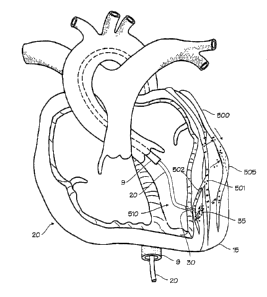
method illustrated in Figure 5 is useful to deliver therapeutic
agents to the coronary arteries, such as the left coronary
artery and its branches, including the left anterior descending
coronary artery, and the right coronary artery and its branches.
As illustrated in Figure 5, catheter system 9 with centrally
located drug delivery catheter 20 implanted at a depth within
the left ventricular apex 15 of the heart 10. Hollow
penetrating structure 30 has penetrated the heart muscle from
the endocardial side. The artery to be treated, in this case
the circumflex branch of the left coronary artery 500, courses
over the surface of the heart (chosen for illustration purposes
only). A corresponding epicardial lymphatic vessel 501 runs
nearby, and many sub-epicardial lymphatic vessel such as vessel
502 drain into the epicardial lymphatic vessel. (It should be
noted that the cardiac lymphatic vessels are both numerous and
largely uncharted, and may be highly variable from person to
person). The artery is occluded by an arterial plaque,
cholesterol or stenotic mass 505 which is amendable to treatment
with drug therapies. The artery may have been previously
treated with angioplasty, or a stent may have been placed across
the occlusion. In any case, several drugs are available to
either ameliorate the blockage or prevent restenosis or re-
occlusion after balloon angioplasty and/or stent placement. The
delivery catheter is navigated into the endocardial space of the
left ventricle 510, and secured in place with penetrating
structure 30. A small dose of therapeutic agent, indicated by
18
CA 02347028 2001-04-17
WO 00/24452 PCT/US99/25068
the molecules 35, is injected into the myocardium, and the
penetrating structure is withdrawn. (Withdrawal of the
penetrating structure may be delayed as necessary to prevent the
therapeutic agent from draining back into the ventricular
space.) The molecules of the therapeutic agent are taken up by
the lymphatic system, entering into vessels 501 and 502, and
transported upwardly. The molecules also migrate out of the
lymphatic system and then migrate into the nearby coronary
artery, following multiple paths indicated by the arrows in
Figure 5. The molecules penetrate the adventicia, or outer
layer, of the coronary artery, and thus enter the coronary
artery. Molecules enter the coronary artery along the entire
length that runs near the lymphatic vessels which initially take
up the molecules. Thus, therapeutic agent enters the coronary
blood vessel at the site of occlusion and proximally to the
occlusion, after having been injected into a more distal
location (relative to the coronary artery). The term entering
the artery may include entering the arterial wall without
entering the lumen of the artery, or passing through the
arterial wall into the lumen of the artery. While the method is
illustrated in relation to the left circumflex coronary artery,
it may be used with all the coronary arteries. Also, while
endocardial access is preferred for the method as applied to the
coronary arteries located on the anterior surface of the heart
(left and right coronary arteries). Therapeutic agents may be
deposited into the myocardium through catheters delivered into
the coronary sinus, the coronary veins, and even the coronary
arteries, including the coronary artery subject to treatment by
angioplasty or stent placement. Additionally, while it is
preferable to accomplish the therapy percutaneously, the method
may be accomplished by injection into the heart, epicardially,
during open surgery, or during endoscopic or key-hole surgery
through the chest.
Various therapeutic agents can be delivered to the coronary
arteries using this approach. Anti-restenosis agents may
include agents which inhibit smooth muscle proliferation,
endothelial cell proliferation, and growth of other components
of arterial plaque and stenosis, antioxidant drugs, anti-
19
CA 02347028 2001-04-17
WO 00/24452 PCT/US99/25068
inflammatory drugs, platelet derived growth factor antagonists,
and numerous other proposed compounds. Anti-restenosis agents
also include anti-neoplastic agents such as taxol, statins (such
as Lovastatin and Provastatin), Pemirolast, Tranilast,
Cilostrazol, INOS, ENOS, EC-NOS, and gene therapy formulations.
A11 of these agents may be formulated as time-release or
controlled release formulations for delivering these molecules
by deposition in the myocardium in position for uptake and
eventual migration into a target site in the coronary arteries.
The therapeutic agents may be incorporated into biodegradable
microspheres with a diameter larger than 15 um (and preferably
greater that 50 um) in diameter so that a depot can be placed
distal to the region of the vessel where treatment is desired
for sustained delivery to the target vessel for extended
periods, such as several hours or several of weeks. The
microspheres would elute agents into the myocardium slowly over
a period of time in order to enable the sustained delivery
through the lymphatics of the heart. In many cases the
molecules may be linked to other molecules such as carbohydrates
to prevent their intravasation and connective losses to the
blood. The microspheres, which are sized to restrict their
migration, degrade within the myocardium near the deposition
site and release agents which then migrate through the
lymphatics and migrate from the lymphatics to the adventicia and
cells within the vascular wall within the target region of the
coronary vessel. For other therapies, gene therapy preparations
are delivered to infect the cardiac myocytes in order to
transfect the RNA for production of the therapeutic proteins
locally which will then migrate through the lymphatic walls to
treat the target vessel peri-adventicially.
The microspheres used in this method are preferably sized
to inhibit migration and immediate uptake by the lymphatic
vessels, and are preferably 50 um in diameter and greater, but
perhaps as small as 30 um. Agents could be encapsulated in
liposomal structures with diameters ranging from 50 to 600 nm
which are transported by the lymphatics and designed to break up
at physiological pH such that agents are released which are able
to diffuse through the lymphatic and arterial walls.
CA 02347028 2001-04-17
WO 00/24452 PCT/US99/25068
Anti-angiogenic agents could also be used to limit the
angiogenic response which has been recently associated in the
literature with atherosclerotic plaques. The hypothesis that
anti-angiogenic agents may limit restenosis could be used during
a revascularization procedure in which angiogenic agents are
delivered along with anti-angiogenic agents at the time of stmt
placement. By having the anti-angiogenic agents be the first
delivered they would transport through the lymphatics and to the
region of injury caused by balloon angioplasty or stmt
placement and minimize the restenosis. Although the reservoir
of microspheres containing angiogenic agents may be delivered at
the same catheterization procedure used to accomplish
angioplasty to stmt placement, and potentially at the same
location, they would be released after the anti-angiogenic and
anti-neoplastic agents have had their effect for limiting
restenosis. Thus dosage forms for anti-angiogenic agents and
angiogenic agents could be placed in the heart simultaneously.
One way of doing this would be to have a microsphere in which
the core contains angiogenic agents and the outer shell contains
anti-angiogenic agents. Another method of doing this is to
supply anti-angiogenic agents in solution or in small
microspheres which are immediately taken up in the lymphatic
vessels, while supplying the angiogenic agents in larger
microspheres which will not be taken up. The method thus
comprises treating a coronary blood vessel with stmt placement,
balloon angioplasty, or both, and delivering a dose of
therapeutic agent to the site of treatment, where the
therapeutic agent is delivered to the myocardium at a location
distal to the site of treatment, and the therapeutic agent
includes anti-angiogenic agent to be released in a time frame
shortly after treatment and angiogenic agent to be released in a
time frame after release of the anti-angiogenic agent.
Alternately, the anti-angiogenic agent can be delivered to the
target site with the angioplasty balloon or stent, by coating
the balloon or stmt with the anti-angiogenic agent, while the
angiogenic agent is deposited in the myocardium for delayed
transport to the target site.
21
CA 02347028 2001-04-17
WO 00/24452 PCT/US99/25068
Thus, the method allows the use of the lymphatic vessels
and endogenous lymphatic transport to carry agents from the
myocardially located depot of therapeutic agents to the target
coronary arteries such that agents are delivered through the
target vessel walls peri-adventicially. This provides a means
of delivering therapeutic agents peri-adventicially to the
vessels of the heart that is far superior to surgical placement
of a peri-adventicial controlled release devices, and delivery
of agents to the space between the pericardial space between the
parietal and visceral pericardium.
While the inventions have been described in relation to the
treatment of cardiac tissue, it should be appreciated that the
compounds and methods of treatment may be applied to various
body tissues. Thus, while the preferred embodiments of the
devices and methods have been described in reference to the
environment in which they were developed, they are merely
illustrative of the principles of the inventions. Other
embodiments and configurations may be devised without departing
from the spirit of the inventions and the scope of the appended
claims.
22