Note: Descriptions are shown in the official language in which they were submitted.
CA 02347212 2001-04-20
WO 00/25297 PCT/IB99/01543
1
ULTRASONIC TRANSDUCER HAVING A TILTED SUPPORT, AND METHOD OF MANUFACTURE
BACKGROUND OF THE INVENTION
The present invention relates generally to
ultrasonic imaging catheters, and more particularly, to
improved ultrasound imaging assemblies and methods of making
same.
Intravascular imaging of blood vessels and
surrounding tissues continues to be of great benefit in a wide
range of medical fields. A particularly successful design for
an intravascular imaging catheter is shown in Fig. 1. The
catheter 10 employs a rotatable imaging assembly 12 containing
an ultrasound transducer 14, where the assembly is attached to
the distal end of a flexible drive cable. A flexible sheath
18 is inserted into a patient, and the drive cable and imaging
assembly are inserted into the sheath. The transducer may be
rotated in order to transmit an ultrasound signal and produce
a video image by well-known techniques.
To produce images, it is desirable to have
ultrasound signals 20 transmitted by the transducer pass
through the sheath (as shown by arrow 22) and reflect off of
tissue or fluids. However, a portion of the ultrasound
signals transmitted by the transducer typically are reflected
by the sheath (as shown by arrow 24). The amount of signal
reflected typically is greatest when the angle of incidence
between the signal and the sheath is about 90 degrees. Hence,
as shown in Fig. 1, it is desirable to tilt the transducer
relative to the sheath, thereby reducing the amount of
reflected signal.
Angling the transducer typically involves forming a
hole 16 in the assembly at the desired angle. However, the
formation of an angled hole, particularly in such a diminutive
assembly, presents difficulties. For example, angled holes
typically have irregularities caused by the hole formation
technique. The transducers often are placed and affixed by
SUBSTITUTE SHEET (RULE 26)
CA 02347212 2001-04-20
WO 00/25297 PCT/IB99/01543
2
hand, which can lead to variations in transducer alignment
between otherwise identical assemblies.
It is desirable, therefore, to provide imaging
assemblies with firmly affixed transducers positioned at the
proper angle relative to the sheath. It also is desirable to
provide imaging assemblies that are easier to manufacture, and
have greater consistency from assembly to assembly.
SUMMARY OF THE INVENTION
The present invention provides exemplary ultrasound
imaging assemblies and methods of making same. Imaging
assembles of the present invention have a machined tilt
transducer package to position the transducer at the proper
angle, thereby reducing the amount of reflected ultrasound
signal from the sheath, and to improve consistency from
transducer assembly to transducer assembly.
In one exemplary embodiment of the present invention
an ultrasound imaging assembly includes a housing having a
distal end, a proximal end, and a longitudinal axis. The
distal end defines a receptacle. The imaging assembly
includes a transducer package having a central axis and an
imaging surface positioned at a desired angle relative to the
central axis. The transducer package is at least partially
disposed within the receptacle so that the central axis is
generally perpendicular to the longitudinal axis. Preferably,
the imaging surface is not coaxial with the longitudinal axis.
In this manner, the imaging surface is positioned to permit a
relatively large percentage of the ultrasound signal to pass
through a sheath surrounding the imaging assembly. The use of
a transducer package having the central axis positioned
generally perpendicular to the housing longitudinal axis
further permits the use of receptacles that are generally
perpendicular to the longitudinal axis. In this manner,
assembly-to-assembly variations are reduced, due in part by
eliminating the need to make angled holes in the housing.
In one aspect of the present invention, the imaging
surface has a shape that is generally elliptical. In another
aspect, the transducer package includes an annular array of
SUBSTITUTE SHEET (RULE 26)
CA 02347212 2001-04-20
WO 00/25297 PCTlIB99/01543
3
transducer elements. It will be appreciated by those skilled
in the art that the transducer package may include transducer
elements and imaging surfaces having a variety of different
shapes within the scope of the present invention.
In one particular aspect, the desired angle between
the central axis and the imaging surface is between about 30
degrees and about 150 degrees, and more preferably, between
about 75 degrees and about 105 degrees. In this manner, the
imaging surface is positioned at the proper angle to permit
the transmission of ultrasound signals through the sheath.
Further, for a transducer package that has a central axis
generally perpendicular to the housing longitudinal axis, the
angle between the longitudinal axis and the imaging surface is
between about +60 degrees and about -60 degrees.
In one aspect, the housing includes stainless steel
and may be plated with an electrically conductive material.
The housing may be plated, for example, with gold, silver,
gold over nickel aver copper, and the like. Preferably, the
housing proximal end is adapted to be coupled to a drive
cable. In this manner, the imaging assembly may be rotated
during operation thereof.
In another aspect, the receptacle is a generally
cylindrical shaped receptacle having an inner wall.
Preferably, the transducer package is disposed within the
receptacle to contact the inner wall. In this manner, tight
tolerances can be maintained to help reduce or eliminate
unwanted transducer package movements within the receptacle.
In one aspect, the transducer package includes at
least one matching layer operably attached to a transducer
element. Alternatively, the transducer package includes a
transducer element operably attached to and between a matching
layer and a backing layer. It will be appreciated by those
skilled in the art that a plurality of matching layers, or no
matching layer, may be used within the scope of the present
invention. In one particular aspect, the backing layer is
electrically conductive. Alternatively, the backing layer is
electrically nonconductive.
SUBSTITUTE SHEET (RULE 26)
CA 02347212 2001-04-20
WO 00/25297 PCT/IB99/01543
4
In still another aspect, the imaging assembly
further includes a potting well located in the distal end.
The potting well is adapted to receive an electrically
conductive material so that the material is in contact with
the transducer package, and preferably in contact with the
backing layer.
The present invention further provides an exemplary
ultrasound imaging catheter. The imaging catheter includes an
imaging assembly, ostensibly as previous described, and a
drive cable coupled to the proximal end.
The invention also provides exemplary methods of
making an ultrasound imaging assembly. In one exemplary
method, a first transducer package is provided having an
imaging surface. A cutting device is provided and the first
transducer package is positioned so that the imaging surface
is at a desired angle relative to the cutting device. The
method includes cutting the first transducer package with the
cutting device to form a second transducer package having a
central axis. The central axis is at the desired angle
relative to the imaging surface. The method includes
providing a housing having a distal end, a proximal end and a
longitudinal axis, with the distal end defining a receptacle.
The second transducer package is positioned to be at least
partially disposed within the receptacle so that the central
axis is generally perpendicular to the longitudinal axis. In
this manner, the second transducer package has a machined
angle, thereby permitting use of a receptacle that is
generally perpendicular to the longitudinal axis. In one
aspect, the desired angle between the central axis and imaging
surface is between about 30 degrees and about 150 degrees.
Alternatively, the desired angle is between about 75 degrees
and about 105 degrees.
In one aspect of the present invention, the cutting
device is selected from a group of cutting devices consisting
of a cutting blade, a drill Such as a core drill, a laser, an
end mill, and the like. In another aspect, the receptacle and
second transducer package are generally cylindrical. In still
SUBSTITUTE SHEET (RULE 2f)
CA 02347212 2001-04-20
WO 00/25297 PCT/IB99/01543-
another aspect, the second transducer package is generally
cylindrical and the imaging surface is generally elliptical.
In one particular aspect, the method includes the
step of removing a portion of the matching layer to define an
5 electrical lead attachment point. In one aspect, the lead
attachment point is aligned to be a proximal-most point of the
second transducer package.
The invention further provides exemplary methods of
manufacturing an ultrasound transducer package. One such
method includes providing a transducer element having first
and second electrodes operably attached to first and second
transducer element surfaces. A matching layer is operably
attached to the first electrode and a backing material is
operably attached to the second electrode. A laser,
preferably an excimer laser, is aligned over a desired region
of the matching layer to ablate the desired region. In this
manner, laser ablation accurately creates a notch so that a
lead can be electrically connected to the first electrode.
In one aspect the laser is aligned over a second
desired region located in the backing material and operated to
ablate the second desired region. In one aspect of the
method, the desired region is ablated until the first
electrode is visible. Similarly, the laser is operated to
ablate the second desired region until the second electrode is
visible. In another aspect, the laser is operated for a
desired number of pulses at a power level sufficient to ablate
the matching layer and insufficient to ablate the first
electrode.
Other features and advantages of the invention will
appear from the following description in which the preferred
embodiment has been set forth in detail in conjunction with
the accompanying drawings.
BRIEF DESCRIPTION OF THE DRAWINGS
Fig. 1 depicts a typical imaging catheter;
Figs. 2A-2B depict transducer packages according to
the present invention;
SUBSTITUTE SHEET (RULE 26)
CA 02347212 2001-04-20
WO 00/25297 PCT/IB99/0154~
6
Figs. 3A-3B depict methods of making transducer
packages in accordance with the present invention;
Figs. 3C-3D are cross-sectional views of the
transducer package depicted in Fig. 3B;
Fig. 4A is a side cross sectional view of an
exemplary imaging catheter according to the present invention;
Fig. 4B is a top overall view of the catheter shown
in Fig. 4A;
Fig. 5 is a side cross sectional view of an imaging
catheter of the present invention within a sheath;
Figs. 6A-6C are overall top views of alternative
transducer packages according to the present invention;
Fig. 6D depicts a side cross sectional view of
another transducer package according to the present invention;
Figs. 7A-7B depict a notch formed in the transducer
package to permit electrical lead attachment to the electrode;
Fig. 7C depicts an alternative electrode for use
with the transducer package of the present invention; and
Fig. 8 is a schematic showing laser ablation of
notches in the transducer package of Fig. 7.
DETAILED DESCRIPTION OF THE PREFERRED EMBODIMENT
Figs. 2A-2B depict a first transducer package 40
containing a matching layer 42, a transducer element 44, and a
backing material 46. Transducer element 44 may comprise a
variety of the materials, including piezocomposite materials,
piezoceramics (such as PZT), piezoplastics, and the like. A
first electrode 48 and a second electrode 50 are operably
attached to opposing sides of transducer element 44.
Electrodes 48 and 50 typically cover one entire surface of
transducer element 44, as shown in Fig. 2A. However,
electrodes 48 and 50 may cover a smaller or greater portion of
transducer element 44. Electrodes 48 and 50 preferably
include gold, gold over titanium, gold over nickel, gold over
chromium, and the like. As shown in Fig. 2B, matching layer
42, transducer element 44 with electrodes 48 and 50, and
backing material 46 are operably connected in a stacked
configuration using an epoxy or the like. First transducer
SUBSTITUTE SHEET (RULE 26)
CA 02347212 2001-04-20
WO 00/25297 PCT/IB99/01543-
7
package 40 then can be manufactured in accordance with the
steps described in conjunction with Fig. 3.
As shown in Fig. 3A, first transducer package 40 is
positioned at an angle 54 relative to a cutting device 56.
Cutting device 56 may comprise a cutting blade, a drill such
as a core drill, a laser, an end mill, and the like. In one
particular embodiment, cutting device 56 comprises an end mill
having a width of between about 0.010 inch (10 thousandths of
an inch) and about 0.050 inch (50 thousandths of an inch).
While first transducer package 40 is positioned at angle 54
relative to cutting device 56, cutting device 56 cuts first
transducer package 40 along a cut 58 indicated by dashed lines
in Fig. 3A. This forms a second transducer package 60 as
depicted to Fig. 3B.
Second transducer package 60 has a central axis 62
and an imaging surface 66. Central axis 62 typically will be
coaxial with cut 58 formed by cutting device 56. In the event
cutting device 56 is a cylindrical shape cutting device, or is
used to provide a cylindrical-shaped cut 58, second transducer
package 60 has a cylindrical cross-section as shown in Fig.
3C. Due to angle 54, imaging surface 66 will have an
elliptical shape as shown in Fig. 3D. In another embodiment
(not shown), second transducer package is formed so that
imaging surface 66 has a circular shape. Second transducer
package 60 may have an excess portion 64 which can be removed
either prior to or after insertion of second transducer
package 60 into a housing receptacle as further described in
conjunction with Fig. 4.
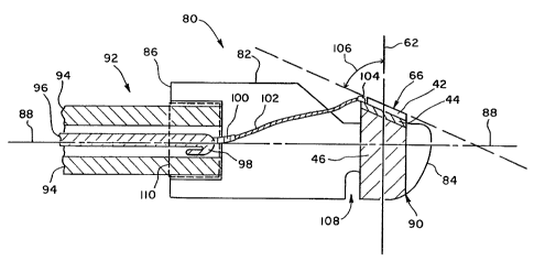
Turning now to Figs. 4A-4B, an exemplary imaging
assembly configured with an exemplary imaging catheter will be
described. Fig. 4A depicts an imaging assembly 80 having a
housing 82. Housing 82 has a distal end 84, a proximal end
86, and a longitudinal axis 88. Housing 82 preferably
comprises stainless steel plated with an electrically
conductive material. For example, housing 82 may be plated
with gold, silver, gold over nickel over copper, and the like.
Plating housing 82 with electrically conductive material
provides housing 82 with an electrically conductive surface
SUBSTITUTE SHEET (RULE 26)
CA 02347212 2001-04-20
WO 00/25297 PCT/IB99/01543
8
which can be used, for example, as a negative or ground
connection for transducer element 44. Housing 82 includes a
receptacle 90 defined within distal end 84. As shown in Fig.
4A receptacle 90 is filled with second transducer package 60.
Prior to or after inserting second transducer package 60 into
receptacle 90, excess portion 64 of backing material 46 may be
removed to provide distal end 84 with a smooth surface.
Imaging assembly 80 is operably attached to a drive
cable 92. Preferably, an epoxy as shown by dashed lines 110
operably attaches drive cable 92 to proximal end 84. In one
particular embodiment, drive cable 92 includes a counterwound
94 surrounding a mini coaxial cable 96. Exemplary drive
cables for use with the present invention are further
described in U.S. Patent Application Serial No. 09/017,578
entitled "Integrated Coaxial Transmission Line and Flexible
Drive Cable", the disclosure of which is incorporated herein
by reference. Mini coaxial cable 96 includes a shield 98
surrounding an insulated lead 100. As shown in Fig. 4A,
shield 98 is folded back to reveal insulated lead 100.
Insulated lead 100 is striped of insulation to reveal lead 102
which is used as a connection with transducer 44. As shown in
Fig. 4A, lead 102 preferably is connected to first electrode
48 by an attachment point 104. Attachment point 104 may
comprise electrically conductive epoxy (e. g., silver epoxy)
and the like.
As described in conjunction with Fig. 3, second
transducer package 60 has been manufactured to position
imaging surface 66 at a desired angle relative to central axis
62. Preferably, desired angle 106 as shown in Fig. 4A is
between about 30 degrees and about 150 degrees, and more
preferably between about 75 degrees and about 105 degrees. In
the embodiment shown in Fig. 4A, a potting well 108 is formed
in housing distal end 84. Potting well 108 preferably is
filled with an electrically conductive material, such as
silver epoxy, gold epoxy, conductive silicone, conductive
urethane, and the like. In this manner, the conductive
material-filled potting well 108 is in contact with backing
material 46. For second transducer package 60 having an
SUBSTITUTE SHEET (RULE 26)
CA 02347212 2001-04-20
WO 00/25297 PCT/IB99/01543
9
electrically conductive backing material 46, potting well 108
provides an electrical connection as well as a mechanical
connection between backing material 46 and the surface of
housing 82. Such a potting well 108 may be necessary to form
electrical and mechanical connections due to the tight
tolerances between second transducer package 60 and receptacle
90.
As previously indicated, housing 82 preferably is
plated with electrically conductive material. By using an
electrically conductive epoxy 110 such as silver epoxy or gold
epoxy, or conductive silicone, conductive urethane, or the
like, an electrically conductive path is established between
second electrode 50, backing material 46, conductive material-
filled potting well 108, distal housing 82, and shield 98.
For the embodiment shown in Fig. 4A, a negative connection is
provided to second electrode 50. Lead 102 provides a positive
electrical connection to first electrode 48 via attachment
point 104. Alternatively the polarities may be reversed
within the scope of the present invention.
Fig. 4B depicts a top view of imaging assembly 80.
Lead 102 is shown operably attached to the proximal-most
portion of transducer 44. In this embodiment, imaging surface
66 is elliptical in shape. It will be appreciated by those
skilled in this art, and as shown by way of example in Fig. 6,
that a variety of shapes for imaging surface 66 and different
locations for attachment point 104 may be used within the
scope of the present invention.
As shown is Fig. 5, imaging assembly 80 of the
present invention provides imaging surface 66 with the
appropriate angle relative to a sheath 120 into which imaging
assembly 80 is disposed. In this manner, a large percentage
of the ultrasound signal passes through sheath 120 compared to
that reflected by sheath 120.
The manufacture of receptacle 90 in distal end 82
also is advantageous. For example, the distal housing shown
in Fig. 1 had a saw-tooth configuration due to machining
limitations. Machines used to make the distal housing
typically have cutting tools that are at right angles with
SUBSTITUTE SHEET (RULE 26)
CA 02347212 2001-04-20
WO 00/25297 PCT/IB99/OI543
respect to the longitudinal axis of the housing. In order to
machine an angled hole, the machinist typically uses an end
mill having a smaller cross section than the receptacle cross
section. The machinist must step the end mill in and make
5 multiple cuts as the housing is advanced. The resulting
angled hole typically is jagged or sawtoothed, and can result
in proximal-to-distal movement of the transducer package
placed therein.
In contrast, receptacle 90 can be made with a single
10 cut or stroke without producing a jagged or sawtooth
receptacle. The cutting device used also can have the same
cross section as the desired receptacle 90. The production of
a smooth-sided receptacle 90 that is generally perpendicular
to housing longitudinal axis 88 can be achieved by a variety
of techniques, including drilling, milling, machining, and the
like.
Further, by producing desired angle 106 during
second transducer package 60 manufacture, increased assembly-
to-assembly consistency is achieved, in part by avoiding the
need to closely monitor the desired angle while creating
receptacle 90 in stainless steel housing 82.
Alternative embodiments of second transducer
packages will be described in conjunction with Fig. 6. For
example, Fig. 6A depicts a second transducer package 160
having an elliptical imaging surface 130 and Fig. 6B depicts
imaging surface 130 as generally rectangular in shape. It
will be appreciated by those skilled in the art that a variety
of shapes for imaging surface 130 may be used within the scope
of the present invention. Further, second transducer package
may comprise an annular array 180 of transducer elements as
shown in Fig. 6C and as further described in U.S. Patent
Application Serial No. 09/017,581 entitled "Annular Array
Ultrasound Catheter", the complete disclosure of which is
incorporated herein by reference.
In addition to second transducer package 60 having
one matching layer 42 as shown in Figs. 2-4, it will be
appreciated by those skilled in the art that the number of
matching layers may vary within the scope of the present
SUBSTITUTE SHEET (RULE 26)
CA 02347212 2001-04-20
WO 00/Z5297 PCT/IB99/01543
11
invention. For example as shown in Fig. 6D, second transducer
package 160 may have first and second matching layers 132 and
134, a transducer element 136 and a backing layer 138.
As shown in Fig. 4, in some embodiments it is
desirable to attach lead 102 to first electrode 48 to permit
the transmission of electrical signals to and from transducer
element 44. As described in conjunction with Figs. 7 and 8,
the present invention provides exemplary methods of creating a
notch 140 for lead 102 to use to attach to first electrode 48.
Figs. 7A and 7B depict notch 140 formed in matching
layer 42 to permit lead 102 to be attached to first electrode
48 by attachment point 104. While Figs. 7A-7C depict notch
140 being formed in first transducer package 40, notch 140
also can be formed after manufacture of second transducer
package 60 within the scope of the present invention.
Further, while notch 140 is depicted formed in a single
matching layer 42, notch 140 also can be formed through
multiple matching layers for those transducer package
embodiments employing more than one matching layer.
Typically, the formation of notch 140 is problematic
due in part to the diminutive size of transducer package 40.
Further, the creation of notch 140 by hand, such as with a
knife or razor blade, can cause variations in notch 140 size
between otherwise identical transducer packages 40. Further,
in some embodiments it also is desirable to precisely locate
notch 140, for example when creating notch 140 to contact a
star-shaped electrode 150 as shown in Fig. 7C.
To overcome at least some of these problems and to
precisely create notches 140, the system depicted in Fig. 8
can be used. System 200 uses a laser 160, preferably an
excimer laser 160, to create notch 140. Excimer laser 160
operates to dissociate the bonds of the material comprising
the matching layer 42. Further, laser 160 can be used to
remove the resin or epoxy (not shown) used to affix matching
layer 42 to first electrode 48. In this manner, electrode 48
can be exposed to permit lead 102 attachment thereto.
At least part of the present invention involves the
recognition that laser ablation can precisely create notch 140
SUBSTITUTE SHEET (RULE 26)
CA 02347212 2001-04-20
WO 00/25297 PCT/IB99/01543
12
having the desired size. Since matching layer 42 operates to
reduce the amount of unwanted ultrasound wave reflection, it
is desirable to only remove as much matching layer 42 material
as is necessary to permit lead 102 attachment. Operating
levels of laser 160 can be set to ablate the appropriate
amount of matching layer 42 material to create notch 140
having the desired size. Laser 160 further permits
consistency in notch 140 manufacture for a number of
transducer packages 40.
Laser 160 also can create notch 140 at a desired
location on matching layer 42. In this manner, notch 140 can
be used to help correctly align second transducer package in
receptacle 90. For example, in the embodiment shown in Fig.
4B, notch 140 is created at the highest point on second
transducer package 60. By then positioning second transducer
package 60 within receptacle 90 so that notch 140 is the
proximal-most point of second transducer package 60, a forward
tilt imaging surface 66 can be established.
By way of example, for matching layer 42 comprising
silver filled epoxy, other filled epoxies or adhesives, and
the like, laser 160 can be operated at about 1.0
Joules/centimeterz (J/cm2) for about 10 to about 50 pulses of
about 0.25 nanoseconds duration to ablate matching layer 42
that is about 0.0003 inches to about 0.007 inches thick.
Similarly, energy levels to ablate epoxy connecting matching
layer 42 to first electrode 48 are about one to about 1.4
J/cmz. However, the energy levels needed to ablate a metal,
such as gold used for first electrode 48 are considerably
higher. For example, gold requires about 7 to about 20 J/cm2
to be ablated. Such a difference in ablation energies allows
the operation of laser 160 at energy levels sufficient to
ensure ablation of matching layer 42 and epoxy, without
causing damage to first electrode 48. In this manner, the
ablation of matching layer 42 can occur by calculating the
number of pulses needed to completely ablate matching layer
42, and operating laser 160 at the required energy levels and
pulses. Additional pulses also can be used to ensure complete
ablation of matching layer 42, yet occur at power levels low
SUBSTITUTE SHEET (RULE 26)
CA 02347212 2001-04-20
WO 00/25297 PCT/IB99/01543
13
enough to avoid damaging electrode 48. Alternatively, laser
160 can be operated until a visual inspection reveals
electrode 48 has been exposed.
Similarly, as shown in Fig. 8, laser 160 can be used
to ablate a notch 170 in backing material 46. Notch 170 would
be particularly useful in the event backing material 46 is
nonconductive. In this manner, notch 170 can be used to
provide an electrical contact to electrode 50. It may be
desirable to create notch 170 at an angle to minimize the
amount of backing material 46 ablated.
It will be appreciated by those skilled in the art
that while the present invention has been described with notch
140 formed to expose first electrode 48, alternative methods
may be used to mount lead 102 to first electrode 48 within the
scope of the present invention. For example, off aperture
mounting techniques disclosed in U.S. Application Serial No.
09/127,994, entitled "PZT Off-Aperture Bonding Technique"
(Attorney Reference No. 12553-007100), and U.S. Application
Serial No. 09/127,089, entitled "Off Aperture Electrical
Connection for Ultrasonic Transducer" (Attorney Reference No.
12553-007600) may be used, the complete disclosures of which
are incorporated herein by reference.
The invention has now been described in detail.
However, it will be appreciated that certain changes and
modifications may be made. For example, the use of laser
ablation to create notch 140 can be applied to a wide range of
transducer configurations, including those described in
09/127,694, entitled "Method of Tuning Ultrasonic Transducer
Matching Layer", the complete disclosure of which are
incorporated herein by reference. Therefore, the scope and
content of this invention are not limited by the foregoing
description. Rather, the scope and content are to be defined
by the following claims.
SUBSTITUTE SHEET (RULE 26)