Note: Descriptions are shown in the official language in which they were submitted.
CA 02347649 2001-04-19
WO 00/24913 PCTIUS99/24819
MN GENE AND PROTEIN
FIELD OF THE INVENTION
The present invention is in the general area of medical genetics and in
the fields of biochemical engineering, immunochemistry and oncology. More
specifically, it relates to the MN gene - a cellular gene considered to be an
oncogene,
which encodes the oncoprotein now known alternatively as the MN protein, the
MN/CA IX isoenzyme or the MN/G250 protein.
BACKGROUND OF THE INVENTION
Zavada et al., International Publication Number WO 93/18152
(published 16 September 1993) and U.S. Patent No. 5,387,676 (issued February
7,
1996), describe the elucidation of the biological and molecular nature of MaTu
which
resulted in the discovery of the MN gene and .protein. The MN gene was found
to be
present in the chromosomal DNA of all vertebrates tested, and its expression
to be
strongly correlated with tumorigenicity.
The MN protein was first identified in HeLa cells, derived from a human
carcinoma of cervix uteri. It is found in many types of human carcinomas
(notably
uterine cervical, ovarian, endometrial, renal, bladder, breast, colorectal,
lung,
esophageal, and prostate, among others). Very few normal tissues have been
found to
express MN protein to any significant degree. Those MN-expressing normal
tissues
include the human gastric mucosa and gallbladder epithelium, and some other
normal
tissues of the alimentary tract. Paradoxically, MN gene expression has been
found to
be lost or reduced in carcinomas and other preneoplastidneoplastic diseases in
some
tissues that normally express MN, e.g., gastric mucosa.
In general, oncogenesis may be signified by the abnormal expression of
MN protein. For example, oncogenesis may be signified: (1) when MN protein is
present in a tissue which normally does not express MN protein to any
significant
degree; (2) when MN protein is absent from a tissue that normally expresses
it; (3) when
MN gene expression is at a significantly increased level, or at a
significantly reduced
1
CA 02347649 2001-04-19
WO 00/24913 PCT/US99/24879
level from that normally expressed in a tissue; or (4) when MN protein is
expressed in
an abnormal location within a cell.
Zavada et al., WO 93/18152 and Zavada et al., WO 95/34650 (published
21 December 1995) disclose how the discovery of the MN gene and protein and
the
strong association of MN gene expression and tumorigenicity led to the
creation of
methods that are both diagnostic/prognostic and therapeutic for cancer and
precancerous conditions. Methods and compositions were provided therein for
identifying the onset and presence of neoplastic disease by detecting or
detecting and
quantitating abnormal MN gene expression in vertebrates. Abnormal MN gene
expression can be detected or detected and quantitated by a variety of
conventional
assays in vertebrate samples, for example, by immunoassays using MN-specific
antibodies to detect or detect and quantitate MN antigen, by hybridization
assays or by
PCR assays, such as RT-PCR, using MN nucleic acids, such as, MN cDNA, to
detect or
detect and quantitate MN nucleic acids, such as, MN mRNA.
Zavada et al, WO 93/18152 and WO 95/34650 describe the production
of MN-specific antibodies. A representative and preferred MN-specific
antibody, the
monoclonal antibody M75 (Mab M75), was depcsited at the American Type Culture
Collection (ATCC) in Manassus, VA (USA) under ATCC Number HB 11128. The M75
antibody was used to discover and identify the MN protein and can be used to
identify
readily MN antigen in Western blots, in radicimmunoassays and
immunohistochemically, for example, in tissue samples that are fresh, frozen,
or
formalin-, alcohol-, acetone- or otherwise fixed and/or paraffin-embedded and
deparaffinized. Another representative and preferred MN-specific antibody, Mab
MN12, is secreted by the hybridoma MN 12.2.2, which was deposited at the ATCC
under the designation HB 11647. Example 1 of Zavada et al., WO 95/34650
provides
representative results from immunohistochemical staining of tissues using MAb
M75,
which results support the designation of the MN gene as an oncogene.
Many studies have confirmed the diagnostic/prognostic utility of MN.
The following articles discuss the use of the MN-specific MAb M75 in
diagnosing/prognosing precancerous and cancerous cervical lesions: Leff, D.
N., "Half
a Century of HeLa Cells: Transatlantic Antigen Enhances Reliability of
Cervical Cancer
Pap Test, Clinical Trials Pending," BioWorld Today: The Daily Biotechnology
2
CA 02347649 2001-04-19
WO 00/24913 PCT/US99/24879
Newspaper, 9(55) (March 24, 1998); Stanbridge, E. J., "Cervical marker can
help
resolve ambigous Pap smears," Diagnostics Intelligence, 10(5): 11 (1998); Liao
and
Stanbridge, "Expression of the MN Antigen in Cervical Papanicolaou Smears Is
an Early
Diagnostic Biomarker of Cervical Dysplasia," Cancer Epidemiology, Biomarkers &
Prevention, 5: 549-557 (1996); Brewer et at., "A Study of Biomarkers in
Cervical
Carcinoma and Clinical Correlation of the Novel Biomarker MN," Gynecologic
Oncology, 63: 337-344 (1996); and Liao et al., "Identification of the MN
Antigen as a
Diagnostic Biomarker of Cervical Intraepithelial Squamous and Glandular
Neoplasia
and Cervical Carcinomas," American lournal of Pathology, 145(3): 598-609
(1994).
Premalignant and Malignant Colorectal Lesions. MN has been detected
in normal gastric, intestinal, and biliary mucosa. [Pastorekova et al.,
Gastroenterology.
112: 398-408 (1997).] Immunohistochemical analysis of the normal large
intestine
revealed moderate staining in the proximal colon, with the reaction becoming
weaker
distally. The staining was confined to the basolateral surfaces of the cryptal
epithelial
cells, the area of greatest proliferative capacity. As MN is much more
abundant in the
proliferating cryptal epithelium than in the upper part of the mucosa, it may
play a role
in control of the proliferation and differentiation of intestinal epithelial
cells. Cell
proliferation increases abnormally in premalignarit and malignant lesions of
the
colorectal epithelium, and therefore, is considered an indicator of colorectal
tumor
progression. [Risio, M., 1. Cell Biochem. 16G: 79-87 (1992); and Moss et al.,
Gastroenterology. 111: 1425-1432 (1996).]
The MN protein is now considered to be the first tumor-associated
carbonic anhydrase (CA) isoenzyme that has been described. Carbonic anhydrases
(CAs) form a large family of genes encoding zinc metalloenzymes of great
physiological
importance. As catalysts of reversible hydration of carbon dioxide, these
enzymes
participate in a variety of biological processes, including respiration,
calcification, acid-
base balance, bone resorption, formation of aqueous humor, cerebrospinal
fluid, saliva
and gastric acid [reviewed in Dodgson et al., The Carbonic Anhydrases, Plenum
Press,
New York-London, pp. 398 (1991)]. CAs are widely distributed in different
living
organisms.
In mammals, at least seven isoenzymes (CA I-VII) and a few CA-related
proteins (CARP/CA VIII, RPTP-(3, RPTP-T) had been identified [Hewett-Emmett
and
3
CA 02347649 2001-04-19
WO 00/24913 PCT/US99/24819
Tashian, Mol. Phyl. Evol.. 5: 50-77 (1996)], when analysis of the MN deduced
amino
acid sequence revealed a striking homology between the central part of the MN
protein
and carbonic anhydrases, with the conserved zinc-binding site as well as the
enzyme's
active center. Then MN protein was found to bind zinc and to have CA activity.
Based
on that data, the MN protein is now considered to be the ninth carbonic
anhydrase
isoenzyme - MN/CA IX. [Opavsky et al., Genomics, 33: 480-487 (May 1996)]. [Lqg
also, Hewett-Emmett, supra, wherein CA IX is suggested as a nomenclatural
designation.]
CAs and CA-related proteins show extensive diversity in both their tissue
distribution and in their putative or established biological functions
[Tashian, R. E., Adv.
in Genetics, 30: 321-356 (1992)]. Some of the CAs are expressed in almost all
tissues
(CA II), while the expression of others appears to be more restricted (CA VI
and CA VII
in salivary glands). In cells, they may reside in the cytoplasm (CA I, CA II,
CA III, and
CA VII), in mitochondria (CA V), in secretory granules (CA VI), or they may
associate
with membrane (CA IV). Occasionally, nuclear localization of some isoenzymes
has
been noted [Parkkila et al., Gut 35: 646-650 (1994); Parkkilla et al.,
Histochem. I., 27:
133-138 (1995); Mori et al., Gastroenterol., 105: 820-826 (1993)].
The CAs and CA-related proteins also differ in kinetic properties and
susceptibility to inhibitors [Sly and Hu, Annu. Rev. Biochem., 64: 375-401
(1995)]. In
the alimentary tract, carbonic anhydrase activity is involved in many
important
functions, such as saliva secretion, production of gastric acid, pancreatic
juice and bile,
intestinal water and ion transport, fatty acid uptake and biogenesis in the
liver. At least
seven CA isoenzymes have been demonstrated in different regions of the
alimentary
tract. However, biochemical, histochemical and immunocytochemical studies have
revealed a considerable heterogeneity in their levels and distribution
[Swensen, E. R.,
"Distribution and functions of carbonic anhydrase in the gastrointestinal
tract," In: The
Carbonic Anhydrases. Cellular Physiology and Molecular Genetics. (Dodgson et
al.
eds.) Plenum Press, New York, pages 265-287 (1991); and Parkkila and Parkkila,
Scan
1. Gastroenterol., 31: 305-317 (1996)]. While CA II is found along the entire
alimentary canal, CA IV is linked to the lower gastrointestinal tract, CA I,
III and V are
present in only a few tissues, and the expression of CA VI and VII is
restricted to
4
CA 02347649 2004-11-26
salivary glands [Parkkila et al., Gut. 35: 646-650 (1994); Fleming et al., J.
Clin. Invest.,
96: 2907-2913 (1995); Parkkila et al., Hepatology. 24: 104 (1996)].
MN/CA IX has a number of properties that distinguish it from other known
CA isoenzymes and evince its relevance to oncogenesis. Those properties
include its
density dependent expression in cell culture (e.g., HeLa cells), its
correlation with the
tumorigenic phenotype of somatic cell hybrids between HeLa and normal human
fibroblasts, its close association with several human carcinomas and its
absence from
corresponding normal tissues [ems., Zavada et at., Int. J. Cancer. 54: 268-274
(1993);
Pastorekova et al., Virology. 187: 620-626 (1992); Liao et al., Am. J.
Pathol.. 145:
598-609 (1994); Pastorek et al., Oncogene. 9: 2788-2888 (1994); Cote, Women's
Health Weekly: News Section, p. 7 (March 30, 1998); Liao et at., Cancer Res..
57:
2827 (1997); Vermylen et al., "Expression of the MN antigen as a biomarker of
lung
carcinoma and associated precancerous conditions," Proceedings AACR. 39: 334
(1998); McKieman et al., Cancer Res.. 57: 2362 (1997); and Turner et at., Hum.
Pathol.. 28(6): 740 (1997)]. In addition, the in vitro transformation
potential of MN/CA
IX cDNA has been demonstrated in NIH 3T3 fibroblasts [Pastorek et at., id.J.
The MN protein has also been identified with the G250 antigen. Uemura
et at., "Expression of Tumor-Associated Antigen MN/G250 in Urologic Carcinoma:
Potential Therapeutic Target, " J. Urol.. 157 (4 Suppl.): 377 (Abstract 1475;
1997)
states: "Sequence analysis and database searching revealed that G250 antigen
is
identical to MN, a human tumor-associated antigen identified in cervical
carcinoma
(Pastorek et al., 1994)."
SUMMARY OF THE INVENTION
Identified herein is the location of the MN protein binding site. Of
particular importance is the region within the proteoglycan-like domain, as 61-
96 (SEQ
ID NO: 97) which contains a 6-fold tandem repeat of 6 amino acids, and within
which
the epitope for the M75 MAb resides in at least two copies, and within which
the MN
binding site is considered to be located. An alternative MN binding site may
be located
in the CA domain.
5
CA 02347649 2001-04-19
WO 00/24913 PCT/US99/24879
Also identified are MN proteins and MN polypeptides that compete for
attachment to cells with immobilized MN protein. Such MN proteins/polypeptides
prevent cell-cell adhesion and the formation of intercellular contacts.
Disclosed herein are cell adhesion assay methods that are used to identify
binding site(s) on the MN protein to which vertebrate cells, preferably
mammalian
cells, more preferably human cells, bind. Such a MN binding site is then
identified as a
therapeutic target which can be blocked with MN-specific antibodies, or
inorganic or
organic molecules, preferably organic molecules, more perferably
proteins/polypeptides
that specifically bind to said site.
Further disclosed are therapeutic methods to treat patients with
preneoplastidneoplastic disease associated with or characterized by abnormal
MN
expression, which methods are based on blocking said MN binding site with
molecules, inorganic or organic, but preferably organic molecules, more
preferably
proteins/polypeptides, that bind specifically to said binding site. The growth
of a
vertebrate preneoplastidneoplastic cell that abnormally expresses MN protein
can be
inhibited by administering such organic or inorganic molecules, preferably
organic
molecules, more preferably proteins/polypeptides in a therapeutically
effective amount
in a physiologically acceptable formulation. Such a preferred therapeutic
protein/polypeptide is herein considered to comprise an amino acid sequence
selected
from the group consisting of SEQ ID NOS: 107-109. Such heptapeptides are
considered to be comprised by MN protein partner(s). Blocking the interaction
between MN protein and its binding partner(s), is expected to lead to a
decrease of
tumor growth.
Further provided are other therapeutic methods wherein the growth of a
vertebrate, preferably mammalian, more preferably human, preneoplastic or
neoplastic
cell that abnormally expresses MN protein is inhibited. Said methods comprise
transfecting said cell with a vector comprising an expression control sequence
operatively linked to a nucleic acid encoding the variable domains of an MN-
specific
antibody, wherein said domains are separated by a flexible linker peptide,
preferably
SEQ ID NO: 116. Preferably said expression control sequence comprises the MN
gene
promoter.
6
CA 02347649 2001-04-19
WO 00/24913 PCT/US99/24879
Still further therapeutic methods comprise transfecting said cell with a
vector comprising a nucleic acid that encodes a cytotoxic protein/polypeptide,
such as
HSVtk, operatively linked to the MN gene promoter. Such a therapeutic vector
may
also comprise a nucleic acid encoding a cytokine, such as, IL-2 or IFN.
Aspects of the instant invention disclosed herein are described in more
detail as follows. The therapeutic use of organic or inorganic molecules,
preferably
organic molecules, is disclosed. Preferred such molecules bind specifically to
a site on
MN protein to which vertebrate cells adhere in a cell adhesion assay, wherein
said
molecule when tested in vitro inhibits the adhesion of cells to MN protein.
Further
preferred are such molecules, which when in contact with a vertebrate
preneoplastic or
neoplastic cell that abnormally expresses MN protein, inhibit the growth of
said cell.
Said vertebrate cells are preferably mammalian and more preferably human.
Preferably such a molecule is organic, and more preferably such a
organic molecule is a protein or a polypeptide. Still further preferably, said
protein or
polypeptide comprises an amino acid sequence selected from the group
consisting of
SEQ ID NOS: 107, 108, 109, 137 and 138. Even more preferably, said polypeptide
is
selected from the group consisting of SEQ ID NOS: 107, 108, 109, 137 and 138.
The site on MN proteins to which vertebrate cells adhere in said cell
adhesion assay is preferably within the proteoglycan-like domain [SEQ ID NO:
50] or
within the carbonic anhydrase domain [SEQ ID NO: 51] of the MN protein.
Preferably
that site comprises an amino acid sequence selected from the group consisting
of SEQ
ID NOS: 10 and 97-106. Still further preferably, that site has an amino acid
sequence
selected from the group consisting of SEQ ID NOS: 10 and 97-106.
Another aspect of this invention concerns MN proteins and MN
polypeptides which mediate attachment of vertebrate cells in a cell adhesion
assay,
wherein said MN protein or MN polypeptide when introduced into the
extracellular
fluid environment of vertebrate cells prevents the formation of intercellular
contacts and
the adhesion of said vertebrate cells to each other. Such MN proteins and MN
polypeptides may be useful to inhibit the growth of vertebrate preneoplastic
or
neoplastic cells that abnormally express MN protein, when such MN proteins or
MN
polypeptides are introduced into the extracellular fluid environment of such
vertebrate
cells. Said vertebrate cells are preferably mammalian, and more preferably
human.
7
CA 02347649 2001-04-19
WO 00/24913 PCT/US99/24819
Said MN proteins or MN polypeptides which mediate attachment of
vertebrate cells in a cell adhesion assay, preferaWy have amino acid sequences
from
SEQ ID NO: 97, from SEQ ID NO: 50, or from SEQ ID NO: 51, more preferably from
SEQ ID NO: 50. Still more preferably such MN proteins or MN polypeptides
comprise
amino acid sequences selected from the group consisting of SEQ ID NOS: 10 and
97-
106. Alternatively, said MN polypeptides are selected from the group
consisting of
SEQ ID NOS: 10 and 97-106.
Representative MN proteins and MN polypeptides which mediate
attachment of vertebrate cells in a cell adhesion assay, are specifically
bound by either
the M75 monoclonal antibody that is secreted from the hybridoma VU-M75, which
was
deposited at the American Type Culture Collection under ATCC No. HB 11128, or
by
the MN12 monoclonal antibody that is secreted from the hybridoma MN 12.2.2,
which
was deposited at the American Type Culture Collection under ATCC No. HB 11647,
or
by both said monoclonal antibodies.
Another aspect of the instant invention is a method of identifying a site on
an MN protein to which vertebrate cells adhere by testing a series of
overlapping
polypeptides from said MN protein in a cell adhesion assay with vertebrate
cells, and
determining that if cells adhere to a polypeptide from said series, that said
polypeptide
comprises a site on said MN protein to which vertebrate cells adhere.
Still another aspect of the instant invention is a vector comprising an
expression control sequence operatively linked to a nucleic acid encoding the
variable
domains of a MN-specific antibody, wherein said domains are separated by a
flexible
linker polypeptide, and wherein said vector, when transfected into a
vertebrate
preneoplastic or neoplastic cell that abnormally expresses MN protein,
inhibits the
growth of said cell. Preferably said expression control sequence comprises the
MN
gene promoter operatively linked to said nucleic acid. Further preferably,
said flexible
linker polypeptide has the amino acid sequence of SEQ ID NO: 116, and even
further
preferably, said MN gene promoter has the nucleotide sequence of SEQ ID NO:
27.
Another further aspect of the instant invention concerns a vector
comprising a nucleic acid that encodes a cytotoxic protein or cytotoxic
polypeptide
operatively linked to the MN gene promoter, wherein said vector, when
transfected
into a vertebrate preneoplastic or neoplastic cell that abnormally expresses
MN protein,
8
CA 02347649 2001-04-19
WO 00/24913 PCT/US99/24879
inhibits the growth of said cell. In one preferred embodiment said cytotoxic
protein is
HSV thymidine kinase. Preferably, said vector further comprises a nucleic acid
encoding a cytokine operatively linked to said MN gene promoter. In
alternative and
preferred embodiments, said cytokine is interferon or interleukin-2.
The MN gene promoter is characterized herein. The identification of the
binding site for a repressor of MN transcription is disclosed. Mutational
analysis
indicated that the direct repeat AGGGCacAGGGC [SEQ ID NO: 143] is
required for efficient repressor binding.
Identification of the protein that binds to the repressor and modification
of its binding properties is another route to modulate MN expression leading
to cancer
therapies. Suppression of MN expression in tumor cells by over expression of a
negative regulator is expected to lead to a decrease of tumor growth. A
repressor
complex comprising at least two subunits was found to bind to SEQ ID NO: 115
of the
MN gene promoter. A repressor complex, found to be in direct contact with SEQ
ID
NO: 115 by UV crosslinking, comprised two proteins having molecular weights of
35
and 42 kilodaltons, respectively.
Abbreviations
The following abbreviations are used herein:
as - amino acid
ATCC - American Type Culture Collection
bp - base pairs
BLV - bovine leukemia virus
BSA - bovine serum albumin
BRL - Bethesda Research Laboratories
CA - carbonic anhydrase
CAM - cell adhesion molecule
CARP - carbonic anhydrase related protein
CAT - chloramphenicol acetyltransferase
Ci - curie
cm - centimeter
CMV - cytomegalovirus
9
CA 02347649 2001-04-19
WO 00/24913 PCT/US99/24879
cpm - counts per minute
C-terminus - carboxyl-terminus
CTL - cytotoxic T lymphocytes
0C - degrees centigrade
DEAE - diethylaminoethyl
DMEM - Dulbecco modified Eagle medium
ds - double-stranded
EDTA - ethyl ened i am i netetraacetate
EGF - epidermal growth factor
EIA - enzyme immunoassay
ELISA - enzyme-linked immunosorbent assay
EMSA - electrophoretic mobility shift assay
F - fibroblasts
FACS - cytofluorometric study
FCS - fetal calf serum
FITC - fluorescein isothiocyanate
FTP - DNase 1 footprinting analysis
GST-MN - fusion protein MN glutathione S-transferase
GVC - ganciclovir
H - HeLa cells
WE - haematoxylin-eosin
HEF - human embryo fibroblasts
HeLa K - standard type of HeLa cells
HeLa S - Stanbridge's mutant HeLa D98/AH.2
H/F-T - hybrid Hela fibroblast cells that are tumorigenic; derived from
HeLa D98/AH.2
H/F-N - hybrid HeLa fibroblast cells that are nontumorigenic; derived from
HeLa D98/AH.2
HPV - Human papilloma virus
HRP - horseradish peroxidase
HSV - Herpes simplex virus
IC - intracellular
CA 02347649 2001-04-19
WO 00/24913 PCT/US99/24819
IFN - interferon
IL-2 - interleukin-2
Inr - initiator
IPTG - isopropyl-Beta-D-thiogalacto-pyranoside
kb - kilobase
kbp - kilobase pairs
kd or kDa - kilodaltons
KS - keratan sulphate
LCMV - lymphocytic choriomeningitis virus
LTR - long terminal repeat
M - molar
mA - milliampere
MAb - monoclonal antibody
MCSF - macrophage colony stimulating factor
ME - mercaptoethanol
MEM - minimal essential medium
min. - minute(s)
mg - milligram
ml - milliliter
mM - millimolar
MMC - mitomycin C
mmol - millimole
MLV - murine leukemia virus
N - normal concentration
NEG - negative
ng - nanogram
nm - nanometer
nt - nucleotide
N-terminus - amino-terminus
ODN - oligodeoxynucleotide
ORF - open reading frame
PA - Protein A
11
CA 02347649 2001-04-19
WO 00/24913 PCT/US99/24819
PBS - phosphate buffered saline
PCR - polymerase chain reaction
PEST - combination of one-letter abbreviations for proline, glutamic acid,
serine, threonine
PG - proteoglycan
pl - isoelectric point
PMA - phorbol 12-myristate 13-acetate
POS - positive
Py - pyrimidine
RACE - rapid amplification of cDNA ends
RCC - renal cell carcinoma
RIA - radioimmunoassay
RIP - radioimmunoprecipitation
RIPA - radioimmunoprecipitation assay
RNP - RNase protection assay
RT-PCT - reverse transcription polymerase chain reaction
SAC - Staphylococcus aureus cells
S. aureus - Staphylococcus aureus
sc - subcutaneous
SDRE - serum dose response element
SDS - sodium dodecyl sulfate
SDS-PAGE - sodium dodecyl sulfate-polyacrylamide gel electrophoresis
SINE - short interspersed repeated sequence
SP - signal peptide
SP-RIA - solid-phase radioimmunoass-ay
SSDS - synthetic splice donor site
SSH - subtractive suppressive PCR
SSPE - NaCl (0.18 M), sodium phosphate (0.01 M), EDTA (0.001 M)
TBE - Tris-borate/EDTA electrophoresis buffer
TC - tissue culture
TCA - trichloroacetic acid
TC media - tissue culture media
12
CA 02347649 2001-04-19
WO 00/24913 PCT/US99/24879
TC - tissue culture
tk - thymidine kinase
TM - transmembrane
TMB - tetramethylbenzidine
Tris - tris (hydroxymethyl) aminomethane
pCi - microcurie
Ng - microgram
pI - microliter
NM - micromolar
VSV - vesicular stomatitis virus
VV - vaccinia virus
X-MLV - xenotropic murine leukemia virus
Cell Lines
AGS - cell line derived from a primary adenogastric carcinoma
[Barranco and Townsend, Cancer Res., 3: 1703 (1983) and
Invest. New Drugs, 1: 117 (1983)]; available from the ATCC
under CRL-1739;
BL-3 - bovine B lymphocytes [ATCC CRL-8037; leukemia cell
suspension; I. Natl. Cancer Inst. (Bethesda) 40: 737 (1968)];
C33 - a cell line derived from a human cervical carcinoma biopsy
[Auersperg, N., I. Nat'l. Cancer Inst. (Bethesda), 32: 135-148
(1964)]; available from the ATCC under HTB-31;
C33A - human cervical carcinoma cells [ATCC HTB-31; I. Natl. Cancer
Ins I. (Bethesda) 32: 135 (1964)];
COS - simian cell line [Gluzman, Y., Cell, 23: 175 (1981)];
13
CA 02347649 2001-04-19
WO 00/24913 PCT/US99/24819
HeLa K - standard type of HeLa cells; aneuploid, epithelial-like cell line
isolated from a human cervical adenocarcinoma [Gey et al.,
Cancer Res., 12: 264 (1952); Jones et al., Obstet. Gynecol., 38:
945-949 (1971)] obtained from Professor B. Korych, [Institute of
Medical Microbiology and Immunology, Charles University;
Prague, Czech Republic];
HeLa - Mutant HeLa clone that is hypoxanthine
D98/AH.2 guanine phosphoribosyl transferase-deficient (HGPRT-) kindly
(also HeLa s) provided by Eric J. Stanbridge [Department of Microbiology,
College of Medicine, University of California, Irvine, CA (USA)]
and reported in Stanbridge et al., Science, 215: 252-259 (15
Jan. 1982); parent of hybrid cells H/F-N and H/F-T, also
obtained from E.J. Stanbridge;
KATO III - cell line prepared from a metastatic form of a gastric carcinoma
[Sekiguichi et al., lapan 1. Exp. Med., 48: 61 (1978)]; available
from the ATCC under HTB-103;
NIH-3T3 - murine fibroblast cell line reported in Aaronson, Science, 237:
178 (1987);
QT35 - quail fibrosarcoma cells [ECACC: 93120832; Cell. 11: 95
(1977)];
Raj - human Burkitt's lymphoma cell line [ATCC CCL-86; Lancet. 1:
238 (1964)];
Rat2TK- - cell line (rat embryo, thymidine kinase mutant) was derived
from a subclone of a 5'-bromo-deoxyuridine resistant strain of
the Fischer rat fibroblast 3T3-like cell line Rat1; the cells lack
14
CA 02347649 2001-04-19
WO 00/24913 PCT/US99/24879
appreciable levels of nuclear thymidine kinase [Ahrens, B.,
Virology. 113: 408 (1981)];
SiHa - human cervical squamous carcinoma cell line [ATCC HTB-35;
Friedl et al., Proc. Soc. Exp. Biol. Med., 135: 543 (1990)];
XC - cells derived from a rat rhabdomyosarcoma induced with Rous
sarcoma virus-induced rat sarcoma [Svoboda, J., Natl. Cancer
Center Institute Monograph No. 17, IN: "International
Conference on Avian Tumor Viruses" Q.W. Beard ed.), pp. 277-
298 (1964)], kindly provided by Jan Svoboda [Institute of
Molecular Genetics, Czechoslovak Academy of Sciences;
Prague, Czech Republic]; and
CGL1 - H/F-N hybrid cells (HeLa D98/AH.2 derivative);
CGL2 - H/F-N hybrid cells (HeLa D98/AH.2 derivative);
CGL3 - H/F-T hybrid cells (HeLa D98/AH.2 derivative);
CGL4 - H/F-T hybrid cells (HeLa D98/Ah.2 derivative).
Nucleotide and Amino Acid Sequence Symbols
The following symbols are used to represent nucleotides herein:
Base
Symbol Meanin
A adenine
C cytosine
G guanine
T thymine
U uracil
I inosine
CA 02347649 2001-04-19
WO 00/24913 PCT/US99/24879
M AorC
R AorG
W A or T/U
S CorG
Y CorT/U
K G orT/U
V AorCorG
H A or C or T/U
D A or G or T/U
B C or G or T/U
N/X AorCorGorT/U
There are twenty main amino acids, each of which is specified by a
different arrangement of three adjacent nucleotides (triplet code or codon),
and which
are linked together in a specific order to form a characteristic protein. A
three-letter or
one-letter convention is used herein to identify said amino acids, as, for
example, in
Figure 1 as follows:
3 Ltr. 1 Ltr.
Amino acid name Abbrev. Abbrev.
Alanine Ala A
Arginine Arg R
Asparagine Asn N
Aspartic Acid Asp D
Cysteine Cys C
Glutamic Acid Glu E
Glutamine GIn Q
Glycine Gly G
Histidine His H
Isoleucine Ile I
Leucine Leu L
Lysine Lys K
16
CA 02347649 2001-04-19
WO 00/24913 PCT/US99/24879
Methionine Met M
Phenylalanine Phe F
Proline Pro P
Serine Ser S
Threonine Thr T
Tryptophan Trp W
Tyrosine Tyr Y
Valine Val V
Unknown or other X
BRIEF DESCRIPTION OF THE FIGURES
Figure 1A-C provides the nucleotide sequence for a MN cDNA [SEQ ID
NO: 1] clone isolated as described herein. Figure 1 A-C also sets forth the
predicted
amino acid sequence [SEQ ID NO: 2] encoded by the cDNA.
Figure 2A-F provides a 10,898 bp complete genomic sequence of MN
[SEQ ID NO: 5]. The base count is as follows: 2654 A; 2739 C; 2645 G; and 2859
T.
The 11 exons are in general shown in capital letters, but exon 1 is considered
to begin
at position 3507 as determined by RNase protection assay.
Figure 3 is a restriction map of the full-length MN cDNA. The open
reading frame is shown as an open box. The thick lines below the restriction
map
illustrate the sizes and positions of two overlapping cDNA clones. The
horizontal
arrows indicate the positions of primers R1 [SEQ ID NO: 7] and R2 [SEQ ID NO:
8]
used for the 5' end RACE. Relevant restriction sites are BamHI (B), EcoRV (V),
EcoRI
(E), Pstl (Ps), Pvull (Pv).
Figure 4 schematically represents the 5' MN genomic region of a MN
genomic clone wherein the numbering corresponds to transcription initiation
sites
estimated by RACE.
Figure 5 provides an exon-intron map of the human MN/CA IX gene. The
positions and sizes of the exons (numbered, cross-hatched boxes), Alu repeat
elements
(open boxes) and an LTR-related sequence (first unnumbered stippled box) are
adjusted
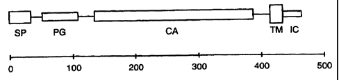
to the indicated scale. The exons corresponding to individual MN/CA IX protein
domains are enclosed in dashed frames designated PG (proteoglycan-like
domain), CA
17
CA 02347649 2004-11-26
(carbonic anhydrase domain), TM (transmembrane anchor) and IC
(intracytoplasmic
tail). Below the map, the alignment of amino acid sequences illustrates the
extent of
homology between the MN/CA IX protein PG region (aa 53-111) [SEQ ID NO: 50]
and
the human aggrecan (aa 781-839) [SEQ ID NO: 54].
Figure 6 is a nucleotide sequence for the proposed promoter of the
human MN gene [SEQ ID NO: 27]. The nucleotides are numbered from the
transcription initiation site according to RNase protection assay. Potential
regulatory
elements are overlined. Transcription start sites are indicated by asterisks
(RNase
protection) and dots (RACE) above the corresponding nucleotides. The sequence
of
the 1st exon begins under the asterisks. FTP analysis of the MN4 promoter
fragment
revealed 5 regions (I-V) protected at both the coding and noncoding strands,
and two
regions (VI and VII) protected at the coding strand but not at the noncoding
strand.
Figure 7 provides a schematic of the alignment of MN genomic clones
according to their position related to the transcription initiation site. All
the genomic
fragments except Bd3 were isolated from a lambda FIX II genomic library
derived from
HeLa cells. Clone Bd3 was derived from a human fetal brain library.
Figure 8 schematically represents the MN protein structure. The
abbreviations are the same as used in Figure 5. The scale indicates the number
of
amino acids.
DETAILED DESCRIPTION
The terms "MN/CA IX" and "MN/CA9" are herein considered to be
synonyms for MN. Also, the G250 antigen is considered to refer to MN
protein/polypeptide. [Uemura et al., J. Urol.. 157 (4 Suppl.): 377 (Abstract
1475;
1997).]
MN/CA IX was first identified in HeLa cells, derived from human
carcinoma of cervix uteri, as both a plasma membrane and nuclear protein with
an
apparent molecular weight of 58 and 54 kilodaltons (kDA) as estimated by
Western
blotting. It is N-glycosylated with a single 3kDa carbohydrate chain and under
non-
reducing conditions forms S-S-linked oligomers [Pastorekova et al., Virology.
187: 620-
626 (1992); Pastorek et al., Oncogene. 9: 2788-2888 (1994)]. MN/CA IX is a
18
CA 02347649 2001-04-19
WO 00/24913 PCT/US99/24879
transmembrane protein located at the cell surface, although in some cases it
has been -
detected in the nucleus [Zavada et al., Int. I. Cancer. 54: 268-274 (1993);
Pastorekova
et al., sura.
MN is manifested in HeLa cells by a twin protein, p54/58N.
Immunoblots using a monoclonal antibody reactive with p54/58N (MAb M75)
revealed
two bands at 54 kd and 58 kd. Those two bands may correspond to one type of
protein
that most probably differs by post-translational processing. Herein, the
phrase "twin
protein" indicates p54/58N.
Zavada et al., WO 93/18152 and/or WO 95/34650 disclose the MN
cDNA sequence (SEQ ID NO: 1) shown herein in Figure 1A-1C, the MN amino acid
sequence (SEQ ID NO: 2) also shown in Figure 1A-1C, and the MN genomic
sequence
(SEQ ID NO: 5) shown herein in Figure 2A-2F. The MN gene is organized into 11
exons and 10 introns.
The first thirty seven amino acids of the MN protein shown in Figure 1A-
1C is the putative MN signal peptide [SEQ ID NO: 6]. The MN protein has an
extracellular domain [amino acids (aa) 38-414 of Figure 1A-1C (SEQ ID NO:
87)], a
transmembrane domain [aa 415-434 (SEQ ID NO: 52)] and an intracellular domain
[aa
435-459 (SEQ ID NO: 53)]. The extracellular domain contains the proteoglycan-
like
domain [aa 53-111 (SEQ ID NO: 50)] and the carbonic anhydrase (CA) domain [aa
135-
391 (SEQ ID NO: 511.
Anticancer Drugs and Antibodies that Block
Interaction of MN Protein and Receptor Molecules
MN protein is considered to be a uniquely suitable target for cancer
therapy for a number of reasons including the following. (1) It is localized
on the cell
surface, rendering it accessible. (2) It is expressed in a high percentage of
human
carcinomas (e.g., uterine cervical, renal, colon, breast, esophageal, lung,
head and neck
carcinomas, among others), but is not normally expressed to any significant
extent in
the normal tissues from which such carcinomas originate.
(3) It is normally expressed only in the stomach mucosa and in some
epithelia of the digestive tract (epithelium of gallbladder and small
intestine). An
anatomic barrier thereby exists between the MN-expressing
preneoplastic/neoplastic
19
CA 02347649 2001-04-19
WO 00/24913 PCT/US99/24879
and MN-expressing normal tissues. Drugs, including antibodies, can thus be
administered which can reach tumors without interfering with MN-expressing
normal
tissues.
(4) MAb M75 has a high affinity and specificity to MN protein. (5) MN
cDNA and MN genomic clones which encompass the protein-coding and gene
regulatory sequences have been isolated. (6) MN-specific antibodies have been
shown
to have among the highest tumor uptakes reported in clinical studies with
antitumor
antibodies in solid tumors, as shown for the MN-specific chimeric antibody
G250 in
animal studies and in phase I clinical trials with renal carcinoma patients.
[Steffens et
al., I. Clin. Oncol.. 15: 1529 (1997).] Also, MN-specific antibodies have low
uptake in
normal tissues.
Data, e.g. as presented herein, are consistent with the following theory
concerning how MN protein acts in normal tissues and in
preneoplastic/neoplastic
tissues. In normal tissues (e.g., in stomach mucosa), MN protein is considered
to be a
differentiation factor. It binds with its normal receptor S (for stomach).
Stomach
carcinomas have been shown not to contain MN protein.
Ectopic expression of MN protein in other tissues causes malignant
conversion of cells. Such ectopic expression is considered to be caused by the
binding
of MN protein with an alternative receptor H (for HeLa cells), coupled to a
signal
transduction pathway leading to malignancy. Drugs or antibodies which block
the
binding site of MN protein for receptor H would be expected to cause reversion
of
prenoplastic/neoplastic cells to normal or induce their death.
Design and Development of MN-Blocking Drugs or Antibodies
A process to design and develop MN-blocking drugs, e.g., peptides with
high affinity to MN protein, or antibodies, has several steps. First, is to
test for the
binding of MN protein to receptors based on the cell adhesion assay described
infra.
That same procedure would also be used to assay for drugs blocking the MN
protein
binding site. In view of the alternative receptors S and H, stomach epithelial
cells or
revertants (containing preferentially S receptors), HeLa cells (containing the
H receptor
and lacking the S receptor) would be used in the cell adhesion assay.
CA 02347649 2001-04-19
WO 00/24913 PCT/US99/24879
To identify the receptor binding site of MN protein, deletion variants of -
MN protein lacking different domains can be used to identify region(s)
responsible for
interaction of MN protein with a receptor. Example 2 identifies and
illustrates how to
detect other binding sites on MN protein. A preferred MN binding site is
considered to
be closely related or identical to the epitope for MAb M75, which is located
in at least
2 copies within the 6-fold tandem repeat of 6 amino acids [aa 61-96 (SEQ ID
NO: 97)]
in the proteoglycan-like domain of the MN protein. Smaller deletion variants
can be
prepared within that relevant domain, e.g., fusion proteins with only small
segments of
MN protein can be prepared. Also, controlled digestion of MN protein with
specific
proteases followed by separation of the products can be performed.
Further, peptides comprising the expected binding site can be
synthesized. All of those products can be tested in cell adhesion assays, as
exemplified
below. See, e.g., Pierschbacher and Ruoslahti, PNAS, 81:5985 (1984); Ruoslahti
and
Pierschbacher, Science, 238: 491.]
Molecules can be constructed to block the MN receptor binding site. For
example, use of a phage display peptide library kit [as Ph.D 7 Peptide 7-Mer
Library
Kit from New England Biolabs; Beverly, MA (USA)] as exemplified in Examples 2
and 3,
can be used to find peptides with high affinity to the target molecules.
Biologic activity
of the identified peptides will be tested in vitro by inhibition of cell
adhesion to MN
protein, by effects on cell morphology and growth characteristics of MN-
related tumor
cells (HeLa) and of control cells. [Symington, I. Biol. Chem.. 267: 25744
(1992).] In
vivo screening will be carried out in nude mice that have been injected with
HeLa
cells.
Peptides containing the binding site of the MN protein will be prepared
[e.g. MAPs (multiple antigen peptides); Tam, J.P., PNAS (USA) D5: 5409 (1988);
Butz et
al., Peptide Res., 7: 20 (1994)]. The MAPs will be used to immunize animals to
obtain
antibodies (polyclonal and/or monoclonal) that recognize and block the binding
site.
See, e.g., Brooks et al., Cell. 79: 1157 (1994).] "Vaccination" would then be
used to
test for protection in animals. Antibodies to the MN binding site could
potentially be
used to block MN protein's interaction(s) with other molecules.
Computer modeling can also be used to design molecules with specific
affinity to MN protein that would mediate steric inhibition between MN protein
and its
21
CA 02347649 2001-04-19
WO 00/24913 PCT/US99/24879
receptor. A computer model of the MN binding site for the receptor will
contain
spatial, electrostatic, hydrophobic and other characteristics of this
structure. Organic
molecules complementary to the structure, that best fit into the binding site,
will be
designed. Inorganic molecules can also be similarly tested that could block
the MN
binding site.
The use of oncoproteins as targets for developing new cancer
therapeutics is considered conventional by those of skill in the art. See,
e.g.,
Mendelsohn and Lippman, "Growth Factors," pp. 114-133, IN: DeVita et al.
(eds.),
Cancer: Principles and Practice of Oncology (4th Ed.; Lippincott;
Philadelphia, 1993).] In
its broadest sense, the design of blocking drugs can be based in competitive
inhibition
experiments. Such experiments have been used to invent drugs since the
discovery of
sulfonamides (competitive inhibitors of para-aminobenzoic acid, a precursor of
folic
acid). Also, some cytostatics are competitive inhibitors (e.g., halogenated
pyrimidines,
among others).
However, the application of such approaches to MN is new. In
comparison to other tumor-related molecules (e.g. growth factors and their
receptors),
MN has the unique property of being differentially expressed in
preneoplastidneoplastic and normal tissues, which are separated by an anatomic
barrier.
MN Gene - Cloning and Sequencing
Figure 1A-C provides the nucleotide sequence for a full-length MN cDNA
clone isolated as described below [SEQ ID NO: 11. Figure 2A-F provides a
complete
MN genomic sequence [SEQ ID NO: 5]. Figure 6 shows the nucleotide sequence for
a
proposed MN promoter [SEQ ID NO: 27].
It is understood that because of the degeneracy of the genetic code, that
is, that more than one codon will code for one amino acid [for example, the
codons
TTA, TTG, CTT, CTC, CTA and CTG each code for the amino acid leucine (Ieu)],
that
variations of the nucleotide sequences in, for example, SEQ ID NOS: 1 and 5
wherein
one codon is substituted for another, would produce a substantially equivalent
protein
or polypeptide according to this invention. All such variations in the
nucleotide
22
CA 02347649 2001-04-19
WO 00/24913 PCT/US99/24879
sequences of the MN cDNA and complementary nucleic acid sequences are included
within the scope of this invention.
It is further understood that the nucleotide sequences herein described
and shown in Figures 1, 2 and 6, represent only the precise structures of the
cDNA,
genomic and promoter nucleotide sequences isolated and described herein. It is
expected that slightly modified nucleotide sequences will be found or can be
modified
by techniques known in the art to code for substantially similar or homologous
MN
proteins and polypeptides, for example, those having similar epitopes, and
such
nucleotide sequences and proteins/ polypeptides are considered to be
equivalents for
the purpose of this invention. DNA or RNA having equivalent codons is
considered
within the scope of the invention, as are synthetic nucleic acid sequences
that encode
proteins/polypeptides homologous or substantially homologous to MN
proteins/polypeptides, as well as those nucleic acid sequences that would
hybridize to
said exemplary sequences [SEQ. ID. NOS. 1, 5 and 27] under stringent
conditions, or
that, but for the degeneracy of the genetic code would hybridize to said cDNA
nucleotide sequences under stringent hybridization conditions. Modifications
and
variations of nucleic acid sequences as indicated herein are considered to
result in
sequences that are substantially the same as the exemplary MN sequences and
fragments thereof.
Stringent hybridization conditions are considered herein to conform to
standard hybridization conditions understood in the art to be stringent. For
example, it
is generally understood that stringent conditions encompass relatively low
salt and/or
high temperature conditions, such as provided by 0.02 M to 0.15 M NaCl at
temperatures of 50 C to 70 C. Less stringent conditions, such as, 0.15 M to
0.9 M salt
at temperatures ranging from 20 C to 55 C can be made more stringent by adding
increasing amounts of formamide, which serves to destabilize hybrid duplexes
as does
increased temperature.
Exemplary stringent hybridization conditions are described in Sambrook
et al., Molecular Cloning: A Laboratory Manual, pages 1.91 and 9.47-9.51
(Second
Edition, Cold Spring Harbor Laboratory Press; Cold Spring Harbor, NY; 1989);
Maniatis
et al., Molecular Cloning: A Laboratory Manual, pages 387-389 (Cold Spring
Harbor
23
CA 02347649 2001-04-19
WO 00/24913 PCTIUS99/24879
Laboratory; Cold Spring Harbor, NY; 1982); Tsuchiya et al., Oral Surgery. Oral
Medicine. Oral Pathology, 71(6): 721-725 (June 1991).
Zavada et al., WO 95/34650 described how a partial MN cDNA clone, a
full-length MN cDNA clone and MN genomic clones were isolated and sequenced.
Also, Zavada et al., Int. I. Cancer, 54: 268 (1993) describes the isolation
and
sequencing of a partial MN cDNA of 1397 bp in length. Briefly attempts to
isolate a
full-length clone from the original cDNA library failed. Therefore, the
inventors
performed a rapid amplification of cDNA ends (RACE) using MN-specific primers,
R1
and R2 [SEQ ID NOS: 7 and 8], derived from the 5' region of the original cDNA
clone.
The RACE product was inserted into pBluescript, and the entire population of
recombinant plasmids was sequenced with an MN-specific primer ODN1 [SEQ ID NO:
3]. In that way, a reliable sequence at the very 5' end of the MN cDNA as
shown in
Figure 1 [SEQ ID NO: 1] was obtained.
Specifically, RACE was performed using 5' RACE System [GIBCO BRL;
Gaithersburg, MD (USA)] as follows. 1 ,ug of mRNA (the same as above) was used
as a
template for the first strand cDNA synthesis which was primed by the MN-
specific
antisense oligonucleotide, R1 (5'-TGGGGTTCTTGAGGATCTCCAGGAG-3') [SEQ ID
NO: 71. The first strand product was precipitated twice in the presence of
ammonium
acetate and a homopolymeric C tail was attached to its 3' end by TdT. Tailed
cDNA
was then amplified by PCR using a nested primer, R2 (5'-
CTCTAACTTCAGGGAGCCCTCTTCTT-3') [SEQ ID NO: 8] and an anchor primer that
anneals to the homopolymeric tail (5'-CUACUACUACUAGGCCACGCGTCGAC
TAGTACGGGI IGGGIIGGGIIG-3') [SEQ ID NO: 91. The amplified product was
digested with BamHI and Sall restriction enzymes and cloned into pBluescript
II KS
plasmid. After transformation, plasmid DNA was purified from the whole
population of
transformed cells and used as a template for sequencing with the MN-specific
primer
ODN1 [SEQ ID NO: 3; a 29-mer 5' CGCCCAGTGGGTCATCTTCCCCAGAAGAG 3'1.
To study MN regulation, MN genomic clones were isolated. One MN
genomic clone (Bd3) was isolated from a human cosmid library prepared from
fetal
brain using both MN cDNA as a probe and the MN-specific primers derived from
the 5'
end of the cDNA ODN1 [SEQ ID NO: 3, su ra and ODN2 [SEQ. ID NO.: 4; 19-mer
(5' GGAATCCTCCTGCATCCGG 3')]. Sequence analysis revealed that that genomic
24
CA 02347649 2001-04-19
WO 00/24913 PCT/US99/24879
clone covered a region upstream from a MN transcription start site and ending
with the
BamHl restriction site localized inside the MN cDNA. Other MN genomic clones
can
be similarly isolated.
Figure 7 provides a schematic of the alignment of MN genomic clones
according to the transcription initiation site. Plasmids containing the A4a
clone and the
XE1 and XE3 subclones were deposited at the American Type Culture Collection
(ATCC) on June 6, 1995, respectively under ATCC Deposit Nos. 97199, 97200, and
97198.
Exon-Intron Structure of Complete MN Genomic Region
The complete sequence of the overlapping clones contains 10,898 bp
(SEQ ID NO: 5). Figure 5 depicts the organization of the human MN gene,
showing
the location of all 11 exons as well as the 2 upstream and 6 intronic Alu
repeat
elements. All the exons are small, ranging from 27 to 191 bp, with the
exception of the
first exon which is 445 bp. The intron sizes range from 89 to 1400 bp. The CA
domain
is encoded by exons 2-8, while the exons 1, 10 and 11 correspond respectively
to the
proteoglycan-like domain, the transmembrane anchor and cytoplasmic tail of the
MN/CA IX protein. Table 1 below lists the splice donor and acceptor sequences
that
conform to consensus splice sequences including the AG-GT motif [Mount,
Nucleic
Acids Res. 10: 459-472 (1982)].
CA 02347649 2004-11-26
TABLE 1
Exon-Intron Structure of the Human MN Gene
Genomic SEQ 5'splice SEQ
Exon Size Position** ID NO donor ID NO
1 445 *3507-3951 28 AGAAG gtaagt 67
2 30 5126-5155 29 TGGAG gtgaga 68
3 171 5349-5519 30 CAGTC gtgagg 69
4 143 5651-5793 31 CCGAG gtgagc 70
5 93 5883-5975 32 TGGAG gtacca 71
6 67 7376-7442 33 GGAAG gtcagt 72
7 158 8777-8934 34 AGCAG gtgggc 73
8 145 9447-9591 35 GCCAG gtacag 74
9 27 9706-9732 36 TGCTG gtgagt 75
10 82 10350-10431 37 CACAG gtatta 76
11 191 10562-10752 38 ATAAT end
Genomic SEQ 3'splice SEQ
Intron Size Position ** ID NO acceptor ID NO
1 1174 3952-5125 39 atacag GGGAT 77
2 193 5156-5348 40 ccccag GCGAC 78
3 131 5520-5650 41 acgcag TGCAA 79
4 89 5794-5882 42 tttcag ATCCA 80
5 1400 5976-7375 43 ccccag GAGGG 81
6 1334 7443-8776 44 tcacag GCTCA 82
7 512 8935-9446 45 ccctag CTCCA 83
8 114 9592-9705 46 ctccag TCCAG 84
9 617 9733-10349 47 tcgcag GTGACA 85
10 130 10432-10561 48 acacag AAGGG 86
** positions are related to nt numbering in whole genomic sequence including
the 5'
flanking region [Figure 2A-F]
* number corresponds to transcription initiation site determined below by
RNase
protection assay
26
CA 02347649 2001-04-19
WO 00/24913 PCT/US99/24879
Mapping of MN Gene Transcription Initiation and Termination Sites
Zavada et al., WO 95/34650 describes the process of mapping the MN
gene transcription initiation and termination sites. A RNase protection assay
was used
for fine mapping of the 5' end of the MN gene. The probe was a uniformly
labeled 470
nucleotide copy RNA (nt -205 to + 265) [SEQ ID NO: 55], which was hybridized
to
total RNA from MN-expressing HeLa and CG1..3 cells and analyzed on a
sequencing
gel. That analysis has shown that the MN gene transcription initiates at
multiple sites,
the 5' end of the longest MN transcript being 30 nt longer than that
previously
characterized by RACE.
Characterization of the 5' Flanking Region
The Bd3 genomic clone isolated from human fetal brain cosmid library
was found to cover a region of 3.5 kb upstream from the transcription start
site of the
MN gene. It contains no significant coding region. Two Alu repeats are
situated at
positions -2587 to -2296 [SEQ ID NO: 56] and -1138 to -877 [SEQ ID NO: 57]
(with
respect to the transcription start determined by RNP).
Nucleotide sequence analysis of the DNA 5' to the transcription start
(from nt -507) revealed no recognizable TATA box within the expected distance
from
the beginning of the first exon. However, the presence of potential binding
sites for
transcription factors suggests that this region might contain a promoter for
the MN gene.
There are several consensus sequences for transcription factors AP1 and AP2 as
well as
for other regulatory elements, including a p53 binding site [Locker and
Buzard, J., DNA
Sequencing and Mapping; 1: 3-11 (1990); Imagawa et al. Cell. 51: 251-260
(1987); El
Deiry et al., Nat. Genet.. 1: 44-49 (1992)]. Although the putative promoter
region
contains 59.3% C+G, it does not have additional attributes of CpG-rich islands
that are
typical for TATA-less promoters of housekeeping genes [Bird, Nature, 321: 209-
213
(1986)]. Another class of genes lacking TATA box utilizes the initiator (Inr)
element as a
promoter. Many of these genes are not constitutively active, but they are
rather
regulated during differentiation or development. The Inr has a consensus
sequence of
PyPyPyCAPyPyPyPyPy [SEQ ID NO: 231 and encompasses the transcription start
site
[Smale and Baltimore, Cell. 57: 103-113 (1989)]. There are two such consensus
27
CA 02347649 2001-04-19
WO 00/24913 PCT/US99/24879
sequences in the MN putative promoter; however, they do not overlap the
transcription-
start (Figure 6).
An interesting region was found in the middle of the MN gene. The
region is about 1.4 kb in length [nt 4,600-6,000 of the genomic sequence; SEQ
ID NO:
49] and spans from the 3' part of the 1st intron to the end of the 5th exon.
The region
has the character of a typical CpG-rich island, with 62.8% C+G content and 82
CpG:
131 GpC dinucleotides. Moreover, there are multiple putative binding sites for
transcription factors AP2 and Sp1 [Locker and Buzard, supra; Briggs et al.,
Science, 234:
47-52 (1986)] concentrated in the center of this area. Particularly the 3rd
intron of 131
bp in length contains three Sp1 and three AP2 consensus sequences. That data
indicates the possible involvement of that region in the regulation of MN gene
expression. However, functionality of that region, as well as other regulatory
elements
found in the proposed 5' MN promoter, remains to be determined.
MN Promoter
Study of the MN promoter has shown that it is TATA-less and contains
regulatory sequences for AP-1, AP-2, as well as two p53 binding sites. The
sequence of
the 5' end of the 3.5 kb flanking region upstream of the MN gene has shown
extensive
homology to LTR of HERV-K endogenous retroviruses. Basal transcription
activity of
the promoter is very weak as proven by analyses using CAT and neo reporter
genes.
However, expression of the reporter genes is severalfold increased when driven
from
the 3.5 kb flanking region, indicating involvement of putative enhancers.
Functional characterization of the 3.5 kb MN 5' upstream region by
deletion analysis lead to the identification of the [-173, + 31 ] fragment
[SEQ I D NO: 21 ]
(also alternatively, but less preferably, the nearly identical -172, + 31
fragment [SEQ ID
NO: 91]) as the MN promoter. In vitro DNase I footprinting revealed the
presence of
five protected regions (PR) within the MN promoter. Detailed deletion analysis
of the
promoter identified PR 1 and 2 (numbered from the transcription start) as the
most
critical for transcriptional activity. PR4 [SEQ ID NO: 115] negatively
affected
transcription as its deletion led to increased promoter activity and was
confirmed to
function as a promoter-, position- and orientation-independent silencer
element.
Mutational analysis indicated that the direct repeat AGGGCacAGGGC [SEQ ID NO:
28
CA 02347649 2001-04-19
WO 00/24913 PCT/US99/24879
143] is required for efficient repressor binding. Two components of the
repressor
complex (35 and 42 kDa) were found to be in direct contact with PR4 by UV
crosslinking. Increased cell density, known to induce MN expression, did not
affect
levels of PR4 binding in HeLa cells. Significantly reduced repressor level
seems to be
responsible for MN up-regulation in the case of tumorigenic CGL3 as compared
to
non-tumorigenic CGL1 HeLa x normal fibroblast hybrid cells.
Utility of MN Promoter as a Tumor-Specific
Promoter for Gene Therapy
Being investigated is whether the MN gene promoter can be used as a
tumor-specific promoter to drive the expression of a suicide gene Ithymidine
kinase (tk)
of HSV)] and mediate the direct and bystander killing of tumor cells. HSVtk
gene
transferred to tumor cells converts nucleoside analogue ganciclovir (GCV) to
toxic
triphosphates and mediates the death of transduced and also neighboring tumor
cells.
The control of HSVtk by the MN gene promoter would allow its expression only
in
tumor cells, which are permissive for the biosynthesis of MN protein, and
selectively
kill such tumor cells, but not normal cells in which MN expression is
repressed.
A plasmid construct in which HSVtk was cloned downstream of the MN
promoter region Bd3, containing both proximal and distant regulatory elements
of MN,
was prepared. That plasmid pMN-HSVtk was transfected to Rat2TK- cells and C33
human cervical carcinoma cells using calcium phosphate precipitation and
lipofection,
respectively. Transfectants were tested for expression of HSVtk and GVC
sensitivity.
Analysis of the transfectants has shown the remarkable cytotoxic in vitro
effect of GVC
even in low concentrations (up to 95% of cells killed).
Polyclonal rabbit antiserum against HSVtk, using fusion protein with GST
in pGEX-3X, has been prepared to immunodetect HSVtk synthesized in transfected
cells. This model system is being studied to estimate the bystander effect,
the inhibition
of cloning efficiency and invasiveness of transduced and GVC-treated cells to
collagen
matrices. A recombinant retroviral vector with the MN promoter-driven HSVtk is
to be
prepared to test its in vivo efficacy using an animal model (e.g., SCID-
mouse).
29
CA 02347649 2001-04-19
WO 00/24913 PCT/US99/24879
MN Promoter Analysis
Since the MN promoter is weak, a classical approach to study it would be
limited due to the relatively low efficiency of transient transfections (up to
10%).
Therefore, stable clonal cell lines expressing constructs containing the MN
promoter
fused to the CAT gene were prepared. In such clonal lines, 100% of the cells
express
the CAT gene driven from the MN promoter, and thus, the activity of the
promoter is
detectable easier than in transient experiments. Also, the promoter activity
can be
analysed repeatedly in the same cells under different conditions or treated by
different
factors and drugs. This approach allows for the study of the mechanisms
underlying
MN regulation at the level of transcription initiation.
Several types of transfections with promoter constructs linked to a
reporter CAT gene (calcium precipitation, DEAE dextran combined with DMSO
shock
and/or chloroquine, as well as electroporation), different methods of CAT
activity assay
(scintillation method, thin layer chromatography) and several recipient cell
lines
differing in the level of MN expression and in transfection efficiency (HeLa,
SiHa,
CGL3, KATO 111, Rat2TK' and C33 cells). Activity of the MN promoter was
detected
preferably by the electroporation of CGL3 cells and thin layer chromatography.
Further
preferably, C33 cells cotransfected with MN promoter-CAT constructs and
pSV2neo
were used.
1. To detect basal activity of the MN promoter and to estimate the
position of the core promoter, expression of the CAT gene from constructs pMN1
to
pMN7 after transfection to CGL3 cells was analyzed. Plasmids with progressive
5'
deletions were transfected into CGL3 cells and activity was analyzed by CAT
assay. [8
g of DNA was used for transfection in all cases except pBLV-LTR (2,ug).]
Only very weak CAT activity was detected in cells transfected by pMN1
and pMN2 (containing respectively 933 bp and 600 bp of the promoter sequence).
A
little higher activity was exhibited with the constructs pMN3, pMN4 and pMN6
(containing respectively 446 bp, 243 bp and 58 bp of the promoter). A slight
peak of
activity was obtained with pMN5 (starting at position -172 with respect to the
transcription start.) Thus, the function of the MN core promoter can be
assigned to a
region of approximately 500 bp immediately upstream from the MN transcription
initiation site.
CA 02347649 2001-04-19
WO 00/24913 PCT/US99/24879
Interestingly, the activity of the large Bd3 region (covering 3.5 kbp
upstream of the transcription start) was severalfold higher than the activity
of the core
promoter. However, its level was still much lower than that exhibited by a
positive
control, i.e., BLV-LTR transactivated by Tax, and even lower than the activity
of BLV-
LTR without transactivation. That the activity of Bd3 was elevated in
comparison to the
core promoter suggests the presence of some regulatory elements. Such elements
are
most probably situated in the sequence between pMN1 and Bd3 (i.e. from -1 kbp
to -
3.5 kbp) [SEQ ID NO: 58]. The cloning and transfection of several deletion
versions of
Bd3 covering the indicated region can be used to determine the location of the
putative
regulatory elements.
Similar results were obtained from transfecting KATO III cells with Bd3
and pMN4. The transfected cells expressed a lower level of MN than the CGL3
cells.
Accordingly, the activity of the MN promoter was found to be lower than in
CGL3
cells.
2. In a parallel approach to study the MN promoter, an analysis based on
G418 selection of cells transfected by plasmids containing the promoter of
interest
cloned upstream from the neo gene was made. This approach is suitable to study
weak
promoters, since its sensitivity is much higher than that of a standard CAT
assay. The
principle underlying the method is as follows: an active promoter drives
expression of
the neo gene which protects transfected cells from the toxic effect of G418,
whereas an
inactive promoter results in no neo product being made and the cells
transfected
thereby die upon the action of G418. Therefore, the activity of the promoter
can be
estimated according to the number of cell colonies obtained after two weeks of
selection with G418. Three constructs were used in the initial experiments -
pMN1 neo, pMN4neo and pMN7neo. As pMN7neo contains only 30 bp upstream of
the transcription start site, it was considered a negative control. As a
positive control,
pSV2neo with a promoter derived from SV40 was used. Rat2TK- cells were chosen
as
the recipient cells, since they are transfectable with high efficiency by the
calcium
precipitation method.
After transfection, the cells were subjected to two weeks of selection.
Then the medium was removed, the cells were rinsed with PBS, and the colonies
were
rendered visible by staining with methylene blue. The results obtained from
three
31
CA 02347649 2001-04-19
WO 00/24913 PCT/US99/24879
independent experiments corroborated the data from the CAT assays. The
promoter
construct pMN4neo exhibited higher transcriptional activity than pMN1 neo.
However,
the difference between the positive control and pMN4neo was not so striking as
in the
CAT assay. That may have been due to both lower promoter activity of pSV2neo
compared to Tax-transactivated pBLV-LTR and to different conditions for cell
growth
after transfection. From that point of view, stable transfection is probably
more
advantageous for MN expression, since the cells grow in colonies with close
cell to cell
contact, and the experiment lasts much longer, providing a better opportunity
to detect
promoter activity.
3. Stable transfectants expressing MN promoter-CAT chimeric genes
were prepared by the cotransfection of relevant plasmids with pSV2neo. As
recipient
cells, HeLa cells were used first. However, no clones expressing the promoter-
CAT
constructs were obtained. That negative result was probably caused by
homologic
recombination of the transfected genomic region of MN (e.g. the promoter) with
the
corresponding endogenous sequence. On the basis of that experience, C33 cells
derived from a HPV-negative cervical carcinoma were used. C33 cells do not
express
MN, since during the process of tumorigenesis, they lost genetic material
including
chromosomal region 9p which contains the MN gene. In these experiments, the
absence of the MN gene may represent an advantage as the possibility of
homologic
recombinations is avoided.
C33 Cells Transfected with MN Promoter-CAT Constructs
C33 cells expressing the CAT gene under MN promoter regions Bd3 (-
3500/+ 31) [SEQ ID NO: 90] and MN5 (-172/+31) [SEQ ID NO: 91] were used for
initial experiments to analyze the influence of cell density on the
transcriptional activity
of the MN promoter. The results indicated that signals generated after cells
come into
close contact activate transcription of the CAT protein from the MN promoter
in
proportion to the density of the cell culture. Interestingly, the data
indicated that the
MN protein is not required for this phase of signal transduction, since the
influence of
density is clearly demonstrated in MN-negative C33 cells. Rather, it appears
that MN
protein acts as an effector molecule produced in dense cells in order to
perform a
certain biological function (i.e., to perturb contact inhibition). Also
interestingly, the
32
CA 02347649 2001-04-19
WO 00/24913 PCT/US99/24819
MN promoter activity is detectable even in very sparse cell cultures
suggesting that MN
is expressed at a very low level also is sparse subconfluent culture.
Deletion Variants. Deletion variants of the Bd3-CAT promoter construct
were then prepared. The constructs were cotransfected with pSV2neo into C33
cervical
cells. After selection with G418, the whole population of stably transfected
cells were
subjected to CAT ELISA analysis. Expression of the deletion constructs
resulted in the
synthesis of similar levels of CAT protein to that obtained with the Bd3-CAT
construct.
On the basis of that preliminary data, the inventors proposed that sequences
stimulating
transcription of MN are located between -3506 and -3375 bp [SEQ ID NO: 92]
upstream from the transcription start. That is the sequence exhibiting
homology to
HERV-K LTR.
However, transient transfection studies in CGL3 cells repeatedly revealed
that the LTR region is not required for the enhancement of basal MN promoter
activity.
Further, results obtained in CGL3 cells indicate that the activating element
is localized
in the region from -933 to -2179 [SEQ ID NO: 110] with respect to
transcription
initiation site (the position of the region having been deduced from
overlapping
sequences in the Bd3 deletion mutants).
Interaction of Nuclear Proteins with MN Promoter Sequences
In order to identify transcription factors binding to the MN promoter and
potentially regulating its activity, a series of analyses using an
electrophoretic mobility
shift assay (EMSA) and DNase I footprinting analysis (FTP) were performed.
EM SA
In the EMSA, purified promoter fragments MN4 (-243/+ 31) [SEQ ID NO:
93], MN5 (-172/+31) [SEQ ID NO: 91], MN6 (-58/+31) [SEQ ID NO: 94] and pMN7
(-30/+ 31) [SEQ I D NO: 951, labeled at the 3' ends by Klenow enzyme, were
allowed
to interact with proteins in nuclear extracts prepared from CGL1 and CGL3
cells. [40
g of nuclear proteins were incubated with 30,000 cpm end-labeled DNA fragments
in
the presence of 2 g poly(dldC).] DNA-protein complexes were analysed by PAGE
(native 6%), where the complexes created extra bands that migrated more slowly
than
33
CA 02347649 2001-04-19
WO 00/24913 PCT/US99/24979
the free DNA fragments, due to the shift in mobility which independent on the
moiety
of bound protein.
The EMSA of the MN4 and MN5 promoter fragments revealed several
DNA-protein complexes; however, the binding patterns obtained respectively
with
CGL1 and CGL3 nuclear extracts were not identical. There is a single CGL-1
specific
complex.
The EMSA of the MN6 promoter fragment resulted in the formation of
three identical complexes with both CGL1 and CGL3 nuclear extracts, whereas
the
MN7 promoter fragment did not bind any nuclear proteins.
The EMSA results indicated that the CGL1 nuclear extract contains a
specific factor, which could participate in the negative regulation of MN
expression in
CGL1 cells. Since the specific DNA-protein complex is formed with MN4
(-243/+31) [SEQ. ID NO.: 93] and MN 5 (-172/+31) [SEQ. ID NO.: 911 promoter
fragments, but not with MN6 (-58/+ 31) [SEQ ID NO: 94], it appears that the
binding
site of the protein component of that specific complex is located between -173
and -58
bp [SEQ. ID NO.: 96] with respect to transcription initiation.
The next step was a series of EMSA analyses using double stranded (ds)
oligonucleotides designed according to the protected regions in FTP analysis.
A ds
oligonucleotide derived from the protected region PR2 [covering the sequence
from -72
to -56 bp (SEQ ID NO: 111)] of the MN promoter provided confirmation of the
binding
of the AP-1 transcription factor in competitive EMSA using commercial ds
olignucleotides representing the binding site for AP-1.
EMSA of ds oligonucleotides derived from the protected regions of PR1 [-
46 to -24 bp (SEQ ID NO: 112)], PR2 [-72 to -56 bp (SEQ ID NO: 111)], PR3 [-
102 to -
85 (SEQ ID NO: 113)] and PR5 [-163 to -144 (SEQ ID NO: 114)] did not reveal
any
differences in the binding pattern of nuclear proteins extracted from CGL1 and
CGL3
cells, indicating that those regions do not bind crucial transcription factors
which
control activation of the MN gene in CGL3, or its negative regulation in CGL1.
However, EMSA of ds oligonucleotides from the protected region PR4 [-133 to -
108;
SEQ ID NO: 115] repeatedly showed remarkable quantitative differences between
binding of CGL1 and CGL3 nuclear proteins. CGL1 nuclear proteins formed a
substantially higher amount of DNA-protein complexes, indicating that the PR4
region
34
CA 02347649 2001-04-19
WO 00/24913 PCT/US99/24879
contains a binding site for specific transcription factor(s) that may
represent a negative
regulator of MN gene transcription in CGL1 cells. That fact is in accord with
the
previous EMSA data which showed CGL-1 specific DNA-protein complex with the
promoter fragments pMN4 (-243/+31; SEQ ID NO: 93) and pMN5 (-172/+31; SEQ ID
NO: 91), but not with pMN6 (-58/+ 31; SEQ ID NO: 94).
To identify the protein involved or the formation of a specific complex
with the MN promoter in the PR4 region, relevant ds oligonucleotides
covalently
bound to magnetic beads will be used to purify the corresponding transcription
factor.
Alternatively the ONE Hybrid System [Clontech (Palo Alto, CA (USA)] will be
used to
search for and clone transcription factors involved in regulation of the
analysed
promoter region. A cDNA library from HeLa cells will be used for that
investigation.
FTP
To determine the precise location of cis regulatory elements that
participate in the transcriptional regulation of the MN gene, FTP was used.
Proteins in
nuclear extracts prepared respectively from CGL1 and CGL3 cells were allowed
to
interact with a purified ds DNA fragment of the MN promoter (MN4, -243/+ 31)
[SEQ
ID NO: 93] which was labeled at the 5' end of one strand. [MN4 fragments were
labeled either at Xhol site (-243/+ 31 *) or at Xbal site (*-243/+ 31).] The
DNA-protein
complex was then subjected to DNase I attack, which causes the DNA chain to
break at
certain bases if they are not in contact with proteins. [A control used BSA
instead of
DNase.] Examination of the band pattern of the denatured DNA after gel
electrophoresis [8% denaturing gel] indicates which of the bases on the
labeled strand
were protected by protein.
FTP analysis of the MN4 promoter fragment revealed 5 regions (I-V)
protected at both the coding and noncoding strand, as well as two regions (VI
and VII)
protected at the coding strand but not at the noncoding strand. Figure 6
indicates the
general regions on the MN promoter that were protected.
The sequences of the identified protected regions (PR) were subjected to
computer analysis using the SIGNALSCAN program to see if they corresponded to
known consensus sequences for transcription factors. The data obtained by that
computer analyses are as follows:
CA 02347649 2001-04-19
WO 00/24913 PCT/US99/24879
PR I - coding strand - AP-2, p53, GAL4 noncoding strand - JCV-repeated
PR II - coding strand - AP-1, CGN4 noncoding strand - TCF-1, dFRA, CGN4
PR III - coding strand - no known consensus sequence, only partial overlap of
AP1 noncoding strand - 2 TCF-1 sites
PR IV - coding strand - TCF-1, ADR-1 noncoding strand - CTCF, LF-A1, LBP-1
PR V - coding strand - no known consensus motif noncoding strand - JCV
repeated
PR VI - coding strand - no known consensus motif noncoding strand - T antigen
of SV 40, GAL4
PR VII - coding strand - NF-uE4, U2snRNA.2 noncoding strand - AP-2, IgHC.12,
MyoD.
In contrast to EMSA, the FTP analysis did not find any differences
between CGL1 and CGL3 nuclear extracts. However, the presence of specific DNA-
protein interactions detected in the CGL1 nuclear extracts by EMSA could have
resulted
from the binding of additional protein to form DNA protein-protein complex. If
that
specific protein did not contact the DNA sequence directly, its presence would
not be
detectable by FTP.
EMSA Supershift Analysis
The results of the FTP suggests that transcription factors AP-1, AP-2 as
well as tumor suppressor protein p53 are potentially involved in the
regulation of MN
expression. To confirm binding of those particular proteins to the MN
promoter, a
supershift analysis using antibodies specific for those proteins was
performed. For this
analysis, DNA-protein complexes prepared as described for EMSA were allowed to
interact with MAbs or polyclonal antibodies specific for proteins potentially
included in
the complex. The binding of antibody to the corresponding protein results in
an
additional shift (supershift) in mobility of the DNA-protein-antibody complex
which is
PAGE visualized as an additional, more slowly migrating band.
By this method, the binding of AP-2 to the MN promoter was confirmed.
However, this method did not evidence binding of the AP-1 transcription
factor. It is
36
CA 02347649 2001-04-19
WO 00/24913 PCT/US99/24879
possible that MN protein binds AP-1-related protein, which is antigenically
different
from the AP-1 recognized by the antibodies used in this assay.
Also of high interest is the possible binding of the p53 tumor suppressor
protein to the MN promoter. It is well known that wt p53 functions as a
transcription
factor, which activates expression of growth-restricting genes and down-
modulates,
directly or indirectly, the expression of genes that are required for ongoing
cell
proliferation. Transient co-transfection experiments using the pMN4-CAT
promoter
construct in combination with wt p53 cDNA and mut p53 cDNA, respectively,
suggested that wt p53, but not mut p53, negatively regulates expression of MN.
In
addition, one of two p53-binding sites in the MN promoter is protected in FTP
analysis
(Figure 6), indicating that it binds to the corresponding protein. Therefore,
supershift
analysis to prove that p53 binds to the MN promoter with two p53-specific
antibodies,
e.g. Mabs 421 and DO-1 [the latter kindly provided by Dr. Vojtesek from
Masaryk
Memorial Cancer Institute in Brno, Czech Republic] are to be performed with
appropriate nuclear extracts, e.g. from MCF-7 breast carcinoma cells which
express wt
p53 at a sufficient level.
Regulation of MN Expression and MN Promoter
MN appears to be a novel regulatory protein that is directly involved in
the control of cell proliferation and in cellular transformation. In HeLa
cells, the
expression of MN is positively regulated by cell density. Its level is
increased by
persistent infection with LCMV. In hybrid cells between HeLa and normal
fibroblasts,
MN expression correlates with tumorigenicity. The fact that MN is not present
in
nontumorigenic hybrid cells (CGL1), but is expressed in a tumorigenic
segregant
lacking chromosome 11, indicates that MN is negatively regulated by a putative
suppressor in chromosome 11.
Evidence supporting the regulatory role of MN protein was found in the
generation of stable transfectants of NIH 3T3 cells that constitutively
express MN
protein. As a consequence of MN expression, the NIH 3T3 cells acquired
features
associated with a transformed phenotype: altered morphology, increased
saturation
density, proliferative advantage in serum-reduced media, enhanced DNA
synthesis and
capacity for anchorage-independent growth. Further, flow cytometric analyses
of
37
CA 02347649 2001-04-19
WO 00/24913 PCT/US99/24879
asynchronous cell populations indicated that the expression of MN protein
leads to
accelerated progression of cells through G1 phase, reduction of cell size and
the loss of
capacity for growth arrest under inappropriate conditions. Also, MN expressing
cells
display a decreased sensitivity to the DNA damaging drug mitomycin C.
Nontumorigenic human cells, CGL1 cells, were also transfected with the
full-length MN cDNA. The same pSG5C-MN construct in combination with pSV2neo
plasmid as used to transfect the NIH 3T3 cells was used. Out of 15 MN-positive
clones
(tested by SP-RIA and Western blotting), 3 were chosen for further analysis.
Two MN-
negative clones isolated from CGL1 cells transfected with empty plasmid were
added as
controls. Initial analysis indicates that the morphology and growth habits of
MN-
transfected CGL1 cells are not changed dramatically, but their proliferation
rate and
plating efficiency is increased.
MN Promoter - Sense/Antisense Constructs
When the promoter region from the MN genomic clone, isolated as
described above, was linked to MN cDNA and transfected into CGL1 hybrid cells,
expression of MN protein was detectable immediately after selection. However,
then it
gradually ceased, indicating thus an action of a feedback regulator. The
putative
regulatory element appeared to be acting via the MN promoter, because when the
full-
length cDNA (not containing the promoter) was used for transfection, no
similar effect
was observed.
An "antisense" MN cDNA/MN promoter construct was used to transfect
CGL3 cells. The effect was the opposite of that of the CGL1 cells transfected
with the
"sense" construct. Whereas the transfected CGL1 cells formed colonies several
times
larger than the control CGL1, the transfected CGL3 cells formed colonies much
smaller
than the control CGL3 cells. The same result was obtained by antisense MN cDNA
transfection in SiHa and HeLa cells.
For those experiments, the part of the promoter region that was linked to
the MN cDNA through a BamHI site was derived from a col - BamHI fragment of
the
MN genomic clone [Bd3] and represents a region a few hundred bp upstream from
the
transcription initiation site. After the ligation, the joint DNA was inserted
into a pBK-
CMV expression vector [Stratagene]. The required orientation of the inserted
sequence
38
CA 02347649 2001-04-19
WO 00/24913 PCT/US99/24879
was ensured by directional cloning and subsequently verified by restriction
analysis.
The tranfection procedure was the same as used in transfecting the NIH 3T3
cells, but
co-transfection with the pSV2neo plasmid was not necessary since the neo
selection
marker was already included in the pBK-CMV vector.
After two weeks of selection in a medium containing G418, remarkable
differences between the numbers and sizes of the colonies grown were evident
as
noted above. Immediately following the selection and cloning, the MN-
transfected
CGL1 and CGL3 cells were tested by SP-RIA for expression and repression of MN,
respectively. The isolated transfected CGL1 clones were MN positive (although
the
level was lower than obtained with the full-length cDNA), whereas MN protein
was
almost absent from the transfected CGL3 clones. However, in subsequent
passages, the
expression of MN in transfected CGL1 cells started to cease, and was then
blocked
perhaps evidencing a control feedback mechanism.
As a result of the very much lowered proliferation of the transfected
CGL3 cells, it was difficult to expand the majority of cloned cells (according
to SP-RIA,
those with the lowest levels of MN), and they were lost during passaging.
However,
some clones overcame that problem and again expressed MN. It is possible that
once
those cells reached a higher quantity, that the level of endogenously produced
MN
mRNA increased over the amount of ectopically expressed antisense mRNA.
Identification of Specific Transcription
Factors Involved in Control of MN Expression
Control of MN expression at the transcription level involves regulatory
elements of the MN promoter. Those elements bind transcription factors that
are
responsible for MN activation in tumor cells and/or repression in normal
cells. The
identification and isolation of those specific transcription factors and an
understanding
of how they regulate MN expression could result in their therapeutic utility
in
modulating MN expression.
EMSA experiments indicate the existence of an MN gene repressor.
Using the One Hybrid System [Clontech (Palo Alto, CA); an in vivo yeast
genetic assay
for isolating genes encoding proteins that bind to a target, cis-acting
regulatory element
or any other short DNA-binding sequence; Fields and Song, Nature, 340: 245
(1989);
39
CA 02347649 2001-04-19
WO 00/24913 PCT/US99/24879
Wu et at., EMBO I., 13: 4823 (1994)] and subtractive suppressive PCR (SSH).
SSH
allows the cloning of genes that are differentially expressed under conditions
which are
known to up or down regulate MN expression such as density versus sparsity of
HeLa
cells, and suspension versus adherent HeLa cells.
In experiments with HPV immobilized cervical cells (HCE 16/3), it was
found that the regulation of MN expression differs from that in fully
transformed
carcinoma cells. For example, glucocorticoid hormones, which activate HPV
transcription, negatively regulate MN expression in HCE, but stimulate MN in
HeLa and
SiHa. Further keratinocyte growth factors, which down regulates transcription
of HPV
oncogenes, stimulates MN expression in suspension HCE but not in adherent
cells.
EGF and insulin are involved in the activation of MN expression in both
immortalized and carcinoma cells. All the noted facts can be used in the
search for
MN-specific transcription factors and in the modulation of MN expression for
therapeutic purposes.
Deduced Amino Acid Sequence
The ORF of the MN cDNA shown n Figure 1 has the coding capacity for
a 459 amino acid protein with a calculated molecular weight of 49.7 kd. The
overall
amino acid composition of the MN/CA IX protein is rather acidic, and predicted
to have
a pi of 4.3. Analysis of native MN/CA IX protein from CGL3 cells by two-
dimensional
electrophoresis followed by immunoblotting has shown that in agreement with
computer prediction, the MN/CA IX is an acidic protein existing in several
isoelectric
forms with pis ranging from 4.7 to 6.3.
As assessed by amino acid sequence analysis, the deduced primary
structure of the MN protein can be divided into four distinct regions. The
initial
hydrophobic region of 37 amino acids (aa) corresponds to a signal peptide. The
mature
protein has an N-terminal or extracellular part of 377 amino acids [aa 38-414
(SEQ ID
NO: 87], a hydrophobic transmembrane segment of 20 amino acids [aa 415-434
(SEQ
ID NO: 52)] and a C-terminal region of 25 amino acids [aa 435-459 (SEQ ID NO:
53)].
The extracellular part is composed of two distinct domains: (1) a
proteoglycan-like domain [aa 53-111 (SEQ ID NO: 50)]; and (2) a CA domain,
located
CA 02347649 2001-04-19
WO 00/24913 PCT/US99/24879
close to the plasma membrane [aa 135-391 (SEQ ID NO: 51)]. [The amino acid
numbers are keyed to those of Figure 1.]
More detailed insight into MN protein primary structure disclosed the
presence of several consensus sequences. One potential N-glycosylation site
was
found at position 346 of Figure 1. That feature, together with a predicted
membrane-
spanning region are consistent with the results, in which MN was shown to be
an N-
glycosylated protein localized in the plasma membrane. MN protein sequence
deduced from cDNA was also found to contain seven S/TPXX sequence elements
[SEQ
ID NOS: 25 AND 26] (one of them is in the signal peptide) defined by Suzuki, l
Mol.
Biol., 207: 61-84 (1989) as motifs frequently found in gene regulatory
proteins.
However, only two of them are composed of the suggested consensus amino acids.
Experiments have shown that the MN protein is able to bind zinc cations,
as shown by affinity chromatography using Zn-charged chelating sepharose. MN
protein immunoprecipitated from HeLa cells by Mab M75 was found to have weak
catalytic activity of CA. The CA-like domain of MN has a structural
predisposition to
serve as a binding site for small soluble domains. Thus, MN protein could
mediate
some kind of signal transduction.
MN protein from LCMV-infected HeLA cells was shown by using DNA
cellulose affinity chromatography to bind to immobilized double-stranded
salmon
sperm DNA. The binding activity required both the presence of zinc cations and
the
absence of a reducing agent in the binding buffer.
CA Domain Required for Anchorage
Independence But for Increased
Proliferation of Transfected NIH 3T3 Fibroblasts
In transfected NIH 3T3 fibroblasts, MN protein induces morphologic
transformation, increased proliferation and anchorage independence. The
consequences of constitutive expression of two MN-truncated variants in NIH
3T3 cells
were studied. It was found that the proteoglycan-like region is sufficient for
the
morphological alteration of transfected cells and displays the growth-
promoting activity
presumably related to perturbation of contact inhibition.
41
CA 02347649 2001-04-19
WO 00/24913 PCTIUS99/24879
The CA domain is essential for induction of anchorage independence,
whereas the TM anchor and IC tail are dispensable for that biological effect.
The MN
protein is also capable of causing plasma membrane ruffling in the transfected
cells and
appears to participate in their attachment to the solid support. The data
evince the
involvement of MN in the regulation of cell proliferation, adhesion and
intercellular
communication.
Sequence Similarities
Computer analysis of the MN cDNA sequence was carried out using
DNASIS and PROSIS (Pharmacia Software packages). GenBank, EMBL, Protein
Identification Resource and SWISS-PROT databases were searched for all
possible
sequence similarities. In addition, a search for proteins sharing sequence
similarities
with MN was performed in the MIPS databank with the FastA program [Pearson and
Lipman, PNAS (USA), 85: 2444 (1988)].
The proteoglycan-like domain [aa 53-111 (SEQ ID NO: 50)], which is
between the signal peptide and the CA domain, shows significant homology (38%
identity and 44% positivity) with a keratan sulphate attachment domain of a
human
large aggregating proteoglycan aggrecan [Doege et al., I. Biol. Chem., 266:
894-902
(1991)].
The CA domain [aa 135-391 (SEQ ID NO: 51)] is spread over 265 as and
shows 38.9% amino acid identity with the human CA VI isoenzyme [Aldred et al.,
Biochemistry, 30: 569-575 (1991)]. The homology between MN/CA IX and other
isoenzymes is as follows: 35.2% with CA II in a 261 as overlap [Montgomery et
al.,
Nucl. Acids. Res., 15: 4687 (1987)], 31.8% with CA I in a 261 as overlap
[Barlow et
al., Nucl. Acids Res., 15: 2386 (1987)], 31.6% with CA IV in a 266 as overlap
[Okuyama et al., PNAS (USA) 89: 1315-1319 (1992)], and 30.5% with CA III in a
259
as overlap (Lloyd et al., Genes. Dev.. 1: 594-602 (1987)].
In addition to the CA domain, MN/CA IX has acquired both N-terminal
and C-terminal extensions that are unrelated to the other CA isoenzymes. The
amino
acid sequence of the C-terminal part, consisting of the transmembrane anchor
and the
intracytoplasmic tail, shows no significant homology to any known protein
sequence.
42
CA 02347649 2001-04-19
WO 00/24913 PCTIUS99/24819
The MN gene was clearly found to be a novel sequence derived from the -
human genome. The overall sequence homology between the cDNA MN sequence
and cDNA sequences encoding different CA isoenzymes is in a homology range of
48-
50% which is considered by ones in the art to be low. Therefore, the MN cDNA
sequence is not closely related to any CA cDNA sequences.
Only very closely related nt sequences having a homology of at least 80-
90% would hybridize to each other under stringent conditions. A sequence
comparison of the MN cDNA sequence shown in Figure 1 and a corresponding cDNA
of the human carbonic anhydrase II (CA II) showed that there are no stretches
of identity
between the two sequences that would be long enough to allow for a segment of
the
CA II cDNA sequence having 25 or more nucleotides to hybridize under stringent
hybridization conditions to the MN cDNA or vice versa.
A search for nt sequences related to MN gene in the EMBL Data Library
did not reveal any specific homology except for 6 complete and 2 partial Alu-
type
repeats with homology to Alu sequences ranging from 69.8% to 91 % (jurka and
Milosavljevic, I. Mol. Evol. 32: 105-121 (1991)]. Also a 222 bp sequence
proximal to
the 5' end of the genomic region is shown to be closely homologous to a region
of the
HERV-K LTR.
In general, nucleotide sequences that are not in the Alu or LTR-like
regions, of preferably 25 bases or more, or still more preferably of 50 bases
or more,
can be routinely tested and screened and found to hybridize under stringent
conditions
to only MN nucleotide sequences. Further, not all homologies within the Alu-
like MN
genomic sequences are so close to Alu repeats as to give a hybridization
signal under
stringent hybridization conditions. The percent of homology between MN Alu-
like
regions and a standard Alu-J sequence are indicated as follows:
Region of Homology within
MN Genomic Sequence 5 % Homology to
LSEQ ID NO: 5; Q Entire Alu-l
Figure 2A-F1 NOS. Sequence
921-1212 59 89.1%
2370-2631 60 78.6%
4587-4880 61 90.1%
43
CA 02347649 2001-04-19
WO 00/24913 PCT/US99/24879
6463-6738 62 85.4%
7651-7939 63 91.0%
9020-9317 64 69.8%
% Homology to
One Half of
Alu-I Sequence
8301-8405 65 88.8%
10040-10122 66 73.2%.
MN Proteins and/or Polypeptides
The phrase "MN proteins and/or polypeptides" (MN
proteins/polypeptides) is herein defined to mean proteins and/or polypeptides
encoded
by an MN gene or fragments thereof. An exemplary and preferred MN protein
according to this invention has the deduced amino acid sequence shown in
Figure 1.
Preferred MN proteins/polypeptides are those proteins and/or polypeptides that
have
substantial homology with the MN protein shown in Figure 1. For example, such
substantially homologous MN proteins/ polypeptides are those that are reactive
with the
MN-specific antibodies of this invention, preferably the Mabs M75, MN 12, MN9
and
MN7 or their equivalents.
A "polypeptide" or "peptide" is a chain of amino acids covalently bound
by peptide linkages and is herein considered to be composed of 50 or less
amino acids.
A "protein" is herein defined to be a polypeptide composed of more than 50
amino
acids. The term polypeptide encompasses the terms peptide and oligopeptide.
MN proteins exhibit several interesting features: cell membrane
localization, cell density dependent expression in HeLa cells, correlation
with the
tumorigenic phenotype of HeLa x fibroblast somatic cell hybrids, and
expression in
several human carcinomas among other tissues. MN protein can be found directly
in
tumor tissue sections but not in general in counterpart normal tissues
(exceptions noted
infra as in normal gastric mucosa and gallbladder tissues). MN is also
expressed
sometimes in morphologically normal appearing areas of tissue specimens
exhibiting
dysplasia and/or malignancy. Taken together, these features suggest a possible
44
CA 02347649 2001-04-19
WO 00/24913 PCTIUS99/24819
involvement of MN in the regulation of cell proliferation, differentiation
and/or
transformation.
It can be appreciated that a protein or polypeptide produced by a
neoplastic cell in vivo could be altered in sequence from that produced by a
tumor cell
in cell culture or by a transformed cell. Thus, MN proteins and/or
polypeptides which
have varying amino acid sequences including without limitation, amino acid
substitutions, extensions, deletions, truncations and combinations thereof,
fall within
the scope of this invention. It can also be appreciated that a protein extant
within body
fluids is subject to degradative processes, such as, proteolytic processes;
thus, MN
proteins that are significantly truncated and MN polypeptides may be found in
body
fluids, such as, sera. The phrase "MN antigen" is used herein to encompass MN
proteins and/or polypeptides.
It will further be appreciated that the amino acid sequence of MN
proteins and polypeptides can be modified by genetic techniques. One or more
amino
acids can be deleted or substituted. Such amino acid changes may not cause any
measurable change in the biological activity of the protein or polypeptide and
result in
proteins or polypeptides which are within the scope of this invention, as well
as, MN
muteins.
The MN proteins and polypeptides of this invention can be prepared in a
variety of ways according to this invention, for example, recombinantly,
synthetically or
otherwise biologically, that is, by cleaving longer proteins and polypeptides
enzymatically and/or chemically. A preferred method to prepare MN proteins is
by a
recombinant means. Particularly preferred methods of recombinantly producing
MN
proteins are described below for the GST-MN, MN 20-19, MN-Fc and MN-PA
proteins.
Recombinant Production of MN Proteins and Polypeptides
A representative method to prepare the MN proteins shown in Figure 1 or
fragments thereof would be to insert the full-length or an appropriate
fragment of MN
cDNA into an appropriate expression vector as exemplified below. In Zavada et
al.,
WO 93/18152, supra" production of a fusion protein GEX-3X-MN (now termed GST-
MN) using the partial cDNA clone (described above) in the vector pGEX-3X
(Pharmacia)
CA 02347649 2001-04-19
WO 00/24913 PCT/US99/24879
is described. Nonglycosylated GST-MN (the MN fusion protein MN glutathione S-
transferase) from XL1-Blue cells.
Zavada et al., WO 95/34650 describes the recombinant production of
both a glycosylated MN protein expressed from insect cells and a
nonglycosylated MN
protein expressed from E. coli using the expression plasmid pEt-22b [Novagen
Inc.;
Madison, WI (USA)]. Recombinant baculovirus express vectors were used to
infect
insect cells. The glycosylated MN 20-19 protein was recombinantly produced in
baculovirus-infected sf9 cells [Clontech; Palo Alto, CA (USA)]. The MN 20-19
protein
misses the putative signal peptide (aas 1-37) of SEQ ID NO: 6 (Figure 1), has
a
methionine (Met) at the N-terminus for expression, and a Leu-Glu-His-His-His-
His-His-
His [SEQ. ID NO.: 22] added to the C-terminus for purification.
In order to insert the portion of the MN coding sequence for the GST-MN
fusion protein into alternate expression systems, a set of primers for PCR was
designed.
The primers were constructed to provide restriction sites at each end of the
coding
sequence, as well as in-frame start and stop codons. The sequences of the
primers,
indicating restriction enzyme cleavage sites and expression landmarks, are
shown
below.
Primer #20:N-terminus
Translation start
5'GTCGCTAGCTCCATGGGTCATATGCAGAGGTTGCCCCGGATGCAG 3'
Nhel Ncol Ndel L.MN cDNA #1 [SEQ. ID. NO. 17]
Primer #1 9:C-terminus
Translation stop
5'GAAGATCTCTTACTCGAGCATTCTCCAAGATCCAGCCTCTAGG 3'
BgIII Xhol L.MN cDNA [SEQ. ID. NO. 18]
The SEQ ID NOS: 17 and 18 primers were used to amplify the MN coding sequence
present in the GEX-3X-MN vector using standard PCR techniques. The resulting
PCR
product (termed MN 20-19) was electrophoresed on a 0.5% agarose/1 X TBE gel;
the 1.3
46
CA 02347649 2004-11-26
TM
kb band was excised; and the DNA recovered using the Gene Clean II kit
according to
the manufacturer's instructions [Biol01; Lajolla, CA (USA)].
Identification of MN Protein Partner(s)
A search for protein(s) interacting with MN was initiated using expression
cloning of the corresponding cDNA(s) and a MN-Fc fusion protein as a probe.
The
chimerical MN-Fc cDNA was constructed in pSG5C vector by substitution of MN
cDNA sequences encoding both the transmembrane anchor and the intracellular
tail of
MN protein with the cDNA encoding Fc fragment of the mouse IgG. The Fc
fragment
cDNA was prepared by RT-PCR from the mouse hybridoma producing IgG2a antibody.
The chimerical MN-Fc cDNA was expressed by transient transfection in
COS cells. COS cells were transfected using leptofection. Recombinant MN-Fc
protein
was released to TC medium of the transfected cells (due to the lack of the
transmembrane region), purified by affinity chromatography on a Protein A
Sepharose
and used for further experiments.
Protein extracts from mock-transfected cells and the cells transfected with
pSG5C-MN-Fc were analysed by immunoblotting using the M75 MAb, SwaM-Px and
ECL detection [ECL - enhanced chemoluminescent system to detect
phosphorylated
tyrosine residues; Amersham; Arlington, Hts., IL (USA)]. The size of MN-Fc
protein
expressed from the pSG5C vector corresponds to its computer predicted
molecular
weight.
35S-labeled MN-Fc protein was employed in cell surface binding assay. It
was found to bind to several mammalian cells, e.g., HeLa, Raji, COS, QT35,
BL3.
Similar results were obtained in cell adhesion assay using MN-Fc protein
dropped on
bacterial Petri dishes. These assays revealed that KATO III human stomach
adenocarcinoma cell line is lacking an ability to interact with MN-Fc protein.
This
finding allowed us to use KATO III cells for expression cloning and screening
of the
cDNA coding for MN-binding protein.
The cDNA expression library in pBK-CMV vector was prepared from
dense HeLa cells and used for transfection of KATO III cells. For the first
round of
screening, KATO III cells were transfected by electroporation. After two days
of
incubation, the ligand-expressing cells were allowed to bind to MN-Fc protein,
then to
47
CA 02347649 2001-04-19
WO 00/24913 PCT/US"/24879
Protein A conjugated with biotin and finally selected by pulling down with
streptavidin=
coated magnetic beads. Plasmid DNA was extracted from the selected cells and
transformed to E. coli. Individual E. coli colonies were picked and pools of 8-
10 clones
were prepared. Plasmid DNA from the pools was isolated and used in the second
round of screening.
In the second round of screening, KATO III cells were transfected by
DEAE dextran method. To identify the pool containing the cDNA for MN-binding
protein, an ELISA method based on the binding of MN-Fc to the transfected
cells, and
detection using peroxidase labelled Protein A were used. Pools are selected by
ability
to bind MN-Fc.
In the third round of screening, plasmid DNAs isolated from individual
bacterial colonies of selected pools are transfected to KATO III cells. The
transfected
cells are subjected to binding with MN-Fc and detection with Protein A as
before. Such
exemplary screening is expected to identify a clone containing the cDNA which
codes
for the putative MN protein partner. That clone would then be sequenced and
the
expression product confirmed as binding to MN protein by cell adhesion assay.
(Far-
Western blotting, co-precipitation etc.) Hybridomas producing Mabs to the
expression
product would then be prepared which would allow the analysis of the
biological
characteristics of the protein partner of MN.
Preparation of MN-Specific Antibodies
The term "antibodies" is defined herein to include not only whole
antibodies but also biologically active fragments of antibodies, preferably
fragments
containing the antigen binding regions. Further included in the definition of
antibodies
are bispecific antibodies that are specific for MN protein and to another
tissue-specific
antigen.
Zavada et al., WO 93/18152 and WO 95/34650 describe in detail
methods to produce MN-specific antibodies, and detail steps of preparing
representative MN-specific antibodies as the M75, MN7, MN9, and MN12
monoclonal
antibodies. Preferred MN antigen epitopes comprise: as 62-67 (SEQ ID NO: 10);
as
61-66, as 79-84, as 85-90 and as 91-96 (SEQ 1D NO: 98); as 62-65, as 80-83, as
86-89
and as 92-95 (SEQ ID NO: 99); as 62-66, as 80-84, as 86-90 and as 92-96 (SEQ
ID
48
CA 02347649 2001-04-19
WO 00/24913 PCT/US99/24819
NO: 100); as 63-68 (SEQ I D NO: 101); as 62-68 (SEQ I D NO: 102); as 82-87 and
as
88-93 (SEQ ID NO: 103); as 55-60 (SEQ ID NO: 11); as 127-147 (SEQ ID NO: 12);
as
36-51 (SEQ ID NO: 13); as 68-91 (SEQ ID NO: 14); as 279-291 (SEQ ID NO: 15);
and as 435-450 (SEQ ID NO: 16). Example 2 provides further description
concerning
preferred MN antigen epitopes.
Bispecific Antibodies. Bispecific antibodies can be produced by
chemically coupling two antibodies of the desired specificity. Bispecific MAbs
can
preferably be developed by somatic hybridization of 2 hybridomas. Bispecific
MAbs
for targeting MN protein and another antigen can be produced by fusing a
hybridoma
that produces MN-specific MAbs with a hybridoma producing MAbs specific to
another
antigen. For example, a cell (a quadroma), formed by fusion of a hybridoma
producing
a MN-specific MAb and a hybridoma producing an anti-cytotoxic cell antibody,
will
produce hybrid antibody having specificity of the parent antibodies. See e.g.,
Immunol. Rev. (1979); Cold Spring Harbor Symposium Quant. Biol.. 41: 793
(1977);
van Dijk et al., Int. I. Cancer. 43: 344-349 (1989).] Thus, a hybridoma
producing a
MN-specific MAb can be fused with a hybridoma producing, for example, an anti-
T3
antibody to yield a cell line which produces a MN/T3 bispecific antibody which
can
target cytotoxic T cells to MN-expressing tumor cells.
It may be preferred for therapeutic and/or imaging uses that the
antibodies be biologically active antibody fragments, preferably genetically
engineered
fragments, more preferably genetically engineered fragments from the VH and/or
VL
regions, and still more preferably comprising the hypervariable regions
thereof.
However, for some therapeutic uses bispecific antibodies targeting MN protein
and
cytotoxic cells would be preferred.
E i~ topes
The affinity of a MAb to peptides containing an epitope depends on the
context, e.g. on whether the peptide is a short sequence (4-6 aa), or whether
such a
short peptide is flanked by longer as sequences on one or both sides, or
whether in
testing for an epitope, the peptides are in solution or immobilized on a
surface.
Therefore, it would be expected by ones of skill in the art that the
representative
49
CA 02347649 2001-04-19
WO 00/24913 PCT/US99/24879
epitopes described herein for the MN-specific MAbs would vary in the context
of the
use of those MAbs.
The term "corresponding to an epitope of an MN protein/polypeptide"
will be understood to include the practical possibility that, in some
instances, amino
acid sequence variations of a naturally occurring protein or polypeptide may
be
antigenic and confer protective immunity against neoplastic disease and/or
anti-
tumorigenic effects. Possible sequence variations include, without limitation,
amino
acid substitutions, extensions, deletions, truncations, interpolations and
combinations
thereof. Such variations fall within the contemplated scope of the invention
provided
the protein or polypeptide containing them is immunogenic and antibodies
elicited by
such a polypeptide or protein cross-react with naturally occurring MN proteins
and
polypeptides to a sufficient extent to provide protective immunity and/or anti-
tumorigenic activity when administered as a vaccine.
Epitope for M75 MAb
The M75 epitope is considered to be present in at least two copies within
the 6X tandem repeat of 6 amino acids [aa 61-96 (SEQ ID NO: 97)] in the
proteglycan
domain of the MN protein. Exemplary peptides representing that epitope
depending on
the context may include the following peptides from that tandem repeat: EEDLPS
(SEQ
ID NO: 10; as 62-67); GEEDLP (SEQ ID NO: 98; as 61-66; as 79-84; as 85-90; as
91-
96); EEDL (SEQ ID NO: 99; as 62-65; as 80-83; as 86-89; as 92-95); EEDLP (SEQ
ID
NO. 100; as 62-66; as 80-84; as 86-90; as 92-96); EDLPSE (SEQ ID NO: 101; as
63-
68); EEDLPSE (SEQ ID NO: 102; as 62-68); and DLPGEE (SEQ ID NO: 103; as 82-87,
as 88-93).
Three synthetic peptides from the deduced as sequence for the EC
domain of the MN protein shown in Figure 1 were prepared. Those synthetic
peptides
are represented by as 51-72 (SEQ ID NO: 104), as 61-85 (SEQ ID NO: 105) and as
75-
98 (SEQ ID NO.: 106). Each of those synthetic peptides contains the motif
EEDLP (SEQ
I D NO: 100) and were shown to be reactive with the M75 MAb.
CA 02347649 2004-11-26
Other Epit=
Mab MN9. Monoclonal antibody MN9 (Mab MN9) reacts to the same
epitope as Mab M75, as described above. As Mab M75, Mab MN9 recognizes both
the
GST-MN fusion protein and native MN protein equally well.
Mabs corresponding to Mab MN9 can be prepared reproducibly by
screening a series of mabs prepared against an MN protein/polypeptide, such
as, the
GST-MN fusion protein, against the peptides representing the epitope for Mabs
M75
TM
and MN9. Alternatively, the Novatope system [Novagen] or competition with the.
deposited Mab M75 could be used to select mabs comparable to Mabs M75 and MN9.
Mab MN12. Monoclonal antibody MN12 (Mab MN12) is produced by
the mouse lymphocytic hybridoma MN 12.2.2 which was deposited under ATCC HB
11647. Antibodies corresponding to Mab MN12 can also be made, analogously to
the
method outlined above for Mab MN9, by screening a series of antibodies
prepared
against an MN protein/polypeptide, against the peptide representing the
epitope for
Mab MN 12. That peptide is as 55 - as 60 of Figure 1 [SEQ ID NO: 11 J. The
Novatope
system could also be used to find antibodies specific for said epitope.
Mab MN7. Monoclonal antibody MN7 (Mab MN7) was selected from
mabs prepared against nonglycosylated GST-MN as described above. It recognizes
the
epitope represented by the amino acid sequence from as 127 to as 147 [SEQ ID
NO:
12] of the Figure 1 MN protein. Analogously to methods described above for
Mabs
MN9 and MN12, mabs corresponding to Mab MN7 can be prepared by selecting mabs
prepared against an MN protein/polypeptide that are reactive with the peptide
having
SEQ ID NO: 12, or by the stated alternative means.
MN-Specific Intrabodies - Targeted Tumor Killing Via Intracellular
'Expression of MN-Specific Antibodies to
Block Transport of MN Protein to Cell Surface
The gene encoding antibodies can be manipulated so that the antigen-
binding domain can be expressed intracellularly. Such "intrabodies" that are
targeted
to the lumen of the endoplasmic reticulum provide a simple and effective
mechanism
for inhibiting the transport of plasma membrane proteins to the cell surface.
[Marasco,
W.A., "Review - Intrabodies: turning the humoral immune system outside in or
51
CA 02347649 2001-04-19
WO 00/24913 PCTIUS99/24819
intracellular immunization," Gene Therapy, 4: 11-15 (1997); Chen et al.,
"Intracellular
antibodies as a new class of therapeutic molecules for gene therapy," Hum.
Gene Ther.,
5(5): 595-601 (1994); Mhashilkar et al., EMBO I., 14: 1542-1551 (1995);
Mhashilkar et
al., 1. Virol.. 71: 6486-6494 (1997); Marasco (Ed.), Intrabodies: Basic
Research and
Clinical Gene Therapy Applications, (Springer Life Sciences 1998; ISBN 3-540-
64151-3)
(summarizes preclinical studies from laboratories worldwide that have used
intrabodies); Zanetti and Capra (Eds.), "Intrabodies: From Antibody Genes to
Intracellular Communication," The Antibodies: Volume 4. [Harwood Academic
Publishers; ISBN 90-5702-559-0 (Dec. 1997)]; Jones and Marasco, Advanced Drug
Delivery Reviews, 31 (1-2): 153-170 (1998); Pumphrey and Marasco, Biodrugs
9(3):
179-185 (1998); Dachs et al., Oncology Res., 9(6-7); 313-325 (1997); Rondon
and
Marasco, Ann. Rev. Microbiol., 51: 257-283 (1997)]; Marasco, W.A.,
Immunotechnology, 1(1): 1-19 (1995); and Richardson and Marasco, Trends in
Biotechnology, 13(8): 306-310 (1995).]
MN-specific intrabodies may prevent the maturation and transport of MN
protein to the cell surface and thereby prevent the MN protein from
functioning in an
oncogenic process. Antibodies directed to MN's EC, TM or IC domains may be
useful
in this regard. MN protein is considered to mediate signal transduction by
transferring
signals from the EC domain to the IC tail and then by associating with other
intracellular
proteins within the cell's interior. MN-specific intrabodies could disrupt
that
association and perturb that MN function.
Inactivating the function of the MN protein could result in reversion of
tumor cells to a non-transformed phenotype. [Marasco et al. (1997), supra.]
Antisense
expression of MN cDNA in cervical carcinoma cells, as demonstrated herein, has
shown that loss of MN protein has led to growth suppression of the transfected
cells. It
is similarly expected that inhibition of MN protein transport to the cell
surface would
have similar effects. Cloning and intracellular expression of the M75 MAb's
variable
region is to be studied to confirm that expectation.
Preferably, the intracellularly produced MN-specific antibodies are single-
chain antibodies, specifically single-chain variable region fragments or sFv,
in which
the heavy- and light-chain variable domains are synthesized as a single
polypeptide and
are separated by a flexible linker peptide, preferably (Gly4 Ser)3 [SEQ ID NO:
116].
52
CA 02347649 2001-04-19
WO 00/24913 PCT/US99/24879
MN-specific intracellularly produced antibodies can be used
therapeutically to treat preneoplastic/neoplastic disease by transfecting
preneoplastic/neoplastic cells that are abnormally expressing MN protein with
a vector
comprising a nucleic acid encoding MN-specific antibody variable region
fragments,
operatively linked to an expression control sequence. Preferably said
expression
control sequence would comprise the MN gene promoter.
Antibody-Mediated Gene Transfer Using MN-Specific
Antibodies or Peptides for Targeting MN-Expressing Tumor Cells
An MN-specific antibody or peptide covalently linked to polylysine, a
polycation able to compact DNA and neutralize its negative charges, would be
expected to deliver efficiently biologically active DNA into an MN-expressing
tumor
cell. If the packed DNA contains the HSVtk gene under control of the MN
promoter,
the system would have double specificity for recognition and expression only
in MN-
expressing tumor cells. The packed DNA could also code for cytokines to induce
CTL
activity, or for other biologically active molecules. The M75 MAb (or, for
example, as a
single chain antibody, or as its variable region) is exemplary of such a MN-
specific
antibody.
The following examples are for purposes of illustration only and are not
meant to limit the invention in any way.
Examplel
Transient Transformation of
Mammalian Cells by MN Protein
This example (1) examines the biological consequences of transfecting
human or mouse cells with MN-cDNA inserted into expression vectors, mainly
from the
viewpoint of the involvement of MN protein in oncogenesis; (2) determines if
MN
protein exerts carbonic anhydrase activity, and whether such activity is
relevant for
morphologic transformation of cells; and (3) tests whether MN protein is a
cell adhesion
molecule (CAM).
53
CA 02347649 2001-04-19
WO 00/24913 PCT/US99/24879
Synopsis
Methods: MN-cDNA was inserted into 3 expression vectors and was
used for transfecting human or mouse cells. MN protein was detected by Western
blotting, radioimmunoassay or immunoperoxidase staining; in all tests the MN-
specific
monoclonal antibody M75 (MAb M75) was used. Carbonic anhydrase activity was
determined by the acidification velocity of carbonate buffer in CO2
atmosphere.
Results: (1) Cells (human CGL-1 and mouse NIH3T3 cells) transfected
with MN-cDNA showed morphologic transformation, but reverted to normal
phenotype
after 4-5 weeks. (2) This reversion was not due to the loss, silencing or
mutation of the
MN insert. (3) MN protein has the enzyme activity of a carbonic anhydrase,
which can
be inhibited with acetazolamide; however, the inhibition of the carbonic
anhydrase
enzyme activity did not affect transformation. (4) MN protein is an adhesion
protein,
involved in cell-to-cell contacts.
Background
This example concerns transformation of mammalian cells by MN-cDNA
inserted into expression vectors derived from retroviruses. Such vectors are
suitable for
efficient and stable integration into cellular DNA and for continuous
expression of MN
protein. Cells transfected with these constructs showed morphologic
transformation,
but after some time, they reverted to normal phenotype.
Sulfonamides, including acetazolamide, are very potent inhibitors of
known carbonic anhydrases [Maren and Ellison, Mol. Pharmacol., 3: 503-508
(1967)].
Acetazolamide was tested to determine if it inhibited also the MN-carbonic
anhydrase,
and if so, whether inhibition of the enzyme affected cell transformation.
There are reasons to believe that MN protein could be involved in direct
cell-to-cell interactions: A) previous observations indicated a functional
resemblance of
MN protein to surface glycoproteins of enveloped viruses, which mediate virus
adsorption to cell surface receptors, and MN participated in the formation of
phenotypically mixed virions of vesicular stomatitis virus. B) Inducibility of
MN protein
expression by growing HeLa cells in densely packed monolayers suggests that it
may be
involved in direct interactions between cells. C) Finally, there is a
structural similarity
between the MN protein and receptor tyrosine phosphatase (3, which also
contains
54
CA 02347649 2001-04-19
WO 00/24913 PCT/US99/24879
proteoglycan and carbonic anhydrase domains; those domains mediate direct
contacts -
between cells of the developing nervous system [Peles et at., Cell, 82: 251-
260 (1995)].
Therefore, MN protein was tested to see if it bound to cell surface receptors;
the result
was clearly positive that it does.
Materials and Methods
Cell Lines
Cells used in this example were: CGL1 and CGL3 - respectively non-
tumorigenic and tumorigenic HeLa x fibroblast hybrids [Stanbridge et al.,
Somat. Cell
Genet.. 7: 699-712 (1981)], mouse cell line NIH3T3, HeLa cells and monkey Vero
cells. The NIH3T3 cells were seeded at very low density to obtain colonies
started
from single cells. The most normal appearing colony, designated subclone 2,
was
picked for use in the experiments reported in this example.
Expression Vectors
Full-length MN cDNA was acquired from a pBluescript subclone
[Pastorek et at., Oncogene, 9: 2877-2888 (1994)]. To remove 5' and 3'
noncoding
sequences, that might reduce subsequent gene expression, a polymerase chain
reaction
(PCR) was performed. The 5' primer
TAGACAGATCTACGATGGCTCCCCTGTGCCCCAG [SEQ ID NO: 88] encompasses a
translation start site and Bg1 II cloning site, and the 3' primer
ATTCCTCTAGACAGTTACCGGCTCCCCCTCAGAT [SEQ ID NO: 89] encompasses a
stop codon and Xbal cloning site. Full-length MN-cDNA as a template and Pfu
DNA
Polymerase [Stratagene; La)olla, CA (USA)] were used in the reaction.
The PCR product was sequenced and found to be identical with the
template; it carried no mutations. The PCR product harbouring solely the MN
coding
sequence was inserted into three vectors: 1. pMAMneo [Clontech; Palo Alto, CA
(USA)] plasmid allowing dexamethasone-inducible expression driven by the MMTV-
Long Terminal Repeat (LTR) promoter and containing a neo gene for selection of
transformants in media supplemented with Geneticin (G418) antibiotics. 2.
Retroviral
expression vector pGD [Daley et al., Science, 247: 824-829 (1990); kindly
provided by
Prof. David Baltimore, New York-Cambridge)] containing MLV-LTR promoter and
neo
CA 02347649 2004-11-26
gene for G418 antibiotics selection. 3. Vaccinia. virus expression vector pSC1
1
[Chakrabarti et at., Mol. Cell. Biol., 5: 3403-3409 (1985)]. Transfection was
performed
via a calcium-phosphate precipitate according to Sambrook et al. (eds.),
Molecular
cloning. A laboratory manual. 2nd ed., Cold Spring Harbor Laboratory Press
(1989).
Vaccinia virus strain Praha clone 13 was used as parental virus [Kutinova
et at., Vaccine. 13: 487-493 (1995)]. Vaccinia virus recombinant was prepared-
by a
standard procedure [Perkus et al., Virology. 152: 285-297 (1986)]. Recombinant
viruses were selected and plaque purified twice in rat thymidine-kinase-less
RAT2 cells
[Topp, W. C., Virology, 113: 408-411 (1981)] in the presence of 5'-
bromodeoxyuridine
(100 Ng/mI). Blue plaques were identified by overlaying with agar containing 5-
bromo-
4-chloro-3-indolyl-(3-D-galactopyranoside (X-Gal) (200 /fg/ml).
CA Assay
Carbonic anhydrase activity was measured by a micro-method [Brion et
al., Anal. Biochem.. 175: 289-297 (1988)]. In principle, velocity of the
reaction CO2 +
H2O H2CO3 is measured by the time required for acidification of carbonate
buffer,
detected with phenol red as a pH indicator. This reaction proceeds even in
absence of
the enzyme, with to - control time (this was set to 60 seconds). Carbonic
anhydrase
reduces the time of acidification to t; one unit of the enzyme activity
reduces the time
to one half of control time: t/to - 1/2.
For the experiment, MN protein was immunoprecipitated with Mab M75
T
from RIPA buffer (1 % Triton X-100, 0.1 % deoxycholate, 1 mM
phenylmethylsulfonyl-
TM
fluoride and 200 trypsin-inhibiting units/ml of Trasylol in PBS, pH 7.2)
extract of Vero
cells infected with vaccinia-MN construct, after the cells developed
cytopathic effect, or
with "empty" vaccinia as a control. The MN + antibody complex was subsequently
adsorbed to protein A - Staphylococcus aureus cells [Kessler, S. W., 1.
Immunol.. 115:
1617-1624 (1975)] and rinsed 2x with PBS and 2x with 1 mM carbonate buffer, pH
8Ø
The precipitate was resuspended in the same buffer and added to the reaction
mixture.
Acetazolamide (Sigma) was tested for inhibition of carbonic anhydrase [Maren
and
Ellison, su ra . In extracts of infected cells used for immunoprecipitation,
the
concentration of total proteins was determined by the Lowry method [Lowry et
al., L
56
CA 02347649 2001-04-19
WO 00/24913 PCT/US99/24879
Biol. Chem., 193: 265-275 (1951)] and that of MN protein by a competition
radioimmunoassay as described in Zavada et at., Int. I. Cancer, 54: 268-274
(1993).
Western Blots
Western blotting and development of the blots using 1251-labelled M75
and autoradiography was performed as before [Pastorekova et al., Virology.
187: 620-
626 (1992); and Zavada (1993), su ra .
Adhesion Assay
For the adhesion assay [Hoffman S., "Assays of cell adhesion," IN: Cell-
cell Interactions, (Stevenson et al. eds.) pp. 1-30 (iRL Press at Oxford
University Press;
Oxford, N.Y., Tokyo; 1992)], 25 l aliquots MN protein (affinity purified pGEX-
3X MN)
[Zavada et al. (1993), s ra or of control proteins were spotted on 5 cm-
diameter
bacteriological Petri dishes and allowed to bind for 2 hours at room
temperature. This
yielded circular protein-coated areas of 4-5 mm diameter. MN protein was
diluted to
10 /.cg/ml in 50 mM carbonate buffer, pH 9.2. Patches of adsorbed control
proteins
were prepared similarly. Those included collagens type I and IV, fibronectin,
laminin
and gelatin (Sigma products), diluted and adsorbed according to the
manufacturer's
recommendations; FCS and BSA were also included. After aspiration of the
drops, the
dishes were rinsed 2x with PBS and saturated for 1 hour with DMEM supplied
with 5%
FCS. The plates were seeded with 5 x 10⁵ cells in 5 ml of DMEM + 5% FCS and
incubated overnight at 37 C. The plates were rinsed with PBS, and the attached
cells
were fixed with formaldehyde, post-fixed with methanol and Giemsa stained.
Results
1. Transformation and reversion of CGL1 cells transfected with MN-cDNA
Since the expression of MN protein correlated with the tumorigenicity of
HeLa x fibroblast hybrids [Zavada et al. (1993), supra , the non-tumorigenic
hybrid
CGL1 cells were first tested. Those cells, transfected with the pMAM.MN
construct,
after selection with Geneticin, formed colonies with varying degrees of
transformation;
some of them appeared normal. While normal CGL1 cells are contact inhibited,
growing in a parallel orientation, the transformed cells formed very dense
colonies,
57
CA 02347649 2001-04-19
WO 00/24913 PCT/US99/24879
showing the loss of contact inhibition. Such colonies grew more slowly than
the
original CGL 1.
After subcloning, the cells isolated from transformed colonies segregated
revertants. The reversion was a gradual, step-wise process; there were
colonies with
different degrees of reversion. After 2 passages, all the cell population
became a
morphologically indistinguishable from normal CGL1. This was due to the
reversion of
some cells and to the selective advantage of the revertants, which grew faster
than the
transformed cells. Despite repeated attempts, not even one single stably
transformed
cell clone was obtained. No transformed colonies were found in CGL1 cells
transfected with an "empty" pMAM control plasmid. Growth of the CGL1 +
pMAM.MN revertants in media supplied with 5 g/ml of dexamethasone for 7 days
enhanced the production of MN protein, but the morphology of the cells did not
return
to transformed.
2. Rescue of transforming MN from the revertants
The reversion of MN-transformed cells to normal phenotype could have at
least 4 causes: A) loss of the MN insert; B) silencing of the MN insert, e.g.,
by
methylation; C) mutation of the MN insert; D) activation of a suppressor gene,
coding
for a product which neutralizes transforming activity of MN protein; E) loss
of a MN-
binding protein. To decide among those alternatives, the following experiment
was
designed.
MN-cDNA was inserted into pGD, a vector derived from mouse leukemia
virus - MLV. A defective virus was thereby engineered, which contained the MN
gene
and the selective marker neo instead of genes coding for viral structural
proteins. With
this construct, mouse NIH3T3 cells were transfected. In media supplied with
Geneticin, the cells formed colonies with phenotypes ranging from strongly
transformed
to apparently normal. All of the transformed colonies and about 50% of the
normal
colonies expressed MN protein. Contrasting with normal NIH3T3 cells, the
transformants were also able to form colonies in soft agar, reflective of the
loss of
anchorage dependence, characteristic of cell transformation. Upon passaging,
the cells
isolated from transformed colonies reverted to normal morphology, and at the
same
time, they lost the capacity to form colonies in soft agar, while still
expressing the MN
58
CA 02347649 2001-04-19
WO 00/24913 PCT/US99/24879
protein. This permanent presence of MN protein in revertants ruled out
alternatives A)
and B) supra, that is, loss or silencing of the MN gene as a cause of
reversion.
To decide among the other 3 alternatives, the revertants were
superinfected with live, replication competent MLV. This virus grows in NIH3T3
cells
without any morphologic manifestations, and it works as a "helper" for the
pGD.MN
construct. Virus progeny from MLV-infected revertants represents an artificial
virus
complex [pGD.MN + MLV]. This consists of 2 types of virions: of standard type
MLV
particles and virions containing the pGD.MN genome, enveloped in structural
proteins
provided by the "helper" virus. This virus complex was infectious for fresh
NIH3T3
cells; it again induced in them morphologic transformation and the capacity to
form
agar colonies.
Contrasting with NIH3T3 transfected with pGD.MN, all the colonies of
cells infected with [pGD.MN + MLV] complex, which grew in the presence of
Geneticin, were uniformly transformed and contained MN proteins. The
transformants
once more reverted to normal phenotype although they kept producing infectious
[pGD.MN + MLV] complex, which induced transformation in fresh NIH3T3 cells.
This
cycle of infection-transformation-reversion was repeated 3 times with the same
result.
This ruled out alternative C) - mutation of MN-cDNA as a cause of reversion.
Normal NIH3T3 cells formed a contact inhibited monolayer of flat cells,
which did not stain with Mab M75 and immu.ioperoxidase. Cells infected with
[pGD.MN + MLV] complex were clearly transformed: they grew in a chaotic
pattern
and showed loss of contact inhibition. Some of the cells showed signs of
apoptosis.
Two passages later, the cell population totally reverted to original phenotype
as a result
of frequent emergence of revertants and of their selective advantages (faster
growth and
a higher efficiency of plating). In fact, the revertants appeared to grow to a
somewhat
lower saturation density than the original NIH3T3 cells, showing a higher
degree of
contact inhibition.
The control NIH3T3 cells did not contain any MN protein (Western blot);
while both transformed cells and revertants contained the same amount and the
same
proportion of 54 and 58 kDa bands of MN protein. In a non-reducing gel, MN
protein
was present in the form of oligomers of 153 kDa. Consistently, by competition
RIA,
59
CA 02347649 2001-04-19
WO 00/24913 PCTIUS99/24819
approximately 40 ng MN/mg total protein was found in both-of the transformed
cells
and revertants.
3. Carbonic anhydrase activity and its inhibition
Since the carbonic anhydrase domain represents a considerable part of the
MN protein (see Figure 8), tests were performed to determine whether it is
indeed
enzymatically active. Vero cells infected with the vaccinia.MN construct,
which
contained more of the MN protein than other cells used in the present
experiments,
served as a source of MN protein. The cells were extracted with RIPA buffer,
and MN
protein was concentrated and partially purified by precipitation with MAb M75
and
SAC. The immunoprecipitate was tested for CA activity. 78 Sul of precipitate
contained
1 unit of the enzyme. From the extract, the concentration of total proteins
and of MN
protein was determined; 1 unit of enzyme corresponded to 145 ng of MN protein
or to
0.83 mg of total protein. The immunoprecipitate from Vero cells infected with
control
virus had no enzyme activity. Activity of MN carbonic anhydrase was inhibited
by
acetazolamide; 1.53 x 10$M concentration of the drug reduced enzyme activity
to
50%.
Preliminary tests showed that confluent cultures of HeLa or of NIH3T3
cells tolerated 10-5 - 10-3M concentration of acetazolamide for 3 days without
any signs
of toxicity and without any effect on cell morphology. In sparse cultures,
10'5M acetazolamide did not inhibit cell growth, but 10AM already caused a
partial
inhibition. Thus, 10-5M acetazolamide was added to NIH3T3 cells freshly
transformed
with the [pGD.MN + MLV] complex. After 4 days of incubation, the colonies were
fixed and stained. No difference was seen between cells growing in the
presence or
absence of acetazolamide; both were indistinguishable from correctly
transformed
NIH3T3 cells. Thus, the enzymatic activity of carbonic anhydrase is not
relevant for the
transforming activity of MN protein.
4. Cell adhesion assay
To determine whether or not MN protein is a cell adhesion molecule
(CAM), adhesion assays were performed in plastic bacteriological Petri dishes
(not
treated for use with tissue culture). Cells do not adhere to the surfaces of
such dishes,
CA 02347649 2001-04-19
WO 00/24913 PCT/US99/24879
unless the dishes are coated with a binding protein. NIH3T3 cells adhered,
spread and
grew on patches of adsorbed MN protein. Only very few cells attached outside
the
areas coated with MN protein.
Other variants of the experiment demonstrated that NIH3T3 cells adhered
and spread on patches of adsorbed collagen I and IV, fibronectin and laminin.
NIH3T3
cells did not attach to dots of adsorbed gelatin, FCS or BSA.
CGL1, HeLa and Vero cells also adhered to MN protein, but 3 leukemia
cell lines showed no adherence. CGL3 cells, strongly expressing MN protein
adhered
less efficiently to MN protein dots then did CGL1. The presence of 10'M
acetazolamide in the media did not affect the cell adhesion.
To confirm the specificity of adhesion, MN protein was absorbed with
SAC loaded with MAb M75 (directed to MN) or MAb M67, directed to an unrelated
antigen (Pastorekova et al., supra), before it was applied to the surface of
the Petri
dishes. Absorption with the SAC-M75 complex totally abrogated the cell binding
activity, whereas absorption with SAC-M67 was without any effect.
Additional Cell Adhesion Results
A shortened MN, missing TM and IC segments, is shed into the medium
by 5ET1 cells (a HeLa X fibroblast hybrid, analogous to CGL3 cells that
express MN
protein abundantly) or by Vero cells infected with VV carrying MN-cDNA with
deleted
TM and IC sequences. The shed MN protein was purified from the media, and
tested in
cell adhesion assays. The cells adhered, spread and grew only on the patches
covered
with adsorbed complete MN protein, but not on the dots of MN lacking TM and IC
regions. Analogous results have been described Also for some other adhesion
molecules. A variety of cells (NIH3T3, CGL1, CGL3, HeLa, XC) attached to MN
protein dots suggesting that the MN receptor(s) is common on the surface of
vertebrate
cells.
Tests were also performed with extracellular matrix proteins or control
proteins dotted on nitrocellulose. The dot blots were treated with MN protein
solution.
Bound MN protein was detected with MAb M75. MN protein absorbed to the dots of
collagen I and IV, but not to fibronectin, laminin, gelatine or BSA.
61
CA 02347649 2001-04-19
WO 00/24913 PCTIUS99/24819
Prospects for therapy. There are many new principles of cancer therapy
employing oncoproteins or molecules that interact with them as targets
[Mendelsohn
and Lippman, "Principles of molecular cell biology of cancer: growth factors,"
In:
DeVita et al., eds., Cancer: principles and practice of oncology, pp. 114-133
4th ed.,
Philadelphia: Lippinocott (1993); DeVita et al., eds., Biologic therapy of
cancer, 2nd
ed., Philadelphia: Lippinocott (1995)]. The MN protein and at least some of
its ligands
(or receptors) appear to be particularly suitable for such purposes.
Example 2
Identification of MN's Binding Site
MN protein is a tumor-associated cell adhesion molecule (CAM). To
identify its binding site, a series of overlapping oligopeptides, spanning the
N-terminal
domain of the MN protein were synthesized. The N-terminal domain is homologous
to
that of proteoglycans and contains a tandem repeat of six amino acids.
The series of oligopeptides were tested by the cell adhesion assay
procedure essentially as described above in Example 1. The synthetic
oligopeptides
were immobilized on hydrophobic plastic surfaces to see if they would mediate
the
attachment, spreading and growth of cells. Also investigated were whether the
oligopeptides or antibodies inhibited attachment of cells (NIH3T3, HeLa and
CGL1) to
purified MN protein coated onto such plastic surfaces. The MN protein was
affinity
purified on agarose covalently linked to sulfonamide, as the MN protein
encompasses a
CA domain.
Several of the oligopeptides were found to be biologically active: (i) when
immobilized onto the plastic, they mediate attachment of cells (NIH3T3, HeLa
and to
CGL1); (ii) when added to the media, they compete for attachment to cells with
the
immobilized MN protein; (iii) these oligopeptides, present in the media do not
inhibit
attachment of cells to TC plastic, but they prevent cell-cell adhesion and
formation of
intercellular contacts; (iv) treatment of immobilized MN protein and of active
peptides
with MAb M75 abrogates their affinity for the cells; and (v) the binding site
of MN was
determined to be closely related or identical to the epitope for MAb M75, at
least two
copies of which are located in the 6-fold tandem repeat of 6 amino acids [aa
61-96
(SEQ ID NO: 97)] in the proteoglycan-like domain of MN protein.
62
CA 02347649 2001-04-19
WO 00/24913 PCTIUS99/24819
It was concluded that ectopically expressed MN protein most likely
participates in oncogenesis by intervention into normal cell-cell contacts.
MN's
binding site represents a potential target for which therapeutic agents can be
designed.
Materials and Methods
Affinity chromatography of MN/CA IX. MN/CA IX was purified by a single
cycle of adsorption - elution on sulfonamide-agarose, as described for other
CAs
[Falkbring et al., FEBS Letters. 24: 229 (1972)]. We used columns of
p-aminoethyIbenzenes ulfonamide-agarose (Sigma). Columns with adsorbed MN/CA
IX
were extensively washed with PBS (NaCl 8.0 g/l, KCI 0.2 g/l, KH2PO4 0.2 g/l,
Na2HPO4
1.15 g/l, pH = 7.2) and eluted with 0.1 mM acetazolamide (Sigma). All steps of
purification were carried out at 0 - 5 C, pH 7.2, at physiological
concentration of salts.
Complete MN/CA IX+ was extracted with 1 % Triton X-100 in PBS from Vero cells
infected with vaccinia virus containing an insert of complete coding region of
MN/CA
IX as described in Zavada et al., Int. I. Oncol., 10: 857 (1997). Before
chromatography, the extract was diluted 1:6 with PBS and centrifuged for 1 h
at
1500 xg. Truncated MN/CA IX ATM AIC was produced from an analogous construct
except that the 3' downstream primer for PCR was: 5' CGT CTA GAA GGA ATT CAG
CTA GAC TGG CTC AGC A 3' [SEQ ID NO: 117]. MN/CA IX A was shed into the
medium, from which it was affinity purified after centrifugation as above. All
steps of
purification were monitored by dot-blots.
Cells and media. The following cell lines were used: HeLa, CGL1 =
non-tumorigenic hybrid HeLa x fibroblast, CGL3 = tumorigenic segregant from
this
hybrid, NIH3T3 cells = mouse fibroblasts. The origin of the cells and growth
media
are described in Zavada et al., Int. I. Cancer. 54: 268 (1993) and Zavada et
al., Int. I.
Oncol.. 10: 857 (1997). In addition, we used also HT29, a cell line derived
from
colorectal carcinoma (ATCC No. HBT-38).
Cell adhesion assay. The conditions of the assay are basically as described
in Example 1. Briefly, 1Ng/ml of purified MN/CA IX in 50 mM mono/bicarbonate
buffer,
pH 9.2, was adsorbed in 30NI drops on the bottom of bacteriological 5 cm Petri
dishes
for 1.5 hr. Then the drops were removed by aspiration and the dishes were 3x
rinsed
with PBS and blocked with 50% FCS in culture medium for 30 min. There were two
63
CA 02347649 2004-11-26
variants of the test. In the first one, the whole bottom of the Petri dish was
blocked with
50% FCS, and the dishes were seeded with 5 ml of cell suspension (105
cells/ml). After
overnight incubation, the cultures were rinsed with PBS, fixed and stained. In
the other
variant, only the area of adsorbed MN/CA IX was blocked and on top of MN/CA IX
dots
were added 30 NI drops of cell suspension in growth medium, containing added
oligopeptides (or control without peptides). After incubation, rinsing and
fixation, the
cultures were stained with 0.5% Trypan blue in 50 mM Tris buffer pH 8.5 for 1
h,
rinsed with water and dried. Stained areas of attached cells were extracted
with 10%
acetic acid, the extracts transferred to 96-well plates and absorbance was
measured at
630 nm on microplate reader.
ELISA. Purified GST-MN [Zavada et al. (1993), su ra at concentration 10
TM
ng /ml in carbonate buffer pH 9.2 was adsorbed for 3 h in Maxisorb strips
(NUNC).
After washing and blocking (1 h) with 0.05% Tween 20 in PBS, SO JI/well of the
antibody + antigen mixtures were added. Final dilution of MAb 75 ascites fluid
was
10'6; concentration of the peptides varied according to their affinity for M75
so as to
allow determination of 50% end-point. These mixtures were adsorbed for 1.5 h,
TM
followed by washing with Tween-PBS. Bound antibody was detected by antimouse
IgG
conjugate with peroxidase (SwAM-Px, SEVAC, Prague), diluted 1:1000. In the
color
reaction OPD (o-phenylenediamine dihydrochloride, Sigma) 1 mg/ml in 0.1 M
citrate
buffer pH 5.0 was used. To this H2O2 was added to final concentration 0.03%.
This
system is balanced so as to allow assay for antigen competing for M75 as well
as for
peptides binding to the epitope of immobilized GST-MN.
Peptides. The peptides used in this study were prepared by the solid phase
method [Merrifield et at., IN: Gutte, B. (ed.), Peptides: Synthesis, Strucures
and
Applications, pp. 93-169 (San Diego; Academic Press; 1995)] using the Boc/BzI
strategy. The peptide acids were prepared on PAM-resin.and peptide amides on
MeBHA resin. Deprotection and splitting from the resin was done by liquid
hydrogen
fluoride. The peptides were purified by C18 RP HPLC and characterized by amino
acid
analysis and FAB MS spectroscopy.
Western blots. MN/CA IX antigens from PAGE gels were transferred to
PVDF membranes (Immobilon P, Millipore) and developed with M75, followed by
64
CA 02347649 2001-04-19
WO 00/24913 PCT/US99/24879
SwAM-Px (see above) and diaminobenzidine (Sigma) with H202. For dot-blots we
used
nitrocellulose membranes.
Phage dis lay. Ph.D.-7 Phage Display Peptide Library kit was used for
screening as recommended by manufacturer (New England Biolabs). 96-well plate
was
coated with peptide SEQ ID NO: 106. Biopanning was carried out by incubating
2x10" phage with target coated plate for 1 h. Unbound phages were washed away
with
TBST (50mM Tris-HCI pH 7.5, 150 mM NaCI, 0.1 % Tween-20) and specifically
bound
phages were eluted with M75 antibody (2Ng in 100 NI of TBS/well). Eluted phage
was
amplified and used for additional binding and amplification cycles to enrich
the pool
in favour of binding sequence. After 5 rounds, individual clones were picked,
amplified and sequenced using T7 sequencing kit (Pharmacia).
Results
Affinity chromatography of MN/CA IX protein. For purification of MN/CA
IX protein we decided to use affinity chromatography on sulfonamide-agarose
column,
described previously for other CAs [Falkbring et al., su ra J. Thadvantages of
this
method are simplicity and the fact that the whole procedure is carried out
under
non-denaturing conditions. Vaccinia virus vector with an insert of the
complete
MN/CA9 cDNA, or with truncated cDNA (lacking transmembrane and intracellular
domains) was employed as a source of MN/CA IX protein.
A single cycle of adsorption - elution yielded relatively pure proteins:
MN/CA IX+ gave 2 bands of 54 and 58 kDa, MN/CA IXL of 54.5 and 56 kDa. These
proteins strongly reacted with MAb M75 on Western blots. In extracts from
HeLa,
CGL3 and HT29 the blot revealed 2 bands of the same size as MN/CA IX+ purified
from vaccinia virus construct.
Adhesion of cells to MN/CA IX protein. MN/CA IX immobilized on
hydrophobic plastic enabled attachment, spreading and growth of cells.
Extremely low
concentrations of MN/CA IX corresponding to 1 Ng/ml of purified protein in
adsorption
buffer were sufficient to cause this effect; other cell adhesion molecules are
used in 10 -
50x higher concentrations. Only complete MN/CA IX protein was active in cell
adhesion test, truncated MN/CA IX did not support cell adhesion at all or it
showed
only a low adhesion activity and in some instances it even acted as a cell
"repellent".
CA 02347649 2001-04-19
WO 00/24913 PCTIUS99/24819
Treatment of the dots of immobilized MN/CA IX with MAb M75
abrogated its capacity to attach the cells, but the control MAb M16,
irrelevant for
MN/CA IX had no effect. Blocking of cell attachment by M75 shows that the
epitope is
identical to or overlapping with the binding site of MN/CA IX for cell
receptors.
Identification of the epito ep recognized by Mab M75. Preliminary
mapping of M75 epitope employing partial sequences of extracellular parts of
MN/CA9
cDNA expressed from bacterial vectors and tested on Western blots located it
in PG
region. For exact mapping, our strategy was to synthesize partially
overlapping
oligopeptides of 15-25 as covering the PG domain and test them in competition
ELISA
with M75. According to the results, this was followed by a series of 6-12 as
oligopeptides. A major part of the PG domain consists of a 6-fold tandem
repeat of 6
as (aa 61 - 96) [SEQ ID NO: 97]; 4 repeats are identical (GEEDLP) ISEQ ID NO:
98] and
2 contain 2 as exchanged (SEEDSP [SEQ ID NO: 141] and REEDPP [SEQ ID NO:
142]).
Following are the results of competition ELISA with recombinant MN/CA
IX and oligopetides synthesized according to partial sequences of the PG
region.
MN/CA IX+ and L produced in mammalian cells possessed a higher serological
activity than any other protein or peptide included in this experiment; fusion
protein
GST-MN synthesized in bacteria was less active. The following peptides span
the PG
region: GGSSGEDDPLGEEDLPSEEDSPC (aa 51-72) [SEQ ID NO: 104];
GEEDLPSEEDSPREEDPPGEEDLPGEC (aa 61-85) [SEQ ID NO: 105];
EDPPGEEDLPGEEDLPGEEDLPEVC (aa 75-98) [SEQ ID NO: 106]; and
EVKPKSEEEGSLKLE (aa 97 - 111) [SEQ ID NO: 118]. SEQ ID NOS: 104 and 106
caused 50% inhibition at 1 ng/ml. Those 2 oligopeptides are mutually non-
overlapping,
thus the epitope is repeated in both of them. SEQ ID NO: 105 was 1000x less
active,
probably due to a different conformation. SEQ ID NO: 118 was inactive; thus it
does
not contain the M75 epitope.
The next step for identifying the epitope was to synthesize oligopeptides
containing all circular permutations of the motif GEEDLP [SEQ ID NO: 98]
repeated
twice. Al16 of the following dodecapeptides [SEQ ID NOS: 119-124] were
serologically active (2 more and 4 less so): GEEDLPGEEDLP [SEQ ID NO: 119];
EEDLPGEEDLPG [SEQ ID NO: 120]; EDLPGEEDLP [SEQ ID NO: 121]; DLPGEEDLPGEE
[SEQ ID NO: 122]; LPGEEDLPGEED [SEQ ID NO: 123]; and PGEEDLPGEEDL [SEQ ID
66
CA 02347649 2001-04-19
WO 00/24913 PCT/US99/24879
NO: 124]. The following series of 7 as sequences, flanked by alanine on both
ends
were tested: APGEEDLPA [SEQ ID NO: 125]; AGEEDLPGA [SEQ ID NO: 126];
AEEDLPGEA [SEQ ID NO: 127]; AEDLPGEEA [SEQ ID NO: 128]; ADLPGEEDA [SEQ ID
NO. 129]; and ALPGEEDLA [SEQ ID NO: 130]. The results showed that the minimum
serologically active sequence is the oligopeptide APGEEDLPA [SEQ ID NO: 125].
SEQ
ID NOS: 127-130 proved negative in competition at 100 Ng/ml. Further, none of
the
following still shorter oligopeptides (6 + 2aa) competed in ELISA for M75:
AGEEDLPA
[SEQ ID NO: 131]; AEEDLPGA [SEQ ID NO: 132]; AEDLPGEA [SEQ ID NO: 133];
ADLPGEEA [SEQ ID NO: 134]; ALPGEEDA [SEQ ID NO: 135]; and APGEEDLA [SEQ
ID NO: 136].
In the oligopeptides of SEQ ID NOS: 104, 105, 106 and 118, the C-
terminal amino acid was present as an acid, whereas in all the other
oligopeptides, the
C-terminal amino acid was present as an amide. It is clear that the affinity
between
these oligopeptides and MAb M75 very strongly increases with the size of
peptide
molecule.
Attempts to demonstrate adhesion of cells to immobilized olieopeptides.
Our initial plan was to follow the pioneering work of Piersbacher and
Ruoslahti, PNAS,
81: 5985 (1984). They linked tested oligopeptides to adsorbed bovine serum
albumin
by cross-linking agent SPDP (N-succinimidyl 3[pyridylhydro] propionate). This
is why
we added onto the C-end of oligopeptides SEQ ID NOS: 104-106 cysteine, which
would enable oriented linking to adsorbed albumin. We demonstrated linking of
the
peptides directly in Petri dishes by immunoperoxidase staining with M75.
Unfortunately, CGL1 or CGL3 cells adhered to control albumin treated with SPDP
and
blocked with ethanolamine (in place of oligopeptides) as strongly as to BSA
dots with
linked oligopeptides. We were unable to abrogate this non-specific adhesion.
Oligopeptides SEQ ID NOS: 104-106 adsorb only very poorly to bacteriological
Petri
dishes, thereby not allowing the performance of the cell adhesion assay.
Alternatively, we tested inhibition of cell adhesion to MN/CA IX dots by
oligopeptides added to the media together with the cell suspension, as
described by
Piersbacher and Ruoslahti, supra.. All peptides SEQ ID NOS: 104-106 and 118-
136,
were tested at concentrations of 100 and 10 prg/m1. None of them inhibited
reproducibly the adhesion of CGL1 cells.
67
CA 02347649 2001-04-19
WO 00/24913 PCT/US99/24879
Oligopeptides with affinity to M75 epitope which inhibit cell adhesion to
MN/CA IX. As an alternative to monoclonal antibodies, we set out to select
oligopeptides exerting affinity to M75 epitope as well as to MN/CA IX receptor
binding
site from a phage display library of random heptapeptides - Ph.D.-7. Our aim
was to
select phages containing the desired heptapeptides by panning on immobilized
peptide
SEQ ID NO: 106 and subsequent elution with M75. Eluted phage was multiplied in
appropriate bacteria and subjected to 4 more cycles of panning and elution.
From the
selected phage population, 10 plaques were picked, amplified and the
heptapeptide-coding region was sequenced. Only 3 heptapeptides were
represented.
Those three heptapeptides, after adding alanine on both sides, are the
following
nonapeptides: AKKMKRRKA [SEQ ID NO: 137]; AITFNAQYA [SEQ ID NO: 138]; and
ASASAPVSA [SEQ ID NO: 139]. The last heptapeptide, synthesized again with
added
terminal alanines as nonapeptide AGQTRSPLA [SEQ ID NO: 1401, was identified by
panning on GST-MN and eluted with acetazolamide. This last peptide has
affinity to the
active site of MN/CA IX carbonic anhydrase. We synthesized these peptides of 7
+ 2
as and tested them in competition ELISA and in cell adhesion inhibition. Both
tests
yielded essentially consistent results: peptide SEQ ID NO: 138 showed the
highest
activity, peptide SEQ ID NO: 137 was less active, peptide SEQ ID NO: 139 was
marginally positive only in ELISA, and peptide SEQ ID NO: 140 was inactive. In
all of
those 4 nonapeptides, the C-terminal amide was present as amide.
Discussion
Purification of transmembrane proteins like MN/CA IX often poses
technical problems because they tend to form aggregates with other membrane
proteins
due to their hydrophobic TM segments. To avoid this, we engineered truncated
MN/CA
IX AICATM, which is secreted into the medium. Indeed, truncated MN/CA IX was
obtained in higher purity than MN/CA IX+. Unfortunately, this protein was of
little use
for our purposes, since it was inactive in the cell adhesion assay. Such a
situation has
also been described for other cell adhesion molecules: their shed, shortened
form
either assumes an inactive conformation, or it adsorbs to hydrophobic plastic
"upside
down," while complete proteins adsorb by hydrophobic TM segments in the
"correct"
position.
68
CA 02347649 2001-04-19
WO 00/24913 PCT/US99/24879
MN/CA IX protein forms oligomers of 150 kDa, linked by disulfidic
bonds. It was not known whether these are homo- or hetero-oligomers, but PAGE
and
Western blot analysis suggest that these are probably homo-oligomers, most
likely
trimers, since on the gel stained with Coomassie Blue no additional bands of
intensity
comparable to 2 bands specific for MN/CA IX appeared. It is also unlikely that
there
could exist an additional protein co-migrating with one of the 2 major MN/CA
IX bands,
since the intensity of their staining on the gel and on Western blots is well
comparable.
There can be no doubt on the specificity of cell attachment to purified
MN/CA IX+. It is abrogated by specific MAb M75, at a dilution 1:1000 of
ascites fluid.
This is a correction to our previous report in Zavada et al., Int. I. Oncol..
10: 857 (1997)
in which we observed that MN/CA IX produced by vaccinia virus vector and
fusion
protein GST-MN support cell adhesion, but we did not realize that GST anchor
itself
contains another binding site, which is not blocked by M75.
MAb M75 reacts excellently with MN/CA IX under any circumstances -
with native antigen on the surface of living cells, with denatured protein on
Western
blots and with antigen in paraffin sections of biopsies fixed with
formaldehyde,
suggesting that the epitope is small and contiguous. In competition ELISA the
smallest
sequence reactive with M75 was 7 + 2 aa, but the affinity between M75 and
tested
peptides strongly depended on their molecular weight. Complete MN/CA IX was
100,000x more active than the smallest serologically active peptide in terms
of
weight/volume concentration. In terms of molar concentration this difference
would be
150,000,000x. Oligopeptides of intermediate size also showed intermediate
activities.
It remains to be elucidated whether such differences in activity are due to
the
conformation depending on the size of the molecule, or to the fact that
complete
MN/CA IX contains several copies of the epitope, but the smallest molecule
only one.
Considering the possibility that the epitope is identical with the cell
adhesion structure in MN/CA IX, we can understand why we failed to detect
inhibition
of cell adhesion by the oligopeptides. The binding site is just not as simple
as the
prototype peptide, RGD [Winter, J., IN Cleland and Craik (eds.), Protein
Engineering.
Principles and Practice, pp. 349-369 (N.Y.; Wiley-Liss; 1996)].
Naturally, one can argue that the size of MN/CA IX is about the same as of
immunoglobulin molecule, and that binding of M75 to its epitope may sterically
hinder
69
CA 02347649 2001-04-19
WO 00/24913 PCT/US99/24879
a different sequence of cell attachment site. This objection has been made
unlikely by -
blocking of both M75 epitope and of cell binding site by nonapeptides 7 + 2
aa. That
result strongly suggests that the epitope and the binding site are indeed
identical.
MN/CA IX and its PG region in particular appears to be a potential target
molecule for therapy for the following reasons: (i) it is exposed on the cell
surface; (ii) it
is present in high percentage of certain human carcinomas; (iii) it is
normally expressed
MN/CA IX in the mucosa of alimentary tract which is not accessible to
circulating
antibodies, in contrast with the tumors; (iv) it is not shed (or only
minimally) into the
body fluids; (v) the motif GEEDLP [SEQ ID NO: 98] is repeated 18 x on the
surface of
every MN/CA IX molecule. Oligopeptide display libraries are being employed in
the
first steps to develop new drugs [Winter, J., su ra . Selected oligopeptides
can serve as
lead compounds for the computerized design of new molecules, with additional
properties required from a drug [DeCamp et al., IN Cleland and Craik (eds.),
supra at
pp. 467-505].
Example 3
Identification of Peptides Binding to
MN Protein Using Phage Display
(a) To identify peptides that are recognized by MN protein, a heptapeptide
phage display library [Ph.D. -7 Peptide 7-mer Library Kit (phage display
peptide library
kit); New England Biolabs; Beverly, MA (USA)] was screened. In screening the
library,
a selection process, i.e., biopanning [Parmley and Smith, Gene. 73: 308
(1988); Noren,
C.J., NEB Transcript, 8(1): 1 (1996)] was carried out by incubating the phages
encoding
the peptides with a plate coated with MN protein, washing away the unbound
phage,
eluting and amplifying the specifically bound phage.
The target MN protein in this process was a glutathione-S-transferase
(GST) MN fusion protein (GST-MN). GST-MN is a recombinantly produced fusion
protein expressed from pGEX-3X-MN containing the cDNA for the MN protein
without
the signal peptide. GST-MN was produced in bacteria under modified cultivation
conditions (decreased optical density, decreased temperature). Such
cultivation
prevented premature termination of translation and resulted in synthesis of
the protein
molecules which were in vast majority of the full length. The GST-MN protein
was
CA 02347649 2001-04-19
WO 00/24913 PCT/US99/24819
used for coating of the wells and binding the relevant phages: The bound
phages were
then eluted by acetazolamide, amplified and used for two additional rounds of
screening.
After sequencing of several independent phage clones obtained after the
third round of screening, the following heptapeptides were obtained:
(1) GETRAPL (SEQ ID NO: 107)
(2) GETREPL (SEQ ID NO: 108)
(3) GQTRSPL (SEQ ID NO: 109)
41
(4) GQTRSPL (
(5) GQTRSPL (
(6) GQTRSPL ( )
(7) GQTRSPL ( N )
The heptapeptides show very similar or identical sequences indicating that the
binding
is specific. The fact that phages bearing these heptapeptides were eluted by
acetazolamide, an inhibitor of carbonic anhydrase activity, indicates that the
peptides
bind to the CA domain of MN protein.
(b) Analogous screening of the heptapeptide phage display library is done
using collagen I, shown to bind MN protein, for elution of phages. Different
peptide(s)
binding to different part(s) of the MN protein molecule are expected to be
identified.
After identifying such MN-binding peptides, the corresponding synthetic
peptides shall
then be analysed for their biological effects.
Example 4
Accessibility In Vivo of MN Protein Expressed in
Tumor Cells and in Stomach
Lewis rats (384g) carrying a BP6 subcutaneous tumor (about 1 cm in
diameter) expressing rat MN protein were injected intraperitoneally (i.p.)
with 1211-M75
Mab (2.5 x 10⁶ cpm). Five days later, 0.5-1 g pieces of the tumor and organs
were
weighed and their radioactivity was measured by a gamma counter.
Table 2? summarizes the results. The highest radioactivity was present in
the tumor. Relatively high radioactivity was found in the liver and kidney,
apparently
reflecting the clearance of mouse IgG from the blood. The stomach continued a
71
CA 02347649 2001-04-19
WO 00/24913 PCT/US99/24879
relatively low level of radioactivity, indicating that the M75 Mab had only
limited
access to MN protein exposed in the gastric mucosa.
TABLE 2
Distribution of radioactivity of 1251-M75 in rat organs and in the tumor
Oran cpml-g
Kidney 2153 2184
Spleen 653 555
Liver 1993 1880
Lung 1183 1025
Blood 1449
Heart 568 477
Stomach 1184 1170
Testis 812 779
Tail 647
Tumor 3646 4058 3333 8653 3839
Example 5
FACS Analysis of MN Protein Expression
in CGL3 Cells - Apoptosis
A FACS investigation was designed to determine the conditions that
influence the synthesis of MN protein and to analyse the cell cycle
distribution of MN-
positive versus MN-negative cells in a CGL3 population stimulated to
apoptosis.
Previous Western blotting analyses have shown CGL3 cells to express a
relatively high
amount of MN protein under different cultivation conditions. CGL3 cells are
considered a constitutive producer of MN proteins. However, Western blotting
does
not recognize small differences in the level of protein. In contrast FACS
allows the
detection of individual MN-positive cells, a calculation of their percentage
in the
analysed population, an estimation of the level of MN protein in the cells,
and a
determination of the cell cycle distribution.
72
CA 02347649 2001-04-19
WO 00/24913 PCT/US99/24879
To study the effect of cultivation conditions on MN expression in CGL3
cells, the CGL3 cells were plated in different relative densities and serum
concentrations. Three days after plating, the cells were collected, surface
labeled by
M75 Mab followed by FITC-conjugated anti-mouse IgG and immediately analysed by
FACS.
The analysis showed that in adherent cells, MN expression is dependent
on cell density as is HeLa cells. However, low density cultures still produced
detectable amounts of MN protein. In low density cultures, serum concentration
does
not seem to play a role. In relatively high density cultures, a decreasing
serum
concentration resulted in slightly diminished MN expression, probably due to a
lower
density that the cells were able to reach during the three days of
cultivation.
The effect of the actual cell density is remarkable, and MN expression
(detectable in 15-90% of the cells) represents a very sensitive monitoring
factor. In all
experiments, there was about a 5% higher percentage of cycling cells in the MN-
positive part of the population, compared to the MN-negative part. That fact
prompted
the analysis of the cell cycle distribution of MN-positive CGL3 cells under
unfavorable
growth conditions, that is, after induction of apoptosis.
Apoptosis
CGL3 cells were stimulated to apoptotic death by several drugs, including
cycloheximide, actimonycin D and dexamethasone. The FACS study showed that the
onset of apoptosis is delayed in MN-positive cells suggesting a protective
role of MN in
this process. It was also observed that the induction of apoptosis resulted in
the down-
regulation of MN expression in a time-dependent manner. That same phenomenon
was described for Bcl-2 anti-apoptotic protein, and there is existing opinion
that the
down-regulation of certain regulatory genes during apoptosis sensitizes the
cells to
undergo apoptotic death. To prove the role of MN in apoptosis, a similar study
with
cells transfected by MN cDNA is to be performed.
The preliminary results indicate the possible involvement of MN in the
suppression of apoptosis. The recent view that tumors arise both as a
consequence of
increased proliferation and decreased cell death 4ppears to be consistent with
the
association of the MN protein with tumors in vivo.
73
CA 02347649 2004-11-26
ATCC Deposits
The materials listed below were deposited with the American Type
Culture Collection (ATCC) now at 10810 University Blvd., Manassus, Virginia
20110-
2209 (USA). The deposits were made under the provisions of the Budapest Treaty
on
the International Recognition of Deposited Microorganisms for the Purposes of
Patent
Procedure and Regulations thereunder (Budapest Treaty). Maintenance of a
viable
culture is assured for thirty years from the date of deposit. The hybridomas
and
plasmids will be made available by the ATCC under the terms of the Budapest
Treaty,
and subject to an agreement between the Applicants and the ATCC which assures
unrestricted availability of the deposited hybridomas and plasmids to the
public upon
the granting of patent from the instant application. Availability of the
deposited strain is
not to be construed as a license to practice the invention in contravention of
the rights
granted under the authority of any Government in accordance with its patent
laws.
Hvbridoma Deposit Date ATCC #
VU-M75 September 17, 1992 HB 11128
MN 12.2.2 June 9, 1994 HB 11647
Plasmid . Deposit Date ATCC #
A4a June 6, 1995 97199
XE1 June 6, 1995 97200
XE3 June 6, 1995 97198
The description of the foregoing embodiments of the invention have been
presented for purposes of illustration and description. They are not intended
to be
exhaustive or to limit the invention to the precise form disclosed, and
obviously many
modifications and variations are possible in light of the above teachings. The
embodiments were chosen and described in order to explain the principles of
the
invention and its practical application to enable thereby others skilled in
the art to
utilize the invention in various embodiments and with various modifications as
are
suited to the particular use contemplated.
74
CA 02347649 2001-04-19
WO 00/24913 PCT/US99/24879
SEQUENCE LISTING
<110> Zavada, Jan
Pastorekova, Silvia
Pastorek, Jaromir
<120> MN Gene and Protein
<130> D-0021.5 PCT
<140>
<141>
<150> 09/177,776
<151> 1998-10-23
<150> 09/178,115
<151> 1998-10-23
<160> 143
<170> Patentln Ver. 2.0
<210> 1
<211> 1522
<212> DNA
<213> HUMAN
<220>
<221> CDS
<222> (13)..(1389)
<220>
<221> mat peptide
<222> (124)..(1389)
<400> 1
acagtcagcc gc atg get ccc ctg tgc ccc age ccc tgg ctc cct ctg ttg 51
Net Ala Pro Lou Cys Pro Bar Pro Trp Lou Pro Lou Lou
-35 -30 -25
ate ccg gcc cct get cca ggc ctc act gtg caa ctg ctg ctg tca ctg 99
Ile Pro Ala Pro Ala Pro Gly Lou Thr Val Gln Lou Lou Lou Bar Lou
-20 -15 -10
ctg ctt ctg atg cct gtc cat ccc cag agg ttg ccc egg atg cag gag 147
Lou Lou Lou Net Pro Val His Pro Gln Arg Lou Pro Arg Net Gln Glu
-5 -1 1 5
1
CA 02347649 2001-04-19
WO 00/24913 PCT/US"/24879
gat tea ccc ttg gga gga ggc tct tct ggg gaa gat gac can. ctg ggc 195
Asp Bar Pro Lou Gly Gly Gly Ser Ser Gly Glu Asp Asp Pro Lou Gly
15 20
gag gag gat ctg ccc agt gaa gag gat tca ccc aga gag gag gat cca 243
Glu Glu Asp Lou Pro Bar Glu Glu Asp Ser Pro Arg Glu Glu Asp Pro
25 30 35 40
ccc gga gag gag gat cta act gga gag gag gat cta cct gga gag gag 291
Pro Gly Glu Glu Asp Lau Pro Gly Glu Glu Asp Lau Pro Gly Glu Glu
45 50 55
gat cta act gaa gtt aag act aaa tca gaa gaa gag ggc tcc ctg aag 339
Asp Lau Pro Glu Val Lys Pro Lys Bar Glu Glu Glu Gly Ser Lau Lys
60 65 70
tta gag gat cta cct act gtt gag get act gga gat act can. gas ace 387
Lou Glu Asp Lau Pro Thr Val Glu Ala Pro Gly Asp Pro Gln Glu Pro
75 80 85
cag sat ant gcc cac agg gac aaa gaa ggg gat gac cag agt cat tgg 435
Gln Asn Ann Ala His Arg Asp Lys Glu Gly Asp Asp Gln Ser His Trp
90 95 100
cgc tat gga ggc gac ccg ccc tgg ccc cgg gtg tcc cca gcc tgc gcg 483
Arg Tyr Gly Gly Asp Pro Pro Trp Pro Arg Val Bar Pro Ala Cys Ala
105 110 115 120
ggc cgc ttc cag tcc ccg gtg gat etc cgc ccc cag etc gcc gcc ttc 531
Gly Arg Phe Gin Bar Pro Val Asp Ile Arg Pro Gin Lou Ala Ala Phe
125 130 135
tgc cog gcc ctg cgc ccc ctg gaa etc ctg ggc ttc cag etc ccg ccg 579
Cys Pro Ala Lou Arg Pro Lou Glu Lou Lou Gly Phe Gln Lou Pro Pro
140 145 150
etc cca gaa ctg cgc ctg cgc aac not ggc cac agt gtg can. ctg acc 627
Lou Pro Glu Lou Arg Lou Arg Asn Asn Gly His Bar Val Gln Lou Thr
155 160 165
ctg cat act ggg cta gag atg get ctg ggt ccc ggg cgg gag tac cgg 675
Lou Pro Pro Gly Lou Glu Not Ala Lou Gly Pro Gly Arg Glu Tyr Arg
170 175 180
get ctg cag ctg cat ctg cac tgg ggg get gca ggt cgt ccg ggc tcg 723
Ala Lou Gin Lau His Lou His Trp Gly Ala Ala Gly Arg Pro Gly Bar
185 190 195 200
2
CA 02347649 2001-04-19
WO 00/24913 PCT/US99/24879
gag cac act gtg gaa ggc cac cgt ttc act gcc gag ate cac gtg gtt 771
Glu His Thr Val Glu Gly His Arg Phe Pro Ala Glu Ile His Val Val
205 210 215
cac ctc age acc gcc ttt gcc ago gtt gac gag gcc ttg ggg cgc ccg 819
His Lou Bar Thr Ala Phe Ala Arg Val Asp Glu Ala Lou Gly Arg Pro
220 225 230
gga ggc ctg gcc gtg ttg gcc gcc ttt ctg gag gag ggc ccg gaa gaa 867
Gly Gly Leu Ala Val Lou Ala Ala Pha Lau Gin Glu Gly Pro Glu Glu
235 240 245
one agt gcc tat gag cag ttg ctg tct cgc ttg gaa gaa ate get gag 915
Ann Bar Ala Tyr Glu Gln Lou Lou Bar Arg Lou Glu Glu Ile Ala Glu
250 255 260
gaa ggc tca gag act cag gto cca gga ctg gac ata tct gca ctc ctg 963
Glu Gly Sex Glu Thr Gln Val Pro Gly Lou Asp Ile Bar Ala Lau Lou
265 270 275 280
ccc tct gac ttc agc cgc tac ttc can. tat gag ggg tct ctg act aca 1011
Pro Bar Asp Phe Bar Arg Tyr Phe Gin Tyr Glu Gly Bar Lou Thr Thr
285 290 295
ccg ccc tgt gcc cag ggt gtc ate tgg act gtg ttt aac cag aca gtg 1059
Pro Pro Cys Ala Gln Gly Val Ile Try Thr Val Phe Ann Gin Thr Val
300 305 310
atg ctg agt get sag cag ctc cac acc ctc tct gac acc ctg tgg gga 1107
Met Lau Bar Ala Lys Gln Lau His Thr Lou Bar Asp Thr Lau Trp Gly
315 320 325
cct ggt gac tct cgg cta cag ctg aac ttc cga gag acg cog cct ttg 1155
Pro Gly Asp Bar Arg Lou Gln Lou Ann Ph* Arg Ala Thr Gln Pro Lou
330 335 340
aat ggg cga gtg att gag gcc tcc ttc cct get gga gtg gac age agt 1203
Asn Gly Arg Val Ile Glu Ala Bar Pha Pro Ala Gly Val Asp Oar Bar
345 350 355 360
cct cgg get get gag cca gtc cag ctg not tcc tgc ctg get get ggt 1251
Pro Arg Ala Ala Glu Pro Val Gln Lou Asn Bar Cys Lou Ala Ala Gly
365 370 375
gac ate cta gcc ctg gtt ttt ggc ctc ctt ttt get gtc ace age gtc 1299
Asp Ile Leu Ala Lou Val Phe Gly Lou Lou Phe Ala Val Thr Bar Val
380 385 390
3
CA 02347649 2001-04-19
WO 00/24913 PCT/US99/24879
gcg ttc ctt gtg cag atg aga agg cag cac aga agg gga acc aaa ggg 1347
Ala Phe Lou Val Gln Net Arg Arg Gin His Arg Arg Gly Thr Lys Gly
395 400 405
ggt gtg agc tac cgc cca gca gag gta gcc gag act gga gcc 1389
Gly Val Bar Tyr Arg Pro Ala Glu Val Ala Glu Thr Gly Ala
410 415 420
tagaggctgg atcttggaga atgtgagaag ccagccagag gcatctgagg gggagccggt 1449
aactgtcctg tcctgctcat tatgccactt ccttttaact gccaagaaat tttttaaaat 1509
aaatatttat eat 1522
<210> 2
<211> 459
<212> PRT
<213> HUMAN
<400> 2
Met Ala Pro Lou Cys Pro Bar Pro Trp Lou Pro Lou Lou Ile Pro Ala
-35 -30 -25
Pro Ala Pro Gly Lou Thr Val Gin Lou Lou Lou Bar Lou Lou Lou Lou
-20 -15 -10
Met Pro Val His Pro Gin Arg Lou Pro Arg Met Gln Glu Asp Bar Pro
-5 -1 1 5 10
Lau Gly Gly Gly Bar Bar Gly Glu Asp Asp Pro Lou Gly Glu Glu Asp
15 20 25
Lou Pro Ser Glu Glu Asp Bar Pro Arg Glu Glu Asp Pro Pro Gly Glu
30 35 40
Glu Asp Lou Pro Gly Glu Gin Asp Lou Pro Gly Glu Glu Asp Lou Pro
45 50 55
Glu Val Lys Pro Lys Bar Glu Glu Glu Gly Sar Leu Lys Lou Glu Asp
60 65 70 75
Lou Pro Thr Val Glu Ala Pro Gly Asp Pro Gln Glu Pro Gin Asn Asn
80 85 90
Ala His Arg Asp Lys Glu Gly Asp Asp Gln Bar His Trp Arg Tyr Gly
95 100 105
4
CA 02347649 2001-04-19
WO 00/24913 PCT/US99/24879
Gly Asp Pro Pro Trp Pro Arg Val Bar Pro Ala Cys Ala Gly Arg Phe
110 115 120
Gln Bar Pro Val Asp Ile Arg Pro Gin Lau Ala Ala Pha Cys Pro Ala
125 130 135
Lou Arg Pro Lou Glu Lou Lou Gly Phe Gin Lou Pro Pro Lou Pro Glu
140 145 150 155
Lou Arg Lou Arg Ann Asn Gly His Bar Val Gln Lau Thr Lou Pro Pro
160 165 170
Gly Lou Glu Not Ala Lou Gly Pro Gly Arg Glu Tyr Arg Ala Lou Gin
175 180 185
LOU His Lou His Trp Gly Ala Ala Gly Arg Pro Gly Bar Glu His Thr
190 195 200
Val Glu Gly His Arg Pha Pro Ala Glu Ile His Val Val His Lou Bar
205 210 215
Thr Ala Phe Ala Arg Val Asp Glu Ala Lou Gly Arg Pro Gly Gly Lou
220 225 230 235
Ala Val Lou Ala Ala Pha Lau Glu Glu Gly Pro Glu Glu Asn Bar Ala
240 245 250
Tyr Glu Gin Lou Lou Bar Arg Lou Glu Glu Ile Ala Glu Glu Gly Bar
255 260 265
Glu Thr Gin Val Pro Gly Lou Asp Ile Bar Ala Lou Lau Pro Bar Asp
270 275 280
Pha Bar Arg Tyr Pha Gln Tyr Glu Gly Bar Lou Thr Thr Pro Pro Cys
285 290 295
Ala Gln Gly Val Ile Trp Thr Val Ph* Asn Gln Thr Val Met Lou Bar
300 305 310 315
Ala Lys Gln Lou His Thr Lou Bar Asp Thr Lou Trp Gly Pro Gly Asp
320 325 330
Bar Arg Lou Gln Lou Asn Phe Arg Ala Thr Gln Pro Lou Asn Gly Arg
335 340 345
Val Ile Glu Ala Bar Pha Pro Ala Gly Val Any Bar Bar Pro Arg Ala
350 355 360
CA 02347649 2001-04-19
WO 00/24913 PCT/US99/24879
Ala Glu Pro Val Gln Lou ken Ser Cys Lou Ala Ala Gly Asp Ile Lou
365 370 375
Ala Lou Val Phe Gly Lou Lou Phe Ala Val Thr Ser Val Ala Phe Lou
380 385 390 395
Val Gln Met Arg Arg Gin His Arg Arg Gly Thr Lys Gly Gly Val Ser
400 405 410
Tyr Arg Pro Ala Glu Val Ala Glu Thr Gly Ala
415 420
<210> 3
<211> 29
<212> DNA
<213> HUMAN
<400> 3
cgcccagtgg gtcatcttcc ccagaagag 29
<210> 4
<211> 19
<212> DNA
<213> HUMAN
<400> 4
ggaatcctcc tgcatccgg 19
<210> 5
<211> 10898
<212> DNA
<213> HUMAN
<220>
<221> gene
<222> (1)..(10898)
<400> 5
ggatcctgtt gactcgtgac cttaccccca accctgtgct ctctgaaaca tgagctgtgt 60
ccactcaggg ttaaatggat taagggcggt gcaagatytg ctttgttaaa cagatgcttg 120
aaggcagcat gctcgttaag agtcatcacc aatccctaat ctcaagtaat cagggacaca 180
6
CA 02347649 2001-04-19
WO 00/24913 PCT/US99/24879
aacactgcgg aaggccgcag ggtcctctgc ctaggaaaac cagagacctt tgttcacttg 240
tttatctgac cttccctcca ctattgtcca tgaccctgcc aaatceccct ctgtgagaaa 300
cacccaagaa ttatcaataa aaaaataaat ttaaaaaaaa aatacaaaaa aaaaaaaaaa 360
aaaaaaaaaa gacttacgaa tagttattga taaatgaata gctattggta aagccaagta 420
aatgatcata ttcaaaacca gacggccatc atcacagctc aagtctacct gatttgatct 480
ctttatcagt gtcattcttt ggattcacta gattagtcat catcctcaaa attctccccc 540
aagttctaat tacgttccaa acatttaggg gttacatgaa gcttgaacct actaccttct 600
ttgcttttga gccatgagtt gtaggaatga tgagtttaca ccttacatgc tggggattaa 660
tttaaacttt acctctaagt cagttgggta gcctttggct tatttttgta gctaattttg 720
tagttaatgg atgcactgtg aatcttgcta tgatagtttt cctccacact ttgccactag 780
gggtaggtag gtactcagtt ttcagtaatt gcttacctaa gaccctaagc cctatttctc 840
ttgtactggc ctttatctgt aatatgggca tatttaatac aatataattt ttggagtttt 900
tttgtttgtt tgtttgtttg tttttttgag acggagtctt gcatctgtca tgcccaggct 960
ggagtagcag tggtgccatc tcggctcact gcaagctcca cctcccgagt tcacgccatt 1020
ttcctgcctc agcctcccga gtagctggga ctacaggcgc ccgccaccat gcccggctaa 1080
ttttttgtat ttttggtaga gacggggttt caccgtgtta gccagaatgg tctcgatctc 1140
ctgacttcgt gatccacccg cctcggcctc ccaaagttct gggattacag gtgtgagcca 1200
ccgcacctgg ccaatttttt gagtctttta aagtaaaaat atgtcttgta agctggtaac 1260
tatggtacat ttccttttat taatgtggtg ctgacggtca tataggttct tttgagtttg 1320
gcatgcatat gctacttttt gcagtccttt cattacattt ttctctcttc atttgaagag 1380
catgttatat cttttagctt cacttggctt aaaaggttct ctcattagcc taacacagtg 1440
tcattgttgg taccacttgg atcataagtg gaaaaacagt caagaaattg cacagtaata 1500
cttgtttgta agagggatga ttcaggtgaa tctgacacta agaaactccc ctacctgagg 1560
tctgagattc ctctgacatt gctgtatata ggcttttcct ttgacagcct gtgactgcgg 1620
7
CA 02347649 2001-04-19
WO 00/24913 PCT/US99/24879
actatttttc ttaagcaaga tatgctaaag ttttgtgagc ctttttccag agagaggtct 1680
catatctgca tcaagtgaga acatataatg tctgcatgtt tccatatttc aggaatgttt 1740
gcttgtgttt tatgctttta tatagacagg gaaacttgtt cctcagtgac ccaaaagagg 1800
tgggaattgt tattggatat catcattggc ccacgctttc tgaccttgga aacaattaag 1860
ggttcataat ctcaattctg tcagaattgg tacaagaaat agctgctatg tttcttgaca 1920
ttccacttgg taggaaataa gaatgtgaaa ctcttcagtt ggtgtgtgtc cctngttttt 1980
ttgcaatttc cttcttactg tgttaaaaaa aagtatgatc ttgctctgag aggtgaggca 2040
ttcttaatca tgatctttaa agatcaataa tataatcctt tcaaggatta tgtctttatt 2100
ataataaaga taatttgtct ttaacagaat caataatata atcccttaaa ggattatatc 2160
tttgctgggc gcagtggctc acacctgtaa tcccagcact ttgggtggcc aaggtggaag 2220
gatcaaattt gcctacttct atattatctt ctaaaagaga attcatctct cttccctcaa 2280
tatgatgata ttgacagggt ttgccctcac tcactagatt gtgagctcct gctcagggca 2340
ggtagcgttt tttgtttttg tttttgtttt tcttttttga gacagggtct tgctctgtca 2400
cccaggccag agtgcaatgg tacagtctca gctcactgca gcctcaaccg cctcggctca 2460
aaccatcatc ccatttcagc ctcctgagta gctgggacta caggcacatg ccattacacc 2520
tggctaattt ttttgtattt ctagtagaga cagggtttgg ccatgttgcc cgggctggtc 2580
tcgaactcct ggactcaagc aatccaccca cctcagcctc ccaaaatgag ggaccgtgtc 2640
ttattcattt ccatgtccct agtccatagc ccagtgctgg acctatggta gtactaaata 2700
aatatttgtt gaatgcaata gtaaatagca tttcagggag caagaactag attaacaaag 2760
gtggtaaaag gtttggagaa aaaaataata gtttaatttg gctagagtat gagggagagt 2820
agtaggagac aagatggaaa ggtctattgg gcaaggtttt gaaggaagtt ggaagtcaga 2880
agtacacaat gtgcatatcg tggcaggcag tggggagcca atgaaggctt ttgagcagga 2940
gagtaatgtg ttgaaaaata aatataggtt aaacctatca gagcccctct gacacataca 3000
cttgcttttc attcaagctc aagtttgtct cccacatacc cattacttaa ctcaccctcg 3060
8
CA 02347649 2001-04-19
WO 00/24913 PCT/US99/24879
ggctccccta gcagcctgcc ctacctcttt acctgcttcc tggtggagtc agggatgtat 3120
acatgagctg ctttccctct cagccagagg acatgggggg ccccagcccc cctgcctttc 3180
cccttctgtg cctggagctg ggaagcaggc cagggttagc tgaggctggc tggcaagcag 3240
ctgggtggtg ccagggagag cctgcatagt gccaggtggt gccttgggtt ccaagctagt 3300
ccatggcccc gataaccttc tgcctgtgca cacacctgcc cctcactcca cccccatcct 3360
agctttggta tggagaagag ggcacagggc cagacaaacc tgtgagactt tggctccatc 3420
tctgcaaaag ggcgctctgt gagtcagcct gctcccctcc aggcttgctc ctcccccacc 3480
cagctctcgt ttccaatgca cgtacagccc gtacacaccg tgtgctggga caccccacag 3540
tcagccgcat ggctcccctg tgccccagcc cctggctccc tctgttgatc ccggcccctg 3600
ctccagccct cactgtgaaa ctgctgctgt cactgctgct tctggtgcct gtccatcccc 3660
agaggttgcc ccggatgcag gaggattccc ccttgggagg aggctcttct ggggaagatg 3720
acccactggg cgaggaggat ctgcccagtg aagaggattc aaccagagag gaggatccac 3780
ccggagagga ggatctacct ggagaggagg atctacctgg agaggaggat ctacctgaag 3840
ttaagcctaa atcagaagaa gagggctccc tgaagttaga ggatctacct actgttgagg 3900
ctcctggaga tcctcaagaa ccccagaata atccccacag ggacaaagaa ggtaagtggt 3960
catcaatctc caaatccagg ttccaggagg ttcatgactc ccctcccata ccccagccta 4020
ggctctgttc actcagggaa ggaggggaga ctgtactccc cacagaagcc cttccagagg 4080
tcccatacca atatccccat ccccactatc ggaggtagaa agggacagat gtggagagaa 4140
aataaaaagg gtgcaaaagg agagaggtga gctggatgag atgggagaga agggggaggc 4200
tggagaagag aaagggatga gaactgcaga tgagagaaaa aatgtgcaga cagaggaaaa 4260
aaataggtgg agaaggagag tcagagagtt tgaggggaag agaaaaggaa agcttgggag 4320
gtgaagtggg taccagagac aagcaagaag agctggtaga agtcatctca tcttaggcta 4380
caatgaggaa ttgagaccta ggaagaaggg acacagcagg tagagaaacg tggcttcttg 4440
actcccaagc caggaatttg gggaaagggg ttggagacca tacaaggcag agggatgagt 4500
9
CA 02347649 2001-04-19
WO 00/24913 PCT/US99/24879
ggggagaaga aagaagggag aaaggaaaga tggtgtactc actcatttgg gactcaggac 4560
tgaagtgccc actcactttt tttgtatttt tttttgagac aaactttcac ttttgttgcc 4620
caggctggag tgcaatggcg cgatctcggc tcactgcaac ctccacctcc cgggttcaag 4680
tgattctcct gcctcagcct ctagccaagt agctgcgatt acaggcatgc gccaccacgc 4740
ccggctaatt tttgtatttt tagtagagac ggggtttcgc catgttggtc aggctggtct 4800
cgaactcctg atctcaggtg atccaaccac cctggcctcc caaagtgctg ggattatagg 4860
cgtgagccac agcgcctggc ctgaagcagc cactcacttt tacagaccct aagacaatga 4920
ttgcaagctg gtaggattgc tgtttggccc acccagctgc ggtgttgagt ttgggtgcgg 4980
tctcctgtgc tttgcacctg gcccgcttaa ggcatttgtt acccgtaatg ctcctgtaag 5040
gcatctgcgt ttgtgacatc gttttggtcg ccaggaaggg attggggctc taagcttgag 5100
cggttcatcc ttttcattta tacaggggat gaccagagtc attggcgcta tggaggtgag 5160
acacccaccc gctgcacaga cccaatctgg gaacccagct ctgtggatct cccctacagc 5220
cgtccctgaa cactggtccc gggcgtccca cccgccgccc accgtcccac cccctcacct 5280
tttctacccg ggttccctaa gttcctgacc taggcgtcag acttcctcac tatactctcc 5340
caccccaggc gacccgccct ggccccgggt gtccccagcc tgcgcgggcc gcttccagtc 5400
cccggtggat atcccccccc agctcgccgc cttctgcccg gccctgcgcc ccctggaact 5460
cctgggcttc cagctcccgc cgctcccaga actgcgcctg cgcaacaatg gccacagtgg 5520
tgagggggtc tccccgccga gacttgggga tggggcgggg cgcagggaag ggaaccgtcg 5580
cgcagtgcct gcccgggggt tgggctggcc ctaccgggcg gggccggctc acttgcctct 5640
ccctacgcag tgcaactgac cctgcctcct gggctagaga tggctctggg tcccgggcgg 5700
gagtaccggg ctctgcagct gcatctgcac tggggggctg caggtcgtcc gggctcggag 5760
cacactgtgg aaggccaccg tttccctgcc gaggtgagcg cggactggcc gagaaggggc 5820
aaaggagcgg ggcggacggg ggccagagac gtggccctct cctaccctcg tgtccttttc 5880
agatccacgt ggttcacctc agcaccgcct ttgccagagt tgacgaggcc ttggggcgcc 5940
CA 02347649 2001-04-19
WO 00/24913 PCTIUS99/24819
cgggaggcct ggccgtgttg gccgcctttc tggaggtacc agatcctgga caccccctac 6000
tccccgcttt cccatcccat gctcctcccg gactctatcg tggagccaga gaccccatcc 6060
cagcaagcgc actcaggccc ctggctgaca aactcattca cgcactgttt gttcatttaa 6120
cacccactgt gaaccaggca ccagccccca acaaggattc tgaagctgta ggtccttgcc 6180
tctaaggagc ccacagccag tgggggaggc tgacatgaca gacacatagg aaggacatag 6240
taaagatggt ggtcacagag gaggtgacac ttaaagcctt cactggtaga aaagaaaagg 6300
aggtgttcat tgcagaggaa acagaatgtg caaagactca gaatatggcc tatttaggga 6360
atggctacat acaccatgat tagaggaggc ccagtaaagg gaagggatgg tgagatgcct 6420
gctaggttca ctcactcact tttatttatt tatttatttt tttgacagtc tctctgtcgc 6480
ccaggctgga gtgcagtggt gtgatcttgg gtcactgcaa cttccgcctc ccgggttcaa 6540
gggattctcc tgcctcagct tcctgagtag ctggggttac aggtgtgtgc caccatgccc 6600
agctaatttt tttttgtatt tttagtagac agggtttcac catgttggtc aggctggtct 6660
caaactcctg gcctcaagtg atccgcctga ctcagcctac caaagtgctg attacaagtg 6720
tgagccaccg tgcccagcca cacccactga ttctttaatg ccagccacac agcacaaagt 6780
tcagagaaat gcctccatca taacatgaca atatgttcat actcttaggt tcatgatgtt 6840
cttaacatta ggttcataag caaaataaga aaaaagaata ataaataaaa gaagtggcat 6900
gtcaggacct cacctgaaaa gccaaacaca gaatcatgaa ggtgaatgca gaggtgacac 6960
caacacaaag gtgtatatat ggtttcctgt ggggagtatg tacggaggca gcagtgagtg 7020
agactgcaaa cgtcagaagg gcacgggtca ctgagagcct agtatcctag taaagtgggc 7080
tctctccctc tctctccagc ttgtcattga aaaccagtcc accaagcttg ttggttcgca 7140
cagcaagagt acatagagtt tgaaataata cataggattt taagagggag acactgtctc 7200
taaaaaaaaa aacaacagca acaactaaaa gcaacaacca ttacaatttt atgttccctc 7260
agcattctca gagctgagga atgggagagg actatgggaa cccccttcat gttccggcct 7320
tcagccatgg ccctggatac atgcacacat ctgtcttaca atgtcattcc cccaggaggg 7380
11
CA 02347649 2001-04-19
WO 00/24913 PCT/US99/24879
cccggaagaa aacagtgcct atgagcagtt gctgtctcgc ttggaagaaa tcgctgagga 7440
aggtcagttt gttggtctgg ccactaatct ctgtggccta gttcataaag aatcaccctt 7500
tggagcttca ggtctgaggc tggagatggg ctccctccag tgcaggaggg attgaagcat 7560
gagccagcgc tcatcttgat aataaccatg aagctgacag acacagttac ccgcaaacgg 7620
ctgcctacag attgaaaacc aagcaaaaac cgccgggcac ggtggctcac gcctgtaatc 7680
ccagcacttt gggaggccaa ggcaggtgga tcacgaggtc aagagatcaa gaccatcctg 7740
gccaacatgg tgaaacccca tctctactaa aaatacgaaa aaatagccag gcgtggtggc 7800
gggtgcctgt aatcccagct actcgggagg ctgaggcagg agaatggcat gaacccggga 7860
ggcagaagtt gcagtgagcc gagatcgtgc cactgcactc cagcctgggc aacagagcga 7920
gactcttgtc tcaaaaaaaa aaaaaaaaaa gaaaaccaag caaaaaccaa aatgagacaa 7980
aaaaaacaag accaaaaaat ggtgtttgga aattgtcaag gtcaagtctg gagagctaaa 8040
ctttttctga gaactgttta tctttaataa gcatcaaata ttttaacttt gtaaatactt 8100
ttgttggaaa tcgttctctt cttactcact cttgggtcat tttaaatctc acttactcta 8160
ctagaccttt taggtttctg ctagactagg tagaactctg cctttgcatt tcttgtgtct 8220
gttttgtata gttatcaata ttcatattta tttacaagtt attcagatca ttttttcttt 8280
tctttttttt tttttttttt ttttttacat ctttagtaga gacagggttt caccatattg 8340
gccaggctgc tctcaaactc ctgaccttgt gatccaccag cctcggcctc ccaaagtgct 8400
gggattcatt ttttcttttt aatttgctct gggcttaaac ttgtggccca gcactttatg 8460
atggtacaca gagttaagag tgtagactca gacggtcttt cttccttcct tctcttcctt 8520
cctcccttcc ctcccacctt cccttctctc cttccttcct ttcttcctct cttgcttcct 8580
caggcctctt ccagttgctc caaagccctg tacttttttt tgagttaacg tcttatggga 8640
agggcctgca cttagtgaag aagtggtctc agagttgagt taccttggct tctgggaggt 8700
gaaactgtat ccctataccc tgaagcttta agggggtgca atgtagatga gaccccaaca 8760
tagatcctct tcacaggctc agagactcag gtcccaggac tggacatatc tgcactcctg 8820
12
CA 02347649 2001-04-19
WO 00/24913 PCTIUS99/24819
ccctctgact tcagccgcta cttccaatat gaggggtctc tgactacacc gccctgtgcc 8880
cagggtgtca tctggactgt gtttaaccag acagtgatgc tgagtgctaa gcaggtgggc 8940
ctggggtgtg tgtggacaca gtgggtgcgg gggaaagagg atgtaagatg agatgagaaa 9000
caggagaaga aagaaatcaa ggctgggctc tgtggcttac gcctataatc ccaccacgtt 9060
gggaggctga ggtgggagaa tggtttgagc ccaggagttc aagacaaggc ggggcaacat 9120
agtgtgaccc catctctacc aaaaaaaccc caacaaaacc aaaaatagcc gggcatggtg 9180
gtatgcggcc tagtcccagc tactcaagga ggctgaggtg ggaagatcgc ttgattccag 9240
gagtttgaga ctgcagtgag ctatgatccc accactgcct accatcttta ggatacattt 9300
atttatttat aaaagaaatc aagaggctgg atggggaata caggagctgg agggtggagc 9360
cctgaggtgc tggttgtgag ctggcctggg acccttgttt cctgtcatgc catgaaccca 9420
cccacactgt ccactgacct ccctagctcc acaccctctc tgacaccctg tggggacctg 9480
gtgactctcg gctacagctg aacttccgag cgacgcaacc tttgaatggg cgagtgattg 9540
aggcctcctt ccctgctgga gtggacagca gtcctcgggc tgctgagcca ggtacagctt 9600
tgtctggttt ccccccagcc agtagtccct tatcctccca tgtgtgtgcc agtgtctgtc 9660
attggtggtc acagcccgcc tctcacatct cctttttctc tccagtccag ctgaattcct 9720
gcctggctgc tggtgagtct gcccctcctc ttggtcctga tgccaggaga ctcctcagca 9780
ccattcagcc ccagggctgc tcaggaccgc ctctgctccc tctccttttc tgcagaacag 9840
accccaaccc caatattaga gaggcagatc atggtgggga ttcccccatt gtccccagag 9900
gctaattgat tagaatgaag cttgagaaat ctcccagcat ccctctcgca aaagaatccc 9960
cccccctttt tttaaagata gggtctcact ctgtttgccc caggctgggg tgttgtggca 10020
cgatcatagc tcactgcagc ctcgaactcc taggctcagg caatcctttc accttagctt 10080
ctcaaagcac tgggactgta ggcatgagcc actgtgcctg gccccaaacg gcccttttac 10140
ttggctttta ggaagcaaaa acggtgctta tcttacccct tctcgtgtat ccaccctcat 10200
cccttggctg gcctcttctg gagactgagg cactatgggg ctgcctgaga actcggggca 10260
13
CA 02347649 2001-04-19
WO 00/24913 PCTIUS99/24819
ggggtggtgg agtgcactga ggcaggtgtt gaggaactct gcagacccct cttccttccc 10320
aaagcagccc tctctgctct ccatcgcagg tgacatccta gccctggttt ttggcctcct 10380
ttttgctgtc accagcgtcg cgttccttgt gcagatgaga aggcagcaca ggtattacac 10440
tgaccctttc ttcaggcaca agcttccccc acccttgtgg agtcacttca tgcaaagcgc 10500
atgcaaatga gctgctcctg ggccagtttt ctgattagcc tttcctgttg tgtacacaca 10560
gaaggggaac caaagggggt gtgagctacc gcccagcaga ggtagccgag actggagcct 10620
agaggctgga tcttggagaa tgtgagaagc cagccagagg catctgaggg ggagccggta 10680
actgtcctgt cctgctcatt atgccacttc cttttaactg ccaagaaatt ttttaaaata 10740
aatatttata ataaaatatg tgttagtcac ctttgttccc caaatcagaa ggaggtattt 10800
gaatttccta ttactgttat tagcaccaat ttagtggtaa tgcatttatt ctattacagt 10860
tcggcctcct tccacacatc actccaatgt gttgctcc 10898
<210> 6
<211> 37
<212> PRT
<213> HU1 N
<400> 6
Not Ala Pro Lou Cys Pro Ser Pro Trp Lou Pro Lou Lou Ile Pro Ala
1 5 10 15
Pro Ala Pro Gly Lou Thr Val Gin. Lou Lou Lou Bar Lau Lou Lou Lou
20 25 30
Not Pro Val His Pro
<210> 7
<211> 25
<212> DNA
<213> HUMAN
<400> 7
tggggttctt gaggatctcc aggag 25
14
CA 02347649 2001-04-19
WO 00/24913 PCT/US99/24879
<210> 8
<211> 26
<212> DNA
<213> HUMAN
<400> 8
ctctaacttc agggagccct cttctt 26
<210> 9
<211> 48
<212> DNA
<213> HUMAN
<220>
<221> primer - bind
<222> (1) .. (48)
<400> 9
cuacuacuac uaggccacgc gtcgactagt acgggnnggg nngggnng 48
<210> 10
<211> 6
<212> PRT
<213> HUMAN
<400> 10
Glu Glu Asp Lou Pro Ser
1 5
<210> 11
<211> 6
<212> PRT
<213> HUMAN
<400> 11
Gly Glu Asp Asp Pro Lou
1 5
<210> 12
<211> 21
<212> PRT
<213> HUMAN
<400> 12
CA 02347649 2001-04-19
WO 00/24913 PCT/US99/24879
Ann Ann Ala His Arg Asp Lys Glu Gly Asp Asp Gin Ser His Trp Arg
1 5 10 15
Tyr Gly Gly Asp Pro
<210> 13
<211> 16
<212> PRT
<213> HUMAN
<400> 13
His Pro Gin Arg Lou Pro Arg Met Gin Glu Asp Ser Pro Lou Gly Gly
1 5 10 15
<210> 14
<211> 24
<212> PRT
<213> HUMAN
<400> 14
Glu Glu Asp Ser Pro Arg Glu Glu Asp Pro Pro Gly Glu Glu Asp Lou
1 5 10 15
Pro Giy Glu Glu Asp Lou Pro Gly
<210> 15
<211> 13
<212> PRT
<213> HUMAN
<400> 15
Lou Glu Glu Gly Pro Glu Glu Ann Ser Ala Tyr Glu Gln
1 5 10
<210> 16
<211> 16
<212> PRT
<213> HUMAN
<400> 16
Not Arg Arg Gin His Arg Arg Gly Thr Lys Gly Gly Val Ser Tyr Arg
1 5 10 15
16
CA 02347649 2001-04-19
WO 00/24913 PCTIUS99/24879
<210> 17
<211> 45
<212> DNA
<213> HUNAN
<400> 17
gtcgctagct ccatgggtca tatgcagagg ttgccccgga tgcag 45
<210> 18
<211> 43
<212> DNA
<213> HUNAN
<400> 18
gaagatctct tactcgagca ttctccaaga tccagcctct agg 43
<210> 19
<211> 10
<212> DNA
<213> HUNAN
<400> 19
ctccatctct 10
<210> 20
<211> 10
<212> DNA
<213> HUNAN
<400> 20
ccacccccat 10
<210> 21
<211> 205
<212> DNA
<213> HUNAN
<400> 21
acctgcccct cactccaccc ccatcctagc tttggtatgg gggagagggc acagggccag 60
acaaacctgt gagactttgg ctccatctct gcaaaagggc gctctgtgag tcagcctgct 120
17
CA 02347649 2001-04-19
WO 00/24913 PCT/US99/24879
ccccaccaag cttgctcctc ccccacccag ctctcgtttc caatgcacgt acagcccgta 180
cacaccgtgt gctgggacac cccac
205
<210> 22
<211> 8
<212> PRT
<213> HUMAN
<400> 22
Lou Glu His His His His His His
1 5
<210> 23
<211> 10
<212> DNA
<213> HUMAN
<220>
<221> misc feature
<222> (1)..(10)
<400> 23
yyycayyyyy 10
<210> 24
<211> 10
<212> DNA
<213> HUMAN
<300>
<301> Locker and Buzard,
<303> DNA Sequencing and Mapping
<304> 1
<306> 3-11
<307> 1990
<400> 24
tgtgagactt 10
<210> 25
<211> 4
<212> PRT
<213> HUMAN
18
CA 02347649 2001-04-19
WO 00/24913 PCT/US99/24879
<220>
<221> SITE
<222> (1)..(4)
<400> 25
Ser Pro lea lea
1
<210> 26
<211> 4
<212> PET
<213> HUMAN
<220>
<221> SITE
<222> (1) .. (4)
<400> 26
Thr Pro Xaa Ice
1
<210> 27
<211> 540
<212> DNA
<213> HUMAN
<220>
<221> promoter
<222> (1)..(540)
<400> 27
cttgcttttc attcaagctc aagtttgtct cccacatacc cattacttaa ctcaccctcg 60
ggctccccta gcagcctgcc ctacctcttt acctgcttcc tggtggagtc agggatgtat 120
acatgagctg ctttccctct cagccagagg acatgggggg ccccagctcc cctgcctttc 180
cccttctgtg cctggagctg ggaagcaggc cagggttagc tgaggctggc tggcaagcag 240
ctgggtggtg ccagggagag cctgcatagt gccaggtggt gccttgggtt ccaagctagt 300
ccatggcccc gataaccttc tgcctgtgca cccacctgcc cctcactcca cccccatcct 360
agctttggta tgggggagag ggcacagggc cagacaaacc tgtgagactt tggctccatc 420
19
CA 02347649 2001-04-19
WO 00/24913 PCT/US99/24879
tctgcaaaag ggcgctctgt gagtcagcct gcccccctcc aggcttgctc ctcccccacc 480
cagctctcgt ttccaatgca cgtacagccc gtacacaccg tgtgctggga caccccacag 540
<210> 28
<211> 445
<212> DNA
<213> HUMAN
<220>
<221> axon
<222> (1)
<223> 1st 1W axon
<400> 28
gcccgtacac accgtgtgct gggacacccc acagtcagcc gcatggctcc 50
cctgtgcccc agcccctggc tccctctgtt gatcccggcc cctgctccag 100
gcctcactgt gcaactgctg ctgtcactgc tgcttctggt gcctgtccat 150
ccccagaggt tgccccggat gcaggaggat tcccccttgg gaggaggctc 200
ttctggggaa gatgacccac tgggcgagga ggatotgccc agtgaagagg 250
attcacccag agaggaggat ccacccggag aggaggatct acctggagag 300
gaggatctac ctggagagga ggatctacct gaagttaagc ctaaatcaga 350
agaagagggc tccctgaagt tagaggatct acctactgtt gaggctcctg 400
gagatcctca agaaccccag aataatgccc acagggacaa agaag 445
<210> 29
<211> 30
<212> DNA
<213> HUMAN
<220>
<221> axon
<222> (1)
<223> 2nd MN axon
<400> 29
gggatgacca gagtcattgg cgctatggag 30
CA 02347649 2001-04-19
WO 00/24913 PCT/US99/24879
<210> 30
<211> 171
<212> DNA
<213> HUMAN
<220>
<221> anon
<222> (1)
<223> 3rd MT axon
<400> 30
gcgacccgcc ctggccccgg gtgtccccag cctgcgaggg ccgcttccag 50
tccccggtgg atatccgccc ccagctcgcc gccttctgcc cggccctgcg 100
ccccctggaa ctcctgggct tccagctccc gccgctccca gaactgcgcc 150
tgcgcaacaa tggccacagt g 171
<210> 31
<211> 143
<212> DNA
<213> HUMAN
<220>
<221> axon
<222> (1)
<223> 4th MN axon
<400> 31
tgcaactgac cctgcctcct gggctagaga tggctctggg tccccggagg 50
gagtaccggg ctctgcagct gcatctgcac tggggggctg caggtcgtcc 100
gggctaggag cacactgtgg aaggccaccg tttccctgcc gag 143
<210> 32
<211> 93
<212> DNA
<213> HUMAN
<220>
<221> axon
<222> (1)
<223> 5th MN axon
21
CA 02347649 2001-04-19
WO 00/24913 PCT/US99/24879
<400> 32
atccacgtgg ttcacctcag caccgccttt gccagagttg acgaggectt 50
ggggcgcccg ggaggcctgg ccgtgttggc cgcctttctg gag 93
<210> 33
<211> 67
<212> DNA
<213> HUMAN
<220>
<221> axon
<222> (1)
<223> 6th MN axon
<400> 33
gagggcccgg aagaaaacag tgcctatgag cagttgctgt ctcgcttgga 50
agaaatcgct gaggaag 67
<210> 34
<211> 158
<212> DNA
<213> HUMAN
<220>
<221> axon
<222> (1)
<223> 7th MN axon
<400> 34
gctcagagac tcaggtccca ggactggaca tatctgcact cctgccctct 50
gacttcagcc gctacttcca atatgagggg tctctgacta caccgccctg 100
tgcccagggt gtcatctgga ctgtgtttaa ccagacagtg atgctgagtg ctaagcag 158
<210> 35
<211> 145
<212> DNA
<213> HUMAN
<220>
<221> axon
22
CA 02347649 2001-04-19
WO 00/24913 PCT/US99/24879
<222> (1)
<223> Sth 1 axon
<400> 35
ctccacaccc tctctgacac cctgtgggga cctggtgact ctcggctaca 50
gctgaacttc cgagcgacgc agcctttgaa tgggcgagtg attgaggcct 100
ccttccctgc tggagtggac agcagtcctc gggctgctga gccag 145
<210> 36
<211> 27
<212> DNA
<213> HUM N
<220>
<221> axon
<222> (1)
<223> 9th MN axon
<400> 36
tccagctgaa ttcctgcctg gctgctg 27
<210> 37
<211> 82
<212> DNA
<213> HUMAN
<220>
<221> axon
c222> (1)
<223> 10th MN axon
<400> 37
gtgacatcct agccctggtt tttggcctcc tttttgctgt caccagcgtc 50
gcgttccttg tgcagatgag aaggcagcac ag 82
<210> 38
<211> 191
<212> DNA
<213> HUNAN
<220>
<221> axon
23
CA 02347649 2001-04-19
WO 00/24913 PCT/US99/24879
<222> (1)
<223> 11th NN axon
<400> 38
aaggggaacc aaagggggtg tgagctaccg cccagcagag gtagccgaga 50
ctggagccta gaggctggat cttggagaat gtgagaagcc agccagaggc 100
atctgagggg gagccggtaa ctgtcctgtc ctgctcatta tgccacttcc 150
ttttaactgc caagaaattt tttaaaataa atatttataa t 191
<210> 39
<211> 1174
<212> DNA
<213> HUNAN
<220>
<221> intron
<222> (1)..(1174)
<223> 1st W intron
<400> 39
gtaagtggtc atcaatctcc aaatccaggt tccaggaggt tcatgactcc cctcccatac 60
cccagcctag gctctgttca ctcagggaag gaggggagac tgtactcccc acagaagccc 120
ttccagaggt cccataccaa tatccccatc cccactctcg gaggtagaaa gggacagatg 180
tggagagaaa ataaaaaggg tgcaaaagga gagaggtgag ctggatgaga tgggagagaa 240
gggggaggct ggagaagaga aagggatgag aactgcagat gagagaaaaa atgtgcagac 300
agaggaaaaa aataggtgga gaaggagagt cagagagttt gaggggaaga gaaaaggaaa 360
gcttgggagg tgaagtgggt accagagaca agcaagaaga gctggtagaa gtcatctcat 420
cttaggctac aatgaggaat tgagacctag gaagaaggga cacagcaggt agagaaacgt 480
ggcttcttga ctcccaagcc aggaatttgg ggaaaggggt tggagaccat acaaggcaga 540
gggatgagtg gggagaagaa agaagggaga aaggaaagat ggtgtactca ctcatttggg 600
actcaggact gaagtgccca ctcacttttt tttttttttt ttttgagaca aactttcact 660
tttgttgccc aggctggagt gcaatggcgc gatctcggct cactgcaacc tccacctccc 720
24
CA 02347649 2001-04-19
WO 00/24913 PCT/US99/24879
gggttcaagt gattctcctg cctcagcctc tagccaagta gctgcgatta caggcatgcg 780
ccaccacgcc cggctaattt ttgtattttt agtagagacg gggtttcgcc atgttggtca 840
ggctggtctc gaactcctga tctcaggtga tccaaccacc ctggcctccc aaagtgctgg 900
gattataggc gtgagccaca gcgcctggcc tgaagcagcc actcactttt acagacccta 960
agacaatgat tgcaagctgg taggattgct gtttggccca cccagctgcg gtgttgagtt 1020
tgggtgcggt ctcctgtgct ttgcacctgg cccgcttaag gcatttgtta cccgtaatgc 1080
tcctgtaagg catctgcgtt tgtgacatcg ttttggtcgc caggaaggga ttggggctct 1140
aagcttgagc ggttcatcct tttcatttat acag 1174
<210> 40
<211> 193
<212> DNA
<213> HUMAN
<220>
<221> intron
<222> (1)..(193)
<223> 2nd MN intron
<400> 40
gtgagacacc cacccgctgc acagacccaa tctgggaacc cagctctgtg gatctcccct 60
acagccgtcc ctgaacactg gtcccgggcg tcccacccgc cgcccaccgt cccaccccct 120
caccttttct acccgggttc cctaagttcc tgacctaggc gtcagacttc ctcactatac 180
tcccccaccc cag 193
<210> 41
<211> 131
<212> DNA
<213> HUNAN
<220>
<221> intron
<222> (1)..(131)
<223> 3rd MN intron
<400> 41
CA 02347649 2001-07-23
TABLE 1
Exon-Intron Structure of the Human MN Gene
Genomic SEQ 5'splice SEQ
Exon Size Position** ID NO acceptor ID NO
1 445 *3507-3951 28 AGAAG gtaagt 67
2 30 5126-5155 29 TGGAG gtgaga 68
3 171 5349-5519 30 CAGTC gtgagg 69
4 143 5651-5793 31 CCGAG gtgagc 70
5 93 5883-5975 32 TGGAG gtacca 71
6 67 7376-7442 33 GGAAG gtcagt 72
7 158 8777-8934 34 AGCAG gtgggc 73
8 145 9447-9591 35 GCCAG gtacag 74
9 27 9706-9732 36 TGCTG gtgagt 75
10 82 10350-70431 37 CACAG gtatta 76
11 191 10562-10752 38 ATAAT end
Genomic SEQ 3'splice SEQ
Intron Size Position **' ID NO acceptor ID NO
1 1 174 3952-5125 39 atacag GGGAT 77
2 193 5156-5348 40 ccccag GCGAC 78
3 131 5520-5650 41 acgcag TGCAA 79
4 89 5794-5882 42 tttcag ATCCA 80
5 1400 5976-7375 43 ccccag GAGGG 81
6 1334 7443-8776 44 tcacag GCTCA 82
7 512 8935-9446 45 ccctag CTCCA 83
8 114 9592-9705 46 ctccag TCCAG 84
9 617 9733-10349 47 tcgcag GTGACA 85
10 130 10432-10561 48 acacag AAGGG 86
** positions are related to nt numbering in whole genomic sequence including
the 5'
flanking region [Figure 2A-F]
* number corresponds to transcription initiation site determined below by
RNase
protection assay
26
CA 02347649 2001-04-19
WO 00/24913 PCT/US99/24879
aaagggaagg gatggtgaga tgcctgctag gttcactcac tcacttttat ttatttattt 480
atttttttga cagtctctct gtcgcccagg ctggagtgca gtggtgtgat cttgggtcac 540
tgcaacttcc gcctcccggg ttcaagggat tctcctgcct cagcttcctg agtagctggg 600
gttacaggtg tgtgccacca tgcccagcta attttttttt gtatttttag tagacagggt 660
ttcaccatgt tggtcaggct ggtctcaaac tcctggcctc aagtgatccg cctgactcag 720
cctaccaaag tgctgattac aagtgtgagc caccgtgccc agccacactc actgattctt 780
taatgccagc cacacagcac aaagttcaga gaaatgcctc catcatagca tgtcaatatg 840
ttcatactct taggttcatg atgttcttaa cattaggttc ataagcaaaa taagaaaaaa 900
gaataataaa taaaagaagt ggcatgtcag gacctcacct gaaaagccaa acacagaatc 960
atgaaggtga atgcagaggt gacaccaaca caaaggtgta tatatggttt cctgtgggga 1020
gtatgtacgg aggcagcagt gagtgagact gcaaacgtca gaagggcacg ggtcactgag 1080
agcctagtat cctagtaaag tgggctctct ccctctctct ccagcttgtc attgaaaacc 1140
agtccaccaa gcttgttggt tcgcacagca agagtacata gagtttgaaa taatacatag 1200
gattttaaga gggagacact gtctctaaaa aaaaaaacaa cagcaacaac aaaaagcaac 1260
aaccattaca attttatgtt ccctcagcat tctcagagct gaggaatggg agaggactat 1320
gggaaccccc ttcatgttcc ggccttcagc catggccctg gatacatgca ctcatctgtc 1380
ttacaatgtc attcccccag 1400
<210> 44
<211> 1334
<212> DNA
<213> RUMUI
<220>
<221> intron
<222> (1)..(1334)
<223> 6th MN intron
<400> 44
gtcagtttgt tggtctggcc actaatctct gtggcctagt tcataaagaa tcaccctttg 60
27
CA 02347649 2001-04-19
WO 00/24913 PCT/US99/24879
gagcttcagg tctgaggctg gagatgggct ccctccagtg caggagggat tgaagcatga 120
gccagcgctc atcttgataa taaccatgaa gctgacagac acagttaccc gcaaacggct 180
gcctacagat tgaaaaccaa gcaaaaaccg ccgggcacgg tggctcacgc ctgtaatccc 240
agcactttgg gaggccaagg caggaggatc acgaggtcaa gagatcaaga ccatcctggc 300
caacatggtg aaaccccatc tctactaaaa atacgaaaaa atagccaggc gtggtggcgg 360
gtgcctgtaa tcccagctac tcgggaggct gaggcaggag aatggcatga acccgggagg 420
cagaagttgc agtgagccga gatcgtgcca ctgcactcca gcctgggcaa cagagcgaga 480
ctcttgtctc aaaaaaaaaa aaaaaaaaga aaaccaagca aaaaccaaaa tgagacaaaa 540
aaaacaagac caaaaaatgg tgtttggaaa ttgtcaaggt caagtctgga gagctaaact 600
ttttctgaga actgtttatc tttaataagc atcaaatatt ttaactttgt aaatactttt 660
gttggaaatc gttctctact tagtcactct tgggtcattt taaatctcac ttactctact 720
agacctttta ggtttctgct agactaggta gaactctgcc tttgcatttc ttgtg ctgt 780
tttgtatagt tatcaatatt catatttatt tacaagttat tcagatcatt ttttcttttc 840
tttttttttt tttttttttt ttttacatct ttagtagaga cagggtttca ccatattggc 900
caggctgctc tcaaactcct gaccttgtga tccaccagcc tcggcctccc aaagtgctgg 960
gattcatttt ttctttttaa tttgctctgg gcttaaactt gtggcccagc actttatgat 1020
ggtacacaga gttaagagtg tagactcaga cggtctttct tctttccttc tcttccttcc 1080
tcccttccct cccaccttcc cttctctcct tcctttcttt cttcctctct tgcttcctca 1140
ggcctcttcc agttgctcca aagccctgta cttttttttg agttaacgtc ttatgggaag 1200
ggcctgcact tagtgaagaa gtggtctcag agttgagtta ccttggcttc tgggaggtga 1260
aactgtatcc ctataccctg aagctttaag ggggtgcaat gtagatgaga ccccaacata 1320
gatcctcttc acag 1334
<210> 45
<211> 512
<212> DNA
28
CA 02347649 2001-04-19
WO 00/24913 PCTIUS99/24819
<213> HUMAN
<220>
<221> intron
<222> (1)..(512)
<223> 7th MN intron
<400> 45
gtgggcctgg ggtgtgtgtg gacacagtgg gtgcggggga aagaggatgt aagatgagat 60
gagaaacagg agaagaaaga aatcaaggct gggctctgtg gcttacgcct ataatcccac 120
cacgttggga ggctgaggtg ggagaatggt ttgagcccag gagttcaaga caaggcgggg 180
caacatagtg tgaccccatc tctaccaaaa aaaccccaac aaaaccaaaa atagccgggc 240
atggtggtat gcggcctagt cccagctact caaggaggct gaggtgggaa gatcgcttga 300
ttccaggagt ttgagactgc agtgagctat gatcccacca ctgcctacca tctttaggat 360
acatttattt atttataaaa gaaatcaaga ggctggatgg ggaatacagg agctggaggg 420
tggagccctg aggtgctggt tgtgagctgg cctgggaccc ttgtttcctg tcatgccatg 480
aacccaccca cactgtccac tgacctccct ag 512
<210> 46
<211> 114
<212> DNA
<213> HUMAN
<220>
<221> intron
<222> (1)..(114)
<223> 8th MN intron
<400> 46
gtacagcttt gtctggtttc cccccagcca gtagtccctt atcctcccat gtgtgtgcca 60
gtgtctgtca ttggtggtca cagcccgcct ctcacatctc ctttttctct ccag 114
<210> 47
<211> 617
<212> DNA
<213> HUMAN
29
CA 02347649 2001-04-19
WO 00/24913 PCT/US99/24879
<220>
<221> intron
<222> (1)..(617)
<223> 9th 1 intron
<400> 47
gtgagtctgc ccctcctctt ggtcctgatg ccaggagact cctcagcacc attcagcccc 60
agggctgctc agaaccgcct ctgctccctc tccttttctg cagaacagac cccaacccca 120
atattagaga ggcagatcat ggtggggatt cccccattgt ccccagaggc taattgatta 180
gaatgaagct tgagaaatct cccagcatcc ctctcgcaaa agaatccccc cccctttttt 240
taaagatagg gtctcactct gtttgcccca ggctggggtg ttgtggcacg atcatagctc 300
actgcagcct cgaactccta ggctcaggca atcctttcac cttagcttct caaagcactg 360
ggactgtagg catgagccac tgtgcctggc cccaaacggc ccttttactt ggcttttagg 420
aagcaaaaac ggtgcttatc ttaccccttc tcgtgtatcc accctcatcc cttggctggc 480
ctcttctgga gactgaggca ctatggggct gcctgagaac tcggggcagg ggtggtggag 540
tgcactgagg caggtgttga ggaactctgc agacccctct tccttcccaa agcagccctc 600
tctgctctcc atcgcag 617
<210> 48
<211> 130
<212> DNA
<213> HUMAN
<220>
<221> intron
<222> (1).. (130)
<223> 10th Dad intron
<400> 48
gtattacact gaccctttct tcaggcacaa gcttccccca cccttgtgga gtcacttcat 60
gcaaagcgca tgcaaatgag ctgctccttg gccagttttc tgattagcct ttcctgttgt 120
gtacacacag 130
<210> 49
CA 02347649 2001-04-19
WO 00/24913 PCT/US99/24879
<211> 1401
<212> DNA
<213> HUMAN
<400> 49
caaactttca cttttgttgc ccaggctgga gtgcaatggc gcgatctcgg ctcactgcaa 60
cctccacctc ccgggttcaa gtgattctcc tgcctcagcc tctagccaag tagctgcgat 120
tacaggcatg cgccaccacg cccggctaat ttttgtattt ttagtagaga cggggtttcg 180
ccatgttggt caggctggtc tcgaactcct gatctcaggt gatccaacca ccctggcctc 240
ccaaagtgct gggattatag gcgtgagcca cagcgcctgg cctgaagcag ccactcactt 300
ttacagaccc taagacaatg attgcaagct ggtaggattg ctgtttggcc cacccagatg 360
cggtgttgag tttgggtgcg gtctcctgtg ctttgcacct ggcccgctta aggcatttgt 420
tacccgtaat gctcctgtaa ggcatctgcg tttgtgacat cgttttggtc gccaggaagg 480
gattggggct ctaagcttga gcggttcatc cttttcattt atacagggga tgaccagagt 540
cattggcgct atggaggtga gacacccacc cgctgcacag acccaatctg ggaaccaagc 600
tctgtggatc tcccctacag ccgtccctga acactggtcc cgggcgtccc acccgccgcc 660
caccgtccca ccccctcacc ttttctaccc gggttcccta agttcctgac ctaggcgtca 720
gacttcctca ctatactctc ccaccccagg cgacccgccc tggccccggg tgtccccagc 780
ctgcgcgggc cgcttccagt ccccggtgga tatccgcccc cagctccccg ccttctgccc 840
ggccctgcgc cccctggaac tcctgggctt ccagctcccg ccgctcccag aactgcgcct 900
gcgcaacaat gcccacagtg gtgagggggt ctacccgaag agacttgggg atggggcggg 960
gcgcagggaa gggaaccgtc gcgcagtgac tgcccggggg ttgggctggc cctaccgggc 1020
ggggccggct cacttgcctc tccctacgca gtgcaactga ccctgcctcc tgggctagag 1080
atggctctgg gtcccgggcg ggagtaccgg gctctgcagc tgcatctgca ctggggggct 1140
gcaggtcgtc cgggctcgga gcacactgtg gaaggccacc gtttccctgc cgaggtgagc 1200
gcggactggc cgagaagggg caaaggagcg gggcggacgg gggccagaga cgtggccctc 1260
tcctaccctc gtgtcctttt cagatccacg tggttcacct cagcaccgcc tttgccagag 1320
31
CA 02347649 2001-04-19
WO 00/24913 PCT/US99/24819
ttgacgaggc cttggggcgc ccgggaggcc tggccgtgtt ggccgccttt ctggaggtac 1380
cagatcctgg acacccccta c 1401
<210> 50
<211> 59
<212> BAT
<213> HUMAN
<400> 50
Ser Ser Gly Glu Asp Asp Pro Lou Gly Glu Glu Asp Lau Pro Ser Gin
1 5 10 15
Gin Asp Ser Pro Arg Glu Glu Asp Pro Pro Gly Glu Glu Asp Lou Pro
20 25 30
Gly Glu Glu Asp Lou Pro Gly Glu Glu Asp Lau Pro Glu Val Lys Pro
35 40 45
Lys 8er Glu Glu Glu Gly Ser Lau Lys Lou Glu
50 55
<210> 51
<211> 257
<212> PRT
<213> HUMAN
<400> 51
Gly Asp Asp Gin Ser His Trp Arg Tyr Gly Gly Asp Pro Pro Trp Pro
1 5 10 15
Arg Val Ser Pro Ala Cys Ala Gly Arg Phe Gin Ser Pro Val Asp Ile
20 25 30
Arg Pro Gin Lou Ala Ala Phe Cys Pro Ala Lou Arg Pro Lou Glu Lou
35 40 45
Lou Gly Phe Gin Lou Pro Pro Lou Pro Giu Lou Arg Lou Arg Asn Asn
50 55 60
Gly His Ser Val Gln Lou Thr Lou Pro Pro Gly Lou Glu Met Ala Lou
65 70 75 80
Gly Pro Gly Arg Glu Tyr Arg Ala Lou Gin Lou His Lou His Trp Gly
85 90 95
32
CA 02347649 2001-04-19
WO 00/24913 PCT/US99/24879
Ala Ala Gly Arg Pro Gly Ser Glu His Thr Val Glu Gly His Arg Pha
100 105 110
Pro Ala Glu Ile His Val Val His Lou Bar Thr Ala Phe Ala Arg Val
115 120 125
Asp Glu Ala Lou Gly Arg Pro Gly Gly Lou Ala Val Lau Ala Ala Phe
130 135 140
Lou Glu Glu Gly Pro Glu Glu Ann gar Ala Tyr Glu Gln Lau Lou gar
145 150 155 160
Arg Lou Glu Glu Ile Ala Glu Giu Gly Sar Glu Thr Gln Val Pro Gly
165 170 175
Lau Asp Ile Ser Ala Lau Lou Pro Bar Asp Phe Ser Arg Tyr Phe Gin
180 185 190
Tyr Gin Gly Bar Lou Thr Thr Pro Pro Cys Ala Gln Gly Val Ile Trp
195 200 205
Thr Val Phe Ann Gln Thr Val Mot Lou Bar Ala Lys Gin Lau His Thr
210 215 220
Lau gar Asp Thr Lou Trp Gly Pro Gly Asp gar Arg Lou Gln Lou Ann
225 230 235 240
Phe Arg Ala Thr Gln Pro Lou Ann Gly Arg Val Ile Glu Ala gar Pho
245 250 255
Pro
<210> 52
<211> 20
<212> PRT
<213> HUMAN
<400> 52
Ile Lou Ala Lou Val Pha Gly Lau Lau Phe Ala Val Thr gar Val Ala
1 5 10 15
Phe Lou Val Gin
33
CA 02347649 2001-04-19
WO 00/24913 PCT/US99/24879
<210> 53
<211> 25
<212> PRT
<213> HUMAN
<400> 53
Met Arg Arg Gin His Arg Arg Gly Thr Lys Gly Gly Val Ser Tyr Arg
1 5 10 15
Pro Ala Glu Val Ala Glu Thr Gly Ala
20 25
<210> 54
<211> 59
<212> PRT
<213> HUMAN
<400> 54
Ser Ala Ser Glu Glu Pro Ser Pro Ser Glu Val Pro Phe Pro Ser Glu
1 5 10 15
Glu Pro Ser Pro Ser Glu Glu Pro Phe Pro Ser Val Arg Pro Phe Pro
20 25 30
Ser Val Val Lou Phe Pro Ser Glu Glu Pro Phe Pro Ser Lys Glu Pro
35 40 45
Ser Pro Ser Glu Glu Pro Ser Ala Ser Glu Glu
50 55
<210> 55
<211> 470
<212> RNA
<213> HUMAN
<400> 55
cauggccccg auaaccuucu gccugugcac acaccugccc cucacuccac ccccauccua 60
gcuuugguau gggggagagg gcacagggcc agacaaaccu gugagacuuu ggcuccaucu 120
cugcaaaagg gcgcucugug agucagccug cuccccucca ggcuugcucc ucccccaccc 180
agcucucguu uccaaugcac guacagcccg uacacaccgu gugcugggac accccacagu 240
cagccgcaug gcuccccugu gccccagccc cuggcucccu cuguugaucc cggccccugc 300
34
CA 02347649 2001-04-19
WO 00/24913 PCT/US99/24879
uccaggccuc acugugcaac ugcugcuguc acugcugcuu cuggugccug uccaucccca 360
gagguugccc cggaugcagg aggauucccc cuugggagga ggcucuucug gggaagauga 420
cccacugggc gaggaggauc ugcccaguga agaggauuca cccagagagg 470
<210> 56
<211> 292
<212> DNA
<213> HUMAN
<400> 56
gtttttttga gacggagtct tgcatctgtc atgcccaggc tggagtagca gtggtgccat 60
ctcgactcac tgcaagctcc acctcccgag ttcacgccat tttcctgcct cagcctcccg 120
agtagctggg actacaggcg cccgccacca tgcccggcta attttttgta tttttggtag 180
agacggggtt tcaccgtgtt agccagaatg gtctcgatct cctgacttcg tgatccaccc 240
gcctcggcct cccaaagttc tgggattaca ggtgtgagcc accgcacctg gc 292
<210> 57
<211> 262
<212> DNA
<213> HUMAN
<400> 57
tttctttttt gagacagggt cttgctctgt cacccaggcc agagtgcaat ggtacagtct 60
cagctcactg cagcctcaac cgcctcggct caaaccatca tcccatttca gcctcctgag 120
tagctgggac tacaggcaca tgccattaca cctggctaat ttttttgtat ttctagtaga 180
gacagggttt ggccatgttg cccgggctgg tctcgaactc ctggactcaa gcaatccacc 240
cacctcagcc tcccaaaatg ag 262
<210> 58
<211> 2501
<212> DNA
<213> HUMAN
<220>
<221> misc_feature
CA 02347649 2001-04-19
WO 00/24913 PCTIUS99/24819
<222> (1)..(2501)
<400> 58
tgttgactcg tgaccttacc cccaaccctg tgctatctga aacatgagct gtgtccactc 60
agggttaaat ggattaaggg cggtgcaaga tgtgctttgt taaacagatg cttgaaggca 120
gcatgctcgt taagagtcat caccaatccc taatctcaag taatcaggga cacaaacact 180
gcggaaggcc gcagggtcct atgcctagga aaaccagaga cctttgttca cttgtttatc 240
tgaccttccc tccactattg tccatgaccc tgccaaatcc ccctctgtga gaaacaccca 300
agaattatca ataaaaaaat aaatttaaaa aaaaaataca aaaaaaaaaa aaaaaaaaaa 360
aaaagactta cgaatagtta ttgataaatg aaaagctatt ggtaaagcca agtaaatgat 420
catattcaaa accagacggc catcatcaca gctcaagtct acctgatttg atctctttat 480
cattgtcatt ctttggattc actagattag tcatcatcct caaaattctc ccccaagttc 540
taattacgtt ccaaacattt aggggttaca tgaagcttga acctactacc ttctttgctt 600
ttgagccatg agttgtagga atgatgagtt tacaccttac atgctgggga ttaatttaaa 660
ctttacctct aagtcagttg ggtagccttt ggcttatttt tgtagctaat tttgtagtta 720
atggatgcac tgtgaatctt gctatgatag ttttcctcca cactttgcca ctaggggtag 780
gtaggtactc agttttcagt aattgcttac ctaagaccct aagccctatt tctcttgtac 840
tggcctttat ctgtaatatg ggcatattta atacaatata atttttggag tttttttgtt 900
tgtttgtttg tttgtttttt tgagacggag tcttgcatct gtcatgccca ggctggagta 960
gcagtggtgc catctcggct cactgcaagc tccacctccc gagttcacgc cattttcctg 1020
actcagcctc ccgagtagct gggactacag gcgcccgcca acatgcccgg ctaatttttt 1080
gtatttttgg tagagacggg gtttcaccgt gttagccaga atggtctcga tctcctgact 1140
tcgtgatcca cccgcctcgg cctcccaaag ttctgggatt acaggtgtga gccaccgcac 1200
ctggccaatt ttttgagtct tttaaagtaa aaatatgtct tgtaagctgg taactatggt 1260
acatttcctt ttattaatgt ggtgctgacg gtcatatagg ttcttttgag tttggcatgc 1320
atatgctact ttttgcagtc ctttcattac atttttctct cttcatttga agagcatgtt 1380
36
CA 02347649 2001-04-19
WO 00/24913 PCT/US99/24879
atatctttta gcttcacttg gcttaaaagg ttctctcatt agcctaacac agtgtcattg 1440
ttggtaccac ttggatcaaa agtggaaaaa cagtcaagaa attgcacagt aatacttgtt 1500
tgtaagaggg atgattcagg tgaatctgac actaagaaac tcccctacct gaggtctgag 1560
attcctctga cattgctgta tataggcttt tcctttgaca gcctgtgact gcggactatt 1620
tttcttaagc aagatatgct aaagttttgt gagccttttt ccagagagag gtctcatatc 1680
tgcatcaagt gagaacatat aatgtctgca tgtttccata tttcaggaat gtttgcttgt 1740
gttttatgct tttatataga cagggaaact tgttcctcag tgacccaaaa gaggtgggaa 1800
ttgttattgg atatcatcat tggcccacgc tttctgacct tggaaacaat taagggttca 1860
taatctcaat tctgtcagaa ttggtacaag aaatagctgc tatgtttctt gacattccac 1920
ttggtaggaa ataagaatgt gaaactcttc agttggtgtg tgtccctngt ttttttgcaa 1980
tttccttctt actgtgttaa aaaaaagtat gatcttgctc tgagaggtga ggcattctta 2040
atcatgatct ttaaagatca ataatataat cctttcaagg attatgtctt tattataata 2100
aagataattt gtctttaaca gaatcaataa tataatccct taaaggatta tatctttgct 2160
gggcgcagtg gctcacacct gtaatcccag cactttgggt ggccaaggtg gaaggatcaa 2220
atttgcctac ttctatatta tcttctaaag cagaattcat ctctcttccc tcaatatgat 2280
gatattgaca gggtttgccc tcactcacta gattgtgagc tcctgctcag ggcaggtagc 2340
gttttttgtt tttgtttttg tttttctttt ttgagacagg gtcttgctct gtcacccagg 2400
ccagagagca atggtacagt ctcagctcac tgcagcctca accgcctcgg ctcaaaccat 2460
catcccattt cagcctcctg agtagctggg actacaggca c 2501
<210> 59
<211> 292
<212> DNA
<213> HUMAN
<220>
<221> inisc feature
<222> (1)
37
CA 02347649 2001-04-19
WO 00/24913 PCT/US99/24879
<400> 59
tttttttgag acggagtctt gcatctgtca tgcccaggct ggagtagcag tggtgccatc 60
tcggctcact gcaagctcca cctcccgagt tcacgccatt ttcctgcctc agcctcccga 120
gtagctggga ctacaggcgc ccgccaccat gcccggctaa ttttttgtat ttttggtaga 180
gacggggttt caccgtgtta gccagaatgg tctcgatctc ctgacttcgt gatccacccg 240
cctcggcctc ccaaagttct gggattacag gtgtgagcca ccgcacctgg cc 292
<210> 60
<211> 262
<212> DNA
<213> HUMAN
<400> 60
ttcttttttg agacagggtc ttgctctgtc acccaggcca gagtgcaatg gtacagtctc 60
agctcactgc agcctcaacc gcctcggctc aaaccatcat cccatttcag cctcctgagt 120
agctgggact acaggcacat gccattacac ctggctaatt tttttgtatt tctagtagag 180
acagggtttg gccatgttgc ccgggctggt ctcgaactcc tggactcaag caatccaccc 240
acctcagcct cccaaaatga gg 262
<210> 61
<211> 294
<212> DNA
<213> HUMAN
<400> 61
tttttttttg agacaaactt tcacttttgt tgcccaggct ggagtgcaat ggcgcgatct 60
cggctcactg caacctccac Ctcccgggtt caagtgattc tcctgcctca gcctctagcc 120
aagtagctgc gattacaggc atgcgccacc acgcccggct aatttttgta tttttagtag 180
agacggggtt tcgccatgtt ggtcaggctg gtctcgaact cctgatctca ggtgatccaa 240
ccaccctggc ctcccaaagt gctgggatta taggcgtgag ccacagcgcc tggc 294
<210> 62
38
CA 02347649 2001-04-19
WO 00/24913 PCT/US99/24879
<211> 276
<212> DNA
<213> HUMAN
<400> 62
tgacagtctc tctgtcgccc aggcgggagt gcagtggtgt gatcttgggt cactgcaact 60
tccgcctccc gggttcaagg gactctcctg cctcagcttc ctgagtagct ggggttacag 120
gtgtgtgcca ccatgcccag ctaatttttt tttgtatttt tagtagacag ggtttcacca 180
tgttggtcag gctggtctca aactcctggc ctcaagtgat ccgcctgact cagcctacca 240
aagtgctgat ttcaagtgag agccaccgtg cccagc 276
<210> 63
<211> 289
<212> DNA
<213> HUMAN
<400> 63
cgccgggcac ggtggctcac gcctgtaatc ccagcacttt gggaggccaa ggcaggtgga 60
tcacgaggtc aagagatcaa gaccatcctg gccaacatgg tgaaacccca tctctactaa 120
aaatacgaaa aaatagccag gcgtggtggc gggtgcctgt aatcccagct actcgggagg 180
ctgaggcagg agaatggcat gaacccggga ggcagaagtt gcagtgagcc gagatcgtgc 240
cactgcactc cagcctgggc aacagagcga gactcttgtc tcaaaaaaa 289
<210> 64
<211> 298
<212> DNA
<213> HUMAN
<400> 64
aggctgggct ctgtggctta cgcctataat cccaccacgt tgggaggctg aggtgggaga 60
atggtttgag cccaggagtt caagacaagg cggggcaaca tagtgtgacc ccatctctac 120
caaaaaaacc ccaacaaaac caaaaatagc cgggcatggt ggtatgcggc ctagtcccag 180
ctactcaagg aggctgaggt gggaagatcg cttgattcca ggagtttgag actgcagtga 240
gctatgatcc caccactgcc taccatcttt aggatacatt tatttattta taaaagaa 298
39
CA 02347649 2001-04-19
WO 00/24913 PCT/US99/24879
<210> 65
<211> 105
<212> DNA
<213> HUMAN
<400> 65
ttttttacat ctttagtaga gacagggttt caccatattg gccaggctgc tctcaaactc 60
ctgaccttgt gatccaccag cctcggcctc ccaaagtgct gggat 105
<210> 66
<211> 83
<212> DNA
<213> HUMAN
<400> 66
cctcgaactc ctaggctcag gcaatccttt caccttagct tctcaaagca ctgggactgt 60
aggcatgagc cactgtgcct ggc 83
<210> 67
<211> 11
<212> DNA
<213> HUMAN
<400> 67
agaaggtaag t 11
<210> 68
<211> 11
<212> DNA
<213> HUMAN
<400> 68
tggaggtgag a 11
<210> 69
<211> 11
<212> DNA
<213> HUMAN
<400> 69
CA 02347649 2001-04-19
WO 00/24913 PCT/US99/24879
cagtcgtgag g 11
<210> 70
<211> 11
<212> DNA
<213> HUNAN
<400> 70
ccgaggtgag c 11
<210> 71
<211> 11
<212> DNA
<213> HUMAN
<400> 71
tggaggtacc a it
<210> 72
<211> 11
<212> DNA
<213> HUMAN
<400> 72
ggaaggtcag t 11
<210> 73
<211> 11
<212> DNA
<213> HUMAN
<400> 73
agcaggtggg c 11
<210> 74
<211> 11
<212> DNA
<213> HUMAN
<400> 74
gccaggtaca g 11
41
CA 02347649 2003-10-30
<210> 75
<211> 11
<212> DNA
<213> HUMAN
<400> 75
tgctggtgag t 11
<210> 76
<211> 11
<212> DNA
<213> HUMAN
<400> 76
cacaggtatt a 11
<210> 77
<211> 11
<212> DNA
<213> HUMAN
<400> 77
atacagggga t 11
<210> 78
<211> 11
<212> DNA
<213> HUMAN
<400> 78
ccccaggcga c 11
<210> 79
<211> 11
<212> DNA
<213> HUMAN
<400> 79
acgcagtgca a 11
<210> 80
<211> 11
<212> DNA
42
CA 02347649 2001-04-19
WO 00/24913 PCT/US99/24819
<213> HUMAN
<400> 80
tttcagatcc a 11
<210> 81
<211> 11
<212> DNA
<213> HUMAN
<400> 81
ccccaggagg g it
<210> 82
<211> 11
<212> DNA
<213> HUMAN
<400> 82
tcacaggctc a 11
<210> 83
<211> 11
<212> DNA
<213> HUMAN
<400> 83
ccctagctcc a 11
<210> 84
<211> 11
<212> DNA
<213> HUMAN
<400> 84
ctccagtcca g 11
<210> 85
<211> 12
<212> DNA
<213> HUMAN
<400> 85
43
CA 02347649 2001-04-19
WO 00/24913 PCT/US99/24879
tcgcaggtga ca 12
<210> 86
<211> 11
<212> DNA
<213> HUMAN
<400> 86
acacagaagg g it
<210> 87
<211> 377
<212> PRT
<213> HUMAN
<400> 87
Gin Arg Leu Pro Arg Met Gln Glu Asp Ser Pro Leu Gly Gly Gly Ser
1 5 10 15
Ser Gly Glu Asp Asp Pro Lou Gly Glu Glu Asp Lou Pro Ser Giu Glu
20 25 30
Any Ser Pro Arg Glu Glu Asp Pro Pro Gly Glu Glu Asp Lou Pro Gly
35 40 45
Glu Glu Asp Lou Pro Gly Glu Glu Asp Lou Pro Glu Val Lys Pro Lys
50 55 60
Ser Glu Glu Glu Gly Ser Lou Lys Lou Glu Asp Lou Pro Thr Val Glu
65 70 75 80
Ala Pro Gly Asp Pro Gln Gin Pro Gln Ann Ann Ala His Arg Asp Lys
85 90 95
Glu Gly Asp Asp Gln Ser His Trp Arg Tyr Gly Gly Asp Pro Pro Trp
100 105 110
Pro Arg Val Ser Pro Ala Cys Ala Gly Arg Phe Gln Ser Pro Val Asp
115 120 125
Ile Arg Pro Gln Lou Ala Ala Phe Cys Pro Ala Leu Arg Pro Lou Glu
130 135 140
Lou Lou Gly Phe Gln Lou Pro Pro Leu Pro Glu Lou Arg Leu Arg Ann
145 150 155 160
44
CA 02347649 2001-04-19
WO 00/24913 PCT/US99/24879
Asn Gly His Ser Val Gln Leu Thr Lou Pro Pro Gly Lau Glu Not Ala
165 170 175
Lou Gly Pro Gly Arg Glu Tyr Arg Ala Lou Gln Lou His Lou His Trp
180 185 190
Giy Ala Ala Gly Arg Pro Gly Sar Glu His Thr Val Glu Gly His Arg
195 200 205
Phe Pro Ala Glu Ile His Val Val His Lou Sar Thr Ala Phe Ala Arg
210 215 220
Val Asp Glu Ala Lau Gly Arg Pro Gly Gly Lou Ala Val Lau Ala Ala
225 230 235 240
Phe Lou Glu Glu Gly Pro Glu Glu Asn Ser Ala Tyr Glu Gln Lau Lou
245 250 255
Ser Arg Lau Glu Glu Ile Ala Gin Glu Gly Ser Glu Thr Gln Val Pro
260 265 270
Gly Lou Asp Ile Ser Ala Lou Leu Pro Ser Asp Phe Ser Arg Tyr Pha
275 280 285
Gin Tyr Glu Gly Bar Lou Thr Thr Pro Pro Cys Ala Gln Gly Val Ile
290 295 300
Trp Thr Val Phe Ann Gln Thr Val Not Lou Sor Ala Lys Gin Leu His
305 310 315 320
Thr Lou Sor Asp Thr Lou Trp Gly Pro Gly Asp Ser Arg Lau Gln Lau
325 330 335
Ann Ph* Arg Ala Thr Gin Pro Lou Asn Gly Arg.Vai Ile Glu Ala Sar
340 345 350
Phe Pro Ala Gly Val Asp Sor Ser Pro Arg Ala Ala Glu Pro Val Gln
355 360 365
Lou Asn Sar Cys Lou Ala Ala Gly Asp
370 375
<210> 88
<211> 34
<212> DNA
<213> HUMAN
CA 02347649 2001-04-19
WO 00/24913 PCT/US99/24879
<400> 88
tagacagatc tacgatggct cccctgtgcc ccag 34
<210> 89
<211> 34
<212> DNA
<213> HUMAN
<400> 89
attcctctag acagttaccg gctccccctc agat 34
<210> 90
<211> 3532
<212> DNA
<213> HUMAN
<220>
<221> misc_feature
<222> (1)..(3532)
<400> 90
tgttgactcg tgaccttacc cccaaccctg tgctctctga aacatgagct gtgtccactc 60
agggttaaat ggattaaggg cggtgcaaga tgtgctttgt taaacagatg cttgaaggca 120
gcatgctcgt taagagtcat caccaatccc taatctcaag taatcaggga cacaaacact 180
gcggaaggcc gcagggtcct ctgcctagga aaaccagaga cctttgttca cttgtttatc 240
tgaccttccc tccactattg tccatgaccc tgccaaatcc ccctctgtga gaaacaccca 300
agaattatca ataaaaaaat aaatttaaaa aaaaaataca aaaaaaaaaa aaaaaaaaaa 360
aaaagactta cgaatagtta ttgataaatg aatagctatt ggtaaagcca agtaaatgat 420
catattcaaa accagacggc catcatcaca gctcaagtct acctgatttg atctctttat 480
cattgtcatt ctttggattc actagattag tcatcatcct caaaattctc ccccaagttc 540
taattacgtt ccaaacattt aggggttaca tgaagcttga acctactacc ttctttgctt 600
ttgagccatg agttgtagga atgatgagtt tacaccttac atgctgggga ttaatttaaa 660
ctttacctct aagtcagttg ggtagccttt ggcttatttt tgtagctaat tttgtagtta 720
atggatgcac tgtgaatctt gctatgatag ttttcctcca cactttgcca ctaggggtag 780
46
CA 02347649 2001-04-19
WO 00/24913 PCT/US99/24879
gtaggtactc agttttcagt aattgcttac ctaagaccct aagccctatt tctcttgtac 840
tagcctttat ctgtaatatg ggcattctta atacaatata atttttggag tttttttgtt 900
tgtttgtttg tttgtttttt tgagacggag tcttgcatct gtcatcccca ggctggagta 960
gcagtggtgc catctcggct cactgcaagc tccacctccc gagttcacgc cattttcctg 1020
cctcagcctc ccgagtagct gggactacag gcgcccgcca ccatgcccgg ctaatttttt 1080
gtatttttgg tagagacggg gtttcaccgt gttagccaga atggtctcga tctcctgact 1140
tcgtgatcca cccgcctcgg cctcccaaag ttctgggatt acaggtgtga gccaccgcac 1200
ctggccaatt ttttgagtct tttaaagtaa aaatatgtct tgtaagctgg taactatggt 1260
acatttcctt ttattaatgt ggtgctgacg gtcatatagg ttcttttgag tttggcatgc 1320
atatgctact ttttgcagtc ctttcattac atttttctct cttcatttga agagcatgtt 1380
atatctttta gcttcacttg gcttaaaagg ttctctcatt agcctaacac agtgtcattg 1440
ttggtaccac ttggatcata agtggaaaaa cagtcaagaa attgcacagt aatacttgtt 1500
tgtaagaggg atgattcagg tgaatctgac actaagaaac tcccctacct gaggtctgag 1560
attcctctga cattgctgta tataggcttt tcctttgaca gcctgtgact gcggactatt 1620
tttcttaagc aagatatgct aaagttttgt gagccttttt ccagagagag gtctcatatc 1680
tgcatcaagt gagaacatat aatgtctgca tgtttccata tttcaggaat gtttgcttgt 1740
gttttatgct tttatataga cagggaaact tgttcctcag tgacccaaaa gaggtgggaa 1800
ttgttattgg atatcatcat tggcccacgc tttctgacct tggaaacaat taagggttca 1860
taatctcaat tctgtcagaa ttggtacaag aaatagctgc tatgtttctt gacattccac 1920
ttggtaggaa ataagaatgt gaaactcttc agttggtgtg tgtccctngt ttttttgcaa 1980
tttcctcctt actgtgttaa aaaaaagtat gatcttgctc tgagaggtga ggcattctta 2040
atcatgatct ttaaagatca ataatataat cctttcaagg attatgtctt tattataata 2100
aagataattt gtctttaaca gaatcaataa tataatccct taaaggatta tatctttgct 2160
gggcgcagtg gctcacacct gtaatcccag cactttgggt ggccaaggtg gaaggatcaa 2220
47
CA 02347649 2001-04-19
WO 00/24913 PCT/US99/24879
atttgcctac ttctatatta tcttctaaag cagaattcat ccctcctccc tcaatatgat 2280
gatattgaca gggtttgccc tcactcacta gattgtgagc tcctgctcag ggcaggtagc 2340
gttttttgtt tttgtttttg tttttctttt ttgagacagg gtcttgctct gtcacccagg 2400
ccagagtgca atggtacagt ctcagctcac tgcagcctca accgcctcgg ctcaaaccat 2460
catcccattt cagcctcctg agtagctggg actacaggca catgccatta cacctggcta 2520
atttttttgt atttctagta gagacagggt ttggccatgt tgcccgggct ggtctcgaac 2580
tcctggactc aagcaatcca cccacctcag cctcccaaaa tgagggaccg tgtcttattc 2640
atttccatgt ccctagtcca tagcccagtg ctggacctat ggtagtacta aataaatatt 2700
tgttgaatgc aatagtaaat agcatttcag ggagcaagaa ctagattaac aaaggtggta 2760
aaaggtttgg agaaaaaaat aatagtttaa tttggctaga gtatgaggga gagtagtagg 2820
agacaagatg gaaaggtctc ttgagcaagg ttttgaagga agttggaagt cagaagtaca 2880
caatgtgcat atcgtggcag gcagtgggga gccaatgaag gcttttgagc aggagagtaa 2940
tgtgttgaaa aataaatata ggttaaacct atcagagccc ctctgacaca tacacttgct 3000
tttcattcaa gctcaagttt gtctcccaca tacccattac ttaactcacc ctcgggctcc 3060
cctagcagcc tgccctacct ctttacctgc ttcctggtgg agtcagggat gtatacatga 3120
gctgctttcc ctctcagcca gaggacatgg ggggccccag ctcccctgcc tttccccttc 3180
tgtgcctgga gctgggaagc aggccagggt tagctgaggc tggctggcaa gcagctgggt 3240
ggtgccaggg agagcctgca tagtgccagg tggtgccttg ggttccaagc tagtccatgg 3300
ccccgataac cttctgcctg tgcacacacc tgccccacac tccaccccca tcctagcttt 3360
ggtatggggg agagggcaca gggccagaca aacctgtgag actttggctc catctatgca 3420
aaagggcgct ctgtgagtca gcctgctccc ctccaggctt gctcctcccc cacccagctc 3480
tcgtttccaa tgcacgtaca gcccgtacac accgtgtgct gggacacccc ac 3532
<210> 91
<211> 204
48
CA 02347649 2001-04-19
WO 00/24913 PCT/US99/24879
<212> DNA
<213> HUMAN
<400> 91
cctgcccctc actccacccc catcctagct ttggtatggg ggagagggca cagggccaga 60
caaacctgtg agactttggc tccatctctg caaaagggcg ctctgtgagt cagcctgctc 120
ccctccaggc ttgctcctcc cccacccagc tctcgtttcc aatgcacgta cagcccgtac 180
acaccgtgtg ctgggacacc ccac
204
<210> 92
<211> 132
<212> DNA
<213> HUMAN
<400> 92
ggatcctgtt gactcgtgac cttaccccca accctgtgct ctctgaaaca tgagctgtgt 60
ccactcaggg ttaaatggat taagggcggt gcaagatgtg ctttgttaaa cagatgcttg 120
aaggcagcat gc 132
<210> 93
<211> 275
<212> DNA
<213> HUMAN
<400> 93
gcatagtgcc aggtggtgcc ttgggttcca agctagtcca tggccccgat aaccttctgc 60
ctgtgcacac acctgcccct cactccaccc ccatcctagc tttggtatgg gggagagggc 120
acagggccag acaaacctgt gagactttgg ctccatctct gcaaaagggc gctctgtgag 180
tcagcctgct cccctccagg cttgctcctc ccccacccag ctctcgtttc caatgcacgt 240
acagcccgta cacaccgtgt gctgggacac cccac 275
<210> 94
<211> 89
<212> DNA
<213> HUMAN
49
CA 02347649 2001-04-19
WO 00/24913 PCT/US99/24819
<400> 94
ctgctcccct ccaggcttgc tcctccccca cccagctctc gtttccaatg cacgtacagc 60
ccgtacacac cgtgtgctgg gacacccca 89
<210> 95
<211> 61
<212> DNA
<213> HUMAN
<400> 95
cacccagctc tcgtttccaa tgcacgtaca gcccgtacac accgtgtgct gggacacccc 60
a 61
<210> 96
<211> 116
<212> DNA
<213> HUMAN
<400> 96
acctgcccct cactccaccc ccatcctagc tttggtatgg gggagagggc acagggccag 60
acaaacctgt gagactttgg ctccatctct gcaaaagggc gctctgtgag tcagcc 116
<210> 97
<211> 36
<212> PRT
<213> HUMAN
<400> 97
Gly Glu Glu Asp Lou Pro Bar Glu Glu Asp Bar Pro Arg Glu Glu Asp
1 5 10 15
Pro Pro Gly Glu Glu Asp Lou Pro Gly Glu Glu Asp Lou Pro Gly Glu
20 25 30
Glu Asp Lou Pro
<210> 98
<211> 6
<212> PRT
<213> HUMAN
CA 02347649 2001-04-19
WO 00/24913 PCT/US99/24879
<400> 98
Gly Glu Glu Asp Lou Pro
1 5
<210> 99
<211> 4
<212> PRT
<213> HUMAN
<400> 99
Glu Glu Asp Lau
1
<210> 100
<211> 5
<212> PRT
<213> HUMAN
<400> 100
Glu Glu Asp Lou Pro
1 5
<210> 101
<211> 6
<212> PRT
<213> HUMAN
<400> 101
Glu Asp Lou Pro Ser Glu
1 5
<210> 102
<211> 7
<212> PRT
<213> HUMAN
<400> 102
Glu Glu Asp Lou Pro Ser Glu
1 5
<210> 103
<211> 6
51
CA 02347649 2001-04-19
WO 00/24913 PCT/US99/24879
<212> PRT
<213> HUMAN
<400> 103
Any Lau Pro Gly Glu Glu
1 5
<210> 104
<211> 22
<212> PRT
<213> HUMAN
<400> 104
Gly Gly Ser Sar Gly Glu Asp Asp Pro Lou Gly Glu Glu Asp LOU Pro
1 5 10 15
Ser Glu Glu Asp Ser Pro
<210> 105
<211> 25
<212> PRT
<213> HUMAN
<400> 105
Gly Glu Glu Asp Lou Pro Sar Glu Glu Asp Bar Pro Arg Glu Glu Asp
1 5 10 15
Pro Pro Gly Glu Glu Asp Lou Pro Gly
20 25
<210> 106
<211> 24
<212> PRT
<213> HUMAN
<400> 106
Glu Asp Pro Pro Gly Glu Glu Asp Lou Pro Gly Glu Glu Asp Lau Pro
1 5 10 15
Gly Glu Glu Asp Lou Pro Glu Val
<210> 107
52
CA 02347649 2001-04-19
WO 00/24913 PCT/US99/24819
<211> 7
<212> PRT
<213> HUMAN
<400> 107
Gly Glu Thr Arg Ala Pro Leu
1 5
<210> 108
<211> 7
<212> PRT
<213> HUMAN
<400> 108
Gly Glu Thr Arg Glu Pro Lou
1 5
<210> 109
<211> 7
<212> PRT
<213> HUMAN
<400> 109
Gly Gln Thr Arg Ser Pro Lou
1 5
<210> 110
<211> 1247
<212> DNA
<213> HUMAN
<220>
<221> misc_feature
<222> (1)..(1247)
<400> 110
tatgctactt tttgcagtcc tttcattaca tttttctctc ttcatttgaa gagcatgtta 60
tatcttttag cttcacttgg cttaaaaggt tctctcatta gcctaacaca gtgtcattgt 120
tggtaccact tggatcataa gtggaaaaac agtcaagaaa ttgcacagta atacttgttt 180
gtaagaggga tgattcaggt gaatctgaca ctaagaaact cccctacctg aggtctgaga 240
ttcctctgac attgctgtat ataggctttt cctttgacag cctgtgactg cggactattt 300
53
CA 02347649 2001-04-19
WO 00/24913 PCT/US99/24879
ttcttaagca agatatgcta aagttttgtg agcctttttc cagagagagg tctcatatct 360
gcatcaagtg agaacatata atgtctgcat gtttccatat ttcaggaatg tttgcttgtg 420
ttttatgctt ttatatagac agggaaactt gttcctcagt gacccaaaag aggtgggaat 480
tgttattgga tatcatcatt ggcccacgct ttctgacctt ggaaacaatt aagggttcat 540
aatctcaatt ctgtcagaat tggtacaaga aatagctgct atgtttcttg acattccact 600
tggtacaaaa taagaatgtg aaactcttca gttggtgtgt gtccctngtt tttttgcaat 660
ttccttctta ctgtgttaaa aaaaagtatg atcttgctct gagaggtgag gcattcttaa 720
tcatgatctt taaagatcaa taatataatc ctttcaagga ttatgtcttt attataataa 780
agataatttg tctttaacag aatcaataat ataatccctt aaaggattat atctttgctg 840
ggcgcagtgg ctcacacctg taatcccagc actttgggtg gccaaggtgg aaggatcaaa 900
tttgcctact tctatattat cttctaaagc agaattcatc tctcttccct caatatgatg 960
atattgacag ggtttgccct cactcactag attgtgagct cctgctcagg gcaggtagcg 1020
ttttttgttt ttgtttttgt ttttcttttt tgagacaggg tcttgctctg tcacccaggc 1080
cagagtgcaa tggtacagtc tcagctcact gcagcctcaa ccgcctcggc tcaaaccatc 1140
atcccatttc agcctcctga gtagctggga ctacaggcac atgccattac acctggctaa 1200
tttttttgta tttctagtag agacagggtt tggccatgtt gcccggg 1247
<210> 111
<211> 17
<212> DNA
<213> HUNAN
<400> 111
ctctgtgagt cagcctg 17
<210> 112
<211> 23
<212> DNA
<213> HUMAN
54
CA 02347649 2001-04-19
WO 00/24913 PCT/US99/24879
<400> 112
aggcttgctc ctcccccacc cag 23
<210> 113
<211> 18
<212> DNA
<213> HUMAN
<400> 113
agactttggc tccatctc 18
<210> 114
<211> 20
<212> DNA
<213> HUMAN
<400> 114
cactccaccc ccatcctagc 20
<210> 115
<211> 26
<212> DNA
<213> HUMAN
<400> 115
gggagagggc acagggccag acaaac 26
<210> 116
<211> 20
<212> PRT
<213> HUMAN
<400> 116
Gly Gly Gly Gly Bar Gly Gly Gly Gly Bar Gly Gly Gly Gly Bar Gly
1 5 10 15
Gly Gly Gly Bar
<210> 117
<211> 34
<212> DNA
<213> HUMAN
CA 02347649 2001-04-19
WO 00/24913 PCT/US99/24879
<400> 117
cgtctagaag gaattcagct agactggctc agca 34
<210> 118
<211> 15
<212> PRT
<213> HUMAN
<400> 118
Glu Val Lys Pro Lys Ser Glu Glu Glu Gly 3er Lou Lys Lou Glu
1 5 10 15
<210> 119
<211> 12
<212> PRT
<213> HUMAN
<400> 119
Gly Glu Glu Asp Lou Pro Gly Glu Glu Asp Lou Pro
1 5 10
<210> 120
<211> 12
<212> PRT
<213> HUMAN
<400> 120
Glu Glu Asp Lou Pro Gly Glu Glu Asp Lou Pro Gly
1 5 10
<210> 121
<211> 10
<212> PRT
<213> HUMAN
<400> 121
Glu Asp Lou Pro Gly Glu Glu Asp Lou Pro
1 5 10
<210> 122
<211> 12
<212> PRT
56
CA 02347649 2001-04-19
WO 00/24913 PCT/US99/24879
<213> HUMAN
<400> 122
Asp Lou Pro Gly Glu Glu Asp Lou Pro Gly Glu Glu
1 5 10
<210> 123
<211> 12
<212> PRT
<213> HUMAN
<400> 123
Lou Pro Gly Glu Glu Asp Lou Pro Gly Glu Glu Asp
1 5 10
<210> 124
<211> 12
<212> PRT
<213> HUMAN
<400> 124
Pro Gly Glu Glu Asp Lou Pro Gly Glu Glu Asp Lou
1 5 10
<210> 125
<211> 9
<212> PRT
<213> HUMAN
<400> 125
Ala Pro Gly Glu Glu Asp Lou Pro Ala
1 5
<210> 126
<211> 9
<212> PRT
<213> HUMAN
<400> 126
Ala Gly Glu Glu Asp Lou Pro Gly Ala
1 5
<210> 127
57
.......... .........
CA 02347649 2001-04-19
WO 00/24913 PCTIUS99/24819
<211> 9
<212> PRT
<213> HUMAN
<400> 127
Ala Glu Glu Asp Lou Pro Gly Glu Ala
1 5
<210> 128
<211> 9
<212> PRT
<213> HUMAN
<400> 128
Ala Glu Asp Lau Pro Gly Glu Glu Ala
1 5
<210> 129
<211> 9
<212> PRT
<213> HUMAN
<400> 129
Ala Asp Lau Pro Gly Glu Glu Asp Ala
1 5
<210> 130
<211> 9
<212> PRT
<213> HUMAN
<400> 130
Ala Lou Pro Gly Glu Glu Asp LOU Ala
1 5
<210> 131
<211> 8
<212> PAT
<213> HUMAN
<400> 131
Ala Gly Glu Glu Asp Lou Pro Ala
1 5
58
CA 02347649 2001-04-19
WO 00/24913 PCTIUS99/24819
<210> 132
<211> 8
<212> PRT
<213> HUMAN
<400> 132
Ala Glu Glu Asp Lou Pro Gly Ala
1 5
<210> 133
<211> 8
<212> PRT
<213> HUMAN
<400> 133
Ala Glu Asp Lou Pro Gly Glu Ala
1 5
<210> 134
<211> 8
<212> PRT
<213> HUMAN
<400> 134
Ala Asp Lou Pro Gly Glu Glu Ala
1 5
<210> 135
<211> 8
<212> PRT
<213> HUMAN
<400> 135
Ala Lou Pro Gly Glu Glu Asp Ala
1 5
<210> 136
<211> 8
<212> PRT
<213> HUMAN
<400> 136
Ala Pro Gly Glu Glu Asp Lou Ala
59
CA 02347649 2001-04-19
WO 00/24913 PCT/US99/24879
1 5
<210> 137
<211> 9
<212> PRT
<213> HUMAN
<400> 137
Ala Lys Lys Net Lys Arg Arg Lys Ala
1 5
<210> 138
<211> 9
<212> PRT
<213> HUMAN
<400> 138
Ala Ile Thr Phe Asn Ala Gln Tyr Ala
1 5
<210> 139
<211> 9
<212> PAT
<213> HUMAN
<400> 139
Ala Ser Ala Ser Ala Pro Val Ser Ala
1 5
<210> 140
<211> 9
<212> PRT
<213> HUMAN
<400> 140
Ala Gly Gin Thr Arg Ser Pro Lou Ala
1 5
<210> 141
<211> 6
<212> PRT
<213> HUMAN
CA 02347649 2001-04-19
WO 00/24913 PCT/US99/24879
<400> 141
Ser Glu Glu Asp Sar Pro
1 5
<210> 142
<211> 6
<212> PRT
<213> NUMAN
<400> 142
Arg Glu Glu Asp Pro Pro
1 5
<210> 143
<211> 12
<212> DNA
<213> HUMAN
<400> 143
agggcacagg gc 12
61