Note: Descriptions are shown in the official language in which they were submitted.
CA 02347727 2001-04-12
WO 00/21436 PCT/US99/22945
PLACING A GUIDE MEMBER INTO THE HEART
BACKGROUND OF THE INVENTION
Field of the Invention
The invention relates to treating heart disease, and more particularly
systems, devices and methods for reestablishing or improving blood flow to the
myocardium.
Description of Related Art
Despite the considerable advances that have been realized in cardiology
and cardiovascular surgery, heart disease remains the leading cause of death
throughout
much of the world. Coronary artery disease, or arteriosclerosis, is the single
leading
cause of death in the United States today. As a result, those in the
cardiovascular field
continue the search fox new and improved treatments.
Coronary artery disease is currently treated by interventional procedures
such as percutaneous translurninal coronary angioplasty (PTCA), atherectomy
and
intracoronary stenting, as well as surgical procedures including coronary
artery bypass
grafting (CABG). The goal of these procedures is to reestablish or improve
blood flow
through occluded (or partially occluded) coronary arteries, which is
accomplished, for
example, by enlarging the blood flow lumen of the artery or by forming a
bypass that
allows blood to circumvent the occlusion. What procedures) is used typically
depends
on the severity and location of the blockages. When successful, these
procedures restore
blood flow to myocardial tissue that had not been sufficiently perfused due to
the
occlusion.
Technological and procedural advances have improved the results obtained
by the medical procedures now used to treat heart disease, and in particular
coronary
artery disease. There is, however, still much room for improvement. For that
reason
there remains a need in the art for new and improved systems, devices and
methods for
treating heart disease such as arteriosclerosis.
CA 02347727 2001-04-12
WO 00/21436 PCT/US99/22945
2
SUMMARY OF 'i HE INVENTION
In a first embodiment, the invention provides a system and method for
placing a guide member through a coronary vessel and the wall of patient's
heart into a
heart chamber. The guide member may then be used to deliver devices into the
heart
chamber to carry out various medical procedures. In one preferred system
constructed
according to this embodiment, an introduces is configured for placement
through the wall
of a patient's heart so as to extend into a heart chamber. The introduces
receives a guide
member sized and configured to be passed through the introduces, the coronary
vessel and
the heart wall into the heart chamber. In another preferred system, the
introduces is
constructed to position the guide member at a desired location within the
heart chamber
that allows the guide member to be removed .from the chamber.
In one preferred method carried out according to this embodiment, a first
end of a guide member is passed through a coronary vessel and the wall of the
heart into a
heart chamber. A second end of the guide member is maintained outside the
heart
chamber, and the first end of the guide member is then passed back out of the
heart
chamber. One of the ends of the guide member may be used to introduce medical
devices
into the heart chamber.
In a second embodiment, the invention provides a system and method for
placing a conduit in the wall of a patient's heart to communicate a coronary
vessel with a
heart chamber. One preferred system constructed according to this embodiment
includes
an introduces configured for placement through the wall of a patient's heart
into a heart
chamber, a guide member sized and configured to be positioned in the
introduces and
placed through the heart wall into the heart chamber, and a conduit. The
conduit is
configured for placement in the heart wall to communicate the heart chamber
with a
coronary vessel.
One preferred method earned out according to this embodiment comprises
positioning a guide member that extends through a coronary vessel and the
heart wall into
a heart chamber. The guide member is used to deliver a conduit into the heart
chamber
and the conduit is positioned in the heart wall to place the coronary vessel
in
communication with the heart chamber, thereby establishing a blood flow path
between
the heart chamber and the vessel.
CA 02347727 2001-04-12
WO 00/21436 PCT/US99/22945
BRIEF DESCRIPTION OF THE DRAWINGS
The invention will be better understood from the following detailed
description of preferred embodiments thereof, taken in conjunction with the
accompanying drawing figures, wherein:
Fig. 1 is a schematic view of a patient prepared to undergo a
cardiovascular surgical procedure, the patient's heart being exposed via a
retractor
positioned in a thoracotomy formed in the patient's chest;
Figs. 2-4 are perspective views sequentially illustrating the use of a system
constructed according to a first embodiment of the invention for placing a
guide member
within a heart chamber, wherein a portion of the heart wall is broken away for
clarity;
Fig. 5 is a perspective view of the heart shown in Fig. 4 after the guide
member has been positioned through the heart wall so as to extend into the
heart chamber
and the system has been removed;
Fig. 6 is a perspective view illustrating the use of another system for
placing a guide member within the heart chamber, wherein a portion of the
heart wall is
broken away for clarity;
Fig. 7 is a perspective view illustrating the use of yet another system for
placing a guide member within the heart chamber, wherein a portion of the
heart wall is
broken away for clarity;
Fig. 8 is a perspective view of the heart shown in Figs. 6 and 7 after the
guide member has been positioned through the heart wall so as to extend into
the heart
chamber and the system has been removed;
Fig. 9 is a perspective view illustrating a conduit placement system
constructed according to a second embodiment of the invention for placing a
conduit in a
heart wall, the system being used with a guide member positioned in the heart
chamber as
shown in Fig. 5;
Fig. 9A is an enlarged view of a portion of the system shown in Fig. 9;
Figs. 10 and 11 are perspective views sequentially illustrating using the
system shown in Fig. 9 to place a conduit in the heart wall;
Figs. 1 OA and 11 A are enlarged sectional views of the system shown in
Figs. 10 and 11, respectively;
Fig. 12 is a perspective view illustrating the conduit placed in the heart
wall by the system shown in Figs. 9-11;
Fig. 12A is an enlarged sectional view of the conduit of Fig. 12;
CA 02347727 2001-04-12
WO 00/21436 PCT/US99122945
4
Fig. 13 is a perspective view illustrating another conduit placement system
for placing a conduit in a heart wall, the system being used with a guide
member
positioned in the heart chamber as shown in Fig. 8;
Fig. 13A is an enlarged view of a portion of the system of Fig. 13;
S Figs. 14, 1 S and 16 are perspective views sequentially illustrating using
the
conduit placement system shown in Fig. 13 to place a conduit in the heart
wall;
Figs. 14A, 15A and 16A are enlarged sectional views of the system shown
in Figs. 14, 15 and 16, respectively;
Fig. 17 is a perspective view illustrating the conduit placed in the heart
wall by the system shown in Figs. 13-16;
Fig. 17A is an enlarged sectional view of the conduit of Fig. 17; and
Figs. 18A-18F are elevation views, in section, sequentially illustrating yet
another conduit and conduit placement system, wherein F'ig. 18F shows the
conduit
positioned in the heart wall.
DESCRIPTION OF THE SPECIFIC EMBODIMENTS
The present invention provides methods and devices for placing a guide
member through a coronary vessel and a heart wall so that the guide member
extends into
a heart chamber containing blood, as well as methods and devices for placing a
conduit in
the heart wall to establish a blood flow path between a coronary vessel and a
heart
chamber. The guide member is preferably introduced through the coronary vessel
which
allows precise control of the guide member. It should be noted that, as used
herein,
coronary vessel refers to any vessel in the vascular structure of the heart,
including but
not limited to the arterial vascular structure including coronary arteries,
septal perforators.
As such, it will be understood that the LAD 30 illustrated in the Figures is
but one
example of a possible vessel which may receive a guide member or be placed in
communication with a heart chamber.
Similarly, in the preferred embodiments the LAD 30 is placed in
communication with a heart chamber that contains blood, which, in the
illustrated
embodiments, is the left ventricle 12. It will be understood, however, that
the invention
may be used to place a conduit in communication with any source of blood
(arterial or
venous}, for example, another heart chamber such as the left atrium, or the
aorta,
pulmonary veins, etc.
CA 02347727 2001-04-12
WO 00/21436 PCTNS99/22945
Fig. 1 schematically depicts a patient who has been prepared to undergo a
cardiovascular surgical procedure. A thoracotomy T formed in the patient's
chest by
making an incision between two ribs (not shown) provides access to the
thoracic cavity.
A retractor, such as the rib retractor R shown in Fig. 1, may be used to
spread the ribs and
increase access to the heart H and great vessels. The retractor is preferably
of a type that
in addition to spreading the sides of the incision along a first plane also
raises one side of
the incision with respect to the other side to increase the working space
around the heart.
Any suitable retractor may be used, for example, one of the commercially
available rib
retractors currently used in minimally invasive cardiac surgery. As shown in
Fig. 1, the
retractor R provides considerable access to the surfaces of the heart H and
great vessels
including the aorta A. The left side of the heart as well as the left coronary
artery LCA is
easily accessible via the thoracotomy T.
Referring now to Figs. 2-5, a first embodiment of the invention provides
methods and devices for placing a guide member through a coronary vessel and a
heart
wall so that the guide member extends into a heart chamber containing blood.
In this
position, the guide member provides a pathway for delivering and guiding
devices into
the heart chamber, for example, a system for placing a conduit to establish a
blood flow
path that communicates the heart chamber with the coronary vessel. Preferably,
the guide
member is positioned in a heart chamber that contains oxygenated blood, i.e.,
blood
containing some level of oxygen.
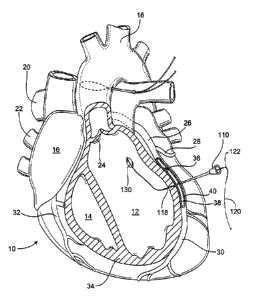
Fig. 2 is an anterior view of a heart 10 showing the left ventricle 12, right
ventricle 14, right atrium 16, aorta 18, pulmonary trunk 20 and pulmonary
veins 22. Fig.
2 shows the heart 10 in the diastolic (relaxed) phase of the heart cycle,
during which the
aortic valve 24 is closed. These views also show the left coronary artery 26
including the
circumflex branch 28 and the anterior descending branch (LAD) 30. The right
coronary
artery 32 is visible as well. The coronary arteries 26, 28, 30, 32 run along
the heart wall
34 and deliver oxygenated blood to the tissue comprising the heart wall, i.e.,
the
epicardium, myocardium, and endocardium. A blockage or occlusion 36 is shown
in the
LAD 30 which results in a partial or complete blockage of the artery lumen, a
condition
often referred to as narrowing of the arteries. This results in inadequate or
no blood flow
to the heart wall tissue fed by the portion of the LAD that is downstream of
the blockage
36.
The embodiment depicted in Figs. 2-5 includes an introducer 110
positioned so as to extend through the outer and inner walls 38, 40 of the LAD
30, and
CA 02347727 2001-04-12
WO 00/21436 PCT/US99/22945
6
through the heart wall 34 into the left ventricle 12. The introduces 110 may
be a small
profile, hollow shaft, for example a stainless steel hypo tube, and preferably
has as small
a diameter as possible in order to minimize damage to the coronary vessel. As
an
example, the introduces 110 may have an OD within the range of from about 0.5
mm to
about 3.0 mm and an ID within the range of from about 0.4 mm to about 2.9 mm.
The
introduces 110 is sized to receive a guide member in a sliding manner and has
an end 118
located in the heart chamber for directing the guide member through the lumen
of the
LAD 30 and into the ventricle 12. As explained below, the guide member is used
to
deliver devices into the heart chamber.
The guide member may be a single member or a plurality of members that
cooperate to guide devices into the heart chamber. The embodiment illustrated
in Figs. 2-
S comprises a first guide member and a second guide member coupled together,
one of
the members remaining positioned in the patient's heart to introduce devices
into the heart
chamber. In the preferred embodiment, the first guide member is a guide wire
120
1 S constructed of any suitable material such as stainless steel, and the
second member is a
catheter 122 coupled to the guide wire 120. Specifically, the proximal end 124
of the
catheter 122 is coupled to the distal end 126 of the guide wire 120 by a
suitable
detachable connection, such as a standard leur lock. The guide wire 120
preferably has a
small diameter, for example, approximately 0.25 mm, as does the catheter 122,
for
example, 2 French.
The distal end 128 of the catheter 122 is provided with a member that will
be engaged by blood flowing from the left ventricle 12 through the aortic
valve 24 into
the aorta 18. In the illustrated and preferred embodiment, the member is a
balloon 130
inflated via an inflation lumen (not shown) that is coupled to a source of
pressurized fluid
such as a syringe pump. An expandable member {e.g., balloon 130) is desirable
because
it may be collapsed for introduction into the heart chamber, thereby allowing
a smaller
opening to be formed through the wall of the coronary vessel. Of course, other
types or
configurations of members that will provide sufficient drag when placed in
normal blood
flow may be used in lieu of a balloon, e.g., an umbrella-shaped member, a soft
plastic
tube or a foam member. The preferred embodiment uses a member that is movable
between collapsed and expanded orientations, but a non-collapsible member
could be
used as well.
Fig. 2 shows the catheter 122 after it has been passed through the
introduces 110 to place the balloon 130 within the ventricle 12, the balloon
being shown
CA 02347727 2001-04-12
WO 00/21436 PCT/US99/22945
7
inflated to its expanded orientation. In Fig. 2 the heart 10 is in diastole;
as such, the
pressure in the left ventricle 12 is relatively low as it receives oxygenated
blood from the
left atrium (not shown). The balloon 130 remains in the left ventricle 12
until the heart
goes into the systolic (contracted) phase of the heart cycle, shown in Fig. 3.
The left
ventricle 12 contracts to expel oxygenated blood into the aorta 18. The
balloon 130 and
catheter 122 are expelled from the ventricle along with the blood. The balloon
130 and
catheter 122 are forced from the left ventricle 12 and pass through the aortic
valve 24 into
the aorta 18, which pulls a portion of the guide wire 120 into the ventricle.
The position
of the guide member may be monitored by ultrasound, TEE, or other means, and
the
guide member may be provided with a steering mechanism (not shown) if desired.
According to this embodiment, a device is provided for removing a portion
of the guide member from the heart chamber. One possible device is shown in
Fig. 3 and
comprises a snare including an introducer sleeve 132 which is placed through
an incision
in the aorta 18. The sleeve 132 receives a thin wire or filament 134
configured to form a
lasso 136. In the illustrated embodiment, the lasso 136 is positioned within
the aorta 18
downstream of the aortic valve 24 so that the balloon 130 and catheter 122
pass through
the lasso upon being forced out of the left ventricle I2 during systole. It
will be
understood that the device for removing a portion of the guide member from the
heart
may be used to retrieve the member from a different location than an opening
in the aorta
as shown in the Figures. For example, a magnet may be used to remove the guide
member.
As shown in Fig. 4, which depicts the heart 10 in diastole, once the balloon
130 and catheter 122 have moved through the lasso 136, the wire 134 is
withdrawn into
the sleeve 132 to securely grasp the catheter 122. The entire assembly of the
sleeve 132,
wire 134, catheter 122 and the guide wire 120 is pulled to a location external
to the heart.
In the illustrated embodiment, as seen in Fig. 4, this is achieved by removing
the
assembly through the incision in the aorta 18, which pulls the distal end 126
of the guide
wire 120 into the left ventricle 12 and then out of the aorta I8.
The above step is earned out while maintaining a portion of the guide wire
j0 I20 outside the LAD 30 and the left ventricle 12. In the illustrated
embodiment, a
proximal section of the guide wire 120 including proximal end 138 is
maintained outside
the heart. As shown in Fig. 5, this embodiment of the invention results in a
guide
member (e.g., wire 120) extending through the wall of a coronary vessel (e.g.,
LAD 30)
and the heart wall, into a heart chamber (e.g., left ventricle 12), and out of
the heart
CA 02347727 2001-04-12
WO 00/21436 PCT/US99/22945
8
chamber to a location external to the heart. In this position the guide member
provides a
pathway for delivering medical devices into the heart chamber for carrying out
medical
procedures.
The guide member and the introducer preferably have small enough
profiles so that only a small, easily repaired incision or opening needs to be
formed in the
walls of the coronary vessel to place the guide member in the heart chamber,
and
preferably an opening that does not need to be closed by sutures. The portion
of the guide
member located external to the heart, e.g., distal end 126 of guide wire 120,
is used to
deliver medical devices into the heart chamber and the coronary vessel without
going
through the outer wall of the coronary vessel. As such, it is not necessary to
form a large
openings) in the wall of the coronary vessel to deliver such devices into the
heart
chamber. In the illustrated embodiment, devices are guided over the distal end
126 of the
guide wire 120 into the aorta 18, and then into the left ventricle 12 to a
desired location.
In one application, the devices are then used to place a conduit (or form a
channel) in the
heart wall to communicate the interior of a coronary vessel with a heart
chamber
containing oxygenated blood.
This embodiment of the invention may of course take various forms and
configurations other than those specifically depicted in Figs. 2-5. For
example, rather
than using a guide member comprising a guide wire and a catheter, one of these
components could be omitted. As an example, in the illustrated embodiment, the
catheter
could be used alone by introducing medical devices over the end of the
catheter with the
balloon. Also, the guide member may be sufficiently stiff to allow its
introduction
through the coronary vessel walls and the heart wall without an introducer,
for example,
by forming a conventional guide wire, catheter, cannula, etc., with a desired
amount of
stiffness or flexibility. The guide member may be formed of one material or
comprise a
composite member, such as a flexible shaft portion and a soft tip.
Additionally, a device may be used to support the walls of the coronary
vessel during introduction of the guide member (and/or the introducer),
thereby
facilitating quick and easy access and penetration of the vessel. Suitable
devices for
supporting the heart wall and/or vessel walls are disclosed in commonly owned,
copending application U.S. Application No. 09/172,098, filed on October 13,
1998 and
entitled "DEVICES AND METHODS FOR USE IN PERFORMING
TRANSMYOCARDIAL CORONARY BYPASS," the subject matter of which is
incorporated herein by reference. The vessel may be supported internally or
externally in
CA 02347727 2001-04-12
WO 00/21436 PCT/US99/22945
9
order to facilitate placement of delivery devices through the vessel walls and
the heart
wall.
An example of another system and method for placing a guide member
through the wall of a heart according to this embodiment is shown in Fig. 6.
As
explained above with respect to Figs. 2-5, an introduces 110A is positioned
through the
walls of the coronary artery LAD and a guide member is slid through the
introduces. The
guide member may include two guide members coupled together, as in the
previous
embodiment, or a single guide member. Fig. 6 shows a single guide member in
the form
of a guide wire 120A with a proximal end 126A and a distal end 138A.
The introduces 1 l0A is configured to direct the guide member to a
particular location within the heart. In the embodiment of Fig. 6, the
introduces 1 l0A is
curved so that the distal end 118A thereof can be positioned to direct the
guide member
into the heart wall at a desired location, such as an area near the apex of
the heart. The
illustrated introduces 110A is generally J-shaped; it may, however, be shaped
differently.
The proximal portion of the introduces is manipulated to aim the distal end
118A in the desired direction and the guide wire 120A is passed through the
introduces.
The distal end 126A of the guide wire 120A exits the end 118A of the
introduces 1 l0A
and passes directly through the heart wall. The guide wire 120A preferably has
sufficient
strength to allow the end 126A to be pushed through the tissue of the heart
wall. This
may be facilitated by locating the distal end 118A of the introduces 1 l0A
relatively close
to the heart wall so that a relatively short length of the guide wire extends
from the
introduces before contacting the surface of the heart wall.
While these steps are being carried out a portion of the guide wire 120A is
maintained outside the LAD 30 and the left ventricle 12. In the illustrated
embodiment, a
proximal portion {including end 138A) of the guide wire 120A is maintained
outside the
heart. After placing the end 126A of the guide wire 120A through the coronary
vessel
and then into and out of the heart chamber, the introduces 110A is removed,
leaving the
guide wire positioned as shown in Fig. 8. One benefit of the system and method
shown in
Fig. 6 is that a snare (or other device) for removing a portion of the guide
member from
the heart chamber is not required. This obviates the need for precisely
directing a portion
of the guide member to a specific location within (or without) the heart
chamber to allow
its removal.
Additionally, while either the introduces or the guide member may be
preshaped to direct the guide member to a particular area within the heart
chamber, it
CA 02347727 2001-04-12
WO 00/21436 PCT/US99/22945
should be noted that the introduces may be in the form of curved hollow needle
that is
sized and configured to be passed through the coronary vessel and the heart
wall into the
heart chamber, and then out of the heart chamber. The introduces thus may
extend from
outside the heart chamber, into the heart chamber, and then out of the heart
chamber. A
guide member or conduit delivery device may then be positioned in the heart
chamber
using the introduces.
In an alternative system and method, shown in Fig. 7, the guide member is
removed from the heart chamber by a snare or like device. An introduces 110 is
used in
the manner described above with respect to Figs. 2-5 to place the distal end
126A of the
10 guide wire 120A within the left ventricle 12 at a desired location, for
example, adjacent
the apex of the heart. The guide wire 120A is preferably preshaped to assume a
desired
position when inserted into the heart chamber. From here the distal end 126A
of the
guide wire 120A is removed to a location external to the heart by a snare
assembly
comprising components 132, 134, 136 that are used to grasp and remove a
portion of the
guide wire 120A from the left ventricle 12. In this and the previous
embodiments, the
guide member may be manipulated or steered to a desired location by a suitable
mechanism (not shown), or formed of a shape memory alloy that directs the
guide
member to the desired location after insertion into the heart.
As in the previous embodiments, a portion of the guide wire 120A is
removed while maintaining another portion of the guide wire outside the LAD 30
and the
left ventricle 12. Specifically, as in the previous embodiment, a proximal
section of the
guide wire 120A including end 138A is preferably maintained outside the heart.
Use of
the system shown in Fig. 7 thus results in a guide member extending through
the wall of
the LAD 30 and the heart wall into the left ventricle 12, and out of the
ventricle to a
location external to the heart, for example, as shown in Fig. 8.
A guide member placed according to the systems and methods described
above provides a pathway for delivering medical devices into the heart chamber
without
passing the devices through the walls of the coronary artery. However, it will
be
recognized that the systems and methods illustrated in Figs. 2-8 are only
exemplary and
that this embodiment of the invention encompasses placing a guide member
within a
patient's heart so that different medical devices may be delivered into a
heart chamber.
The particular procedure carried out or the systems or devices used to place
the guide
member will vary depending on the application.
CA 02347727 2001-04-12
WO 00/21436 PCT/tJS99/22945
11
In one preferred application, a guide member positioned as discussed
above is used to deliver a conduit (or form a channel) that forms a blood flow
path
between a heart chamber and the interior of a coronary vessel. Figs. 9-13A
illustrate a
delivery system and method constructed according to a second embodiment of the
invention for placing a conduit within the heart wall to communicate the left
ventricle 12
with the LAD 30. A delivery system is designated by the reference numeral 140
and
comprises a conduit 142 supported on a delivery device 144. The conduit 142 is
preferably a rigid (i.e., not expandable) tubular element including a body
portion 146
having a first end 148 and a second end 150. The conduit body portion 146
includes one
or more openings 152 passing through the wall thereof. In the preferred
embodiment, the
body portion 146 has a plurality of holes located adjacent the second end 150,
which end
is preferably tapered as shown in Figs. 9 and 9A. Blood flows into the
interior of the
conduit 142 via the first end 148 and out of the conduit through openings 152
into the
interior of the LAD 30.
In the preferred construction, the delivery device comprises a shaft 154
(Fig. 9A) having a substantially complementarily shaped exterior to support
the conduit
142 during introduction into the left ventricle 12 and delivery into the heart
wall 34. The
shaft 154 has an elongated body that extends beyond the length of the conduit
142. The
elongated body is slidable over a guide member (guide wire 120 in the Figures)
that has
been positioned in a manner corresponding to that described above with respect
to Fig. 5.
The body of the shaft 154 has a clamp 158 which secures the shaft to the guide
wire 120
in order to deliver the conduit 142 into the heart wall 34. The clamp 158 has
a rotatable
knob 160 to selectively clamp the shaft 154 to the guide wire 120. Any other
suitable
mechanism may be used to couple the shaft 154 to the guide wire 120. Fig. 9
shows the
shaft 154 secured to the guide wire 120 and ready to be introduced into the
left ventricle
12 via the opening in the aorta 18.
Once the shaft 154 and conduit 142 have been slid over the distal end 126
of the guide wire 120 and secured thereto by the clamp 158, the proximal end
138 of the
guide wire 120 is pulled in the direction of the arrow in Fig. 10. This moves
the guide
wire 120, shaft 154 and conduit 142 into the aorta 18, past the aortic valve
24 and into the
left ventricle 12. As the distal end 126 of the guide wire 120 is pulled
further, the conduit
142 enters the heart wall 34, as shown in Figs. 10-10A. The position of the
conduit 142
relative to the heart wall 34 and the LAD 30 can be controlled by manipulating
the distal
end 126 of the guide wire 120 or the proximal end of the shaft 154 (or end 138
of the
CA 02347727 2001-04-12
WO 00/21436 1'CT/US99/22945
12
wire). The position of the conduit 142 within the heart wall 34 thus can be
selectively
adjusted by pulling or pushing an end of the guide wire 120 (or shaft 154)
with respect to
the heart wall.
Fig. 10A shows the conduit 142 positioned in the heart wall 34 so that a
portion of the second end 150 is located within the lumen of the LAD 30. In
this
embodiment at least some of the openings 152 are located within the LAD 30 and
deliver
blood from the left ventricle 12 into the artery. Once the conduit has been
placed in its
desired position, for example, the position shown in Fig. 10A, the shaft 154
is removed
from the conduit by pulling the shaft into the left ventricle 12 and toward
the aorta 18.
This may be accomplished in various ways. For example, as shown in Fig. 11,
the clamp
158 may be disengaged from the guide wire 120 by rotating the knob 160 to
allow the
shaft 154 to be slid off the proximal end of the wire in the direction of the
arrow.
Alternatively, the shaft 154 and the guide wire 120 may be removed as a unit
by pulling
the elements through the conduit 142, into the left ventricle 12 and out of
the aorta 18,
thereby obviating the need to release the clamp 158 from the wire 120.
In the illustrated embodiment, the second end 150 of the conduit 142
includes a low profile end 162 with a small diameter that is easily passed
through the
outer wall 38 of the LAD 30 after the conduit has been moved to the position
of Figs. 10-
10A. The end 162 preferably comprises a thin-walled section of tubing that
collapses or
folds and conforms to the exterior of the guide wire 120. The end 162 may be
separate
from or integral with the material forming the conduit 142. In the preferred
construction
shown in the Figures, the end 162 is detachable from the conduit 142 by
suitable means,
e.g., a perforated or scored section. As such, when the conduit 142 is
positioned within
the heart wall, the portion of the end that is outside the LAD 30 may be
grasped and
removed by pulling it in the direction of the arrow in Fig. 11, preferably
while holding the
proximal end 138 of the guide wire 120 or the portion of the shaft 154 located
external to
the heart, as shown in Fig. 11. Alternatively, the end 162 may be omitted with
the
conduit 142 open or closed at the end positioned in the artery.
After removing the end 162 from the conduit 142, the opening in the outer
wall 38 of LAD 30 is repaired as shown in Figs. 12-12A. If the shaft 154 has
been
removed previously without removing the guide wire 120, the guide wire is
removed by
pulling it out of the opening in the aorta 18 or the openings in the walls of
the LAD 30.
The opening in the aorta is then repaired. The resulting placement of the
conduit 142
provides a blood flow path between the left ventricle 12 and the LAD 30, which
path is
CA 02347727 2001-04-12
WO 00/21436 PCT/US99/22945
13
located distal to the occlusion 36 so that the myocardial tissue fed by the
distal portion of
the LAD is perfused.
The illustrated conduit 142 is preferably formed of a rigid material that is
strong enough to resist the force exerted by the myocardial tissue during
systole so that
the blood flow path is not blocked when the heart contracts. The conduit 142
may be
formed of various materials, for example, stainless steel, titanium, nitinol,
polymers,
ceramics, etc. Alternatively, the conduit may be constructed of a material
that partially
collapses when the heart contracts during systole and returns to its normal
configuration
during diastole, or the conduit may achieve specific flow characteristics by
regulating
blood flow with a valve or other means.
Additionally, the conduit may have a constant cross-section fully open at
both ends, and it may be provided with any size, shape and number of openings
152
depending on the desired flow characteristics. For example, the openings 152
may be
located along the entire length or only a portion of the length of the conduit
142.
Moreover, the shape of the conduit itself may be varied from that shown in the
Figures.
For example, the conduit 142 may have a constant cross-section over its length
rather
than a tapered portion, or one or both ends of the conduit may have enlarged
portions,
e.g., flanges, extensions or outwardly tapered sides for aiding in engaging
the conduit
with the wall of the heart or the wall of the coronary vessel. As a further
example, the
conduit may have an enlarged central portion with reduced size ends, and the
central
portion may act as a blood reservoir. Further, while the conduit 142 is shown
extended
into the lumen of the LAD 30, it may instead be flush with or slightly below
the inner
wall 40 of the LAD 30 (or flush with the surface of occlusion 36 in the LAD
30).
Similarly, although in the preferred system and method the shaft 154 and
the conduit 142 are coupled to the guide wire 120 so as to move therewith, the
system
may be used without coupling these elements. For example, the guide wire 120
may be
positioned as shown and held while the delivery system 140 is slid over the
wire into the
aorta and then into the left ventricle 12. The conduit 142 may then be pushed
into the
heart wall 12. However, it is preferred to secure the guide wire 120 to the
shaft 154 so
that these components can be pulled into the heart chamber as a unitary
assembly. This
avoids having to push the shaft and conduit over the wire, which exerts force
against the
guide wire and tends to pull the wire toward the chamber or against the walls
of the
artery. Nonetheless, the system and method may be used by sliding or otherwise
moving
the delivery device relative to the guide wire.
CA 02347727 2001-04-12
WO 00/21436 PCT/US99/22945
14
Figs. 13-17A show another preferred system and method for delivering a
conduit into a heart chamber and placing the conduit in the heart wall to
communicate the
heart chamber with the interior of a coronary vessel. As shown in Fig. 13A,
the system is
designated by the reference numeral 170 and comprises a conduit 172 supported
on a
delivery device 174. The conduit 172 is preferably an expandable tubular
member in the
form of a stmt including a plurality of elements 176 that move relative to
each other as
the stmt moves between collapsed and expanded orientations. The conduit 172
preferably comprises a stmt having a plurality of struts that move to a load
supporting
position when the stmt assumes its expanded orientation, the struts defining a
plurality of
open areas through which blood may flow. In the illustrated embodiment, the
conduit
172 is delivered into the left ventricle and placed in the heart wall while in
its collapsed
orientation, and then is opened to its expanded orientation.
The conduit 172 is supported on the delivery device 174 in its collapsed
orientation. In the illustrated embodiment, the conduit 172 is a balloon-
expandable stmt;
thus, the device 174 comprises a balloon 178 that is inflated via an inflation
lumen that
communicates with a source of pressurized fluid (not shown). Alternatively,
the stmt
could be expanded by a non-inflatable mechanism rather than a balloon. As a
further
alternative, the conduit 172 may be in the form of a self expanding stmt that
is retained in
its collapsed orientation by a cover or sleeve disposed around the stmt. In
either case, it
is desirable to cover the conduit 172 during delivery into the left ventricle
and the heart
wall to prevent the elements 176 from snagging or damaging tissue as they are
passed
into heart. As such, if the conduit comprises a balloon-expandable stmt as
illustrated, a
sheath 180 is preferably placed over the conduit to cover the elements 176.
The delivery device 174 comprises an elongated shaft provided with a
mechanism for securing the device to the guide wire 120A. A preferred
mechanism is
supported by a Y-connector 182 and includes a clamp 184 in the form of a
rotatable knob
that is operated as described above with respect to the embodiment of Figs. 9-
12A. The
clamp 184 is used to secure the shaft to the guide wire 120A so that the
components can
be manipulated as a unitary assembly. The Y-connector 182 may be provided with
a leg
186 for being coupled to a source of pressurized fluid, such as a syringe
pump.
The elongated shaft of the delivery device 174 is slidable over the guide
wire 120A which, in the illustrated embodiment, has been positioned in a
manner
corresponding to that described above with respect to Fig. 8. The clamp 184
secures the
shaft 174 to the guide wire 120A in order to deliver the conduit 172 into the
heart wall 34.
CA 02347727 2001-04-12
WO 00/21436 PCT/US99/22945
The clamp 184 has a rotatable knob to selectively clamp the shaft 174 to the
guide wire
120A. Any other suitable mechanism may be used to attach the shaft 174 to the
guide
wire 120A. Fig. 13 shows the shaft 154 secured to the guide wire 120 and in
the process
of being introduced into the left ventricle 12 via the opening in the aorta
18.
5 Once the shaft 174 and conduit 172 have been slid over the proximal end
138A of the guide wire 120A and secured thereto by the clamp 184, the distal
end 126A
of the guide wire is pulled in the direction of the arrow in Fig. 13. This
moves the guide
wire 120A, shaft 174 and conduit 172 into the left ventricle 12. As the distal
end 126A of
the guide wire 120A is pulled further, the conduit 172 enters the heart wall
34 with the
10 end of the sheath 180 dilating the opening, as shown in Figs. 14-14A. The
position of the
conduit 172 relative to the heart wall 34 and the LAD 30 can be controlled by
manipulating the proximal end of the shaft and the distal end 126A of the
guide wire
120A. That is, the position of the conduit 172 within the heart wall 34 can be
selectively
adjusted by pulling one end of the guide wire 120A (or and end of the shaft
154) toward
15 or away from the heart wall.
Fig. 14A shows the conduit 172 positioned in the heart wall 34 so that an
end 188 of the conduit is located within the lumen of the LAD 30. At least
some of the
openings defined between the stmt elements 176 are located within the LAD 30
so that
blood may flow into the LAD through the end and the wall of the conduit. Once
the
conduit has been placed in its desired position, for example, the position
shown in Fig.
14A, the sheath 180 is removed to expose the conduit to the heart wall tissue.
The sheath
may be removed in any suitable manner. In the illustrated embodiment, the
sheath 180
has an end 190 which can be grasped outside the LAD 30 and pulled. The entire
sheath is
preferably formed of a soft, collapsible material to permit the sheath to be
folded or
bunched up in order to pass through the opening in the walls of the LAD 30.
Suitable
materials include urethane, polyethylene, polytetrafluoroethylene, etc.
Fig. 15 shows the sheath 180 after it has been removed from the conduit
172 and the LAD 30. Removing the sheath 180 exposes the conduit 172 and
results in
tissue moving into the spaces between the stmt elements 176, which helps
retains the
conduit in position. Fig. 1 SA shows the conduit 172 once the sheath 180 has
been
removed. Next, in the case of an expandable conduit such as that shown in
Figs. 15-15A,
a mechanism is utilized to expand the stmt elements 176, preferably to their
maximum
strength position. One suitable mechanism is shown in Fig. 16A and includes
the balloon
178. The balloon 178 is inflated by a source ofpressurized fluid, such as
syringe 192,
CA 02347727 2001-04-12
WO 00/21436 PCT/US99/22945
16
coupled to inflation lumen 186. The syringe 192 is actuated to inflate the
balloon 178 and
expand the conduit 172 to the orientation shown in Figs. 16-16A.
Next, the syringe 192 is actuated to take down the balloon 178 for removal
from the interior of the conduit 172. The deflated balloon can be pulled into
the ventricle
S 12 and removed through the opening in the heart wall (adjacent the apex in
the Figures).
This may be accomplished by disengaging the clamp 184 from the guide wire 120A
to
allow the shaft 174 and balloon 178 to be slid off of the wire in the
direction of the arrow
in Fig. 17 (not shown). The guide wire 120A may then be removed by pulling
either end
through the chamber. Alternatively, the shaft 174, balloon 178 and guide wire
120A may
be removed as a unit by pulling the elements (after the balloon has been
deflated) through
the conduit 172 and then into and out of the left ventricle 12.
Figs. 17-17A show the conduit 172 expanded and positioned in the heart
wall after the delivery system has been removed from the heart. After the
conduit 172
has been fully expanded it firmly engages the tissue of the heart wall. The
conduit 172,
because it is constructed as a stmt, remains expanded despite the force
exerted against it
by the heart wall during the systolic phase of the heart cycle. In the
illustrated
embodiment, the size of the conduit is constant over its cross-section and the
ends of the
conduit extend slightly into the lumen of the LAD 30 and the left ventricle
12. It should
be recognized though that the conduit may have a size or shape that varies
over its length,
and may be positioned with one or both ends extending within, beyond or flush
with the
ventricle and coronary surfaces of the heart wall.
Moreover, it will be appreciated that Figs. 9-12A and Figs. 13-17A
respectively illustrate two independent conduit delivery systems and methods
for placing
a conduit within the heart wall to communicate the left ventricle with a
coronary artery.
The conduit delivery system and method of Figs. 9-12A are disclosed in
connection with
a guide member placed as shown in Fig. 5, while the system and method of Figs.
13-17A
are disclosed in connection with a guide member placed as shown in Fig. 8. It
will be
appreciated, however, that such description is for explanatory purposes as
each conduit
delivery system and method may be used with a guide member placed by any
manner
other than those disclosed herein. Similarly, the specific construction and
configuration
of the conduit and delivery system may be different from those specifically
illustrated.
Figs. 18A-21 show systems and methods for placing a conduit in the heart
wall so as to communicate a heart chamber with a coronary vessel according to
a third
embodiment of the invention. In this embodiment, the conduit has a generally
funnel-
CA 02347727 2001-04-12
WO 00/21436 PCT/US99/22945
17
shaped configuration in that one end of the conduit is larger than the other
end of the
conduit. The conduit is positioned in the heart wall to communicate a heart
chamber with
a coronary vessel. The larger end of the conduit is preferably positioned in
the heart
chamber and the smaller end is positioned in the vessel, although one or both
ends could
be flush with or within the heart wall.
Referring to Figs. 18A-18F, a conduit delivery system indicated by
reference numeral 200 includes a conduit 202 supported on a delivery device
204. The
conduit 202 is similar to the conduit 172 in that it is in the form of a stmt
including a
plurality of elements 206 that move relative to each other as the stmt moves
to its
expanded orientation. The conduit 202 preferably assumes a maximum load
supporting
position when fully expanded. Blood may flow through a plurality of open areas
defined
between the stmt elements 206.
The conduit 202 is supported on the delivery device 204 which itself is
supported on a guide member, such as guide wire 208. T'he conduit 202 is
supported in a
collapsed orientation and, in the illustrated embodiment, is expanded by a
balloon 210
that is inflated via an inflation lumen that communicates with a source of
pressurized
fluid (not shown). Alternatively, a non-inflatable mechanism rather than a
balloon could
expand the conduit 202, or it could be in the form of a self expanding stmt
that is retained
in its collapsed orientation by a cover or sleeve. The conduit 202 preferably
is covered
during delivery into the left ventricle and the heart wall to prevent the
elements 206 from
damaging tissue. Thus, a sheath 212 is placed over the conduit 202 to cover
some (and
preferably most or all) of the elements 206.
The delivery device 204 may comprise an elongated shaft provided with a
mechanism for securing the device to the guide wire 208 which extends through
a heart
chamber (such as left ventricle 12) and the wall of a coronary artery (such as
LAD 30).
The conduit 202 is moved into the heart chamber 12 and is placed in the heart
wall 34, as
shown in Fig. 18A. The position of the conduit 202 relative to the heart wall
34 and the
LAD 30 can be controlled and adjusted as explained above.
The conduit 202 is preferably positioned in the sheath 212 so that a portion
214 including end 216 extends beyond the sheath {Fig. 18A). This permits the
portion
214 of the conduit 202 to be expanded to a larger size than the portion of the
conduit
within the sheath 212. As shown in Fig. 18B, the balloon 210 is inflated and
expands the
portion 216 of the conduit 202; however, the sheath 212 prevents or limits
expansion of
the remaining portion of the conduit.
CA 02347727 2001-04-12
WO 00/21436 PCT/US99/22945
18
After this step, the sheath 212 is removed from the conduit 202, for
example, by pulling and end 218 of the sheath through the opening in the wall
38 of the
LAD 30, as shown in Fig. 18C. The sheath is preferably made of a strong yet
soft,
collapsible material that allows the sheath to be folded and removed through
the small
opening in the wall of the artery. The materials described above with respect
to the
sheath 180 of the previous embodiment may be used. The expanded portion 216 of
the
conduit 202 aids in retaining the conduit in the heart wall while the sheath
is removed.
The resulting configuration is shown in Fig. 18D.
Next, the balloon 210 is inflated to expand the entire conduit 202, as
shown in Fig. 18E. The conduit expands along its remaining length (i.e., other
than
expanded portion 216) to the orientation shown in Fig. 1 SF. After the sheath
212 has
been removed and the conduit 202 expanded, the stmt elements 206 are forced
against
the tissue of the heart wall to retain the conduit in position. The expanded
portion 216
provides an enlarged opening through which blood may flow from the left
ventricle 12
into the LAD 30, and also serves to secure the conduit within the heart wall.
When
expanded to the orientation shown in Fig. 18E, the entire conduit is
preferably, though not
necessarily, in its maximum radial strength position.
The balloon 210 is then deflated and removed from the conduit 202, for
example, by moving the delivery device 204 and balloon 210 into the left
ventricle 12 and
then out of the heart (with or without removing the guide wire 208). After
this, the
opening in the wall 38 of left ventricle 12 is repaired leaving the conduit
202 positioned
as shown in Fig. 18F. As can be seen, the conduit 202 assumes a funnel shape
by way of
the sheath restricting expansion of a portion of the conduit; however, it will
be
appreciated that this shape may be obtained in an alternative manner.
It will be understood that the invention encompasses many variations of
the preferred systems and methods described in detail herein. As an example,
rather than,
or in conjunction with, delivering a conduit into the heart wall, a guide
member placed as
described above may be used to deliver a tissue removal device into the
chamber. The
tissue removal device can be used to core a channel in the heart wall and/or
remove a
portion of the wall of the coronary vessel. If used in conjunction with
placing a conduit
in the heart wall, for example, in order to core a channel in the wall that
receives the
conduit, it should be recognized that the tissue removal device may be
separate from or
combined with the conduit delivery device. The device may instead be used to
form a
CA 02347727 2001-04-12
PCTNS99/22945
WO 00/21436
19
channel in the heart wall that remains open to provide a blood flow path
without using a
conduit.
Several suitable tissue removal devices that may be used with a guide
member are disclosed in commonly owned, copending application U.S. Application
No.
09/170,994, filed on October 13, 1998, and entitled "DELIVERING A CONDUIT INTO
A HEART WALL TO PLACE A CORONARY VESSEL IN COMMUNICATION
WITH A HEART CHAMBER AND REMOVING TISSUE FROM THE VESSEL OR
HEART WALL TO FACILITATE SUCH COMMUNICATION," the subject matter of
which is incorporated herein by reference.
Moreover, it will be understood that the surgical approach depicted in Fig.
1 is but one exemplary manner of accessing the heart in order to utilize the
systems,
devices and methods of the invention. The approach illustrated in Fig. 1,
which can be
characterized as minimally invasive in that a thoracotomy is used as opposed
to a median
sternotomy, may be desirable in some applications. However, those skilled in
the art will
recognize that other approaches may be used to access the heart in order to
practice the
invention.
For example, an open surgical procedure including a median sternotomy
may be used, or a minimally invasive procedure utilizing one or more
relatively small
access openings or ports may be used. Endoscopes or thoracoscopes may be used
for
visualization if the procedure is truly minimally invasive. Additionally,
rather than
forming one or more incisions in the patient's chest wall, an endovascular
approach may
be used to guide various inventive devices to the heart through the patient's
vascular
system to the heart, for example, by introducing the devices into a peripheral
vessel such
as the femoral artery.
Further, the exemplary embodiments are described primarily in connection
with their use in a beating heart procedure. Nevertheless, it will be
recognized that the
systems, devices and methods of the invention may be used in stopped-heart
procedures
utilizing cardiopulmonary bypass (CPB), or procedures during which the heart
is
intermittently stopped and started. As a result, the detailed description of
preferred
embodiments set forth in the drawing Figures and accompanying disclosure
should not be
construed as limiting the applications for which the invention may find
utility. The
preferred embodiments of the invention are described above in detail for the
purpose of
setting forth a complete disclosure and for sake of explanation and clarity.
It will be
readily understood that the scope of the invention defined by the appended
claims will
CA 02347727 2001-04-12
WO 00/21436 PCT/US99/22945
encompass numerous changes and modifications to the embodiments disclosed
herein.
As an example, a guide member or conduit delivery device may include
radiopaque
markers for monitoring their position. Also, a guide member or conduit placed
according
to the invention may be used to deliver any medical device, such as tissue
removal
S devices, or any pharmaceutical substance, such as angiogenic growth factors
or other
substances that aid in the perfusion of surrounding myocardial tissue.