Note: Descriptions are shown in the official language in which they were submitted.
CA 02349205 2006-12-22
PROSTHESIS AND METHODS OF INDUCING BONY INGROWTH USING
ULTRASOUND THERAPY
Technical Field
The present disclosure relates to prosthetic implant devices and
methods of utilizing ultrasound to induce bony ingrowth into a prosthetic
device.
More particularly, the present disclosure relates to prostheses which are
adapted
for insertion into the medullary canal of so-called "long bones" (femur,
humerus,
clavicle, radius, ulna, tibia, fibula, metacarpal, metatarsal and phalanges)
and
methods of inducing the soft cancellous bone surrounding the medullary canal
to
grow inwardly, i.e., "bony ingrowth", towards the prosthetic implant to
stabilize the
prosthesis within the medullary canal.
1
CA 02349205 2001-05-03
WD 00/28925 PCT/US99/26265
Background
Joint replacement, or arthroplasty, is a surgical procedure in which
the diseased portions of the joint are removed and replaced with new
artificial
parts called a prosthesis. During typical joint replacement surgery, the
surgeon
removes the diseased portion of the bone, surrounding tissue and cartilage
from
the joint leaving the healthy parts of the joint intact. The surgeon then
replaces
the diseased portion of the joint with new parts which mimic the movement of
the
joint. For example, with hip arthroplasty, the surgeon replaces the head of
the
femur and the acetabulum with artificial parts made of materials which permit
a
natural, gliding motion of the hip joint. This generic type of device includes
prostheses that have femoral components made of alloys, such as
cobalt-chromium-molybdenum and/or titanium-based alloys.
in some cases the surgeon uses a special glue or cement to bond
the new parts of the joint to the existing healthy bone. In other cases the
artificial
parts are made from a porous material which permits the patient's own bone to
grow into the pores of the porous material to hold the new parts in place.
See,
e.g., U.S. Patent Nos. 4,536,894 to Galante et al., 5,018,285 to Zolman et al.
and
5,004,476 to Cook.
Successful replacement of deteriorated, arthritic, and severely
injured joints has contributed to enhanced mobility and comfortable,
independent
living for many people who would otherwise be substantially disabled. New
2
CA 02349205 2001-05-03
WO 00/28925 PCT/US99/26265
technologies involving prosthetic devices for replacement of joints, along
with
advances in surgical techniques, has diminished the risks associated with
these
operations and improved the immediate and long-term outcome of joint
replacement surgery.
Questions remain, however, concerning which prosthetic designs
and materials are most effective for specific groups of patients and which
surgical
techniques and rehabilitation approaches yield the best long-term results. For
example, patients whose joints have been severely damaged due to osteoporosis
tend to suffer from long-term difficulties with the prosthesis due to
insufficient
bone mass surrounding the medullary canal. Osteoporosis causes abnormally
porous and fragile bones due to age, low calcium intake, inadequate physical
activity, certain drugs, estrogen deficiency, hormone disorders, nutritional
disorders, bone disuse and family history of the disease. Implanting a
prosthetic
device into such porous and fragile bones has had limited success and may
require a repeat/revision procedure or long therapy. Issues also exist
regarding
the best indications and approaches for revision surgery.
Nevertheless, further improvements in the total design of joint
prostheses are needed to facilitate more stable fixation of the implanted
prosthesis at the bone/metal interface. For example, with cemented prosthetic
devices fixation problems can occur due to the various stress loads, i.e., the
compression, shear and torsion to which the implanted device is subjected.
3
CA 02349205 2001-05-03
WO 00/28925 PCT/US99/26265
These mechanical forces, especially shear and torsion, as well as other
factors
such as osteoporosis, weaken the bone-cement bond. In addition, it is known
that there is a tendency for bone resorption which also weakens the cement
bond
between the intramedullary canal of the bone and the prosthesis.
By providing a bony ingrowth surface on the prosthetic device
and/or by providing therapy for inducing bony ingrowth into the prosthetic
device,
a more stable fixation can be made. However, with conventional prosthetic
device treatments/ techniques which include bony ingrowth surfaces, sufficient
bony ingrowth for long term stabilization typically requires the prosthesis to
be
stably fixed for at least six weeks after surgery, and any relative motion of
the
prosthesis during that period prevents or minimizes bony ingrowth. This is a
particularly significant problem in view of the difficulty in fitting the
prosthesis with
sufficiently close tolerances to provide large contact areas between the
porous
material and the bone, even where the entire outer surface of the prosthesis
is
fabricated from porous material. For example, it has been reported that an
instance of 10 to 20 percent of femoral stem loosening or failure in total hip
arthropiasty patients followed over five or more years, especially in younger
patients.
The present disclosure also relates to directing ultrasonic energy in
relatively low levels into living tissue to stimulate ingrowth of the soft
bone
surrounding the medullary canal into the prosthesis. The disclosure also
includes
4
CA 02349205 2006-12-22
various techniques for the transdermai delivery of acoustic energy through
body
tissue and/or fluids to propagate resonant waves along the inner cavity and/or
outer surface of the prosthesis to stimulate ingrowth of the surrounding soft
cancellous bone about the periphery of the medullary canal.
Acoustic bone fracture repair techniques and parameter preferences
are discussed in detail in U.S. Patent No. 5,520,612.
Much like bone fracture repair, acoustic techniques to stimulate
bony ingrowth are also subject to a range of values best determined by
professional experience. However, it is nevertheless helpful to list certain
parameter considerations which may play a significant role in promoting
ingrowth:
1) The frequency of surgically non-invasive acoustic delivery into the
body should be carefully calculated and monitored to insure steady standing-
wave development within the medullary canal;
2) The frequency of the transducer (or other carrier) should be
adjustably selectable, with provision for frequency sweeping between adjusted
limits; and
3) The frequency of pulse modulation should be adjustably selectable,
with provision for frequency sweeping between adjusted limits.
CA 02349205 2001-05-03
WO 00/28925 PCT/US99/26r265
SUMMARY
A bone prosthesis includes a first portion for engaging a first bone
segment and at least one channel disposed within the first portion for
propagating
acoustic energy through the channel to the first bone segment. Preferably, the
prosthesis further includes a second portion for engaging a second bone
segment. The channel includes an interior .reflective surface which defines a
resonating chamber disposed through the first portion. In one embodiment, the
resonating chamber includes at least one opening for receiving acoustic
energy.
In another embodiment, the resonating chamber is convoluted.
Another embodiment of the bone prosthesis includes a first portion
for engaging a first bone segment and a second portion for engaging a second
bone segment. At least one of the portions includes at least one means for
propagating acoustic energy to the corresponding bone segment. Preferably, the
propagating means includes: a transducer collar which engages one of the
portions; a transducer disposed adjacent one of the portions; and/or a
piezoelectric/piezoceramic membrane material which is disposed between a
porous material wrapped around the prosthesis and the outer periphery of the
prosthesis.
6
CA 02349205 2001-05-03
WO 00/28925 PCT/US99/26-265
In another embodiment, the bone prosthesis includes a ball portion
for engaging the acetabulum of the pelvic bone and the first portion is an
implant
for engaging the medulfary canal of the femur. In yet another embodiment, the
first portion engages the medullary canal of the humerus and a second portion
engages the medullary canal of the ulna and the first and second portions move
relative to one another about a pivot.
In still another embodiment, the first portion engages the femur and
the second portion engages the tibia. The first and second portions are
movable
relative to one another upon movement of one of the femur and the tibia.
Preferably, the first portion includes at least one dowel which engages a
corresponding bore associated with the femur and the second portion includes
at
least one dowel which engages a corresponding bore associated with the tibia.
The channel includes an interior reflective surface which defines a resonating
chamber disposed through each of the dowels.
The present disclosure also relates to a method for measuring the
stability of an implanted prosthesis and includes the steps of: a) providing a
source having a probe for sending and receiving signals and a comparator for
comparing and analyzing prior signal data with newer signal data; b) placing
the
probe adjacent the prosthesis; c) transmitting an initial signal through the
probe
to the prosthesis; d) receiving a return signal from the probe after the
signal
7
CA 02349205 2001-05-03
WO 00/28925 PCT/US99/26265
propagates and returns through the prosthesis; e) storing the return signal
data;
f) repeating steps (a) through (e); and g) comparing and analyzing stored
return
signal data to determine implant stabilization.
Another embodiment of the present disclosure relates to a method
for measuring the stability of an implanted prosthesis and includes the steps
of: a)
providing a source having a probe for sending signals and a comparator for
comparing and analyzing prior signal data with newer signal data; b) providing
a
receiving sensor which connects to the source and monitors the signals as the
signals propagate through the prosthesis; c) placing the probe adjacent the
prosthesis; d) placing the receiving sensor along the prosthesis; e)
transmitting signals through the probe to the prosthesis; f) monitoring the
signal
with the receiving sensor as the signal propagates through the prosthesis; g)
storing the signal data; h) repeating steps (a) through (g); and i) comparing
and
analyzing stored signal data to determine implant stabilization.
Another embodiment relates to a method for stabilizing an implanted
prosthesis and includes the steps of: a) providing a bone prosthesis having a
first
portion and at least one channel for propagating acoustic energy therethrough;
b)
engaging the first portion within a first bone segment; and c) directing
acoustic
energy at the first portion such that acoustic energy is transmitted through
the
channel to the first bone segment to stimulate bony ingrowth.
8
CA 02349205 2001-05-03
W_O 00/28925 -PCT/US99/26265
BRIEF DESCRIPTION OF THE DRAWINGS
Fig. 1A is a front view of one embodiment of the present disclosure
showing a hip prosthesis implanted within the upper femur with an external
transducer emitting acoustic waves at the prosthesis to stimulate bony
ingrowth;
Fig. 1 B is partial cross-section of the hip prosthesis of Fig. 1
showing an internal reflective surface and a resonating chamber;
FIG. 1 C is partial cross-section of the hip prosthesis of Fig. 1
showing the external transducer emitting acoustic waves at the reflective
surface
which, in turn, directs the waves downward through the resonating chamber;
Fig. 2A is partial cross-section of an alternate embodiment of the hip
prosthesis of Fig. 1 showing an internally disposed transducer mounted within
the
resonating chamber;
Fig. 2B is partial cross-section of the hip prosthesis of Fig. 2A
showing the external transducer emitting acoustic waves at the internal
transducer which, in turn, emits acoustic waves downward through the
resonating
chamber;
9
CA 02349205 2001-05-03
W_0 00/28925 PCT/iJS99/26-265
Fig. 3 is partial cross-section of an alternate embodiment of the hip
prosthesis of Fig. 1 showing a resonating chamber which is configured and
dimensioned in a convoluted form to maximize the interference between the
acoustic wave and the internal walls of the resonating chamber;
Fig. 4 is partial cross-section of an alternate embodiment of the hip
prosthesis of Fig. 1 showing a series of laterally extending slots which
extend
from the interior walls of the resonating chamber to the outermost periphery
of the
prosthesis to conduct energy directly to the inner walls of the medullary
canal;
Fig. 5A is partial cross-section of an alternate embodiment of the hip
prosthesis of Fig. 1 showing a transducer collar which surrounds the
prosthesis
and emits acoustic waves downwardly along the outer periphery of the
prosthesis
to stimulate bony ingrowth from the surrounding bone of the medullary canal;
Fig. 5B is partial cross-section of an altemate embodiment of Fig.
5A showing a hip prosthesis having both an internally disposed resonating
chamber for internally propagating acoustic waves and a transducer collar for
emitting acoustic waves downwardly along the outer periphery of the
prosthesis;
Fig. 6A is a front view of an alternate embodiment of the hip
prosthesis of Fig. I showing a piezoelectric/piezoceramic membrane material
CA 02349205 2001-05-03
W.D 00/28925 PCT/US99/26265
disposed between a porous coating and the outer shell of the hip prosthesis
for
conducting acoustic energy to the medullary canal;
Fig. 6B is a cross section of the Fig. 6A embodiment taken along
line 6B-6B;
Figs. 7A-7E are front views of various embodiments of the hip
prosthesis of Fig. 1 showing various pattems disposed about the outer
periphery
of the prosthesis for promoting acoustic wave propagation to the medullary
canal;
Fig. 8 is a front view of a diagnostic apparatus having a main
transmitting unit and a send/receive probe for monitoring and recording the
acoustic signals propagated through the hip prosthesis and medullary canal;
Fig. 9 is a front view of an alternate embodiment of the diagnostic
apparatus having a main transmitting unit, a probe for sending acoustic waves
through the hip prosthesis and a receiving sensor array probe for monitoring
the
acoustic signals propagated through the hip prosthesis and relaying the
information back to the main unit;
Figs. 10A-10J show various views of an alternate embodiment of the
present disclosure showing a knee joint prosthesis implanted between the lower
11
CA 02349205 2001-05-03
WO 00/28925 PCT/US99/26265
femur and the upper portion of the tibia with an external transducer emitting
acoustic waves at the prosthesis to stimulate bony ingrowth;
Figs. 11A and 11B show an alternate embodiment of a knee joint
prosthesis having a plurality of dowels eadh having an inwardly disposed
resonating chamber for propagating acoustic energy therethrough to stimulate
bony ingrowth;
Figs. 12A-12C show various views of an alternate embodiment of
the present disclosure showing an elbow joint prosthesis implanted between the
humerus and the ulna with an external transducer emitting acoustic waves at
the
prosthesis to stimulate bony ingrowth; and
Figs. 13A-13C show an altemate embodiment of an elbow joint
prosthesis having a plurality of inwardly disposed resonating chambers for
propagating acoustic energy therethrough to stimulate bony ingrowth.
DETAILED DESCRIPTION OF THE PREFERRED EMBODIMENTS
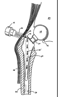
Referring now to Figs. 1A-1C which show one embodiment of the
present disclosure, namely, a hip prosthesis which is generally designated by
reference numeral 10. Hip prosthesis 10 includes a head or ball portion 18
which
is connected to a lower wedge-like implant member 32 by way of a neck portion
12
CA 02349205 2001-05-03
WO 00/28925 PCT/US99/26265
24. Preferably, the lower wedge member 32 is generally tapered such that the
lowermost portion 36 is configured to facilitate insertion of the wedge member
32
into the medullary canal 38 of the femur bone 14.
As best seen in Fig. 1A, the upper end "ball" portion 18 of the
prosthesis is preferably configured and dimensioned to engage the acetabulum
(socket) 16 of the pelvic bone 26 in a cup-like manner. This "ball and socket"
arrangement allows a wide range of motion, including sitting, standing,
walking
and other daily activities. Once the "ball and socket" are engaged, the
muscles
and ligaments 28, 32 of the upper leg, e.g., vastus lateralis and gluteus
muscles,
among other things, cooperate to retain the hip joint in place in much the
same
fashion as the ball and socket-like arrangement of the original hip.
Figs. 1 A and 1 C show the preferred position of the wedge member
32 implanted within the femur 14. More particularly, during hip replacement
surgery, the surgeon removes the diseased portion of the bone, surrounding
tissue and cartilage from the hip joint leaving the healthy parts of the hip
joint
intact. The upper portion of the femur bone 14 is preferably surgically
reconfigured to expose the medullary canal 38 which is a generally centrally
located passageway disposed within the femur 14 which extends the entire
length
of the same. In some cases it may be necessary to excavate the upper most
portion of the canal 38 to facilitate insertion of the wedge member 32 and
accommodate the wider upper portion 20 of the wedge member 32.
13
CA 02349205 2001-05-03
WO 00/28925 PCTIUS99/26265
The surgeon then replaces the head of the femur 14, i.e., ball, and
the acetabulum, i.e., socket, with new, biocompatible artificial parts, e.g.,
ball 18
and socket 16, made of materials which permit a natural, gliding motion of the
hip
joint, e.g., cobalt-chromium-molybdenum and/or titanium-based alloys. In some
cases the surgeon uses a special glue or cement to bond the new parts of the
hip
joint to the existing healthy bone 14. In other cases the artificiai parts are
made
from or include a porous biocompatible material which permits the patient's
own
bone to grow into the pores and hold the new parts in place.
It has been seen, however, that with prostheses that have been
cemented in place the various stress loads, i.e., the compression, shear and
torsion, to which the implanted device is normally subjected may cause the
bone-
cement bond to weaken. Other factors such as osteoporosis, also tend to
weaken the bone cement bond.
The various embodiments of the present disclosure provide
configurations which cooperate with ultrasonic therapies to induce bony
ingrowth
into the prosthetic device and provide a more stable fixation between the
prosthesis and the bone.
As illustrated in Figs. 1 A-1 C, once the prosthesis has been properly
implanted, the upper portion 20 of the wedge member 32 protrudes from the
14
CA 02349205 2001-05-03
WO 00/28925 -PCT/US99/26365
upper portion of the femur 14 to expose an opening 22 disposed proximate the
uppermost portion 20 of the wedge member 32. This opening 22 leads to a
resonating chamber 34 which extends inwardly down the wedge member 32
towards the tapered end portion 36 as best seen in Fig. 1 C.
An external ultrasonic transducer 12 is applied to the outer skin of
the patient (preferably pre-treated with a lotion or gel specifically
developed to
reduce the chances of the skin developing rashes or burning) and emits
acoustic
waves 30 at frequencies of between about 5KHz to about 10kHz which are
transcutaneously delivered through the body tissue and muscle 28, 32 towards
the upper portion 20 of the prosthesis 10. Preferably, the acoustic wave 30 is
focused at opening 22 such that the majority of the wave 30 enters through the
opening 22 and into the resonating chamber 34 which is filled with a fluid to
facilitate propagation of the acoustic wave 30' through the resonating
chamber.
As best seen in Figs. 1 B and 1C, the uppermost portion of the
resonating chamber 34 is equipped with a reflective surface 42 which is
specifically configured and dimensioned to reflect waves 30 downwardly through
the resonating chamber 34. Preferably, the reflected waves 30' resound off the
interior walls of the resonating chamber 34 and cause the prosthesis 10 to
resonate.
CA 02349205 2001-05-03
WO 00/28925 PCT/US99/26265
By resonating the prosthesis 10, or a portion thereof, within the
medullary canal 38 of the femur 14 at specific frequencies for short, e.g., 20
minute, periods of time on a weekly, bi-weekly, daily, or other time-specific
basis,
the energy will stimulate the soft cancellous bone 40 which surrounds the
medullary canal 38 to grow inwardly, i.e., "bony ingrowth", and stabilize the
prosthesis 10 within the femur 14.
It is contemplated that the cross-sectional areas of the resonating
chamber 34 can be varied along the length thereof to adjust the depth of
penetration of the propagated wave. In addition, the excitation values can be
varied to promote bony ingrowth from the distal end 36 to the upper portion
20.
Preferably, the reflective surface 42 can be configured at any
desired angle to resound acoustic waves 30' downward through the resonating
chamber 34 to cause the prosthesis to resonate/vibrate at different
frequencies.
Although it is preferable to utilize a resonating chamber 34 which has a
natural
resonance which responds to the acoustic waves 30' being propagated
therethrough, in some cases it may be desirable to configure the resonating
chamber 34 to have a more cylindrical-like cross-section or some other
geometrically advantageous cross-section to produce a different or specific
desired resonating effect.
16
CA 02349205 2006-12-22
In some cases it may also be preferable to calibrate the transducer
12 to emit one steady frequency to resonate the prosthesis 10, or in other
cases it
may be preferable to sweep the modulating frequency across a wide range of
frequencies to stimulate bony ingrowth. See, e.g., U.S. Patent No. 5,520,612.
Figs. 2A and 2B show an altemate embodiment of the present
disclosure which include a hip prosthesis 110 generally configured,
dimensioned
and operable in the same fashion as the Figs. 1A-1C embodiment with the
exception that this embodiment includes a second acoustically coupled
transducer 144 disposed intemaily within the upper portion 120 of the wedge
member 132 of the prosthesis 110. In use, the extemal transducer 12 emits
acoustic energy (focused or unfocused) at the second transducer 144 which, in
turn, emits acoustic waves 30' downward through the resonating chamber 134.
Preferably, the dimensions of the second transducer 144 are matched to the
frequency to facilitate wave 30' propagation through the fluid-filled
resonating
chamber 134 and/or the Eigen modes are matched to the cavity.
Figs. 3 and 4 show alternate configurations of the resonating
chamber 334, 434 disposed within the wedge member 332, 432 of the hip
prosthesis 310, 410. More particularly, Fig. 3 shows a zig-zag-like resonating
chamber 334 internally disposed within the wedge member 332. It is believed
that configuring the chamber 334 in this fashion will enhance vibration of the
17
CA 02349205 2001-05-03
WO 00/28925 PCT/US99/26265
prosthesis which will, in turn, further stimulate bony ingrowth of the soft
cancellous
bone 40 surrounding the medullary canal 38.
Fig. 4 shows another alternate configuration of the resonating
chamber 434 wherein a series of generally laterally disposed slots 450 extend
from the interior walls of the resonating chamber 434 to the outermost
periphery
of the wedge member 432 to propagate the acoustic energy 30' directly through
the prosthesis 410 to the surrounding soft cancellous bone 40 of the medullary
canal 38. It is further contemplated that structures such as biocompatible
ball
bearings, blades, wires, etc. can be positioned within the slots 450 to
enhance the
propagation of energy to the surrounding bone 40.
Fig. 5A and 5B show two additional alternate embodiments of the
present disclosure incorporating a transducer collar 546 which transfers/emits
acoustic waves 30' downward along the outer periphery of the wedge member
532 to stimulate bony ingrowth. More particularly and with reference to Fig.
5A,
the hip prosthesis 510 of this embodiment includes a generally circular
transducer
collar 546 which surrounds the uppermost portion 520 of the wedge member 532.
Collar 546 is activated and/or energized by the acoustic waves 30 emitted from
external transducer 12 and, in turn, propagates waves 30' downwardly along the
outer periphery of the wedge member 532 to stimulate ingrowth of the soft
cancellous bone 40 surrounding the medullary canal.
18
CA 02349205 2001-05-03
WA 00/28925 -PCT/US99/26365
Preferably, collar 546 can work in combination with other wave
propagation devices. For example, collar 546 can work in combination with a
porous material 560 which wraps the wedge member 532 and helps to propagate
the acoustic wave 30' downwardly to stimulate the soft bone 40. More
particularly, the acoustic wave 30' is trapped- within the porous layer 560 as
it
travels/conducts downward through the wedge member 532 and/or the porous
layer 560 acts as its own waveguide. The medullary canal of this embodiment
may act as a type of waveguide further enhancing/stimulating bony ingrowth.
It is also contemplated to impinge the acoustic wave 30 from the
external transducer 12 directly upon the porous material 560 above the femur
14
such that the acoustic wave 30 travels within the porous layer 560 downward
along the outer periphery of the wedge member 532 without the use of collar
546.
Fig. 5B shows an alternate embodiment of the Fig. 5A embodiment
wherein the wedge member 532 also includes an internally disposed resonating
chamber 534 which is configured and dimensioned similar to the resonating
chamber of Fig. 3 to propagate waves 30' internally through the wedge member
532. As shown in this figure, acoustic waves 30" and 30' are propagated along
the outer periphery of the wedge member 532 and within the resonating chamber
534, respectively, which, it is contemplated, will have a duel effect of
enhancing
bony ingrowth.
19
CA 02349205 2001-05-03
WO 00/28925 PCTIUS99/26265
Although Fig. 5B shows the resonating chamber similar to Fig. 3, it
is contemplated that other embodiments of the resonating chamber described
herein may be used in combination with the transducing collar 546 to enhance
bony ingrowth.
Figs. 6A and 6B show yet another alternate embodiment of the hip
prosthesis 610 which includes a piezoelectric/piezoceramic membrane material
670 disposed between the wedge member 632 and the porous coating 660.
Preferably, the piezoelectric/piezoceramic material is activated externally,
e.g., by
external transducer 12, and operates to propagate acoustic energy downward
along the outer periphery of the wedge member 632 to stimulate bony ingrowth
from the soft bone 40 into the porous coating to stabilize the prosthesis 610.
Figs. 7A-7E show alternate embodiments of hip prosthesis wherein
the outer periphery of the wedge member 732 is patterned to conduct/transmit
acoustic waves 30 directly into the medullary canal 38. For example, the
different
patterns of the wedge member 732 can include grooves or channels (Figs. 7A
and 7B), honeycomb patterns (Fig. 7C), semi-circular/spiral groove patterns
(Fig.
7D) and/or a series of longitudinally or laterally oriented zig-zag patterns
(Fig. 7E).
These patterns have a two-fold effect: 1) to directly conduct acoustic waves
30
into the medullary canal 38 to stimulate bony ingrowth; and 2) to enhance the
fit
of the prosthesis 710 in the medullary canal 38 during implantation.
CA 02349205 2001-05-03
WO 00/28925 PCTIUS99/26265
In use and as best seen in Fig. 7A, an extemal transducer 12 emits
acoustic waves 30 towards the upper portion 720 of the wedge member 732. The
acoustic waves 30, in turn, travel along the outer periphery of the wedge
member
732 and are trapped within the specified pattern thus propagating the waves 30
within the pattern between the wedge member 732 and the medullary canal. It is
contemplated that these patterns promote better ultrasound coverage and, thus,
enhance the coverage of bony ingrowth.
Fig. 7B shows one particular embodiment of the present disclosure
which includes a vertical groove pattern 755 which extends along the outer
periphery of the prosthesis 710 to conduct acoustic waves 30 into the
medullary
canal 38. This particular embodiment also includes a series of downwardly
angled bridges 765 disposed between adjacent grooves 755 which provide
alternate paths for the acoustic energy 30 should a groove 755 become
saturated
with bony ingrowth.
For the purpose of analysis, the etched patterns/grooves 755 on the
outer surface of the prosthesis 710 can be considered to be a unique
collection of
rectangular waveguides, each of dimensions dx and dY and open in the z-
direction. If a particuiar choice of "n" and "m" specifies one of the possible
normal
modes of vibration, then the cutoff frequency (f,) for the nm'" mode is:
21
CA 02349205 2001-05-03
W-0 00/28925 PCT/US99/26265
f =( 2 ) '' )2 +(MY
For n=m=1, the longitudinal velocity of sound cL = 1500 meters per second,
then
the cutoff frequencies for the channels are:
dx=dy=6mm, f, z 177 kHz; dx=dy 3mm, f. z 354 kHz; dx=dy 1.5mm, f, z 707 kHz.
Insonification of the channels 755 at frequencies much lower than f, will
produce
vibrational modes of primariiy shear waves.
Since the femur is generally cylindrical and about one-forth of the
body weight and if the channel 755 is considered to be a about two-thirds of
the
femur length and if the channel 755 is filled with body fluid, blood, and some
tissue debris, then the ultrasound absorption can be assumed to be about 0.3
dB
per MHz per cm. For example, the femur of a person who is six feet tall is
about
eighteen inches in length, thus, the prosthesis 710 should have a grooved
channel 755 about nine inches (23 cm) in length. If the transmitted frequency
is
1.0 MHz, then the maximum absorption incurred for this channel size is about
7dB, or an 80% reduction in acoustic intensity from the proximal to distal
end.
The acoustic power is variable to ensure sufficient spatial average-temporal
average (SATA) intensity levels along the channel 755 to induce bony tissue
ingrowth through ultrasound stimulation.
22
CA 02349205 2001-05-03
WO 00/28925 PCT/US99/26265
If the conventional porous coatings on the prosthesis are distributed
at strategic locations, primarily at the proximal end, an acoustic mode can be
produced to generate shear waves at these specific locations to enhance bony
tissue ingrowth - a type of induced "spot tissue-welding".
Preferably, the frequency of wave 30 and the channel/groove 755
sizes are varied to promote bony ingrowth from the distal to proximal ends of
the
prosthesis. It is contemplated that by promoting bony growth in this fashion,
the
entire prosthesis 710 can fuse within the medullary canal 38 of the bone.
Alternatively, the wedge member 732 can also include an intemally
disposed resonating chamber which can be configured and dimensioned similar
to the resonating chamber of Fig. I to propagate waves intemally through the
wedge member 732. As such, acoustic waves can be propagated along the outer
periphery of the wedge member 732 and within the resonating chamber,
respectively, to enhance the bony ingrowth.
Although the channels 755 shown herein are shown to have a U-
shaped cross-section, it is envisioned that other shapes can be used to which
may promote enhanced fusion of the prosthesis 710 with the bony tissue
ingrowth, e.g., undercut, rectangular and/or hemispherical.
23
CA 02349205 2001-05-03
WO 00/28925 PCT/US99/26265
Figs. 8 and 9 illustrate a reflected diagnostic system for determining,
e.g., whether any one of the aforedescribed ultrasonic therapies are required,
i.e.,
the prosthesis has loosened with the medullary canal. More particularly, Fig.
8
shows a main unit 11 equipped with a send/receive probe 15 which is placed in
contact with the prosthesis 10. A signal is applied from unit 11 through cable
13
to probe 15 and to prosthesis 10. The return signal from the prosthesis is
received by the receive portion of the probe after the signal
propagates/travels
through the prosthesis. Preferably, the main unit 11 includes a leamed neural
net
which compares prior data and actual live data to determine prognosis, i.e.,
the
return signal is analyzed and compared to prior acoustic/signal data taken at
the
time of implantation or last treatment to determine if the prosthesis 10 has
ioosened and/or the extent of bony ingrowth.
Fig. 9 shows an alternative diagnostic system wherein a second
needle and/or sensor array 23 is placed adjacent the bottom of the prosthesis
10
to directly receive the primary signal from the main unit 11 at predetermined
points along the prosthesis. The signal is then analyzed and compared to prior
acoustic/signal data taken at the time of implantation or last treatment to
determine if the prosthesis 10 has loosened. Preferably, the sensor array 23
will
give progressive readings and relay the information back to the main unit 11
via
cable 21.
24
CA 02349205 2001-05-03
WAO 00/28925 -PCT/US99/26265
Figs. 10A-10J show an alternate embodiment of the present
disclosure which includes a knee joint prosthesis 810a having an upper
prosthetic
implant 820a and a lower prosthetic implant 830a which are designed to engage
one another to form the prosthetic joint. Upper implant 820a is generally U-
shaped and dimensioned to receive and encompass the patella 816 of the femur
814. Lower implant 830a is generally T-shaped and dimensioned to fit atop the
distal end of the tibia 818. During knee joint replacement surgery, the
surgeon
removes the diseased portion of the bone, surrounding tissue and cartilage
from
the joint and reshapes the patella 816 and the distal end of the tibia 818 to
receive upper and lower implants 820a and 830a, respectively (see Fig. 10E).
Preferably, the upper implant includes a pair of dowels 842a which
project from the implant 820a and are generally dimensioned to engage a
corresponding pair of bores 872 which are drilled into the patella 816.
Likewise,
the lower implant 830a includes a dowel 832a which engages a corresponding
bore 870 which is drilled into the distal end of the tibia 818. Preferably,
the
dowels 842a and 832a include a plurality of channels or grooves 840a which
extend along the length of the dowels 842a and 832a and which provide a dual
function: 1) facilitate insertion and stability of the dowels within bores 870
and
872; and 2) provide a pathway to propagate acoustic energy 30' into the bone
816, 818 to stimulate bony ingrowth. In some cases the surgeon can use a
special glue or cement to bond the implants 820a, 830a to the existing healthy
bone 816, 818. In other cases the implants 820a, 820b can include a porous
CA 02349205 2001-05-03
WO 00/28925 PCT/US99/26265
biocompatible material which permits the patient's own bone to grow into the
pores and hold the implants 820a, 830a in place.
Preferably, the inner periphery of both the upper and lower implants
820a, 830a also include a plurality of grooves 847a and 845a, respectively,
which
also stabilize the upper and lower implants 820a, 830a atop the bone 816, 818
and provide a pathway for the acoustic energy 30' to stimulate bony ingrowth.
As seen best in Fig. 10B, the top portion of the lower implant 830a
includes a pair of rectilinear recesses 850a which are designed to seat the
lowermost portion of the upper implant 820a in a cradle-like manner. This
permits a natural, rocking motion of the implants 820a, 830a relative to one
another which mimics the natural motion of the original knee joint. Once the
upper and lower implants are engaged, the muscles and ligaments which
surround the knee joint are replaced and cooperate to retain the joint 810a in
place.
Figs. 11A and 11B show an alternate embodiment of a knee
prosthesis of the present disclosure which includes similar upper and lower
implants 820b and 830b which engage one another in a similar cradle-like
fashion
to form the prosthetic knee joint 810b. More particularly, upper and lower
implants 820b, 830b are generally shaped to engage the patella 816 and distal
end of the tibia 818, respectively, in a similar manner as described above
with
26
CA 02349205 2001-05-03
WO 00/28925 PCTIUS99/26265
respect to the Figs. 10A-10H embodiment with the exception that instead of
grooves, each dowel 842b and 832b includes an elongated resonating chamber
843b and 833b, respectively, which extends the length thereof.
As seen best in Fig. 11 B, upper and lower implants 820b and 830b
also include side channels 823b and 822b, respectively, which carry the
acoustic
energy 30' toward the resonating chambers 843b, 833b of the dowels 842b,
832b. Preferably, each side channel 823b, 822b also includes a reflective
surface 845b, 835b, respectively, which directs the acoustic energy 30' into
the
corresponding resonating chambers 843b, 833b. Preferably, the reflected waves
30' resound off the interior walls of the resonating chambers 843b, 833b and
cause the prosthesis 810b to resonate which stimulates bony ingrowth.
By resonating the prosthesis 810a or 810b, or a portion thereof,
within the patella 816 and tibia 818 at specific frequencies for short, e.g.,
20
minute, periods of time on a weekly, bi-weekly, daily, or other time-specific
basis,
the energy will stimulate the soft.cancellous bone which surrounds the dowels
842a,b and 832a,b to grow inwardly and stabilize the upper and lower members
820a,b and 830a,b .
Figs. 12A-12C show yet another embodiment of the present
disclosure which includes an elbow prosthetic implant 910a designed to engage
the lower end of the humerus 914 and upper end of the ulna 916. More
27
CA 02349205 2001-05-03
WO 00/28925 PCT/US99/26265
particularly, the prosthetic device 910a includes a pair of tapered, spike-
like
insertion members 920a and 930a which are pivotally joined to one another
about
a pivot 935a. Upper spike member 920a is dimensioned to insert into bore 922
which is drilled into the medullary canal 928 of the ulna 916 preferably
between
the otecranon process and the coronoid process. Lower spike member 930a is
dimensioned to insert into bore 932 which is drilled through the olecranon
depression 21 and into the medullary canal 938 of the humerus. In much the
same manner as described with the above prosthetic devices, the surgeon can
use special glues or cement to bond the spike members 920a, 930a to the
existing healthy bone 916, 918 or porous materials which permit the patient's
own
bone to grow into the pores and hold the spike members 920a, 930a in place.
Preferably the outer periphery of each of the spike members 920a,
930a includes a plurality of grooves or channels 942a and 940a, respectively,
which facilitate insertion and stabilization of spike members 920a and 930a
within
bores 928, 938 and also provide pathways for the propagation of acoustic
energy
30' into bones 816, 818 to stimulate bony ingrowth. In use and as seen best in
Fig. 12C, an external transducer 12 emits acoustic waves 30 towards the lower
spike member 930a. The acoustic energy 30', in turn, travels along the outer
periphery of the spike member 930a between the spike member 930a and the
medullary canal 938 to stimulate bony ingrowth.
28
CA 02349205 2001-05-03
WO 00/28925 - PCT/US99/26265
As mentioned above, the spike members 920a, 930a are joined to
one another by pivot 935a which permits natural, pivotal motion of the spike
members 920a, 930a relative to one another to mimic the natural motion of the
original elbow joint.
Figs. 13A-13C show an alternate embodiment of an elbow
prosthesis 910b according to the present disclosure which includes similar
upper
and lower spike members 920b and 930b which are joined about pivot 935b to
form the prosthesis 910b. Much like the embodiment shown in Figs. 12A-12C,
spike members 920b and 930b are shaped for insertion into corresponding bores
922, 932, respectively, drilled into the medullary canals 928, 938 of the ulna
916
and the humerus 914. However, instead of grooves disposed along the outer
periphery of the spike members 920b, 930b, each spike member 920b, 930b
includes an elongated resonating chamber 924b and 934b, respectively, which
extend the length thereof towards respective distal ends 926b and 936b.
As seen best in Fig. 13C, external ultrasonic transducer 12 emits
acoustic waves 30 which are transcutaneously delivered through the body tissue
towards the prosthesis 910b. Preferably, the acoustic wave 30 is focused at
openings 923b, 933b such that a majority of the wave energy enters openings
923b, 933b of resonating chambers 924b, 934b and resounds off the interior
walls
of resonating chamber 9244b, 934b and causes each spike member 920b, 930b
to resonate. In some cases it may be preferable to fill resonating chamber
924b,
29
CA 02349205 2001-05-03
WO 00/28925 - PCT/US99/26365
934b with a fluid to facilitate propagation of the acoustic wave 30' through
the
resonating chambers 924b, 934b.
By resonating the spike members 920b, 930b within the medullary
canals 928, 938, the resounding energy will stimulate the soft cancellous bone
which surrounds the canals 928, 938 to grow inwardly and stabilize the
prosthesis
910b within the bones 916, 914.
From the foregoing and with reference to the various figure
drawings, those skilled in the art will appreciate that certain modifications
can also
be made to the present disclosure without departing from the scope of the
present
disclosure. For example, in some case it may be preferable to employ a
converter to convert ultrasound to the power transducer and/or use an audio
signal to activate an internally housed transducer. Although it is preferable
to fill
the resonating chamber with a ultrasound conducting fluid, in some cases it
may
be preferable to shape the resonating chamber as a resonating horn filled with
a
solid material.
Although the various prosthetic devices of Figs. 10-13 show
resonating chambers similar in construction to the resonating chamber of Fig.
3,
it is contemplated that various resonating chambers described herein may be
used in combination with the prosthetic devices of the Figs. 10-13
embodiments.
Likewise, it is contemplated that the various patterns shown on the outer
CA 02349205 2001-05-03
WQ 00/28925 -PCT/US99/26265
periphery of the prostheses shown with respect to Figs. 7A-7E can be employed
on the prosthetic devices shown in Figs. 10-13.
In addition to the internal and external waveguides shown herein, it
is envisioned that the structure can be used in conjunction with porous
coatings of
the type known in the art either arranged in predetermined patterns or
uniformly,
see, e.g., U.S. Patent No. 4,536,854, U.S. Patent No. 5,018,285 and U.S.
Patent
No. 5,004,476.
The various embodiments of the present disclosure provide
configurations which cooperate with ultrasonic therapies to induce bony
ingrowth
into the prosthetic device and provide a more stable fixation between the
prosthesis and the bone. While particular embodiments of the disclosure have
been described, it is not intended that the disclosure be limited thereto, as
it is
intended that the disclosure be as broad in scope as the art will allow and
that the
specification be read likewise. Therefore, the above description should not be
construed as limiting, but merely as exemplifications of preferred
embodiments.
Those skilled in the art will envision other modifications within the scope
and spirit
of the present disclosure.
31