Note: Descriptions are shown in the official language in which they were submitted.
CA 02351323 2001-05-22
WO 00/32129 PCT/US99/28233
INTRACARDIAC GRASP CATHETER
FIELD OF THE INVENTION
The present invention relates to a steerable medical catheter and, more
particularly, to a flexible, electrode-bearing catheter of the type used in
electrophysiological studies for intracardiac electrocardiographic recording,
mapping,
stimulation and ablation.
BACKGROUND OF THE INVENTION
Catheters are often used in medical procedures to provide physical access to
remote locations within a patient via a relatively small passageway, reducing
the need for
traditional invasive surgery. The catheter tube can be inserted into an artery
or other
passageway through a relatively small incision in the patient's body, and
threaded through
the patient's system of blood vessels to reach the desired target.
Various types of catheters are used in various procedures, both diagnostic
and therapeutic. One general type of catheter used for both diagnostic and
therapeutic
applications is a cardiac electrode catheter. The diagnostic uses for a
cardiac electrode
catheter include recording and mapping of the electrical signals generated in
the course of
normal (or abnormal) heart function. Therapeutic applications include pacing,
or
~0 generating and placing the appropriate electrical signals to stimulate the
patient's heart to
beat in a specified manner, and ablation. In an ablation procedure, electrical
or radio-
CA 02351323 2001-05-22
WO 00/32129 PCT/US99I28233
2
frequency energy is applied through an electrode catheter to form lesions in a
desired
portion of the patient's heart, for example the right atrium. When properly
made, such
lesions will alter the conductive characteristics of portions of the patient's
heart, thereby
controlling the symptoms of supra-ventricular tachycardia, ventricular
tachycardia, atrial
flutter, atrial fibrillation, and other arrhythmias.
Such a catheter is typically placed within a desired portion of the patient's
heart or arterial system by making a small incision in the patient's body at a
location where
a suitable artery or vein is relatively close to the patient's skin. The
catheter is inserted
through the incision into the artery and manipulated into position by
threading it through a
sequence of arteries, which may include branches, turns, and other
obstructions.
Once the cardiac electrode catheter has been maneuvered into the region of
interest, the electrodes at the distal end of the catheter are placed against
the anatomical
feature or area sought to be diagnosed or treated. This can be a difficult
procedure. The
electrophysiologist manipulating the catheter typically can only do so by
operating a
system of controls at the proximal end of the catheter shaft. The catheter can
be advanced
and withdrawn longitudinally by pushing and pulling on the catheter shaft, and
can be
rotated about its axis by rotating a control at the proximal end. Both of
these operations
are rendered even more difficult by the likelihood that the catheter must be
threaded
through an extremely tortuous path to reach the target area. Finally, once the
tip of the
catheter has reached the target area, the electrodes at the distal end of the
catheter are
placed in proximity to the anatomical feature, and diagnosis or treatment can
begin.
In the past, the difficulties experienced by electrophysiologists in the use
of
a cardiac electrode catheter have been addressed in a number of different
ways.
To facilitate maneuvering a catheter through a tight and sinuous sequence
of arterial or venous passageways, catheters having a pre-shaped curve at
their distal end
have been developed. To negotiate the twists and branches common in a
patient's arterial
or venous system, the catheter typically is rotatable to orient the pre-shaped
curve in a
desired direction. Although the tip of the catheter may be somewhat flexible,
the curve is
fixed into the catheter at the time of manufacture. The radius and extent of
the curvature
generally cannot be altered. Therefore, extensive pre-surgical planning is
frequently
necessary to determine what curvature of catheter is necessary. If the
predicted curvature
turns out to be incorrect, the entire catheter may need to be removed and
replaced with one
CA 02351323 2001-05-22
WO 00/32129 PCT/US99/28233
3
having the proper curvature. This is an expensive and time-consuming ordeal,
as catheters
are generally designed to be used only once and discarded. Moreover, the
additional delay
may place the patient at same additional risk.
A variation of the pre-shaped catheter uses a deflectable curve structure in
the tip. This type of catheter has a tip that is ordinarily substantially
straight, but is
deflectable to assume a curved configuration upon application of force to the
tip.
However, the tip deflection is not remotely controllable. In a certain
patient's arterial
system, a point may be reached at which the proper force cannot be applied to
the catheter
tip. In such cases, the catheter must be withdrawn and reinserted through a
more
appropriate passage, or another catheter with a different tip configuration
must be used.
Another attempt to facilitate the placement of catheters takes the form of a
unidirectional steering catheter. A typical unidirectional steering catheter
has a steering
mechanism, such as a wire, that extends the length of the catheter to the
distal tip. The
steering mechanism is coupled to the tip in such a way that manipulation of
the proximal
end of the mechanism (e.g., by pulling the steering wire) results in
deflection of the
catheter tip in a single direction. This type of catheter is illustrated, for
example, in U.S.
Patent No. 5,125,896 issued to Hojeibane. The direction of deflection can be
controlled by
embedding a ribbon of wire in the tip; the ribbon is flexible along one
dimension but not in
others. This type of catheter can further be controlled by rotating the entire
shaft of the
catheter; in this manner, the direction of bend within the patient can be
controlled. The
shaft of such a catheter must be strong enough to transmit torque for the
latter form of
control to be possible.
U.S. Patent 5,383,852 to Stevens-Wright describes a steerable
electrocardial catheter including a flexible tip assembly having a proximal
and a distal
section. In this catheter, two steering mechanisms are used to separately
control bending of
either or both the proximal and distal sections. The steering mechanisms for
the proximal
and distal sections include separate steering wires, as described above, which
are coupled
to the proximal and distal sections, respectively.
Bidirectional steering catheters also exist. The distal end of a bidirectional
steering catheter can be maneuvered in two planes, allowing the tip to be
positioned with
greater accuracy. However, bidirectional steering catheters are complex
mechanically and
are often difficult to manipulate.
CA 02351323 2001-05-22
WO 00/32129 PCT/US99/28233
4
Although the foregoing types of catheters address the issue of
maneuverability in different ways, none of them is ideally configured to
maintain contact
with and apply a desired amount of pressure to a desired anatomical feature,
such as an
atrial wall.
One device used for the latter purpose is known as a basket catheter. See,
for example, the HIGH DENSITY MAPPING BASKET CATHETER manufactured by
Cardiac Pathways Corporation. A basket catheter has several spring-biased arms
near the
distal tip. When these arms are unconstrained, they bow outward to define a
basket-like
shape. The arms of the basket are constrained for implantation in a sheath
structure.
When the tip of the catheter has reached the desired location, the sheath is
retracted, or the
arms are advanced out of the sheath.
However, because the tip of the catheter is sheathed, it is not easily
steerable into location, and is not as flexible as one might desire. Moieover,
the sheath
adds bulk to the device, which might significantly limit the range of
applications in which
the basket catheter can be used. The basket has only one shape and size. Once
the arms
are deployed from the sheath, the basket assumes a single configuration
defined upon
manufacture. If the predefined configuration of the basket is not suitable,
then
substantially no correction is possible. Also, known basket catheters are not
indicated for
use in high-energy therapeutic applications, such as ablation.
A variable-geometry sheathed electrode catheter is also known in the art.
This device has a single electrode-bearing tip portion that is initially
disposed within a
relatively inflexible sheath. When the tip portion is advanced with respect to
the sheath,
the tip portion bows out of a slot-shaped aperture in the sheath. The shape of
the tip
portion can be controlled to apply a desired amount of pressure to an
anatomical feature.
However, as a sheath is used around the catheter, the device is not easily
steerable into
location. Moreover, as discussed above, the sheath structure adds undesirable
bulk to the
device.
Radio frequency ablation (RFA) has become the treatment of choice for
specific rhythm disturbances. To eliminate the precise location in the heart
from which an
arrhythmia originates, high frequency radio waves are generated onto the
target tissue,
whereby heat induced in the tissue burns the tissue to eliminate the source of
arrhythmia.
For successful ablation treatment, e.g., to produce a lesion at a given
CA 02351323 2001-05-22
WO 00/32129 PCT/US99/28233
anatomical site, it is generally required that the catheter be accurately
positioned at the
ablation site and that continuous contact be maintained between the electrode
and the
ablation site for the duration of the ablation treatment.
U.S. Patent 5,617,854 to Munsif describes, inter alia, a pre-shaped catheter
particularly useful for ablating in the vicinity of the sinoatriai node, the
left atrium, and up
to the mitral valve. The tip of the catheter is formed of a temperature-
sensitive shape-
memory material, e.g., Nitinol, or is otherwise invoked to assume a segmented
configuration upon reaching a desired position. The segmented configuration
includes a
distal segment which bears an ablation electrode. In operation, the segmented
shape
produces tension which urges the ablation electrode on the distal segment into
contact with
a wall of the Ieft atrium, while other segments are urged against other
tissue. Since the
shape of the catheter tip is f xed, the tip is not easily manipulated.
Further, the tension
produced between the segments of the catheter tip is dependent on the shape
and
dimensions of the ablation site, e.g., the left atrium.
Atrial fibrillation and atrial flutter are the most common type of arrhythmia
found in clinical practice. Although the potential adverse consequences of
these types of
arrhythmia is well known, the basic electrophysiological mechanisms and
certain
management strategies to control these types of arrhythmia have been
understood only
recently.
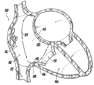
Reference is made to Fig. 1 which schematically illustrates a cross-section
of a human heart 100 showing typical atrial flutter circuits. Such circuits
includes macro-
entrant, counter-clockwise, pathways 120 from the right atrium 102, through
the inter-
atriai septum 114, down the free wall 116, and across the isthmus of tissue
108 between
the inferior vena cava 112 and the tricuspid annulus 106 of the tricuspid
valve 110.
Most electrophysiologists recommend treating atrial flutter by producing a
linear contiguous lesion 118 at the istlunus of tissue 108, between vena cava
112 and the
tricuspid annulus 106. Linear lesion 118 can be produced by RF ablation
electrodes which
are placed in contact with tissue 108. It is contemplated that isthmus tissue
108 is a critical
link of the atrial flutter circuit and, thus, linear lesion 118 is expected to
terminate this
source of arrhythmia and prevent the recurrence of such arrhythmia.
Existing ablation treatment for atrial flutter includes the use of a catheter
bearing at least one single or bi-polar ablation electrode. Unfortunately, an
undue amount
CA 02351323 2001-05-22
WO 00/32129 PCT/US99/28233
6
of time is spent in correctly positioning the ablation electrode of the
catheter against the
site to be treated. Further, in existing electrode catheter configurations,
the catheter must
generally be repeatedly repositioned until an acceptable lesion 118 is
produced. Thus,
lesion 1 I8 is often non-continuous, i.e., there may be gaps in the lesion
line which may
require further repositioning of the ablation catheter. Such repeated
repositioning of the
catheter is time consuming and may result in prolonged, potentially harmful,
exposure of
patients to X-ray radiation.
Accordingly, there is a need for a cardiac electrode catheter that can be
conveniently and quickly steered into secured, operative, engagement with a
preselected
portion of the isthmus of tissue between the inferior vena cava and the
tricuspid annulus,
to produce a predefined, substantially continuous, lesion on this isthmus of
tissue.
The difficulties in steering, positioning and providing secured contact of an
electrode catheter, with reference to the isthmus of tissue between the
interior vena cava
and the tricuspid annulus, are also applicable in mapping and/or ablating
other
intracardiac sites. For example, steering and positioning difficulties may
arise in mapping
and possible ablation in the vicinity of the coronary sinus.
SUMMARY OF THE INVENTION
The present invention seeks to provide a steerable electrode catheter having
a relatively flexible distal end portion accommodating at least one electrode,
for example,
an elongated configuration of mapping/ablation electrodes, that can be
conveniently
guided to a predetermined intracardiac site, for example, to the vicinity of
the tricuspid
valve, and that can be steered into a shape which enables convenient
positioning of at least
one electrode in secure operative engagement with a predetermined mapping
and/or
ablation site, for example, an ablation site along the isthmus of tissue
between the inferior
vena cava and the tricuspid annulus. If ablation of tissue is required, an
electrode catheter
in accordance with the present invention may be used to produce a predefined,
elongated,
substantially continuous, lesion at the ablation site.
According to an embodiment of the present invention, the catheter includes
a flexible distal end portion which may be controlled by one or two steering
mechanisms,
namely, a distal steering mechanism and/or a proximal steering mechanism. The
distal
steering mechanism may be adapted to deflect only the tip of the distal end
portion into a
CA 02351323 2001-05-22
WO 00/32129 PCT/US99/28233
7
hook-shaped configuration. The proximal steering mechanism may be adapted to
deflect
the entire distal end portion. Alternatively, the distal end portion of the
electrode catheter
may have a pre-shaped distal tip, e.g., the tip may be pre-shaped into a
partly deflected
configuration. In this alternative embodiment, a single steering mechanism may
be used
both to deflect the distal end portion and to further shape the pre-shaped
distal tip into the
desired hook-shaped configuration.
In another embodiment of the present invention, the distal end potion of the
electrode catheter may be adapted to be steerable or deflectable at three
regions, namely, a
distal tip deflection region, an intermediate deflection region, and a
proximal deflection
region. The curvature of the distal end portion at the intermediate deflection
region, in
addition to either or both of the distal tip deflection region and the
proximal deflection
region, enables more flexibility in conforming the shape of the distal end
portion of the
catheter to the shape of the target tissue, e.g., the above mentioned isthmus
of tissue,
during mapping and/or ablation of the target tissue. In an embodiment of the
present
invention, the intenmediate deflection region is adapted to be curved towards
the target
tissue, thereby to provide improved contact with the target tissue when the
end portion of
the catheter is urged against the target tissue.
In yet another embodiment of the present invention, the distal end portion is
not deflectable at the intermediate region but, rather, the distal end portion
is formed of a
resilient material and is pre-shaped to have a predetermined curvature at the
intermediate
region. In this embodiment of the invention, when the distal end portion is
urged against
the target tissue, the curvature of the intermediate region changes until the
electrode
configuration on the distal end potion conforms to the shape of the target
tissue. This
ensures urged contact between the at least one electrode and the target tissue
without an
additional steering mechanism.
In still another embodiment of the present invention, at least one
displaceable electrode may be used in addition to or instead of the elongated
configuration
of electrodes described above, to produce an elongated lesion on the target
tissue. A
displaceable electrode that may be suitable for use in conjunction with this
embodiment of
the invention is described in co-pending U.S. Patent Application No.
09/203,922, entitled
"Internal Mechanism for Displacing a Slidable Electrode", filed December 2,
1998, the
disclosure of which is expressly incorporated herein by reference.
CA 02351323 2001-05-22
WO 00/32129 PCTNS99/28233
According to an embodiment of the present invention, at least one ablation
electrode is brought into secured engagement with a target tissue, for
example, the isthmus
of tissue between the inferior vena cava and the tricuspid annulus, as
follows. First, the
distal end of the catheter is guided into the right atrium. As the distal end
of the catheter
advances in the right atrium, the proximal steering mechanism may be activated
to deflect
the entire distal end portion, such that the distal end portion may be
conveniently inserted
into the right ventricle. Once the distal end portion is inside the right
ventricle, the distal
steering mechanism is activated to produce the hook-shape configuration at the
tip of the
distal end portion. Then, the catheter is pulled back, i.e., in the direction
of the right
atrium, until the hook-shaped tip of the distal end is anchored at the
tricuspid annulus. The
catheter may then be pulled further back and the curvature of the distal end
portion may be
adjusted, e.g., using the proximal steering mechanism, until the at least one
ablation
electrode securely engages an ablation site along the isthmus of tissue
between the
tricuspid annulus and the inferior vena cava. Once such secured engagement is
obtained,
the at least one ablation electrode may be activated to produce a
substantially continuous,
linear, lesion at the ablation site.
As mentioned above, a catheter having a pre-shaped distal tip may
alternatively be used. In such case, the catheter may be guided into the right
ventricle and
then pulled back until the pre-shaped tip is anchored at the tricuspid
annulus, obviating the
step of deflecting the distal tip before pulling back the catheter. The
catheter may then be
pulled further back and the curvature of the distal end portion may be
adjusted, e.g., using
the proximal steering mechanism, as described above, until the distal tip
assumes the
desired hook-shaped configuration that provides a firm grip of the tricuspid
annulus and
secure engagement between the at least one ablation electrode and the ablation
site, e.g.,
along the isthmus of tissue between the tricuspid annulus and the inferior
vena cava.
An electrode catheter configuration as described above may be used, in
some preferred embodiments of the invention, for mapping and/or ablation of
tissue at
other intracardiac location where anchoring onto an edge of an orifice may be
helpful in
correctly and securely positioning an electrode catheter. For example, a
configuration as
described above may be useful for mapping and, possibly, ablating of tissue in
the vicinity
of the coronary sinus, by maneuvering the distal end of the catheter into the
coronary sinus
and pulling the catheter back until the distal tip of the catheter is anchored
at an edge of
CA 02351323 2001-05-22
WO 00/32129 PCT/US99/28233
9
the coronary sinus orifice.
BRIEF DESCRIPTION OF THE DRAWINGS
The present invention will be understood and appreciated more fully from
the following detailed description of the preferred embodiment taken in
conjunction with
the accompanying drawings in which:
Fig. 1 is a schematic, cross-sectional, illustration of a human heart showing
an atrial flutter circuit including an isthmus of tissue between the inferior
vena cava and
the tricuspid annulus;
Fig. 2 is a schematic, perspective, illustration of an electrode catheter in
accordance with an embodiment of the present invention;
Fig. 3 is a schematic, side view, cross-sectional, illustration of a distal
end
portion of the electrode catheter of Fig. 2;
Figs. 4A-4C are schematic, front view, cross-sections of the distal end
portion of Fig. 3, taken along section lines A-A, B-B and C-C, respectively;
Fig. 5 is a schematic, cross-sectional, illustration of the human heart,
showing the electrode catheter of Fig. 2 being introduced into the right
atrium;
Fig. 6 is a schematic, cross-sectional, illustration of the human heart,
showing the electrode catheter of Fig. 2 being steered from the right atrium
into the right
ventricle;
Fig. 7 is a schematic, cross-sectional, illustration of the human heart,
showing the tip of the electrode catheter of Fig. 2 being deflected into a
"hook" shape;
Fig. 8 is a schematic, cross-sectional, illustration of the human heart,
showing the electrode catheter of Fig. 2 being pulled back to engage the
isthmus of tissue
between the inferior vena cava and the tricuspid annulus with the tip of the
catheter
anchored at the tricuspid annulus;
Fig. 9 is a schematic illustration of an end portion of an electrode catheter
in accordance with another embodiment of the present invention;
Figs. l0A and l OB are schematic illustrations of part of an electrode
catheter in accordance with yet another embodiment of the present invention,
in a non-
deflected configuration and a deflected configuration, respectively.
Fig. 11 is a schematic, perspective, illustration of a medical device
CA 02351323 2001-05-22
WO 00/32129 PCT/US99/28233
carrying a displaceable electrode which may be used in conjunction with still
another
embodiment of the present invention;
Fig. 12 is a fragmented side view, in enlarged scale, of the displaceable
electrode included in the medical device of Fig. 1;
5 Fig. 13 is a cross-sectional view taken along the line 403-403 of Fig. 2
and looking in the direction of the arrows;
Fig. 14 is a cross-sectional view taken along the line 404-404 of Fig. 2
and looking in the direction of the arrows; and
Fig. 15 is a schematic, perspective, illustration of an alternative
10 embodiment of the medical device of Fig. 11.
DETAILED DESCRIPTION OF PREFERRED EMBODIMENTS
Reference is made to Fig. 2 which schematically illustrates a perspective
view of an ablation andlor mapping catheter 10 in accordance with an
embodiment of the
present invention.
Catheter 10 includes a handle portion 22, electric connectors 24, a tubular
catheter shaft 11 and a distal end portion 12 including an end shaft 13.
Distal end portion
12 includes a distal tip 16, a distal tip deflection region 60 and a proximal
deflection
region 62. According to the present invention, distal end portion 12 can be
steered from a
generally straight configuration, indicated by the solid lines in Fig. 1, to a
deflected
configuration, indicated by the broken lines in Fig. 1. The broken Iine
configuration in Fig.
1 also illustrates how distal deflection region 60 can be deflected into a
hook-shaped
configuration, as described in detail below.
In an embodiment of the present invention, tip 16 may include a sensor or
mapping electrode, as is known in the art, for monitoring the electric
potential of tissue in
contact therewith. This may be helpful in guiding and positioning distal end
portion 12, as
described below. Additionally or alternatively, tip 16 may include an ablation
electrode
for ablating tissue in contact therewith.
Reference is now also made to Fig. 3 which schematically illustrates a
side-view, cross-section, of distal end portion 12. End shaft 13, which is
preferably hollow
as shown in Fig. 3, accommodates an elongated configuration 40 of ablation
electrodes 14.
CA 02351323 2001-05-22
WO 00/32129 PCTNS99128233
11
Elongated configuration 40 may include any number of electrodes 14, with a
predetermined spacing therebetween, or a single elongated electrode, as known
in the art,
adapted to produce a substantially continuous, substantially linear, lesion
when brought
into operative engagement with a target tissue. Electrodes 14 are preferably
all ring-
s electrodes covering the entire circumference of shaft 13. Additionally or
alternatively,
electrodes configuration 40 may include at least one mapping electrode, as is
known in the
art, for monitoring the potential of the tissue in contact with the electrode
configuration.
Reference is now made also to Figs. 4A-4C which schematically illustrate
front-view cross-sections of distal end portion 12 along section lines A-A, B-
B, and C-C,
respectively, in Fig. 3. In accordance with the present invention, catheter 10
includes a
distal steering mechanism which is used to deflect tip I6 of distal end
portion 12, as
mentioned above, by producing a small radius of curvature at region 60.
Catheter 10
further includes a proximal steering mechanism which controls the curvature of
region 62,
between shaft 11 and 13, thereby to control the deflection of the entire
distal end portion
12.
The distal and proximal steering mechanisms may include any suitable
steering mechanisms known in the art, for example, the control mechanisms
described in
U.S. Patent 5,383,852 to Stevens-Wright, the disclosure of which is
incorporated herein
by reference. As shown in Figs. 3-4C, the distal and proximal control
mechanisms may
include control wires 55 and 64, respectively, which extend along the interior
of shaft 11
from handle portion 22 to regions 60 and 62, respectively, of distal end
portion I2. Wire
55 is attached to tip 16 and may extend through middle guiding Loops along
most of the
length of shaft 13, as shown in Figs. 4B and 4C, and then through off center
guiding loops.
at region 60, as shown in Fig. 4A, whereby only a small segment adjacent to
tip 16 is
deflected by wire 55. Wire 64 may extends through off center guiding loops in
shaft 13, as
shown in Fig. 4C, and is attached to end shaft 13 at region 62.
The deflection of distal end portion 12 into a desired configuration is
preferably controlled by an electrophysiologist using control members 26
and/or 27 on
handle portion 22. In the embodiment shown in Fig. 2, control member 26 may
include a
rotatable control member attached to wire 55, such that forward or backward
rotation of
control member 26 results in corresponding movement of wire 55, thereby
controlling the
deflection of end portion 12 at region 60. Control member 27 may include a
slidable
CA 02351323 2001-05-22
WO 00/32129 PCT/US99/28233
12
control member attached to wire 64, such that forward or backward sliding of
control
member 27 results in corresponding movement of wire 64, thereby controlling
the
deflection of end portion 12 at region 62. As known in the art, the
electrophysiologist may
also rotate distal end portion 12 about the longitudinal axis of catheter 10.
Any suitable
rotation mechanism, as is known in the art, can be used to control the
rotation of distal end
portion 12. For example, catheter shaft can be made of a rotationally rigid
material that
transmits the rotation of handle portion 22 to distal end 12. Alternatively,
the rotation of
handle 22 may be transmitted by a rotationally stiff member (not shown)
extending
longitudinally through the interior of catheter shaft 11.
In an embodiment of the present invention, electrodes 14 are addressed,
together or separately, via connectors 24, which are connected to electrodes
14 by
conductors 66. Conductors 66 may extend along the interior of catheter shaft
11 and end
shaft 13, for example, through middle guiding loops, as shown in Figs. 4A-4C.
Using connectors 24, electrodes 14 are connected to an ablation energizing
circuit, which may be activated by user controls as are known in the art. Upon
activation,
the energizing circuit energizes electrodes 14 with radio frequency (RF)
energy, as is
known in the art. Using separate ablation controls, the electrophysiologist
may activate
electrodes 14 together or separately (if selective ablation is desired) to
ablate a target
tissue, as described in detail below.
As known in the art, electrodes 14 may be associated with temperature
sensors (not shown in the drawings) which may be connected to temperature
monitoring
circuitry for monitoring the temperature of the tissue in contact with
electrodes 14. An
output of the temperature monitoring circuitry may be visually displayed to
the
electrophysiologist, as is known in the art, to provide the
electrophysiologist with on-line
indication of the electrode temperatures, which are indicative of adjacent
tissue
temperatures. If temperature sensors are used, they may be connected to the
monitoring
circuitry via connectors 56 and additional conductors (not shown) in catheter
shaft 11.
According to the present invention, catheter 10 is used for ablating tissue
on the endocardium isthmus of tissue between the inferior vena cava and the
tricuspid
annulus of a patient suffering from aberrant heart activity, such as atrial
flutter or
fibrillation, as described below.
Figs. 5-8 schematically illustrate a procedure for introducing catheter 10
CA 02351323 2001-05-22
WO 00/32129 PCTIUS99/28233
13
into the right atrium and subsequently guiding distal end portion 12 to
securely engage a
portion of the endocardium tissue 108 between the inferior vena cava and the
tricuspid
annulus.
As shown in Fig. 5, distal end portion 12 is first guided into the right
atrium
of the patient's heart 100 from the inferior vena cava. Once catheter 10 is
introduced into
the right atrium, the electrophysiologist proceeds to deflect distal end
portion 12 towards
the right ventricle 104, using the proximal steering mechanism of catheter 10.
Distal end
portion 12 enters the right ventricle via the tricuspid valve 110, as shown in
Fig. 6. If
necessary, end shaft 13 may be rotated to assist in the manipulation of distal
end portion
12.
After distal end portion 12 is inserted into the right ventricle, the
electrophysiologist uses the distal steering mechanism to deflect tip 16 into
the hook-
shaped configuration described above, as shown in Fig. 7. Then, the catheter
is pulled
back, i.e., in the direction of inferior vena cava I I2, until a portion of
the tricuspid annulus
106 is grasped by the hook-shaped tip 16, as shown in Fig. 8.
Once tip 16 is anchored at the tricuspid annulus, the catheter may be pulled
further back and the curvature of distal end portion 12 may be adjusted, using
the proximal
steering mechanism, until electrodes I4 of elongated configuration 40 securely
engage a
portion of the isthmus of tissue 108 between tricuspid annulus 106 and
inferior vena cava
1 I2. At this point, the electrophysiologist activates some or all of
electrodes 14 to ablate a
substantially continuous, substantially linear, lesion on the endocardial wall
of the isthmus
of tissue 108.
As described above, electrodes 14 may be associated with temperature
sensors. These sensors may include thermocouples or any other temperature
sensing
means known in the art. Based on the temperatures measured by these optional
temperature sensors, the electrophysiologist may deactivate some or all of
electrodes 14
when the temperature of the ablated tissue site exceeds a predetermined
threshold. Then,
when the temperature of the ablated sites drops below the threshold, the
electrophysiologist may reactivate electrodes 14 if further ablation is
required.
As mentioned above, tip 16 may optionally include a sensor electrode for
monitoring/mapping the electrical potential of tissue adjacent tip 16, e.g.,
to enable more
accurate and/or more efficient positioning of end portion 12 against isthmus
of tissue 108.
CA 02351323 2001-05-22
WO 00/32129 PGT/US99/28233
14
Sensor electrodes may also be included in electrode configuration 40, e.g.,
for mapping the
electrical potential along isthmus of tissue 108, during or between ablation
sessions, to
determine whether further ablation may be necessary.
Reference is now made to Fig. 9 which schematically illustrates a distal end
portion 212 of an ablation catheter in accordance with another embodiment of
the present
invention, having an elongated electrode configuration 240 including a
plurality of
electrodes 214 and a tip 216. In the embodiment of Fig. 9, distal end potion
212 is adapted
to be steerable or deflectable at three regions, namely, a distal tip
deflection region 260, an
intermediate deflection region 250 and a proximal deflection region 262.
Regions 260 and
262 are generally analogous to the distal and proximal deflection regions 60
and 62,
respectively, of distal end portion 12, as described above with reference to
Figs. 2-8.
Intermediate deflection region 250 may be located at a predetermined position
along
electrode configuration 240. The mechanisms for deflecting end portion 212 at
regions
260 and 262 may be similar to those used for deflecting end portion 12 at
regions 60 and
62, respectively, as described in detail above with reference to Figs. 2-8.
The mechanism
for deflecting distal end portion 2I2 at intermediate region 250 may include
any suitable
deflection mechanism, for example, a control wire (not shown) extending
through the
hollow interior of end portion 212, analogous to control wires 55 and 64 in
the
embodiment of Figs. 2-8.
The curvature of end portion 212 at any or all of regions 260, 250 and 262
may be controlled by the electrophysiologist using any suitable controls (not
shown), for
example, handle controls similar to controls 26 and 27 in the embodiment of
Figs. 2-8.
Thus, in this embodiment, the electrophysiologist may control the curvature of
distal end
portion 212 at region 250, in addition to controlling the curvature of distal
and proximal
regions 260 and 262. The addition of intermediate deflection region 250
enables more
flexibility in conforming the shape of distal end portion 212 to the shape of
isthmus of
tissue 108 during ablation treatment. In an embodiment of the present
invention,
intermediate deflection region 250 is adapted to be deflected in the direction
indicated by
arrow 270, so as to provide improved contact with isthmus of tissue 108 when
end portion
212 is urged against the tissue.
In yet another embodiment of the present invention, end portion 212 is not
deflectable at region 250 but, rather, end portion 212 is formed of a
resilient material and
CA 02351323 2001-05-22
WO 00/32129 PCT1US99/28233
is pre-shaped to have a predetermined curvature at region 250, as shown
generally in Fig.
9. In this embodiment of the invention, when end portion 212 is urged against
a target
tissue, such as isthmus of tissue 108, the curvature of region 250 changes
until electrode
configuration 240 conforms to the shape of the target tissue. This ensures
urged contact
5 between electrodes 214 and the target tissue without an additional steering
mechanism.
In still another embodiment of the present invention, end portion 212 is
deflectable only at distal region 260, to assume a hook-shaped conf guration
as described
above, but is not deflectable at proximal. region 262. End portion 212 may
also be pre-
shaped or deflectable at intermediate region 250, as described above. In this
embodiment,
10 once the tricuspid annulus is grasped by the hook-shaped tip of the
catheter, it is primarily
the backward pulling force applied by the electrophysiologist that brings
electrodes 214
into urged contact with the target endocardial tissue.
Reference is now made to Figs. l0A and l OB which schematically illustrate
part of an electrode catheter 300 in accordance with yet another embodiment of
the present
15 invention. Catheter 300 includes a tubular catheter shaft 311 and a distal
end portion 312
including an end shaft 3I3. Distal end portion 312 includes a distal tip 316,
a distal tip
deflection region 360 and a proximal curvature region 362. In this embodiment,
region
360 distal end portion 312 is pre-shaped to have a partly deflected
configuration, as shown
in Fig. 10A, with an inner-curve angle a. Such pre-shaping of region 360 may
be
performed by pre-baking region 360 into the desired configuration, as is known
in the art.
As described below, distal end portion 312 may be steered into a deflected
configuration,
shown in Fig. 10B, wherein proximal curvature region 362 is curved to a
predetermined
extent and distal deflection region 360 is further deflected into a hook-
shaped
configuration, similar to that described above with reference to Figs. 2-8.
Distal end portion 312 has an elongated electrode configuration 340
including a plurality of electrodes 314 and a tip 316. As in the embodiment of
Figs. 2-8,
tip 316 may include a sensor or mapping electrode, as is known in the art, for
monitoring
the electric potential of tissue in contact therewith and/or an ablation
electrode for ablating
tissue in contact therewith. In the embodiment of Figs. l0A and l OB, distal
end potion 312
is adapted to be steerable or deflectable by a single deflection mechanism at
both regions
362 and 360. The mechanism for deflecting end portion 312 at regions 360 and
362 may
include a control wire (not shown), similar to control wire 55 in Figs. 3 and
4A-4C, which
CA 02351323 2001-05-22
WO 00/32129 PCT/US99/28233
16
extends through the hollow interior of end portion 312 . The control wire is
fixedly
attached to tip 316 and may extend through off-center guiding loops, as are
known in the
art, along the entire length of shaft 313. Thus, in contrast to the
embodiments described
above with reference to Figs. 2-8, the curvature of the entire length of
distal end portion
3I2, including regions 362 and 360, is affected upon activation of the
deflection
mechanism, thereby producing the deflected configuration shown in Fig. l OB.
re 55. In an
embodiment of the present invention, shaft 313 is made from a material which
is more
flexible than the material used for shaft 311. The transition between the
materials of shafts
311 and 313 is indicated by numeral 315. The material for shaft 313 may
include
Polyether Block Amide, having a Shore D hardness of 40-S5, available from
Atochem,
Inc., U.S.A., under the trade name of Pebax. It should be appreciated,
however, that wide
range of materials and hardnesses of shaft 313 may be suitable for the present
invention,
depending on specific design requirements. The material used for shaft 31 I
should be at
least slightly harder than that of shaft 313, and preferably has a Shore D
hardness at least 5
higher than that of shaft 313.
It should be appreciated that, in the embodiment of Figs. l0A and IOB,
when the control wire is pulled backwards to deflect end portion 312, region
362 becomes
curved and region 360 is fully deflected into the desired hook-shaped
configuration, as
shown in Fig. I OB. Thus, the deflection of both regions 362 and 360 is
performed in a
single action, obviating the need to use two separate deflection mechanisms,
as in some of
the above described embodiments. This simplifies the deflection procedure to
be executed
by the electrophysiologist.
Catheter 300 may be used for mapping and/or ablating the isthmus of tissue
between the inferior vena cava and the tricuspid annulus, as follows. In
analogy to the
procedure described above with reference to Figs. 5-8, distal end portion 312
is first
guided into the right atrium of the patient's heart from the inferior vena
cava. Once end
portion 312 is introduced into the right atrium, the electrophysiologist
proceeds to steer
distal end portion 312 towards the right ventricle, using the single steering
mechanism
described above. Distal end portion 312 enters the right ventricle via the
tricuspid valve, in
analogy to the description above with reference to Fig. 6. If necessary, end
shaft 313 may
be rotated by the eIectrophysiologist to assist the manipulation of distal end
portion 312.
Since distal end portion 312 is inserted into the right ventricle with a
partly
CA 02351323 2001-05-22
WO 00/32129 PCT/US99/28233
17
deflected distal deflection region 360, there is no need to further deflect
the distal end
portion before anchoring tip 316 at the tricuspid annulus. The
electrophysiologist then
simply pulls back the catheter, in the direction of the inferior vena cava,
until a portion of
the tricuspid annulus is grasped by the partly deflected tip 16, in analogy to
the description
above with reference to Fig. 8.
It has been found by the present inventors that when distal deflection region
360 is pre-shaped to be partly deflected by an inner-curve angle, a , of
between about 20
degrees and about 100 degrees, for example, 40-60 degrees, regions 360 and 362
assume
a final configuration (upon deflection), as shown in Fig. I OB, which is
suitable for
mapping and/or ablating tissue in the vicinity of the tricuspid annulus as
described above.
This finding is empirical and may depend on various parameters and specific
applications
design requirements of catheter 300. For example, the choice of angle a may
depend on
the material used to form shaft 313, the distance between transition point 315
and the
proximal end of electrode configuration 340 (indicated by numeral 356), the
distance
I5 between the center of region 360 (indicated by numeral 355) and distal tip
316, and/or the
length of electrode configuration 340. For example, an angle of 40-60 degrees
has been
found suitable for a distal end portion 312 made from the Pebax material
described above,
wherein the distance between transition 315 and proximal electrode 356 is
approximately
3.5 cm, the distance between center 355 and tip 316 is approximately 1.8 cm,
and the
length of electrode configuration 340 is approximately 2.8 cm.
Once tip 316 is anchored at the tricuspid annulus, the catheter may be
pulled further back and the deflection mechanism described above may be used
to further
curve region 362 and to fully deflect region 360 into the hook-shaped
configuration shown
in Fig. l OB, until electrodes 314 of elongated configuration 340 securely
engage a portion
of the isthmus of tissue between tricuspid annulus and inferior vena cava. At
this point, the
electrophysiologist may activate some or all of electrodes 314 to ablate a
substantially
continuous, substantially linear, lesion on the endocardial wall, as described
above.
It should be understood that electrode catheter configurations as described
above may also be used for mapping and/or ablation of other intracardiac sites
where
anchoring onto an edge of an orifice may be helpful in correctly and securely
positioning
an electrode catheter. For example, a configuration as described above may be
useful for
mapping and, possibly, ablating tissue in the vicinity of the coronary sinus,
by
CA 02351323 2001-05-22
WO 00/32129 PCT/US99/28233
18
maneuvering the distal end of the catheter into the coronary sinus and
subsequently pulling
the catheter back until the distal tip of the catheter is anchored at an edge
of the coronary
sinus orifice. In yet another embodiment of the present invention, the
elongated
configuration of electrodes described thus far may be replaced by (or used in
addition to)
at least one displaceable electrode, as described below. A medical device
incorporating
such a displaceable electrode is described in co-pending U.S. Patent
Application No.
09/203,922, entitled "Internal Mechanism for Displacing a Slidable Electrode",
filed
December 2, 1998, assigned to the assignee of the present application, the
entire
disclosure of which is incorporated herein by reference.
Referring now to Figs. 11-I5, and particularly to Fig. 11, there is shown a
medical device 410 carrying a displaceable electrode 412 which may be used in
some
embodiment of the present invention, instead of or in addition to the
elongated
configuration of electrodes described above with reference to Figs. 1-10.
Displaceable
electrode 412 is slidably mounted over an elongated catheter shaft 414 of the
device 410
and which is selectively movable relative to the catheter shaft in either a
distal or proximal
direction along the catheter shaft 414. An electrode displacement mechanism,
generally
designated 416, is connected to the displaceable electrode 412 and is
operative to displace
the electrode relative to the catheter shaft 414 and thus the medical device
410 as well.
Thus, for example, in an ablation procedure, the device 410 may be manipulated
inside a
patient's body until the electrode 412 is disposed in a desired position,
e.g., in contact with
part of an intracardiac target tissue as described above. Ablation energy may
then be
delivered to the electrode to destroy the adjacent tissue as is known in the
art. The
electrophysiologist may then manipulate the electrode displacement mechanism
416 to
move the electrode 412 relative to the shaft 414 a desired distance, and
ablation energy
may again be delivered to the electrode to ablate the adjacent tissue. The
procedure may
repeated one or more times to create a continuous, substantially linear lesion
on the target
tissue.
Referring to Fig. 12, the medical device 410 in accordance with one
illustrative embodiment of the invention is in the form of a catheter, for
example, an
ablation catheter, mapping catheter, or other diagnostic catheter. It will be
apparent that
the medical device 410 of the present invention can take many different forms,
such as any
medical device having an insertion member to be inserted into a patient's
body. In the
CA 02351323 2001-05-22
WO 00/32129 PCT/US99/28233
19
illustrative embodiment, the catheter includes the catheter shaft 414, which
can be
manipulated internally through a patient's body to a site of interest within
the patient's
body. The catheter shaft defines at least one interior lumen 422 (Fig. 13)
which is sized to
slidably receive a portion of the electrode displacement mechanism 416
therein. In a
preferred embodiment, the catheter shaft defines a plurality of interior
lumens 422 for
passing various components through the respective lumens, as is described in
greater detail
below.
In one embodiment, the catheter includes a control handle 424 for
manipulating the electrode displacement mechanism 416 (Fig. 11 ). The catheter
handle
may take many different farms. One suitable form of control handle is shown in
Fig. 11
and is disclosed in greater detail in U.S. Patent Number 5,462,527 to Stevens-
Wright, the
disclosure of which is hereby expressly incorporated by reference as if fully
set forth
herein. Briefly, the control handle includes a slide actuator 426 which
travels
longitudinally along the control handle in a longitudinal slot (not shown)
formed in the
handle. Each end of the slot defines a stop limiting the extent of travel of
the slide
actuator. The slide actuator is connected to the electrode displacement
mechanism 416
and therefore movement of the slide actuator translates into movement of the
electrode
displacement mechanism and thus the electrode 4I2, as is described in greater
detail
below. Another suitable form of control handle is disclosed in U.S. Patent
Number
5,611,777 to Bowden et al., an illustrative embodiment of which is shown in
Fig. 15 and
which is expressly incorporated herein by reference.
The control handle 424 is connected to a plurality of connectors 423, which
connect to suitable power supplies (not shown) to provide ablation energy to
the slidable
electrode 412, and to diagnostic equipment (not shown) to transmit sensing
signals
generated by the catheter electrodes, as is well known in the art and
described in greater
detail below.
The medical device 410 of the present invention is also preferably a
steerable catheter, and thus the control handle also preferably includes a
rotatable thumb
wheel 425 rotatably mounted in the control handle 424, which can be rotated by
a user to
deflect the distal end portion of the catheter, for example, as described
above with
reference to Figs. 1-10. Thus, the thumb wheel may be engaged to one or more
pull wires
CA 02351323 2001-05-22
WO 00/32129 PCT/US99/28233
429 (Fig. 14) which extend through one or more of the lumens 422 in the
catheter shaft
414 and are connected to the distal end portion of the catheter at an off axis
location,
whereby tension applied to one or more of the pull wires causes the distal
portion of the
catheter to curve in a predetermined direction or directions. The thumb wheel
may be
5 knurled along its periphery or formed with upstanding ribs 435 to facilitate
manipulation
of the thumb wheel by a user's fingers.
In one illustrative embodiment of the invention, the displacement
mechanism 416 may include a relatively.stiff displacing member 430 in the form
of a
mandrel which includes a f rst, proximal end securely connected to the slide
actuator 426
10 inside the control handle 424. The mandrel may be in the form of a shaft,
stiff wire,
hypotube, or the like, and may extends distally from the slide actuator
through the handle
424, through one of the lumens 422, and then extends laterally with respect to
the catheter
shaft and into engagement with the inside surface of the slidable electrode
4I2.
The catheter shaft 414 preferably includes a longitudinal slot 432 formed at
15 a predetermined location on the catheter shaft. The slot preferably extends
into one of the
lumens 422 to create an opening from the lumen to the outer surface of the
catheter shaft
414. A portion of the mandrel 430 extends through the slot 432 for engagement
with the
inside surface of the slidable electrode 412 (Fig. 13). The slot may be formed
with
different dimensions to permit the electrode 4I2 to travel different distances
along the
20 catheter shaft 414. Preferably, the slot is between about one and about
eight centimeters in
length, but may, of course, be of any suitable length, subject to the
dimensions of the
control handle 424. The slot design may also serve to limit blood ingress into
the lumen
422 which receives the mandrel 430. Specifically, as shown in Fig. 14, the
slot 432 may
be formed having a generally V-shaped cross-section to minimize the opening
between the
slot and lumen.
In one embodiment, the mandrel 430 includes an elongated, proximal
segment 434 located inside the control handle 424, a tapered, cylindrical
distal segment
436 extending through the catheter shaft 414, a transitioning segment 438
which extends
distally and laterally outwardly through the catheter shaft 414, and a contact
segment 440
which is sized for slidable receipt within the slot 432 and which may be
connected to the
inside surface of the displaceable electrode 412. The angled segment 438
extends into the
CA 02351323 2001-05-22
WO 00/32129 PCT/US99/28233
21
longitudinal slot 432, and the contact segment 440 travels longitudinally
within the slot.
The distal segment 436 is preferably formed with a smaller cross-sectional
diameter than
the proximal segment 434 to maintain tip flexibility, while acting as a
positive means for
stopping overextended electrode movement.
The mandrel 430 may be formed of electrically conductive material, such
that it serves not only to displace the movable electrode 4I2, but may also
deliver
electrical power to or from the electrode, in the case of either an ablation
electrode or a
sensing electrode. Alternatively, the mandrel may include an interior
passageway through
which one or more conductors extend to the electrode 412. In either case, the
mandrel is
preferably surrounded within a protective sheath which is treated with a hemo-
compatible
coating.
The mandrel 430 may be formed having a relatively high column strength
to selectively displace the electrode 412 distally and proximally. Thus, when
the mandrel
is compressed by movement of the actuator 426 in a distal direction, the
mandrel will
resist bowing and will reliably advance the electrode 412 along the catheter
shaft 414. In
addition, in order to resist bowing, it is preferred to provide a Lumen 422
sized to receive
the mandrel 430 in a relatively tight manner while still allowing relative
movement there
between, such that the lumen walls assist in preventing the mandrel from
bowing to any
significant extent.
The slidable electrode 412 may be a conventional ring electrode having a
suitably sized interior opening for slidable extension over the catheter shaft
414. In one
embodiment, the catheter shaft may include a necked down segment in
registration with
the longitudinal slot 432. The electrode may be formed having a predetermined
outer
diameter so that it is flush with the outer diameter of the enlarged portion
of the catheter
shaft 414. Alternatively, the electrode 4I2 may be formed with an outer
diameter larger
than the outer diameter of the catheter shaft 414 so that it projects
laterally outwardly from
the catheter shaft 414 to provide a high-profiled electrode which facilitates
tissue contact.
In such an embodiment, the electrode 412 has a thickness sufficient to cause
the outer
contact surface thereof to project outwardly from the catheter shaft 414. As a
result, the
contact surface of the electrode generally contacts the patient's tissue
before the catheter
shaft 414 comes into contact with the target tissue, even at locations where
the tissue has
an irregular surface.
CA 02351323 2001-05-22
WO 00/32129 PCT/US99/28233
22
While the slidable electrode 412 described herein is a ring electrode, it will
be apparent that the electrode may take many different forms. For example, the
electrode
may be in the form of a strip electrode connected to the mandrel 430 and
aligned with the
slot 432. As used herein, "ring electrode" is defined as an electrode with a
cylindrical
inner surface for slidable extension over a tubular shaft such as catheter
shaft 414. The
outer surface of the ring electrode can take on any suitable configuration,
depending on the
particular use of the electrode.
In some embodiments, the medical device 410 may include a tip electrode
450 at the catheter distal tip, which may be of conventional design, and one
or more
additional electrodes 452 at spaced apart locations along the catheter shaft.
The
electrodes) may be used for monopolar ablation, bipolar ablation with the
slidable
electrode 412, mapping, and other functions well known to those skilled in the
art.
Typically, the electrodes 452 may be used for sensing, in either a monopolar
or bipolar
fashion, while the tip electrode 450 may be used for making follow-up burns to
fill in any
gaps after the slidable electrode 412 has been used to create a linear lesion.
However,
other uses for the various electrodes are possible, as is well known to those
skilled in the
art. Each of the additional electrodes is mounted on the catheter shaft 414,
and connected
to a respective conductive wire 454 extending through one of the lumens 422 of
the
catheter. Also, each of the electrodes which is intended for use as an
ablation electrode
may be connected to a temperature sensor {not shown), which allows the
clinician to
monitor the temperature of the electrodes to avoid subjecting the tissue to
excessive
temperatures to avoid charnng and coagulum. The temperature sensors can be
thermocouples, thermistors, resistive thermal devices ("RTD"), or the like.
Each
temperature sensor may have an associated conductive lead (not shown) which
extends
through one of the lumens 422 to a signal processor (not shown) for processing
the
electrical signals generated by the respective temperature sensors.
By locating the mandrel 430 inside the medical device 410, a number of
benefits are realized. Firstly, the mandrel is kept out of contact with the
patient's tissue.
Thus, when the slidable electrode 412 is displaced relative to the patient's
tissue, the
mandrel does not rub against the patient's tissue and thus cannot get caught
on that tissue.
In addition, a stiff mandrel may be used, without increasing the diameter of
the overall
device 410.
CA 02351323 2001-05-22
WO 00/32129 PCT/US99/28233
23
In operation, a target tissue may be determined by positioning the distal
portion of the medical device 410 in the heart and sensing the electrical
signals using one
or more of the electrodes 412, 450, and 452, with the signals being
transmitted to an
appropriate diagnostic device via the connectors 423, or by using a different
catheter with
diagnostic capabilities. Once the site is located, one or more of the
electrodes are moved
to the proper locations) and a power supply (not shown) is connected to one of
the
connectors 423 to energize one or more of the electrodes 412, 450, and 452 in
either a
constant voltage, power, or temperature mode as is well known to those skilled
in the art.
The electrodes can be energized simultaneously, sequentially, or in accordance
with some
other pattern. For example, the slidable electrode 412 can be energized and
displaced
relative to the shaft 414 to create a linear lesion, with the tip electrode
450 then being
energized to perform any necessary follow-up burning as is well known in the
art. Radio-
frequency energy, typically in the range of about 250 Khz to 500 Khz, is
delivered to the
electrodes 412, 450, and 452 to ablate the patient's tissue. Energy flows from
the
respective electrodes 412, 450, and 452, through the tissue, to either one of
the other
electrodes (in a bipolar mode) or to a return plate (not shown), which is
connected to the
ground potential of the power supply, to complete the circuit. The flow of
current through
the circuit to the tissue causes heating which results in the destruction of
the tissue near the
electrodes 412, 450, and 452. If performed successfully, permanent
interruption of the
arrhythmia occurs and the patient is cured.
Often, in order to disrupt an arrhythmia, a long, continuous lesion must be
formed. The medical device 410 of the present invention is designed to
facilitate creating
continuous lesions. A clinician may simply manipulate the medical device 410
until the
displaceable electrode 412 comes into contact with the patient's tissue and is
located at
one end of the arrhythmia. Ablation energy, for example, RF energy, is then
delivered to
the electrode 412, and the electrode is left in place for an amount of time
sufficient to
ablate the adjacent tissue. The clinician then manipulates the electrode
displacement
mechanism 416 so that the electrode travel a selected distance. In one
embodiment, this is
achieved by sliding the slide actuator 426 relative to the control handle 424.
Once in the
new location, ablation energy is again delivered to the electrode so that it
ablates the
adjacent tissue. This procedure is repeated one or more times to create the
continuous
lesion, without requiring the clinician to move the catheter shaft 414 or the
entire medical
CA 02351323 2001-05-22
WO 00/32129 PCTNS99/28233
24
device 410. Subsequently, the tip electrode 450 may be used for follow-up
burning as
described above.
From the foregoing, it will be apparent to those skilled in the art that the
present invention provides a medical device which facilitates the creation of
continuous
lesions, without requiring an elongated electrode that hinders the flexibility
of the medical
device. In addition, the medical device of the present invention provides an
easily
actuated mechanism for displacing an electrode to facilitate creating
continuous lesions.
It will be appreciated by persons skilled in the art that the present
invention
may be carried out using any of the above described configurations of
electrodes and/or
deflection regions and/or pre-shaped regions, as well as any other suitable
configuration of
electrodes and/or deflectable/pre-shaped regions.
It should be appreciated that the present invention is not limited to the
specific embodiments described herein with reference to the accompanying
drawing.
Rather, the scope of the present invention is limited only by the claims.