Note: Descriptions are shown in the official language in which they were submitted.
CA 02351616 2001-05-16
U.S. Patent Application of IBRAHIM
PURIFICATION METHOD AND APPARATUS
1. Field of the invention
This invention relates to a method, an apparatus, and kit for performing
purification of nucleic acids, proteins and cells. More specifically, the
invention relates
to an apparatus and methods for purification and concentration of nucleic
acids, proteins
(e.g., antigens and antibodies) and cells without the need of centrifugation,
precipitation
or lengthy incubations. The apparatus and methods can be adapted to non-
specific or
specific capture of nucleic acids, proteins or cells in a biological or
environmental
samples and can be adapted for detection of the captured moiety by enzymatic
colorimetric, fluorescent, luminescent or electrochemical formats with or
without nucleic
acids amplification.
2. Descr'~gtion of Related Art
Nucleic acids preparation and purification is essential to virtually all
molecular
biology. Most methods in use for purifying nucleic acids rely on labor-
intensive organic
extractions and/or centrifugation. In recent years, a new class of analytical
and
purification techniques have been developed which rely on the inherent
biological
affinities between proteins, between enzymes and their substrates, and between
proteins
and nucleic acids.
Affinity techniques are attractive because the desired molecules are rapidly
and
specifically immobilized away from the other contaminating molecules in an
impure
mixture, offering rapid and extensive purification or enrichment levels.
Contaminating
molecules are simply washed away, while target molecules remain firmly
affinity-bound.
Target molecules may be detached from their counterpart molecules simply by
altering
the environment to disfavor the affinity between the two.
In one technique, a solid phase support is used to attach target molecules
from a
sample, such as DNA, RNA, proteins or cells. The solid phase support can also
be coated
CA 02351616 2001-05-16
U.S. Patent Application of IBRAHIM
with specific oligonucleotides, peptide or cell receptors to capture a
specific DNA, RNA
or protein molecules as well as whole cells or microorganisms. Such solid
phase
supports consist generally of material with selective adsorption, ion exchange
and
catalytic properties. When such solid phase supports are formed by deep
reactive ion
etching (DRIE), they can provide exceptionally large surface area, high levels
of activity
and selectivity in a wide range of reactions, for example to nonspecifically
capture
electrically charged molecules, or specifically capture molecules through
affinity binding.
Examples of solid phase supports include silica-based material, synthetic
polymers and a
host of other naturally-occurring or chemically modified elements.
Chemical modification may be achieved by incorporating metal atoms, e.g., Li,
Be, Mg, Co, Fe, Mn, Zn, B, Ga, Fe, Ge, Ti, Au, Pt or As into a solid support
framework
consisting of, for example, Si4+ and A13+. In a typical application of a solid
support
system to directly capture nucleic acids molecules, for example, is to mix a
biological
sample with a guanidine-based lysis/binding solution in the reservoir, the
sample capture
assembly is inserted into the reservoir, sealed, the entire apparatus is
briefly vortexed,
agitated or sonicated, briefly incubated at the appropriate temperature, e.g.,
37°C (the
shaft may also be thermally regulated through an attachment to a miniaturized
thermal
regulator) to allow the released nucleic acids to adsorb or bind to the
capture assembly.
Mechanical disruption (by vortexing, sonication or shaking) or enzymatic
disruption
(e.g., by lysozymes, proteinase K, collagenase) may be required for some
biological
samples to enhance the release of nucleic acids.
After the nucleic acids are released and captured onto the capture assembly by
virtue of electrical charge or affinity binding, the capture assembly is
removed, placed
into another reservoir containing wash buffer with appropriate salt
concentration and
ionic strength (e.g., 1.0 M NaCI, 50 mM MOPS, 15% ethanol, pH 7.0 for DNA),
sealed
and briefly vortexed or agitated. Several washes can be performed in the same
reservoir
by replenishing the wash buffer if multiple washing is necessary to remove
undesirable or
inhibitory material from the captures nucleic acids. The removal of
undesirable or
inhibitory material can enhance subsequent nucleic acids amplification steps.
After washing, the reservoir is replaced with a fresh reservoir containing
elution
buffer with appropriate salt concentration and ionic strength (e.g., 1.25 M
NaCI, 50 mM
CA 02351616 2001-05-16
U.S. Patent Application of IBRAHIM
Tris/HCI, 15 % ethanol, pH 8.5 for DNA), and the capture assembly is inserted
into the
reservoir, incubated at the appropriate temperature, e.g., 65°C for
several minutes (or the
capture assembly is subjected to the appropriate elution temperature through
the thermal
regulator attachment). Alternatively, it is possible to perform thermal
cycling through the
thermal regulator attachment while the DNA is initially bound to the capture
assembly
with the appropriate nucleic acids amplification buffer and reagents placed in
the
reservoir. The Lysis/binding, washing and elution buffer conditions may be
adapted
according to the sample type and the type of the nucleic acids (DNA or RNA).
However, the solid phase supports currently available do not provide vast
surface
area to maximize binding of molecules. In addition, they are expensive to
make, and do
not lend themselves to in-home or field use because of either their size or
configuration.
Furthermore, they do not allow the flexibility of purifying different types of
molecules,
e.g., nucleic acids, proteins or whole cells in a single format with the
ability to capture
such molecules specifically or nonspecifically, and detect such molecules
(specially
nucleic acids) with or without nucleic acids amplification using colorimetric,
fluorescent,
luminescent or electrochemical formats. The present invention, in toto, allows
much
greater flexibility and efficiency and is adaptable to future modification by,
for example,
incorporating thermal cycling amplification (e.g. PCR), isothermal
amplification and
fluorogenic, colorimetric, luminescence or electrochemical detection in the
same device.
The present invention also allows incorporation of specific capture molecules,
e.g.
dendritic (branched) oligonucleotides or peptides to further increase the
capture surface
area and allow the specific capture of nucleic acids, cells or proteins. In
addition, the
invention can be adapted to an arrayable platform to allow high throughput
sample
processing and detection in the same device.
What is lacking in the art is a simple, inexpensive apparatus, flexible kit
and
method for DNA, RNA, protein, antigen, antibody or cell purification that can
be used in
the field, home or laboratory with the flexibility described above. In
particular, what is
needed is an apparatus and method that does not require centrifugation,
precipitation,
lengthy incubations, or extensive equipment and that provides a massive
surface area for
maximum exposure to and binding of target molecules. With an increasing desire
to
perform rapid testing for a variety of infectious disease agents or biological
markers in
CA 02351616 2001-05-16
U.S. Patent Application of IBRAHIM
the home, field or by medical and health care workers, there is a need to
provide a simple,
flexible and easy to use apparatus, kit and methods for purification and
detection.
It is therefore an object of the invention to provide a simple method,
apparatus
and kit for conducting DNA, RNA, protein or cell purification.
It is a further object of the invention to provide an apparatus that is
convenient to
use in the home or in the field that provides high precision and good economy.
It is still a further object of the invention to provide an apparatus that
provides
efficient purification of nucleic acids, proteins, or cells with out the need
for
centrifugation, precipitation or lengthy incubations.
It is a further object of this invention to provide an apparatus that is
adaptable for
direct detection of nucleic acids and proteins by colorimetric, fluorogenic,
luminescence
or electronic means or detection of nucleic acids molecules after nucleic
acids
amplification in such an apparatus.
It is a further object of this invention to provide an apparatus and concept
that is a
adaptable for rapid, flexible high through put screening of biological samples
or
biological products for infectious disease agents and biomarkers.
These and other objects are achieved with the method, apparatus and kit of the
present invention.
The present invention is summarized as an apparatus for purifying
DNA, RNA, proteins (antigens and antibodies) or cells and is adaptable for
detection of
such moieties by a variety of detection formats with a wide range of
applications in the
medical diagnostics, counter bioterrorism and the health care arena. The
apparatus has a
wand and a reservoir tube (e.g., a microfuge tube). The wand is made of a cap,
a sample
collection assembly and an elongated shaft connecting the cap to the sample
collection
assembly. The sample collection assembly has a series of microstructures on
its surface
or microparticles enclosed within it for increasing the surface area of the
sample
collection assembly. The increased surface area permits maximum exposure to
and
binding of target molecules thereto.
CA 02351616 2001-05-16
U.S. Patent Application of IBRAHIM
The reservoir tube associated with the wand has one end defining an opening
and
a second end that is closed. The cap of the wand securely and sealingly
fastens to the
open end of the reservoir tube with the shaft and the sample collection
assembly fitting
easily inside the reservoir tube.
In use, for nucleic acids applications, a sample is placed inside a first
reservoir
tube with a lysis or denaturing solution. Then the wand is inserted into the
first reservoir
tube. The cap of the wand secures and seals closed the first reservoir tube.
The first
reservoir tube is agitated by shaking or vortexing to mix the sample with the
denaturing
solution. During this step, the target molecules bind to the sample collection
assembly's
massive surface area. The wand, which now has target molecules attached to the
sample
collection assembly is then removed from the first reservoir tube and inserted
into a
second reservoir tube which contains a wash buffer.
The second reservoir tube is then securely and sealingly closed with the cap
of the
wand like before. The second reservoir tube is also agitated to mix the sample
with the
wash buffer. The wand is then removed from the second reservoir tube and
inserted into
a third reservoir tube. The third reservoir tube contains an elution buffer.
The third reservoir tube; is incubated and after a short while, the DNA or RNA
is
purified. It can then be recovered and analysed.
A similar process is used for the capture of antigens, antibodies or cells,
however
different reagents or buffers are used.
Agitation or sealing is not required during the incubation steps as long as
the
capture assembly is in contact with the sample. However, agitation may enhance
binding
and sealing would help contain the sample in the reservoir and prevent
accidental loss of
the sample or contaminating the sample from an outside source.
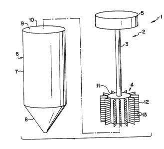
FIG. 1 is a perspective view of a Purification Apparatus according to a first
embodiment of the invention;
CA 02351616 2001-05-16
U.S. Patent Application of IBRAHIM
FIG. la is an enlargement of the flange 12 shown in FIG. 1;
FIG. 2 is a perspective view of a Purification Apparatus according to a second
embodiment of the invention;
FIG. 3 is a perspective view of a Purification Apparatus according to a third
embodiment of the invention;
FIG. 4 is perspective view a screw on cap;
FIG. S is a perspective view of a snap-on cap; and
FIG. 6 is a perspective view of a Purification Apparatus according to the
invention showing a heating unit and a sensing unit.
The present invention is directed to a purification apparatus for purifying
Nucleic acids, proteins, microorganisms or cells.
Refernng to Fig. 1, the purification apparatus 1 has a wand 2 and a reservoir
tube
6. The wand 1 is made of a cap 5, a sample collection assembly 4 and an
elongated shaft
3 connecting the cap 5 to the sample collection assembly 4. The sample
collection
assembly 4 has a series of microstructures 13 in the form of grooves (created
by deep
reactive ion etching or tooling), parallel lanes or cross-etchings on its
surface, or
microparticles 13a (See Fig. 2) enclosed within it for increasing the surface
area of the
sample collection assembly 4. The increased surface area permits maximum
exposure to
and binding of target molecules thereto, allowing concentration of target
molecules or
cells.
The cap 5 of the wand 1 is easily held between the forefinger and the thumb of
a
user. The cap configuration reduces the risk of contamination because the
user's fingers
do not come into contact with the sample capture assembly. The cap fits snugly
into the
open end 9 at the lip 10 of the reservoir tube 6. Referring to Figs. 4 and 5,
the cap 5 can
be formed with screw-on ridges 15 for screwing the cap S into the reservoir
tube 6. In
this embodiment, the reservoir tube has complimentary grooves (not shown)
therein for
receiving a screw-on cap in a sealing engagement. Alternatively, the cap 5 can
have a
stopper lip 16 and can fit into the reservoir tube 6 and be held in place in a
sealed fashion
by the force of friction or by a ridge 18 with a complimentary groove (not
shown) inside
6
CA 02351616 2001-05-16
U.S. Patent Application of IBRAHIM
the reservoir tube for receiving the ridge 18. A tab 17 assists the user in
removing the
wand from the reservoir tube 6 as shown in Fig. 5.
The cap 5 is connected to one end of a shaft 3. The other end of the shaft is
connected to a sample capture assembly 4. The shaft 3 is either solid or
hollow and can
be formed of metal or an inert synthetic material such as plastic. The sample
capture
assembly 4 is designed to increase surface area to a maximum to allow maximum
exposure to and binding of target molecules thereto. Therefore, the sample
capture
assembly 4 has microstructures associated therewith, either on its surface or
within it in
the form of microparticles enclosed inside a mesh enclosure in a form of a
"molecular
sieve". If microparticles are used, further enhancements, e.g., the use of
zeolitic particles,
can be made to allow molecular size selection.
The sample capture assembly 4 is generally a main body 11 having
microstructures on its surface in the form of cross-etched lanes, dimples,
domes, pillars
and/or pores. Such microstructures can be formed by tooling or etching.
Preferably,
cross-etched lanes in the configuration presented herein are used as
microstructures and
are etched to a depth of 0.001-2 mm and preferably 2 mm. The main body 11 can
preferably have one or more flanges 12 protruding radially outward therefrom,
wherein
the microstructures 13 are on an outer surface of the flanges 12. Fig. la
shows an
enlargement of a single flangel2. Alternatively, the main body can have
striations 14,
wherein a cross-section of the main body 11 would reveal a jagged outer edge
as shown
in Fig. 3. The striations increase the surface area and preferably also have
microstructures on their outer surface. The main body 11 can also be porous.
Still further, Fig. 2 shows a wand 2 having a sample capture assembly 4 that
has
microstructures 13a associated therewith within it in the form of
microparticles enclosed
inside a mesh enclosure 13b. The microparticles are made from silica-based
material,
polystyrene or other synthetic polymers and may be coated with a target
specific surface
such as specific oligonucleotides, peptides or cell receptors to capture a
target DNA,
RNA, protein or cell type. They are preferably about 1 to SOO~tm in diameter.
The sample collection assembly 4 may be coated with oligonucleotide probes or
specific proteins to capture specific target molecules. The sample collection
assembly
may also be made of or coated with a material that binds non-specifically with
nucleic
CA 02351616 2001-05-16
U.S. Patent Application of IBRAHIM
acids or proteins. A suitable material for binding non-specifically to nucleic
acids
include silica-based material and synthetic polymers. However, a host of other
naturally
occurring or chemically modified elements that are known to bind non-
specifically to
nucleic acids or proteins may be used. The sample collection assembly can also
be
coated with gold, platimum or other material to enhance electrical or
electrochemical
conductivity. The sample collection assembly can also be coated with singular
or
dendritic oligonucleotide probes, peptide probes or cell receptors to capture
specific
target molecules. The use of dendritic probes in conjunction with the sample
collection
assembly described herein can further significantly increase the capture
surface area and
significantly enhance analytical and clinical sensitivity.
The capture of nucleic acids, proteins or cells either non-specifically or by
affinity
binding onto solid phase supports as well as colorimetric, luminescent,
fluorescent and
electrochemical detection are well known in the art as described in the
following and
other references, of which these are herein incorporated by reference: Ausubel
F., Brent
R., Kingston R.E., Moore D.D., Seidman J.G., Smith J.A., Struhl K., (1987).
Current
Protocols in Molecular Biology. Greene Publishing Associates and Wiley-
Intersciences.
John Wiley & Sons, New York, Chichester, Brisbane, Toronto, Singapore.;
Sambrook J.,
Fritsch EF, Maniatis J. ( 1989). Molecular cloning: A laboratory manual. 2"d
edition,
Cold Spring Harbor laboratory Press, Cold Spring Harbor, New York.; Hornes E.,
Korsnes L. ( 1990). Magnetic DNA hybridization properties of oligonucleotide
probes
attached to superparamagnetic beads and their use in the isolation of poly(A)
mRNA
from eukaryotic cells. Genet. Anal. Tech. Appl. 7:145-150.; Jakobsen K.S.,
Haugen M.,
Saeboe-Larsen S., Hollung K., Espelund M., Hornes E. ( 1994). Direct mRNA
isolation
using magnetic Oligo(dT) beads: A protocol for all types oc cell cultures,
animal and
plant tissues. In: Advances in Biomagnetic Separation, (Ed. Uhlen M., Hornes
E., Olsvik
O) Eaton Publishing pp.61-71.; Rodriguez LR., Chader G.J. (1992). A novel
method for
the isolation of tissue specific genes. Nucleic Acids Res. 18:4833-4842.;
Schussler P.,
Gohr L.G., Sommer G., Kunz W., Grevelding C.G. (1995). Combined isolation of
nucleic acids and proteins from small amounts of tissue. Trends Genet. 11:378-
379.;
Beattie K.L., Fowler R.F. (1991). Solid-phase gene assembly. Nature 352:548-
552.;
Rudi K., Kroken M., Dahlberg O.J., Deggerdal A., Jakobsen K.S., Larsen F. (
1997).
CA 02351616 2001-05-16
U.S. Patent Application of IBRAHIM
Rapid, universal method to isolate PCR-ready DNA using magnetic beads.
BioTechniques 22:506-511.; Collin-Osdoby P., Oursler M.J., Webber D., Osdoby
P.
( 1991 ). Osteoclast-specific monoclonal antibodies coupled to magnetic beads
provide a
rapid and efficient method of purifying avian osteoclasts. J. Bone Mine. Res.
6:1353-
1365.; Cudjoe K.S., Krona R., Olsen E. (1994). IMS: A new selective enrichment
technique for the detection of salmonella in foods. Int. J. Food Microbiol.
23:159-165.;
Elgar G.S., Brenner S. (1992). A novel method for isolation of large insert
DNA from
recombinant lambda DNA. Nucleic Acids Res. 20:4667.; Gabrielsen O.S., Huet J.
(1993). Magnetic DNA affinity purification of yeast transcription factor.
Meth.
Enzymol. 218:508-525.; Hames B.D., Higgins S.J. (1985). Nucleic acid
hybridization:
A practical approach. IRL Press, Oxford, England.; Hawkins R.E., Russell S.J.,
Winter
G. ( 1992). Selection of phage antibodies by binding affinity. Mimicking
affinity
maturation. J. Mol. Biol. 226:889-896.; Boom, R., Sol, C.J., Salimans, M.M.,
Jansen,
C.L., Wertheim-van Dillen, P.M., and van der Noordaa, J. (1990). Rapid and
simple
method for purification of nucleic acids. J. Clin. Microbiol., 28(3):495-503.;
Lundeberg
J., Larsen F. (1995). Solid-phase technology:magnetic beads to improve nucleic
acid
detection and analysis. Biotechnology Annual Review 1:373-401.; Millar D.S.,
Withey
S.J., Tizard M.L.V., Ford J.G., Hermon-Taylor J. (1995). Solid-phase
hybridization
capture of low abundance target DNA sequences: application to the polymerase
chain
reaction detection of Mycobacterium paratuberculosis and Mycobacterium avium
susp.
Silvaticum. Anal. Biochem. 226:325-330.; Vlieger A.M., Medenblik A.M.J.C., Van
Gijlswijk R.P.M., Tanke H.J., Van der Ploeg M., Gratama J.W., Raap A.K.
(1992).
Quantitation of polymerase chain reaction products by hybridization-based
assays with
fluorescent, colorimetric or chemiluminescent detection. Anal. Biochem. 205:1-
7.
The reservoir tube 6 serves as a reservoir for collecting samples, washing the
captured nucleic acids, proteins, antibodies or antigens, and eluting the
captured nucleic
acid or proteins or other molecules. The reservoir tube 6 described herein has
an
elongated body 7 with one end having a lip 10 defining an opening 9 and a
second end 8
that is closed and preferably cone shaped. The second end 8 can also be
rounded or
cylindrical. The cap S of the wand 2 securely and sealingly fastens to the
open end 9 of
the reservoir tube 6 with the shaft 3 and the sample collection assembly 4
fitting easily
9
CA 02351616 2001-05-16
U.S. Patent Application of IBRAHIM
inside the reservoir tube 6. The reservoir tube typically holds 0.5-15 ml of
sample and
preferably is a 1.5 ml reservoir tube. The reservoir tube can be larger or
smaller without
detracting from the spirit of the invention. The reservoir can also be
designed in the form
of a microtiter plate or microtiter plate modules to allow arrayable, modular
configuration.
The reservoir tube is made of a size to enclose the shaft and sample capture
assembly of the wand and sealingly engage the wand's cap. The reservoir and
wand can
be manufactured together and be packaged as a kit with multiple reservoir
(tubes of
different sizes and shapes or microtiter plates) in each kit. The wand can be
manufactured in a size to fit reservoirs of different sizes and shapes that
are commercially
sold on the market and commonly used in biomedical research.
In use, a sample is placed inside a first reservoir tube with a lysis or
denaturing
solution. By the term DNA or RNA sample, it is meant a sample, usually cells,
that
contain DNA or RNA within the cells. Then the wand is inserted into the first
reservoir
tube. The cap of the wand secures and seals closed the first reservoir tube.
The first
reservoir tube is agitated by shaking or vortexing to mix the sample with the
denaturing
solution. The first tube is preferably incubated at 37°C for a period
of 5-15 minutes. The
shaft of the wand can be thermally regulated through an attachment or wire
connection 22
to a heating unit 21 as shown in Fig. 6. During this step, the target
molecules bind to the
massive surface area of the sample collection assembly.
The wand, which now has target molecules bound to the sample collection
assembly is then removed from the first reservoir tube and inserted into a
second
reservoir tube which contains a wash buffer. The second reservoir tube is then
securely
and sealingly closed with the cap of the wand as before. The second reservoir
tube is also
agitated to mix the sample with the wash buffer. One or several washes can be
performed in the same reservoir if multiple washing is necessary to remove
inhibitory
material from the captured nucleic acids. The wand is then removed from the
second
reservoir tube and inserted into a third reservoir tube. The third reservoir
tube contains
an elution buffer. The third reservoir tube is incubated at about 65°C
for about 5-15
minutes with or without agitation or vortexing (agitation or vortexing may
enhance
elution). If the shaft is attached to a heating unit that regulates
temperature in the sample
to
CA 02351616 2001-05-16
U.S. Patent Application of IBRAHIM
capture assembly, elution can be achieved by adjusting the incubation
temperature.
After a short while, the DNA or RNA is purified. It can then be recovered and
analyzed.
It is also possible to perform thermal cycling while the captured DNA is bound
to the
capture assembly, and the elution buffer is replaced with the appropriate
nucleic acid
amplification buffer and reagents.
The sample can be detected by one of several methods. DNA or amplified DNA
can be detected by known colorimetric, luminescent, fluorescent or
electrochemical
methods.
In another embodiment, the wand may further have a sensing unit 24 associated
with it via a sensing contact or wire connection 25 as shown in FIG. 6 for
sensing
electrical or electrochemical signals emitted from the sample on the sample
collection
assembly, following a hybridization and/or an enzymatic reaction. Such a
sensing unit
would detect changes in electrical properties of bound nucleic acids or
protein molecules
either directly or indirectly. Direct detection can be achieved by measuring
changes in
current subsequent to a hybridization reaction. Indirect detection can be
achieved by
including in the hybridization reaction an enzyme and a substrate to drive a
reduction/oxidation reaction resulting in electrical current change which can
be measured
by an electric current sensing device, for example. Alternatively, other
indirect reaction
may involve enzymatic reaction to produce colorimetric, fluorogenic or
luminescence
signal which can be detected with miniature optical devices such as a
flurometer or
spectrometer designed to fit the closed end of the reservoir. In this
embodiment, the tube
would fit into such a detection device wherein the detection would take place.
The purification apparatus of the invention can be used for efficient
purification
of nucleic acids, proteins and cells without the need of centrifugation,
precipitation or
lengthy incubations. It can also be configured to allow nucleic acid
amplification and
detection by integrating the purification apparatus into an instrument that
allows
temperature cycling and detection apparati capable of fluorescent,
colorimetric,
luminescent or electrochemical sensing.
EK~MPLES
Example 1
CA 02351616 2001-05-16
U.S. Patent Application of IBRAHIM
In a typical nucleic acids purification, mix 10-100 pl of sample with 100 p.l
of
lysis/denaturing buffer in a 1.5 ml reservoir tube. Insert the shaft and
sample capture
assembly of the wand into the reservoir tube and close the reservoir tube with
the cap.
Vortex the reservoir tube for about 1 minute. Incubate the reservoir tube at
37°C for
about 5 minutes. Remove the wand, and insert the wand into a fresh reservoir
tube
containing 1000 pl of wash buffer. Vortex the reservoir tube for about 1
minute.
Remove the wand and insert it into a fresh reservoir tube containing 100 pl of
elution
buffer. Heat the reservoir tube to abut 65°C for about 5 minutes. The
DNA or RNA is
now purified and ready for further analysis or processing.
Example 2
Detection of nucleic acids can be performed in a variety of formats. For
example,
after the captured nucleic acids are eluted into the reservoir, a biotin- and
a digoxigenin-
labeled probes can be hybridized, a streptavidin-coated capture assembly is
immersed
into the reservoir to capture they hybrid complex, then an antibody against
digoxigenin is
added to bind to the digoxigenin-labeled probe, then an enzyme labeled
secondary
antibody is added to bind to the primary antibody, then a chemiluminescent or
colorimetric substrate is added to drive a colorimetric or luminescent
reactions which can
then be detected with a colorimeter, photoluminometer or by an electric
current
measuring device. A number of washing steps must be performed between the
addition
of reagents in the same or different reservoirs to remove unbound molecules.
These
hybridization and detection methods are known in the art.
It is also possible to configure the detection assays so that the captured
nucleic on
the sample collection assembly is hybridized in situ to a tagged protein-DNA
probe and
proceed with the detection according to methods that known in the art.
It is also possible to configure the detection assays so that fluorescently-
labeled
probes are used for hybridization and detection according to known methods.
Example 3
12
CA 02351616 2001-05-16
U.S. Patent Application of IBRAHIM
Mix 10-100 pl of sample with 100 ~tl of lysis/denaturing buffer in a 1.5 ml
reservoir tube. Insert the shaft and sample capture assembly (after coating
with
appropriate antibody) of the wand into the reservoir tube and close the
reservoir tube with
the cap. Vortex the reservoir tube for about 1 minute. Incubate the reservoir
tube at 37°C
for about 5-15 minutes. Remove the wand, and insert the wand into a fresh
reservoir tube
containing 1000 pl of blocking buffer. Vortex the reservoir tube for about 1
minute.
Incubate at 37°C for about S-15 minutes. Remove the wand and insert it
into a fresh
reservoir tube containing 100 p.l of conjugate solution. Remove the wand, and
insert the
wand into a fresh reservoir tube containing 1000 p.l of wash buffer. Shake or
agitate for 1
min. Discard wash buffer and repeat the washing step. Remove the wand and
insert it
into a fresh reservoir tube containing 100 ltl of detection reagent. Analyze
the color and
determine the antigen according to a color chart. Alternatively, the color can
be read by
using a spectrophotometer. The detection step can also be modified to allow
electrochemical, luminescent or fluorescent detection using an appropriate
signal
detection attachment.
Example 4
Mix 10-100 ~tl of sample with 100 ~1 of lysis/denaturing buffer in a 1.5 ml
reservoir tube. Insert the shaft and sample capture assembly (after coating
with
appropriate antigen) of the wand into the reservoir tube and close the
reservoir tube with
the cap. Vortex the reservoir tube for about 1 minute. Incubate the reservoir
tube at 37°C
for about 5-15 minutes. Remove the wand, and insert the wand into a fresh
reservoir tube
containing 1000 pl of blocking buffer. Vortex the reservoir tube for about 1
minute.
Incubate at 37°C for about S minutes. Remove the wand and insert it
into a fresh reservoir
tube containing 100 ltl of conjugate solution. Remove the wand, and insert the
wand into
a fresh reservoir tube containing 1000 pl of wash buffer. Discard the wash
buffer and
repeat the washing step. Remove the wand and insert it into a fresh reservoir
tube
containing 100 pl of detection reagent. Analyze the color and determine the
antibody.
13
CA 02351616 2001-05-16
U.S. Patent Application of IBRAHIM
Alternatively, the color can be read by using a spectrophotometer. The
detection step can
also be modified to allow electrochemical, luminescent or fluorescent
detection using an
appropriate signal detection attachment.
The present purification apparatus also has applications in detection of blood
chemistry, detection of chemokines and other disease markers and
identification of
microbial agents.
Having thus described in detail preferred embodiments of the present
invention, it
is to be understood that the invention defined by the appended claims is not
to be limited
by particular details set forth in the above description as many apparent
variations thereof
are possible without departing from the spirit or scope of the present
invention.
14