Note: Descriptions are shown in the official language in which they were submitted.
CA 02351706 2001-05-16
WO 00/32100 PCT/US99/28482
-1 -
REFRACTOMETRIC DEVICE FOR THE IN VIVO DETECTION OF
FERTILE PERIODS OF OVULATING FEMALES
GOVERNMENT RIGHTS STATEMENT
This invention was made with Government support under Grant No. DPE-
3061-A-00-1029-00 awarded by the U.S. Agency of International Development
(US AID). The Government has certain rights in the invention.
CROSS-REFERENCE TO PARENT APPLICATION
This application is a continuation-in-part of eepend~ application
Serial No. 08/804,057 filed on February 21, 1997, the entire content of
which is expressly incorporated hereinto by reference.
FIELD OF INVENTION
The present invention relates generally to refractometric devices.
More particularly, the present invention relates to the field of
refractometric devices which allow in vivo ovulation detection in female
mammals.
BACKGROUND AND SUMMARY OF THE INVENTION
Detecting with reasonable precision the fertile period of the female
reproductive cycle would have significant benefits from a human "family
planning" aspect as well as with animal husbandry. For example, by
detecting the fertile period, a woman would be able to determine when
intercourse would likely lead to conception. Such information could
therefore be used by the woman to either refrain from or engage in
intercourse if contraception or conception is desired, respectively.
Similarly, livestock breeders would be able to maximize successful
CA 02351706 2001-05-16
WO 00/32100 PCT/US99/28482
inseminations with knowledge of the ovulation cycle of the particular
breeding animal.
It is well known that a number of physiological changes ensue
during a woman's menstrual cycle and various tests have been developed
as an aid to family planning. For example, it is known that the water
content of vaginal mucus varies during the menstrual cycle. In this
regard, U.S. Patent No. 4,099,820 to Kopito et al (the entire contend of
which is incorporated expressly hereinto by reference) discloses an in
vitro device which detects ovulation during a menstrual cycle using the
transmissivity and/or diffusivity properties of vaginal mucus. That is,
according to Kopito et a1 '820, a vaginal mucus sample must first be
positioned between a pair of plates at a specific pressure and temperature
in order to provide a mucus stratum of predetermined thickness. A
photometer then measures the optical transmissivity andlor diffusivity in
order to determine the phase of the menstrual cycle.
The techniques disclosed by Kopito et al '820 are not without
problems. Specifically, since the apparatus is quite cumbersome and the
mucus sample must be positioned with great care and accuracy with
respect to the photometer, it is impractical for "home" use, but instead
must be administered by clinical technicians. Furthermore, since the
device is only capable of detecting vaginal.mucus properties in vitro, there
is a real risk that the water content in the mucus will change in the short
time it takes to obtain a sample and to prepare it for testing in the Kopito
et al '820 apparatus, thereby potentially leading to inaccurate readings.
What has been needed in this art, therefore, is a relatively simply
device that can be employed in vivo to detect the ovulation period of
CA 02351706 2001-05-16
WO 00/32100 PCTlUS99/28482
-3-
females. It is towards fulfilling such a need that the present invention is
directed.
Broadly, the present invention relates to a device that detects in
vivo physiological changes in a female's cervical mucus. These changes
correlate with the timing of ovulation and thus provide a marker for the
fertile period of the menstrual cycle. The fertile period is the time interval
during which unprotected intercourse can lead to pregnancy. The period
extends several days before the day of ovulation and ends immediately
after that day. More particularly. the device of the present invention
measures the water content, or hydration of cervical mucus, which has
been shown to increase several days before ovulation. These changes in
mucus hydration correlate with the pre-ovulation rise in luteinizing
hormone (LH) which only precedes ovulation by 24-36 hours, and thus
occurs after the onset of the fertile period. Other hormone changes, e.g.,
those of estradiol or progesterone, are less consistent within and among
women than the increase in mucus hydration and thus are less robust
markers of the fertile period. The device of this invention therefore
measures in vivo the mucus hydration by its direct correlate, the index of
refraction of the mucus. As a result, problems associated with in vitro
measurement of mucus hydration (e.g., possible mucus dehydration when
a sample is removed from the body) are eliminated.
The device of this invention generally includes a refractive index
detector having a light source, a photoreceptor and a light guide
positioned so as to guide light from the light source to the photoreceptor.
The light guide includes at feast one active surface to be wetted by the
cervical mucus. The detector may be planar or curvelinear and may be
embedded within a distal sensing head or extend upright therefrom (e.g.,
CA 02351706 2001-05-16
WO 00/32100 PCT/US99/28482
-4-
so as to somewhat penetrate the external cervical os during use). Most
preferably, the fight guide is fabricated from a fluorocarbon polymer.
Most preferably, the device of this invention will include a proximal
handle which allows the user to manipulate the distal sensing head into
close proximity to the external cervical os. The handle may include a
source of electrical power (e.g., a DC battery pack, solar cell or the like),
a
processor for processing the signal received from the photodetector
indicative of the cervical mucus refractive index, and a human-readable
display (e.g., an alpha-numeric display, light indicator, analog display or
the like). A relatively slender (as compared to the handle) stem
operatively connects the handle to the distal sensing head. The sensing
head may be formed as a one-piece structure with the stem and angled
relative thereto so as to assist in placement of the detector in close
proximity to the external cervical os. Alternatively, the sensing head may
be connected to the stem to allow for relative pivotal articulation to permit
selective adjustment of the sensing head's angular orientation relative to
the stem. The stem itself may be entirely rigid, or may be flexible.
Further aspects and advantages of this invention will become more
clear after careful consideration is given to the detailed description of the
preferred exemplary embodiments thereof which follow.
BRIEF DESCRIPTION OF THE DRAWINGS
Reference will hereinafter be made to the accompanying drawings
wherein like reference numerals throughout the various FIGURES denote
Like structural elements, and wherein;
CA 02351706 2001-05-16
WO 00/32100 PCT/US99/28482
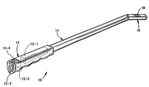
FIGURE 1 is a perspective view of one embodiment of a
refractometric device in accordance with the present invention;
FIGURE 2 is an enlarged top plan view of the distal (detector) end
of the device shown in FIGURE 1;
FIGURE 3 is a longitudinal cross-sectional elevational view through
the detector employed in the device of this invention as taken along lines
3-3 in FIGURE 2;
FIGURE 4 is a latitudinal cross-sectional elevatianal view through
the detector employed in the device of this invention as taken along lines
4-4 in FIGURE 2;
FIGURE 5 is an enlarged perspective view showing a modified
distal (sensing) end in accordance with another embodiment of this
invention;
FIGURE 6 is an enlarged perspective view showing a differently
configured detector that may be employed in the devices of this invention;
FIGURE 7 is a plan view of the detector depicted in FIGURE 6;
FIGURE 8 is an end elevational view of the detector depicted in
FIGURE 7 as taken along line 8-8 therein;
FIGURE 9 is a latitudinal cross-sectional view of the detector
depicted in FIGURE 7 as taken along line 9-9 therein;
CA 02351706 2001-05-16
WO 00!32100 PCT/US99/28482
-6-
FIGURE 10 is a side elevational view of the detector depicted in
FIGURE 7 as taken along line 10-10 therein;
FIGURE 11 is a longitudinal cross-sectional view of the detector
depicted in FIGURE 7 as taken along line 11-11 therein;
FIGURE 12 is an enlarged top plan view showing a modified distal
{sensing) end in accordance with another embodiment of this invention;
FIGURE 13 is a bottom plan view of the embodiment depicted in
FIGURE 12;
FIGURE 14 is a cross-sectional eievational view through the
detector section of the embodiment depicted in FIGURE 12 as taken
along line 14-14 therein;
FIGURE 15 is a top plan view of another embodiment of a
refractometric device in accordance with the present invention;
FIGURE 16 is side elevational view of the device depicted in
FIGURE 15;
FIGURE 17 is an enlarged end elevational view of the device
depicted in FIGURE 15 as taken along line 17-17 in FIGURE 16;
FIGURE 18 is an enlarged perspective view of the distal (detector)
end of the device shown in FIGURE 15;
CA 02351706 2001-05-16
WO 00/32100 PCT/US99/28482
-?~
FIGURE 19 is a side elevational view of the detector depicted in
FIGURE 18;
FIGURES 20 and 21 are each cross-sectional elevational views of
the detector depicted in FIGURE 18 as taken along lines 20-20 and 21-
21, respectively, therein;
FIGURE 22 is a cross-sectional plan view of the detector depicted
in FIGURE 18 as taken along line 22-22 in FIGURE 2~1; and
FIGURE 23 is a graph of the voltage across a photodiode versus
concentration and refractive index of sucrose solutions obtained from the
data of the Example below.
DETAILED DESCRIPTION OF THE PREFERRED
EXEMPLARY EMBODIMENTS
A particulariy preferred embodiment of a device 10 in accordance
with the present invention is depicted in FIGURE 1. As shown, the device
generally includes a proximal handle 12 to allow the device 10 to be
gripped and manually manipulated during use, and a relatively slender (as
compared to the handle) stem 14 extending distally from the handle 12
along a common axis. The stem 14 terminates in a distal sensing head
16 which is angularly oriented with respect to the stem 14 (e.g., 30° t
which, far many women, approximates the angle between their vagina
and external cervical os). Most preferably, the stem 14 and distal sensing
head 16 are formed as a one piece structure from a biomedically
compatible plastics material.
CA 02351706 2001-05-16
WO 00/32100 PCT/US99/28482
.$_
The distal sensing head 16 carries a refractive index detector 18 for
placement against the woman's external cervical os during use. As will be
explained in greater detail below, the detector 18 measures the refractive
index of the woman's cervical mucus which, in turn; is indicative of
ovulation. The detector 18 is connected electrically to an electrical power
source 12-1 (e.g., a battery pack, photocells or the like) and processor
12-2 contained within the handle 12 via wires (not shown) embedded in
the stem 14. A button 12-3 at the proximal end of the handle 12 activates
the detector 18 and causes a signal indicative of the refractive index to be
supplied to the processor 12-2. The processor 12-2 may then display the
signal in a human-readable format via a visual display panel 12-4. In this
regard, the display panel 12-4 may numerically display the detected
refractive index of the cervical mucus and/or may process the signal to
display a light signal indicative of a fertile period should the refractive
index deviate from a predetermined value.
Accompanying FIGURES 2-4 show in greater detail the refractive
index detector 18 that is employed in the device 10 described above with
reference to FIGURE 1. In this regard, the detector 18 includes a light
source 18-1 (e.g., a conventional !-ED) and a photodetector 18-2 (e.g., a
conventional photodiode) axially spaced from the light source 18-3. An
elongated channel having a U-shaped cross-section is formed axially
between the light source 18-1 and photodetector 18-2 by means of
stainless steel support rods 18-3, 18-4 and 18-5. The channel is flfied
with a fluorocarbon polymer {e.g., Teflon FEP fluorocarbon polymer
commercially available from DuPont) which serves as the light guide '18-6
for the detector 7 8. The surface 18-7 of the support rod 18-4 in contact
with the fluorocarbon light guide 18-6 is polished so as to provide a light-
reflective surface interface therebetween.
CA 02351706 2001-05-16
WO 00/32100 PCT/US99/28482
As shown in FIGURES 2 and 3, the upper surfaces of the light
source, 18-1, photodetectar 18-2, support cods 18-3 and 18-5 and the
light guide 18-6 are each substantially co-planar with the upper surface of
the sensing head 16 so as to present a smooth external surface to the
user. In addition, although not shown in the drawings, the upper surfaces
of the light source, 18-1, photodetector 18-2 and support rods 18-3 and
18-5 may be covered by an opaque film coating (e.g., black paint) so as to
mask all available light paths except for that provided by the light guide
18-6.
Virtually any fluorocarbon polymer having a refractive index (ASTM
D-542) of between about 1.335 to about 1.450, and more preferably
between about 1.341 to about 1.347 may be used in the practice of this
invention. The fluorocarbon polymer employed in this invention will also
exhibit, or may be processed to exhibit, an optical clarity of about 98% or
greater, and more preferably about 99% or greater. As used herein and in
the accompanying claim, the term "optical clarity" is 100% minus the
percent haze value as determined by ASTM D 1003-61 {reapproved 1988,
incorporated fully by reference herein).
The term "fluorocarbon polymer" as used herein and in the
accompanying claims is meant to refer to any polymer, copolymer,
terpolymer and the like having at least one (preferably more than one)
fluorocarbon moiety in a repeat unit of its molecular chain. By way of
example, preferred fluorocarbon polymers that may be employed in
accordance with the present invention include copolymers comprised of
tetrafluoroethylene with hexafluoropropylene comonomers,
polychlorotrifluoroethylene, ethylene-tetrafluoroethylene copolymers,
CA 02351706 2001-05-16
WO 00/32100 PCT/US99128482
-10-
polyvinylidene fluoride polymers, and polyvinyl fluoride polymers.
Particularly preferred according to this invention are copolymers
comprised of tetrafluoroethyfene with hexafluoropropylene comonomers
commercially available from DuPont under the registered trademark
Teflon~ FEP fluorocarbon polymers. The preferred fluorocarbon polymers
will exhibit a refractive index (ASTM D-542) of between about 1.341 to
about 1.34?. The optical clarity of the preferred fluorocarbon polymers
may be increased by heating the polymer to greater than its glass ,
transition temperature (T9) followed by rapid quenching of the heat-treated
polymer. The resulting heat-treated and quenched fluorocarbon polymer
will most preferably exhibit an optical clarity of about 98% or greater, and
more preferably about 99% or greater.
Accompanying FIGURE 5 depicts a modification of the device 10
shown above which allows hinged articulation to occur between the
sensing head 16 and the stem 14. In this regard, it will be observed that
the distal end of the stem 14 includes a U-shaped yoke comprised of a
pair of parallel, laterally spaced fngers 14-1, 14-2. The sensing head 16
includes a proximally projecting boss 16-8 which is sandwiched between
the fingers 14-1 and 14-2 so as to be in interference fit therewith.
Mechanical stability may be increased by providing a hinge pin {not
shown) with its ends embedded in the fingers 14-1 and 14-2, and
extending through the boss 16-8 to allow the sensing head 16 to pivot
therearound {arrow A in FIGURE 5). Also, the head 16 may be positioned
between the yoke fingers 14-1, 14-2, in which case the proximally
projecting boss 16-8 may be omitted. Furthermore, detents may be
provided as desired to frictionaily lock the sensing head in one of several
angular orientations relative to the stem 14. In such a manner, the user
may angularly adjust the orientation between the sensing head 16 and the
CA 02351706 2001-05-16
WO 00/32100 PCT/US99/28482
-11 -
stem 14 to ensure proper presentation of the detector 18 to the external
cervical os.
Accompanying FIGURES 6-11 depict another embodiment of a
refractive index detector 20 that may be employed instead of the detector
18 in the device 10 described above. In this regard, it will be observed
that the detector 20 includes a stainless steel support plate 22 which may
be embedded into the sensing head 16 of the device 10. Unlike the
detector 18 discussed previously (vvhich is substantially coplanar with the
exterior surface of the sensing head 16), the detector 20 protrudes
upwardly from the surface of the sensing head 16 and thus provides a pair
of lateral active surfaces or "windows" in the light guide for detecting the
refractive index of the cervical mucus.
The detector 20 includes generally a base section 24 having a
generally triangular cross-section with a convexly protruding upper section
26 extending from its apex. The base section 24 includes a central
stainless steel support member 28 having a triangularly shaped
cross-section embedded within the fluorocarbon light guide 30. A
U-shaped reflector 32 is positioned over the apex of the support member
28 so as to guide light therearound. The surfaces 28-1 and 28-2 are
highly polished reflective surfaces corresponding to the active window
surfaces 30-1 and 30-2 of the fight guide 30.
The structures of the detector 20 shown in FIGURES fi-11 permit
the light source LS {e.g., a fight emitting diode) and photoreceptor PR
(e.g., a photodiode) to be positioned below the base plate 22. Thus, the
light source LS and photoreceptor PR can conveniently be encased
completely by the biomedically compatible plastics material forming the
CA 02351706 2001-05-16
WO 00132100 PCT/US99/28482
-12-
sensing head 16. Furthermore, the convexly protruding upper section 26
provides a convenient tactile positioning aid to the user in locating the
detector within the external cervical os. In such a manner, therefore, the
active surfaces 30-1 and 30-2 of the light guide 30 may be brought into
direct contact with the cervical mucus so that accurate refractive index
readings may be obtained.
Another refractive index detector 40 that may be employed in the
device 10 is depicted in accompanying FIGURES 12-14. As can be
observed, the~detector 40 is generally planar and comprised of a primary
support plate 42 which defines a central window for receiving a secondary
support plate 44, each of which is preferably formed of stainless steel.
The secondary support plate 44 is confgured so as to be relatively thinner
in cross=sectional thickness as compared to the primary support plate 42:
As such, the reflective surface 44a of the secondary support plate 42 is
recessed from the upper surface of the primary support plate 42 so as to
provide space for the fluorocarbon fight guide 46.
The longitudinal edges 44-1, 44-2 and 42-1, 42-2 of the secondary
and primary support plates 44, 42, respectively, define therebetween inlet
and outlet spaces which are filled with a fluorocarbon polymer and thereby
establish inlet and outlet light conduits 46-1 and 46-2, respectively, which
are oriented in a plane that is substantially perpendicular to the active
surface of the light conduit 46. In this regard, the inlet light conduit 46-1
is
generally triangular in cross-section by virtue of the opposed beveled
edges 42-1 and 44-1 of the primary and secondary support plates 42, 44,
respectively. Furthermore, the longitudinally opposed edges 42-3 and 42-
4 of the primary support plate 42 are beveled so as to help reflect fight
into and out of the planar light guide 46.
CA 02351706 2001-05-16
WO 00/32100 PCT/US99128482
-13-
It will be further observed that the light guides associated with the
detectors 18, 20 and 46 are each planar and have a substantially uniform
thickness dimension along their lengths and widths. However, as wilC be
discussed in greater detail below, the light guides may be convexly
curvelinear. Such curvelinear light guides, however, will still have a
substantially uniform thickness dimension along their lengths and widths
similar to the planar light guides already discussed above.
The fight guides of this invention will have an aspect ratio -- that is
tll where t is the thickness of the light guide and I is the wetted length of
the light guide -- of between about 1:10 to about 1:20. The aspect ratios
of the detectors of this invention thus translate into wetted lengths of
between about 0.050 to about 0:300 inch, and more preferably between
about 0.100 to about 0.200 inch. Thus, each of the active surfaces far
paired active surface detectors according to this invention (e.g., the
embodiment depicted in FIGURE 6}, will be one-half of the total wetted
length dimension noted above.
Another embodiment of a device 50 according to the present
invention is depicted in accompanying FIGURES 15-17. Like the device
described previously, the device 50 includes a proximal handle 52 and
a distal sensing head 54. The handle 52 includes a battery pack 52-1, a
processor 52-2, visual display 52-3 and operational button 52-4 which
serve similar functions to those similar components described above with
reference to FIGURE 7.
The sensing head 54 carries a detector 56 {to be described in
greater detail below}. However, unlike the device 10 discussed previously
CA 02351706 2001-05-16
WO 00/32100 PCT/US99/28482
-14-
(which employs an entirely rigid stem 14), the sensing head 54 is
connected to the handle 52 by means ofi a longitudinally flexible, but
torsionally rigid, stem 58. The longitudinal flexibility of the stem 58 thus
allows the device 50 to be configured so as to accommodate individual
anatomical differences in the orientation of the external cervical os and
vagina. At the same time, the torsional rigidity of the stem 58 permits the
user to rotate the sensing head 54 to permit tactile placement of the
detector within the external cervical os so that precise refractive index
readings may be obtained:
In use, a distal portion of the stem 58 is rotationally supported by a
lateral support member 60 (see F1GURE 17) which may be attached to a
user's finger F by means ofi an adjustable elastic {or similar) band 62. As
such, the sensing head 54 is positioned laterally parallel to the user's
finger tip. The user may thus manipulate the handle with one hand and
rotate the sensing head 54 so that the detector 56 is in contact with the
finger tip on the other hand to which the device 50 is attached. The user
may then manually locate their external cervical os and, once located,
may rotate the sensing head 54 in an opposite direction so that the
detector 56 is actually positioned therewithin. Once positioned within the
external cervical os, the user may operate button 52-4 and thereby obtain
a reading indicative of the refractive index of the cervical mucus (which
thus correlates to the user's ovulation period).
Accompanying FIGURES 18-22 depict in greater detail the sensing
head 54 and detector structures employed in device 50. In this regard,
the sensing head 54 is mast preferably formed of a biomedicaily
compatible plastics material in which a stainless steel base plate 64 of the
detector 54 is embedded. The detector 54 extends upwardly from the
CA 02351706 2001-05-16
WO 00/32100 PCT/US99/28482
-15-
base plate 64 and includes a cylindrical sensing post section 66 and a
convexly domed locator head section 68 most preferably unitarily formed
from the same fluorocarbon polymer.
A central stainless steel support member 70 having a generally
rectangular shaped cross-section is embedded within the fluorocarbon
polymer forming the cylindrical post 66. In this regard, the opposed sides
of the central support member 70 are convexly curved so as to
correspond to the curvature of the post 66 and are polished to provide a
reflective surface for the light traveling through the light guide regions 66-
1
and 66-2 {see FIGURE 21 ). A 90° angled reflector 72 having a pair of
support arms 72-1, 72-2 and an angular cap 72-3 is provided so that the
cap 72-3 is positioned over the top of the support member 70. Light being
refracted along the light guide 66-1 will thus be redirected to the light
guide f6-2 by virtue of the angular cap structure 72-3. The light guide
regions 66-1 and 66-2 thus establish surfaces corresponding to the active
window surfaces of the post 66. Placement of the detector 54 in a
woman's external cervical os will therefore permit the cervical mucus to
come into contact with the active surfaces of the light guides 66-1 and 66-
2 so that refractive index measurements may be detected by comparing
the amount of light which is refracted back to the photoreceptor PR to the
amount of light emitted by the light source LS. It will be observed that,
although convexly curved, the light guides 66-1 and 66-2 have a
substantially constant thickness dimension.
Further understanding of this invention will be obtained from the
following non-limiting Example:
CA 02351706 2001-05-16
WO 00/32100 PCTIUS99J28482
_
EXAMPLES
I. Example I
A 2 x 2 inch square of sample material was cut from a sheet of
1000L FEP Teflon~ film (0.010 inches thick) manufactured by DcrPont
High Performance Films, which reportedly was made from NP-40 FEP
resin of Daikin Industries, Ltd. The sample was heat-treated using a
laboratory heat-treatmentlquenching press. The press had a pair o_f
heated platens for melting a sample and a pair of freezing platens for
rapidly quenching the sample. The tested sample was heated for 45
seconds between the hot platens at 657°F, then quenched between the
quench platens immersed in liquid nitrogen (minus 320°F). A transfer
arm
enabled the hot mold containing the sample to be swung rapidly between
the heated and the quench platens.
Each pair of platens included a stationary platen and a platen that
was attached to the piston rod of an air cylinder to allow the platens to be
opened and closed rapidly. The air cylinders also provided a force that
kept the mold flat and in intimate contact with the platens.
The sample itself was contained within a rnoid comprised of a
silverlstainless steel/silver sandwich structure. Initially the sandwich is
loosely held together by small screws at the periphery, but the force of the
platens and the adhesion of the molten sample keep the sandwich
together during processing. The top layer of the sandwich was a polished
sheet of 99.9% silver (Fins Silver) having a thickness of 26 gage (0.016
inches) before polishing and had a rectangular geometry, 3 inches by 3.5
inches which was lapped flat and polished on both sides. one side of the
silver contacted the platen; while the other side acted as the top surface
of the mold for the sample. The side facing the sample must be polished
CA 02351706 2001-05-16
WO 00/32100 PCT/US99/28482
_17_
to ensure that the sample will have a smooth surface after melting and
quenching. The reverse side was polished to give good contact with the
platen for high heat transfer capability. The middle layer of the sandwich
was 0.010 inch thick shim stock (stainless steel alloy AISI 302) having a
2-inch square hole which formed the sides of the mold. The thickness of
the shim stock establishes the final thickness of the sample. The shim
stock extends laterally from the sandwich and was attached rigidly to the
transfer arm. The bottom layer of the sandwich was another sheet of Fine
Silver. It is similar to the above described top sheet except that it formed
the bottom of the mold.
The hot platens were made of aluminum (alloy 6061 ). For each
platen, the surface that contacted the mold sandwich was a 3-inch square
which was lapped and polished for good heat transfer capability. Each
hot platen had (2) 200 watt cartridge heaters and were controlled in
parallel by a rheostat control so that temperature could be adjusted for
optimum conditions. Temperature was measured with a surface contact
thermometer. There was also provision for leveling the lower platen so
that the platen surfaces are parallel when the mold sandwich is squeezed
- between them.
The cold platens were made of copper (alloy 110, 99.9% pure) so
as to provide sufficient heat transfer during quenching. It is important that
there is sufficient mass of copper to absorb the quantity of heat from the
sample and sample mold to achieve quenching. The required heat
capacity is governed by the mass of the pieces and the following thermal
processes: heat of solidification of the sample, cooling of the sample,
cooling of the silver plates and cooling ofi the stainless steel shim stock.
For the apparatus described here, there was a minimum of 1 inch copper
CA 02351706 2001-05-16
WO 00/32100 PCT/US99/28482
-18-
behind the polished surface of each platen. For each platen, the surface
that contacted the mold sandwich was a 3-inch square which was lapped
and polished for high heat transfer capability. The bottom platen resides
in a stainless steel pan.
Before the meltinglquenching process began, the bottom platen
was partially immersed in liquid nitrogen with only the top surface
protruding by approximately 0.25 inches until the nitrogen boiling is .
minimal. The top platen was machined with a 1 inch deep welt in its top
surface. Before the meltinglquenching process began, this well was fiilled
with liquid nitrogen until the nitrogen boiling is minimal. The time
dependent cooling of the platens was characterized using a themocouple
embedded in a Teflon~ fluoropolymer sheet squeezed between the
platens. Whenever the nitrogen boiling became minimal, the measured
temperature was below -315°F. The quench platens and stainless steel
pan of liquid nitrogen were surrounded by a double layer box of 0.775
inch Styrofoam~ (1.55 inches total). Fifteen seconds before sample
transfer, a "window" of Styrofoam~ insulator material was removed to
allow the transfer arm to swing between the quenching platens. There
was also provision for leveling the lower platen so that the platen surfaces
are parallel when the mold sandwich is squeezed between them.
A sample heat treated in the above-described manner and an
untreated sample were tested per ASTM D 1003-61 (reapproved 1988).
As allowed by ASTM D1003-61, each sample was immersed in isopropyl
alcohol prior to testing so as to negate the effects of surface flaws. The
untreated sample had a haze value of 2.6% (corresponding to an optical
clarity of 97.4%) which is inadequate for use in the present invention while
CA 02351706 2001-05-16
WO 00/32100 PGT/US99J28482
_' g _
the heat treated sample had a haze value of 1.0% (corresponding to an
optical clarity of 99.0%) and is satisfactory for use in the present
inventian.
Example ll:
A device employing a detector similar to that depicted in FIGURES
1-4 was constructed in order to investigate refractometric measurements
using sucrose solutions. The detector comprised a transparent light
conduit made of fluorinated ethylene propylene (FEP) positioned between
a light emitting diode (LED) and a photodiode detector.
The light conduit portion of the detector was a portion of a Teflon~
FEP fluorocarbon film (DuPont) measuring 0.010" thick x 0.200" long x
0.200" wide. The clarity of the film had been increased by heating it
above its glass transition temperature followed by a rapid quench into an
aqueous solution of calcium chloride at approximately -49°C. The film
was sandwiched between polished stainless steel sheets during heating
and quenching to give two smooth faces, top and bottom, upon removal
from the sheets. The bottom face of the film was then glued to two
stainless steel supports separated by parallel channel 0.060" wide. The
_. film was unsupported along the channel at its bottom surface thereby
allowing air contact for the length of the film.
A red LED (Panasonic LN21 CVAL(URS)) and a photodiode
(Jameco Company PJN 112168) each of T 1-3/4 style were modified by
cutting off their convex lenses and polishing them flat close to the anode
wire and parallel to the embedded chip. The components were arranged
such that the axis of the LED, axis of the FEP film light conduit and axis of
the photodiode all coincided to allow the maximum fight transmission
through the FEP conduit to the photodiode. The components were
CA 02351706 2001-05-16
WO 00/32100 PCT/US99/28482
- 20 -
epoxied in place. Black paint was used to mask all fight paths from the
LED to the photodiode except far paths through the 0.060" x 0.010" x
0.200" conduit of the FEP film.
The LED was wired in series with a 328 ohm resistor, and during
testing, a regulated 9.00 volts was applied across the pair. Similarly, the
photodiode was wired in series with a 993 ohm resistor, and during
testing, a regulated 9.00 volts was applied across the pair. The vottage
across the LEDlresistor pair remained constant, and the measured
voltage across the resistor (7.23 volts) indicated a current flow of 22.0
milli-amperes through the series combination. The voltage across the
photodiode varied in response to the refractive index of the medium
against the tap face of the FEP film. This voltage was measured by use
of a multimeter (Fluke 8021 B). A separate multimeter (Fluke 73 Series I1)
was used to verify that the source voltage did not vary. Both meters
agreed with each other within one digit of resolution (0.01 volts) over the
range of measurement. Initial drift of the voltage source (BK Precision
mode! #1651 ) by 0.04 volt was eliminated by a two hour warm-up of the
instrument. Na variation of the instrument was measured during testing.
Test solutions were made by mixing sucrose (EM Science PIN
SX1075-1 ) with reverse osmosis deionized (ROD!) water whose purity
exceeds 15 Megohm-cm. The sucrose was dried in an oven at 105°C for
several hours, followed by storage overnight under vacuum at about 40
mini-torr. Solutions were mixed in 10 gram amounts on a weight/weight
basis with milligram accuracy (Mettler model #AT200) into 15 mini-liter
centrifuge tubes (Corning PIN 25310-15) and capped. The following
solutions were made: 0% (unaltered RODI), 1, 2, 4, 6, 8 and 10%.
Refractive index was measured for each solution to five decimal places at
CA 02351706 2001-05-16
WO 00/32100 PCT/US99/28482
-21 -
25°C (Bellingham and Stanley model #RFM 340): The solutions were
measured before and after testing of the detector. Measurements with the
RFM 340 were repeatable to within n = 0.00014.
Characterization of the detector was made in a room whose
temperature was controlled befinreen 25.0°-25.4°C. 100
microliters of a
particular solution was placed on the FEP film and immediately covered
with a large, opaque cup to exclude ambient light. Readings were made
within 30 seconds, and there was no measured change of the reading
after covering. After each sample, the FEP film surface was rinsed with
RODI water and dried with a tissue. The samples were measured in the
following order: 2, 6, 1, 10, 4, 8, 0% (set #1). After one hour, in which the
power supply was left on, the samples were measured in the following
order: 0, 1, 2, 4, 6, 8, 10% (set #2); then immediately followed by: 2, 6,
1, 10, 4, 8, 0% (set #3). Results are plotted in the graph of FIGURE 23.
The graphs of FIGURE 23 shows some variation which may be due
to residual fluid from previous samples or condensate on the underside of
the conduit. The graphs all show a limiting value above refractive index n
= 1.34. These graphs demonstrate that high resolution over a relatively
narrow range of sucrose solutions may be obtained with the detector of
this invention.
*********************
The devices of this invention have been discussed in terms of their
presently preferred embodiment, namely, as a means to detect in vivo
hydration of cervical mucus. However, other end use applications may be
identified by those skilled in this art. Suffice it to say, however, that the
refractive index of the fluorocarbon polymer employed as the light guide
CA 02351706 2001-05-16
WO 00/32100 PCT/US99/28482
- 22 -
must be matched to the refractive index range that is desired to be
measured.
Thus, while the invention has been described in connection with
what is presently considered to be the most practical and preferred
embodiment, it is to be understood that the invention is not to be limited to
fhe disclosed embodiment, but on the contrary, is intended to cover
various modifications and equivalent arrangements included withiwthe
spirit and scope of the appended claims.