Note: Descriptions are shown in the official language in which they were submitted.
CA 02352150 2007-02-05
BASKET FILTER
Field of the Invention
The present invention pertains to the field of intra vena cava filters. In
particular, the
present invention pertains to the retrieval of intra vena cava filters.
Intra vena cava filters are commonly implanted either temporarily or
permanently in
patients at risk for blood clotting.
Background of the Invention
There are a number of situations in the practice of medicine when it becomes
desirable for a physician to place a filter in the vascular system of a
patient. One of the most
common applications for vascular filters is the treatment of Deep Venous
Thrombosis (DVT).
Deep Venous Thrombosis patients experience clotting of blood in the large
veins of the lower
portions of the body. These patients are constantly at risk of a clot breaking
free and traveling
via the inferior vena cava to the heart and lungs. This process is known as
pulmonary
embolization. Pulmonary embolization can frequently be fatal, for example when
a large
blood clot interferes with the life-sustaining pumping action of the heart. If
a blood clot
passes through the heart it will be pumped into the lungs and may
-1-
CA 02352150 2001-03-09
WO 00/16845 PCT/US99/22188
cause a blockage in the pulmonary arteries. A blockage of this type in the
lungs
will interfere with the oxygenation of the blood causing shock or death.
Pulmonary embolization may be successfully prevented by the appropriate
placement of a thrombus filter in the vascular system of a patient's body.
Placement of the filter may be accomplished by performing a laparotomy with
the
patient under general anesthesia. However, intravenous insertion is often the
preferred method of placing a thrombus filter in a patient's vascular system.
Intravenous insertion of a thrombus filter is less invasive and it requires
only a local anesthetic. In this procedure, the thrombus filter is collapsed
within a
delivery catheter. The delivery catheter is introduced into the patients
vascular
system at a point which is convenient to the physician. The delivery catheter
is
then fed further into the vascular system until it reaches a desirable
location for
filter placement. The thrombus filter is then released into the blood vessel
from
the delivery catheter.
In the treatment of Deep Venous Thrombosis, a thrombus filter is placed
in the inferior vena cava of a patient. The inferior vena cava is a large
vessel
which returns blood to the heart from the lower part of the body. The inferior
vena cava may be accessed through the patient's femoral vein.
Thrombus filters may be placed in other locations when treating other
conditions. For example, if blood clots are expected to approach the heart and
lungs from the upper portion of the body, a thrombus filter may be positioned
in
the superior vena cava. The superior vena cava is a large vessel which returns
-2-
CA 02352150 2001-03-09
WO 00/16845 PCT/US99/22188
blood to the heart from the upper part of the body. The superior vena cava may
by accessed through the jugular vein, located in the patient's neck.
Once placed inside a blood vessel, a thrombus filter acts to catch and hold
blood clots. The flow of blood around the captured clots allows the body's
lysing
process to dissolve the clots.
Summary of the Invention
The present invention pertains to an intra vena cava filter implantable
temporarily or permanently, and methods for removal thereof. The filter is
held
in place in a vein or other organ by friction fit between the basket portion
and the
wall of the vein or other organ. Additionally, the struts have sharpened tips
which
engage the wall of the vein or inner surface of another organ to enhance
positional
stability of the filter.
Brief Description of the Drawinjzs
Figure 1 is a view of the filter in accordance with the present invention
disposed within a vessel, and a removal device;
Figure 2 is a detailed view of sharpened ends of struts of the filter of
Figure 1;
Figure 3 is a view of the filter of Figure 1, wherein the removal device is
attached to the filter;
Figure 4 is a view of the filter of Figure 1, wherein the filter is partially
within into the removal device; and
-3-
CA 02352150 2001-03-09
WO 00/16845 PCT/US99/22188
Figure 5 is a view of the filter of Figure 1, wherein a sheath is disposed
over the filter.
Detailed Description of the Invention
The following detailed description should be read with reference to the
drawings, in which like elements in different drawings are numbered
identically.
The drawings which are not necessarily to scale, depict selected embodiments
and
are not intended to limit the scope of the invention. Examples of
constructions,
materials, dimensions, and manufacturing processes are provided for selected
elements. Those skilled in the art will recognize that many of the examples
provided have suitable alternatives which may be utilized.
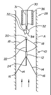
Referring now to the drawings wherein like reference numerals refer to
like elements throughout the several views, Figure 1 is a side view of a
preferred
embodiment of a filter 10 in accordance with the present invention disposed
within vessel or vena cava A. Filter 10 includes a generally tubular hub 12
from
which extends a plurality of struts 14. Two struts 14 are shown in Figure 1,
however, preferably six struts are evenly spaced in a generally conical
formation.
Additionally, each strut 14 can include bends along their length to catch
thrombus
which flows through vessel A in the direction of the arrows. The struts of the
present invention are preferably arranged in a manner similar to the
GreenfieldTM
filter made by Medi-Tech (Watertown, Mass.). The end of each strut preferably
includes a sharpened tip 16 for engagement with the vessel wall to stabilize
filter
10 within vessel A.
-4-
CA 02352150 2007-02-05
Extending from hub 12 opposite struts 14 are preferably, preformed, flattened
wires
18. An end of each wire 18 opposite hub 12 is preferably coupled to a coupling
20. As
shown in Figure 1, two wires 18 are disposed between hub 12 and coupling 20.
There are,
however, preferably four equally spaced wires 18 forming a basket portion of
the filter. The
basket portion of the filter may be generally bulbous in shape. The basket
portion of the filter
may also be ball shaped. In the embodiment of figure 1, the shape of the
basket portion of the
filter may be described as an elliptical rotation. A slider rod 22, having a
stop, or ball tip 24,
is connected to coupling 20 and disposed through hub 12. Slider rod 22 is
preferably, fixably
connected to coupling 20, and slidable longitudinally within hub 12.
In a presently preferred embodiment, wires 18 are made from a shape memory
alloy
such as NiTi alloy. In a presently most preferred embodiment, wires 18 are
preferably preset
to expand radially to meet the walls of vessel A at approximately 37 C (body
temperature)
when placed in vessel A. It is anticipated that wires 18 may be comprised of
other
biocompatible materials.
Embodiments of the present invention have also been envisioned, in which wires
18
are mechanically biased to expand radially toward the walls of vessel A if
unconstrained.
Wires 18 may be comprised of metallic or non-metallic materials. Examples of
metallic
materials which may be suitable in some applications include stainless steel.
Examples of
non-metallic materials which may be suitable in some applications are included
in the list
below which is not exhaustive: polycarbonate, poly(L-lactide) (PLLA), poly(D,L-
lactide)
(PLA),
-5-
CA 02352150 2001-03-09
WO 00/16845 PCTIUS99/22188
polyglycolide (PGA), poly(L-lactide-co-D,L-lactide) (PLLA/PLA), poly(L-
lactide-co-glycolide) (PLLA/PGA), poly(D, L-lactide-co-glycolide) (PLA/PGA),
poly(glycolide-co-trimethylene carbonate) (PGAJPTMC), polyethylene oxide
(PEO), polydioxanone (PDS), polycaprolactone (PCL), polyhydroxylbutyrate
(PHBT), poly(phosphazene), polyD,L-lactide-co-caprolactone) (PLA/PCL),
poly(glycolide-co-caprolactone) (PGA/PCL), polyanhydrides (PAN), poly(ortho
esters), poly(phoshate ester), poly(amino acid), poly(hydroxy butyrate),
polyacrylate, polyacrylamid, poly(hydroxyethyl methacrylate), polyurethane,
polysiloxane and their copolymers.
A removal device 28 is disposed above filter 10 in Figure 1. Device 28
includes a stabilizer 30 and a catheter 36. Catheter 36 could be made in a
manner
similar to a guide catheter. Stabilizing device 30 preferably includes a
tubular
shaft 31 having a proximal end (not shown) and a distal end. Preferably
extending between the proximal end and the distal end are elongate members 32
having a distal end extending beyond the distal end of shaft 31. The distal
end of
members 32 are preferably bent to form a claw as shown. Atraumatic balls 34
can
be disposed at the distal end of members 32. Removal device 28 can be placed
in
the position shown by way of a jugular vein access point.
Figure 2 is a detailed view of the sharpened tips 16 of struts 14 of Figure
1. Sharpened tips 16 are preferably bent relative to the longitudinal axis of
struts
14 such that tips 16 engage the wall of vena cava A approximately
perpendicularly.
-6-
CA 02352150 2001-03-09
WO 00/16845 PCTIUS99/22188
Figure 3 is a view of filter 10 of Figure 1 in which the claw portion of
stabilizing device 30 has been brought into contact with coupling 20.
Atraumatic
balls 34 as shown engaging a portion of coupling 20 to hold filter 10. The
claw
portion of device 30 can be closed to grasp coupling 20 by advancing shaft 31
over members 32 to engage the claw portion forcing balls 34 toward each other.
Once filter 10 is grasped by stabilizer 30, catheter 36 can be advanced into
engagement with wires 18.
Figure 4 shows the filter of Figure 1, wherein catheter 36 has been
advanced further than as shown in Figure 3, to engage struts 14. In Figure 5,
catheter 36 has been advanced yet further to compress struts 14 inwardly to
draw
sharpened tips 16 away from the wall of vessel A. A second catheter 38 has
been
advanced over the entire filter 10 to shield the vessel wall from tips 16
during
subsequent removal of filter 10 in the direction shown by the arrows. It can
be
appreciated by those skilled in the art, that a method substantially similar
to that
shown and described herein with respect to the preceding figures can be
preformed in reverse to place filter 10 within vena cava A.
Numerous characteristics and advantages of the invention covered by this
document have been set forth in the foregoing description. It will be
understood,
however, that this disclosure is, in many respects, only illustrative. Changes
may
be made in details, particularly in matters of shape, size and ordering of
steps
without exceeding the scope of the invention. The invention's scope is, of
course,
defined in the language in which the appended claims are expressed.
-7-