Note: Descriptions are shown in the official language in which they were submitted.
CA 02353014 2001-07-12
METHOD AND APPARATUS FOR DEPTH PROFILE ANALYSIS BY LASER
INDUCED PLASMA SPECTROSCOPY
BACKGROUND OF THE INVENTION
1. Field of the invention
This invention relates to optical instrumentation, and more particularly to a
method and apparatus for depth profile analysis of materials by laser-induced
plasma
spectroscopy (LIPS).
2. Brief Description of the prior art
Coatings and surface modification by the diffusion of elements into materials
are
widely used in industry to give enhanced properties to the; materials. A
knowledge of the
compositional variation in surfaces and interfaces is of primary interest
since interfacial
composition plays a key role in the functional behavior oiEthe; material. The
quality of
these layers can be investigated by a number of techniques depending on the
information
required. Classical analytical chemistry has focused on techniques and methods
giving
information on bulk composition and few are devoted to depth profiling.
Techniques
such as Auger and X-ray photoelectron spectroscopy have been used to study
surface
chemistry on the atomic scale, and can be used to probe into the coating by
removing
material through ion bombardment to yield depth profile .data. Thicker
coatings can be
analyzed with the use of X-ray spectrometry and Rutherford backscattering
techniques.
However, such techniques require working in ultra-high vacuum conditions to
avoid
scattering by molecules in the gas phase, a circumstance that imposes severe
restrictions
on the practical use of these approaches. Glow discharge optical emission
spectrometry
(GD-OES) and glow discharge mass spectrometry (GD-MS) have been used to
measure
coatings over a thickness range 0.01 ~m to over 50 Vim. Measurement times are
about 15
minutes and depth resolution is typically around 100 nm. These techniques
suffer from
poor lateral resolution. Furthermore the specimen shape and thickness is
limited to the
sample chamber configuration.
These and other conventional techniques used in industry for depth profile
analysis require preparation of the sample, are time consuming, and involve
high cost
-1-
CA 02353014 2001-07-12
instrumentation (e.g. Auger, GD-MS). Furthermore, somAe techniques based on X-
ray
fluorescence are also limited in sensitivity.
An emerging method, laser-induced plasma spectroscopy (LIPS), promises to
provide rapid, in-situ compositional analysis of a variety of materials in
hostile
environments and at a distance. Basically, this method includes focusing a
high power
pulsed laser beam on the material, thus vaporizing and ionizing a small volume
of the
material to produce a plasma having an elemental composition which is
representative of
the material composition. The optical emission of the plasma is analyzed with
an optical
spectrometer to obtain its atomic composition.
The great need in industry for fast techniques with on-site capabilities makes
LIPS
a promising technique for in depth profile analysis of layered materials.
However, the
energy distribution within the laser beam (typically a near Gaussian mode in
many laser
systems) has limited the depth resolution achievable with this technique as it
produces
cone-shaped craters with non-negligible edge contribution to the ablated mass.
Several
solutions have been proposed to remedy this problem. Vadillo and Laserna (J.
Anal. At.
Spectrometry, vol. 12, 1997, p. 859) improved the depth :resolution of LIPS
measurements by using a simple two-lens telescope combined with a pinhole mask
to
generate a collimated output of a XeCI excimer laser, resulting in a flat
energy profile.
Beam masking has also been employed to attenuate the shot energy and to
eliminate the
peripheral irregularity of the beam profile (by Kanicky et al., Fresenius J.
Anal. Chem.,
vol. 336, 2000, p. 228). These approaches have solved, to some extent, the
problem of
irregular energy distribution over the beam cross section but have failed to
eliminate the
interaction between the laser and the wall of the crater. I:n fact, the plasma
produced by
the laser also interacts with the wall of the crater and induces some mixing
of material,
which complicates the analysis by LIPS, in particular in the region close to
an interface.
An object of this invention is to provide a tool to overcome this problem and
make
it possible to realize a measurement without being affected by the edge of the
crater.
It is also an object of this invention to enhance the; resolution of depth
profiling by
LIPS. The basics of this technique are known in the art for analysis of
elements present in
a sample and is described, for example, in US patent 5,75~ 1,416, the contents
of which are
incorporated herein by reference.
-2-
CA 02353014 2001-07-12
SUMMARY OF THE INVENTION
An object of our invention is to provide a reliable depth profile analysis of
solid
material. Accordingly, this invention consists in a new method and apparatus
for
measuring the evolution of concentration as a function oi.-' depth and can
achieve a more
accurate measurement than classical instrumentation, without sample
preparation.
The present invention features two different probes. The same laser generates
the
two probes. The first probe produces a reproducible and .controlled ablation
that produces
a first large crater and the second probe, collinear with the first, has a
smaller beam size
and allows generating the analytical plasma inside the crater. The emission of
the plasma
is collected and separated in an optical spectrometer.
Accordingly in a first aspect the present invention provides a method of
spectrochemical
depth-profile analysis of heterogeneous materials, comprising directing a
first burst of
ablation laser pulses in a first beam at a sample to form an ablation crater
with a bottom
and wall; directing a second single pulse or burst of laser pulses in a second
beam having
a smaller width than said first beam at the bottom of said crater so as to
create a plasma
that emits radiation representative of a component in the sample without
significant
contribution from. the wall of the ablation crater, measuring the intensity of
radiation from
said plasma; determining the concentration of said selected component in said
material
from the intensity of said radiation; and evaluating the depth at which said
plasma is
created. The above steps are preferably repeated in order to determine the
evolution of
concentration of the selected component as a function of depth.
Many laser systems produce a near-Gaussian ene~:~gy distribution within the
laser
beam, which limits the depth resolution achievable with the LIPS technique as
it produces
cone-shaped craters with a non-negligible peripheral contribution to the
ablated mass. The
first part of this invention allows obtaining a more homol;enous ablation by
using only the
center of the laser beam. The laser shot number controls l;he ablation depth.
The second
part of this invention allows performing an analysis of the surface at the
bottom of the
crater, without any contribution from the crater wall.
In one aspect of this invention, there is provided am apparatus for depth
spectroscopic analysis of heterogeneous materials, comprising an energy source
for
-3-
CA 02353014 2001-07-12
generating pulses of energy in the form of a first beam of predetermined width
incident
on a sample to cause ablation thereof and thereby form a crater with a bottom
and a wall;
an energy source for generating a single pulse or burst of pulses in a second
beam of laser
light, said second beam having a width less than said first beam and being
directed at the
bottom of said crater so as to form a plasma emitting radiation representative
of a selected
component present in said material without significant contribution from the
wall of the
crater; a detector for measuring the intensity of radiation of said selected
component at
different depths of crater; and a depth profile evaluator for determining the
depth of the
crater for each radiation intensity measurement.
The energy sources can be one or two lasers disposed such that their optical
paths
are substantially collinear. A small deviation from colinearity is acceptable.
The measuring device, e.g. a spectrometer, is-preferably disposed
substantially
colinearly with the optical path of the laser beams.
The dimensions of the laser beam at the focal point is not a significant
factor. The
beam used far ablation must simply be larger than that u:>ed to carry out the
measurement.
Typically, a diameter ratio of 1/3 could be used.
BRIEF DESCRIPTION OF THE DRAWINGS
The objects, features and advantages of the presemt invention will become
apparent from the following detailed description of the invention in
conjunction with the
drawings in which:
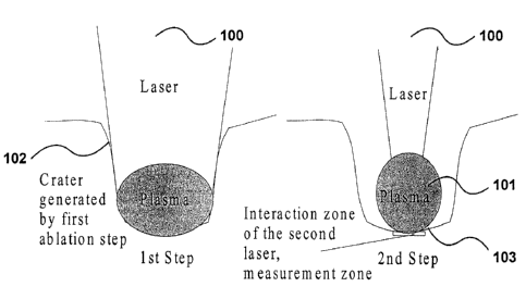
Figure 1 illustrates the principle of operation of the invention;
Figures 2a to 2c are overall block diagram of various emlbodiments of the
invention;
Figures 3a and 3b shows two possible embodiments of tLie depth measuring
system based
on interferometry with a short coherence length source;
Figure 3c shows the envelopes of interference signals from which crater depth
is
determined;
Figure 4 shows two different emission spectra, one characteristic of the
coating
composition, and the second one characteristic of the substrate; and
-4-
CA 02353014 2001-07-12
Figure 5 shows two depth profiles of the zinc emission lime obtained with
classical LIPS
instrumentation and by using the method and apparatus of the present
invention.
DETAILED DESCRIPTION OF THE PREFERRED EMBODIMENTS
In accordance with the principles of the invention two laser pulses or bursts
of
laser pulses of different diameter are used. The first laser pulse or burst
(the number of
laser shots determine the resolution of depth profiling) realizes the
ablation. The second
laser pulse or burst (the number of laser shots increases tlhe precision)
vaporizes a small
volume at the bottom of the crater generated by the first laser pulse or
burst, and produces
plasma of which the optical emission is analyzed with a spectrometer. The
spectrum is
detected through appropriate optics by a gated photodiode array detector, an
intensified
CCD camera, or by an array of photomultipliers each individually positioned to
detect an
emission line representative of a given element.
The material may be opaque or partly transparent. As a result of the high
temperature generated, a small amount of the material is ablated, vaporized
and ionized,
its atoms and ions being brought in excited states, thus allowing species in
the plasma to
be identified by spectrally and temporally resolving the spark light emission.
To perform a reliable depth profile analysis, it is important to ensure a
controlled
and reproducible ablation rate and a well-characterized alblation volume. The
ablation has
to be the same for each shot in terms of radial distribution of the ablated
depth. In order to
obtain this result, the spatial characteristics of the laserbeam have to be
controlled and
the Iaser needs to be stable from shot to shot. Furthermore, to achieve a good
depth
resolution, all parts of the laser beam throughout its cross-section should
sample the
material at approximately the same depth. This condition is difficult to
satisfy with a
near-Gaussian laser beam, which produces cone-shaped craters. Inevitably, for
any given
shot (except the first), the laser will sample material from different depths
along the crater
surface. In view of this, it seems clear that modification of the energy
radial distribution
of the laser beam should be developed to increase the deI>th resolution. To do
so, a
diaphragm is used to select only a homogenous part of the laser beam. A
homogenizer
could be added before the diaphragm to this set-up in order to obtain a better
laser beam
profle. This setup allows a better control of the generated crater shape.
However, in spite
of this technical improvement, the optical emission of the; plasma always
shows a non-
-5-
CA 02353014 2001-07-12
negligible contribution from the wall of the crater. This degrades the
precision of the
result, and in particular increases the apparent spatial extent of the
transition in
composition between a coating and a substrate as shown in Fig. 5.
To overcome this problem, a second smaller laser beam (the analyzing beam) is
focused inside the crater, and generates plasma emission., which is only
dependent on the
composition of the bottom of the crater. The role of the second beam is to
probe in a very
precise way the elementary composition of the thinnest zone also possible
without
contribution from the edge of the crater. The depth resolution also depends on
the number
of ablation shots in the first step, the energy in this laser ibeam, the
wavelength of the
laser, and can be adjusted according to the needs or the nature of the
samples.
Generally the number of ablation shots will be munch higher than the number of
analyzing shots, typically 100 to one. The depth of the small crater generated
by the
analyzing beam can be neglected compared to the depth of the ablation crater.
However,
when high resolution is needed the ratio of ablation shot number to the
analyzing one
could be less than 100. This means the highest resolution corresponds to a
ratio of one,
i.e. one ablation shot is followed by an analyzing shot. Tlhe depth of crater
produced by
analyzing shot cannot be neglected. To overcome this problem, different
solutions are
possible. First, the energy of the analyzing pulse can be reduced in order to
avoid the
surface damage. If the emission signal of the analyzing plasma resulting from
the laser
pulse is too weak, the plasma can be excited with a second laser pulse (US
patent
6,008.897) at appropriate wavelength which could be generated by a wavelength
tunable
laser source. Secondly, a mixed-wavelength pulse can be used as analyzing beam
shot
(Patent pending). The use of mixed-wavelength laser pul,~e damages less the
surface
because of the screening and plasma absorption.
As shown in Figure 1, an ablation beam 100 produces a plasma 101 at the bottom
of the crater 102 generated in a first ablation step. A second laser beam
which has smaller
diameter is used to make a measurement in the interaction zone 103 at the
centre of the
bottom of the crater and produces a second plasma. The emission of this plasma
101 is
analyzed in order to obtain the composition of the interaction zone without
contribution
of the crater edge.
-6-
CA 02353014 2001-07-12
Figures 2a to 2c show three different experimental setups all built on the
same
principle.
In the embodiment shown in Figure 2a, just one laser 205 is employed and the
laser beam 200 passes through a large diaphragm 206 and is reflected by a
mirror 207
through focusing optics and reflected by a dichroic plate 208. A crater is
formed on the
target 209 by focusing the laser beam 200 using a focusing system ideally
composed
preferably of two lenses in order to realize an image of the diaphragm on the
surface with
a chosen magnification. A counter (not shown) allows firing a predetermined
number of
shots to control the ablation depth. Then, a movable diaphragm support (not
shown) is
actuated and the smallest diaphragm 206a is moved in the place of the large
diaphragm
206 on the same optical axis. This allows a measurement:, to be made at the
center of the
first crater. The diaphragm material is preferably made of a light scattering
material and
low absorption material at the laser wavelength, in order to increase the
lifetime of this
component.
With the aid of a lens 211, a reduced image of the plasma is created at the
entrance slit of the spectrometer 212, which is connected to a data processing
unit 213.
The current configuration thus allows efficient collection of the light
emitted by the
plasma along the axis of the.plasma plume using a dichroic plate, or a pierced
mirror.
The optical emission from the plasma is spectrally analy:~ed using typically a
grating
spectrometer equipped with a gated detector such as an io~tensified photodiode
array
detector, CCD camera, or an array of photomultipliers each individually
positioned in the
focal plane to detect, simultaneously and during a specified time period, a
number of
emission lines representative of the different elements in the material to be
analyzed.
Standard techniques are used to properly synchronize the: lasers and detectors
so as to
collect the emission signal during the time window providing the best signal
to noise
ratio, while a fast computer evaluates the measured spectra and calculates the
element
concentrations via calibration procedures which are well known to
spectroscopists.
The set-up shown in Figure 2b includes two optical paths. A 50/50 beamsplitter
220 is located immediately downstream of the laser 205.. The laser beam in
this setup
follows the first optical path 221 (the second path 222 is stopped by a
shutter 225), and as
in the first setup, it passes through a large diaphragm 206 and is reflected
by a mirror 207.
_7_
CA 02353014 2001-07-12
A crater is formed on the target by focusing the laser beam using a focusing
system
ideally composed preferably of two lenses in order to realize an image of the
diaphragm
on the surface with a chosen magnification. A counter allows firing a
predetermined
number of shots to control the ablation depth: After this jFirst step, a
shutter 223 stops the
ablation laser beam 221 and the shutter 225 is opened, in order to allow the
beam to
follow the second path. The same results could be obtained using an electro-
optic cell
with beam sputter 220 being a polarizing beam splitter. Such a device would be
located
immediately after the laser output, and by application of a controlled voltage
will shift the
polarization so the laser beam is sent either along path 221 or 222. Then, in
this new path
is disposed a smaller diaphragm 206a coupled to a focussing system that
focuses the laser
beam into the first crater. A polarized beamsplitter located in this path
(mirror-2) reflects
the first beam and lets pass the second beam when the el~ectro-optic system is
used (half
wave plates are used in both paths to flip the polarization). Otherwise, a
50/50 plate
replaces it. For this setup, a pierced mirror 226 is required. The detection
device is
identical to the first setup.
The third configuration shown in Figure 2c permits a similar result to be
obtained
using two lasers 205, 205a. The first laser beam follows exactly the same path
that is
described in setup (b), and controls the ablation step. A beam homogenizer
could be used
in order to obtain a better laser beam profile. The second Laser 205a is used
in the
measurement step, and it is positioned in order to be focused at the center of
the bottom of
the crater generated by the first laser. For this setup, the ~,zse of a
diaphragm and a
focusing system as already described is preferable but not obligatory, a
simple lens can
replace the diaphragm and focusing system. The only reduirement is that the
diameter on
the target surface of the laser beam 221 is larger than laser beam 222 at the
same position.
For this setup, pierced mirror 226 is used as collection tool, and the
detection arrangement
is identical to the other setups. This embodiment shows also that an optical
profilometer
is integrated with the system and is used to monitor throughout the whole
analysis the
depth of the crater. Preferred configurations of such a profilometer are shown
in FIGs.3a
and 3b.
Independently of the configuration used for the LIPS system, in order to
perform
accurate profilometry, the depth at which each measurerr~ent is made has to be
evaluated.
_g_
CA 02353014 2001-07-12
This evaluation can be performed by taking the sample off the LIPS system and
measuring the crater depth with a profilometer. The profilometer can be based
on
confocal microscopy, laser triangulation or interferometr;y using a short
coherence length
light source (also called white Light interferometry or optical coherence
tomography). In
confocal microscopy, light is sent through a pinhole and 'the light collected
through the
same pinhole after reflection by the object is monitored. 'The surface
location is
determined by noting that the collected light is at maximum when the image of
the
pinhole is at focus on the surface: In laser-triangulation, the light spot at
the surface of the
object is viewed by a linear camera along a direction mal;ing an angle with
the
illumination axis. The position of the spot on the linear c;~mera is dependant
upon the
distance of the surface from the device, which allows monitoring the surface
location. In
interferometry with a short coherence length source, a maximum interference
signal is
observed when the path length along the arm going to thf; object is equal to
that a
reference arm whose length is varied. This variation being calibrated, this
technique also
allows monitoring the surface location.
Crater depth measurement for each composition analysis (or after a certain
number of analyses) requires positioning the sample at th,e same location
under the LIPS
apparatus, which is possible, but generally inconvenient. In some cases, it is
also possible
to calibrate the ablation rate so only one measurement is needed at the end of
analysis.
For example for a layer on top of a substrate, a depth measurement can be
performed on
calibration samples with a layer on top and without a layer. From these
measurements, the
removal rate per laser shot in the layer and in the substrate is evaluated.
From this
calibration, count of the laser shots and final depth measurement, the depths
in the
homogenous zones are readily evaluated. Depth in the transition zone is
performed with a
reasonable accuracy by interpolation. This obviously ass~zmes that the
ablation rate is the
same for the study sample and the calibration samples, which in particular
requires
sufficient laser stability (total power and power distribution). Furthermore
such a
procedure is not applicable on samples with composition variation right from
the surface
or more complex mufti-layer samples. Consequently, it will be much convenient
to have
the depth measurement provision integrated with the LIPS apparatus. The two
following
embodiments show how this can be accomplished by using interferometry with a
short
coherence length source.
-9-
CA 02353014 2001-07-12
Figure 3a shows an embodiment which actually realize a two-wave Michelson
interferometer made of single mode optical fibers. A supra luminescent diode
300 giving
a bandwidth of typically 20nm is used as light source. This diode 300 is
followed by an
optical isolator 30I to prevent feedback from any interface and from the
surface of the
object of affecting its operation. The beam is then fed through a
splitter/mixer 302, which
is a 50-50% bi-directionnal coupler, The reference arm length is varied by
collimating the
beam with lens and mounting the mirror (or a retrorefle<;tor) on a translation
slide. In the
arm going to the object, the beam emerging from the fiber is focused onto the
surface by a
lens and a dichroic mirror mounted on a rotating slide or a galvanometer. This
dichroic
mirror lets the ablation beams to go through, reflects the interferometer
light and allows
scanning across the crater. Assuming that the reference axm scan is much
faster than the
scan across the crater, depth information is obtained for each position across
the crater
from the signal observed at zero path length difference on the detector.
In the second and preferred embodiment, no scanning across the crater is
performed and only two depth measurements are performed, one inside the crater
at the
location of elemental analysis and the other one outside t:he crater in a
region unaffected
by ablation and residual debris.
As shown in Figure 3b, another 50-50% bidirectional coupler 304 is used in the
arm going to the sample to give two secondary light sources that are separated
by a given
distance. A telecentric optical system made of two lenses is then used to
focused them on
the sample, one at the measurement location in the crater and the other one
outside the
crater.
Figure 3c shows two signals (envelopes of the interference signal) from which
the
crater depth is determined, the scan of the reference arm being calibrated.
The two
secondary sources given by the second 50-SO% coupler are not in the same plane
so the
two signals are conveniently separated before the start of any ablation.
Figure 4 shows spectra obtained with the apparatus of Figure 2a by firing on a
1
mm diameter pinhole coupled to focusing optics (lens couple) allowing to
obtain, 500 urn
diameter spot (x2 demagnification) at the surface of an ar;mealed galvanneal
coated steel
sample (containing approximately 9 % of Fe in a Zn matrix). The first spectrum
is
obtained with a single shot of 60 ~uJ energy on the zinc coating, and the
other one after
- 10-
CA 02353014 2001-07-12
several ablation shots have reached the steel substrate (w:ith Fe as main
component). The
comparison of the two optical emission spectra shows the disappearance of the
Zn
emission lines. This information is used to measure the thickness of the Zn
coating.
Figure 5 is a comparison of two depth profiles of zinc obtained by monitoring
the
307.21 nm emission line: The ablation depth is evaluated by interferometry
with a short
coherence length source as described above. The sample is galvannealed steel
annealed
zinc-coated steel. The zinc coating has been analysed by electronic microprobe
(reference
analytical technique for the analysis of solids). The coating thickness is
approximately 7
~.m with an interface length between Zn/steel of less than 2 ~,m.
One of the profiles shown in fig.5 is obtained by using classical LIPS
instrumentation, the laser beam being filtered by a large diaphragm. The
second one is
obtained using the present invention. In the two cases, each point of
measurement
corresponds to 10 measurement shots, after 100 ablation shots, obtained with
the large
diaphragm. It is seen that the profilometry technique according to the present
invention
provides a more accurate measurement of the coating thickness. The interface
is
described with more precision: the beginning of the interface appears in the
same place
with the two systems but ends 2 ~.m sooner with the system according to this
invention.
The Zn emission line falls down to zero quickly using this invention, which is
not the
case with conventional instrumentation where the Zn emiission persists. The
results of
measured thickness and of interface length obtained with this invention are
very close to
those obtained with a conventional electronic microprobe.
The above description of the present invention is susceptible to various
modifications, changes and adaptations, and the same are intended to be
comprehended
within the scope of the appended claims.
-11-