Note: Descriptions are shown in the official language in which they were submitted.
CA 02353483 2007-12-06
72249-113
IL-1 ZETA, IL-1 ZETA SPLICE VARIANTS AND XREC2
DNAS AND POLYPEPTIDES
BACKGROt1ND OF THE INVENTION
Field of the Invention
The invention is directed to novel, purified and isolated IL-1 zeta, IL-1 zeta
splice variants and Xrec2 polypeptides and fragments thereof, the nucleic
acids
encoding such polypeptides, processes for production of recombinant forms of
such
polypeptides, antibodies generated against these polypeptides, fragmented
peptides
derived from these polypeptides, and uses thereof.
Description of Related Art
Interleukin-1 (IL-1) is a member of a large group of cytokines whose primary
function is to mediate immune and inflammatory responses. There are five known
IL-
1 family members which include IL-1 alpha (IL-1 a), IL-1 beta (IL-1(3), IL-1
receptor
antagonist (IL-lra), IL-1 delta (IL-18-as disclosed in W099/35268), and IL-18
(previously known as IGIF and sometimes IL-1 gamma). IL-1 that is secreted by
macrophages is actually a mixture of mostly IL-1 P and some IL-la (Abbas et
al.,
1994). IL-la and IL-1(3, which are first produced as 33 kD precursors that
lack a
signal sequence, are further processed by proteolytic cleavage to produce
secreted
active forms, each about 17 kD. Additionally, the 33 kD precursor of IL-l(X is
also
active. Both forms of IL-1 are the products of two different genes located on
chromosome 2. Although the two forms are less than 30 percent homologous to
each
other, they both bind to the same receptors and have similar activities.
The IL-1 family of ligands binds to a common receptor composed of a ligand
binding chain, the type I IL-1 receptor (IL-1RI), and a required signaling
component,
1
CA 02353483 2001-06-04
WO 00/36108 PCT/US99/29549
the IL-1R accessory protein (AcP) (Sims et al. 1988; Greenfeder et al. 1995;
Cullinan
et al. 1998). A type II IL-1 receptor (IL-1RII) binds and sequesters the
agonist IL-1
(especially IL-1(3) without inducing any signaling response of its own
(McMahan et
al. 1991; Sims et al. 1993; Colotta et al. 1993; Colotta et al. 1994). IL-1
ligands can
also bind to a soluble proteolytic fragment of IL-1RII (sIL-1RII) (Colotta et
al., 1993).
IL-lra, a biologically inactive form of IL-l, is structurally homologous to IL-
1. IL-lra is produced with a signal sequence which allows for efficient
secretion into
the extracellular region (Abbas et al., 1994). Additionally, IL-lra binds to
the type I
IL-i receptor but fails to bring about the subsequent interaction with AcP.
Thus, IL-
lra blocks IL-1RI and prevents the action of the agonist IL-1 (Hannum et al.
1990;
Eisenberg et al. 1990).
The major source of IL-1 is the activated macrophage or mononuclear
phagocyte. Other cells that produce IL-1 include epithelial and endothelial
cells
(Abbas et al., 1994). IL-1 secretion from macrophages occurs after the
macrophage
encounters and ingests gram-negative bacteria. Such bacteria contain
lipopolysaccharide (LPS) molecules, also known as endotoxin, in the bacterial
cell
wall. LPS molecules are the active components that stimulate macrophages to
produce tumor necrosis factor (TNF) and IL-1. In this case, IL-I is produced
in
response to LPS and TNF production. At low concentrations, LPS stimulates
macrophages and activates B-cells and other host responses needed to eliminate
the
bacterial infection; however, at high concentrations, LPS can cause severe
tissue
damage, shock, and even death.
The biological functions of IL-1 include activating vascular endothelial cells
and lymphocytes, local tissue destruction, and fever (Janeway et al., 1996).
At low
levels, IL-1 stimulates macrophages and vascular endothelial cells to produce
IL-6,
upregulates molecules on the surface of vascular endothelial cells to increase
leukocyte adhesion, and indirectly activates inflammatory leukocytes by
stimulating
mononuclear phagocytes and other cells to produce certain chemokines that
activate
inflammatory leukocytes. Additionally, IL-1 is involved in other inflammatory
responses such as induction of prostaglandins, nitric oxide synthetase, and
metalloproteinases. These IL-1 functions are crucial during low level
microbial
2
CA 02353483 2001-06-04
WO 00/36108 PCT/US99/29549
infections. However, if the microbial infection escalates, IL-1 acts
systemically by
inducing fever, stimulating mononuclear phagocytes to produce IL-1 and IL-6,
increasing the production of serum proteins from hepatocytes, and activating
the
coagulation system. Additionally, IL-1 does not cause hemorrhagic necrosis of
tumors, suppress bone marrow stem cell division, and IL-1 is lethal to humans
at high
concentrations.
Given the important function of IL-1, there is a need to identify additional
members of the IL-1 ligand family and the IL-1 receptor family. In addition,
in view
of the continuing interest in protein research and the immune system, the
discovery,
identification, and roles of new proteins and their inhibitors, are at the
forefront of
modern molecular biology and biochemistry. Despite the growing body of
knowledge, there is still a need in the art to discover the identity and
function of
proteins involved in cellular and immune responses.
In another aspect, the identification of the primary structure, or sequence,
of an
unknown protein is the culmination of an arduous process of experimentation.
In
order to identify an unknown protein, the investigator can rely upon a
comparison of
the unknown protein to known peptides using a variety of techniques known to
those
skilled in the art. For instance, proteins are routinely analyzed using
techniques such
as electrophoresis, sedimentation, chromatography, sequencing and mass
spectrometry.
In particular, the unique nature of the composition of a protein with regard
to
its specific amino acid constituents results in unique positioning of cleavage
sites
within the protein. Specific fragmentation of a protein by chemical or
enzymatic
cleavage results in a unique "peptide fingerprint" (D. W. Cleveland et al., J.
Biol.
Chem. 252:1102-1106, 1977; M. Brown et al., J. Gen. Virol. 50:309-316, 1980).
Consequently, cleavage at specific sites results in reproducible fragmentation
of a
given protein into peptides of precise molecular weights. Furthermore, these
peptides
possess unique charge characteristics that determine the isoelectric pH of the
peptide.
These unique characteristics can be exploited using a variety of
electrophoretic and
other techniques (Brock et al., Biology of Microorganisms, pp. 76-77, Prentice
Hall,
6th ed. 1991).
3
CA 02353483 2001-06-04
WO 00/36108 PCT/US99/29549
Fragmentation of proteins is further employed for amino acid composition
analysis and protein sequencing (P. Matsudiara, J. Biol. Chem. 262:10035-
10038,
1987; C. Eckerskorn et al., Electrophoresis 9:830-838, 1988), particularly the
production of fragments from proteins with a"blocked" N-terminus. In addition,
fragmented proteins can be used for immunization, for affinity selection (R.
A.
Brown, U.S. Patent No. 5,151,412), for determination of modification sites
(e.g.
phosphorylation), for generation of active biological compounds (Brock et al.,
Biology of Microorganisms. pp. 300-301, Prentice Hall, 6th ed. 1991), and for
differentiation of homologous proteins (M. Brown et al., J. Gen. Virol. 50:309-
316,
1980).
In addition, when a peptide fingerprint of an unknown protein is obtained, it
can be compared to a database of known proteins to assist in the
identification of the
unknown protein using mass spectrometry (W.J. Henzel et al., Proc. Natl. Acad.
Scf.
USA 90:5011-5015, 1993; D. Fenyo et al., Electrophoresis 19:998-1005, 1998). A
variety of computer software programs to facilitate these comparisons are
accessible
via the Internet, such as Protein Prospector (Internet site:
prospector.uscf.edu),
MultiIdent (Internet site: www.expasy.ch/sprot/multiident.html), PeptideSearch
(Internet site: www.mann.embl-heiedelberg.de...deSearch/FR PeptideSearch
Form.html), and ProFound (Internet site: www.chait-sgi.rockefeller.edu/cgi-
bin/prot-
id-frag.html). These programs allow the user to specify the cleavage agent and
the
molecular weights of the fragmented peptides within a designated tolerance.
The
programs compare these molecular weights to protein molecular weight
information
stored in databases to assist in determining the identity of the unknown
protein.
Accurate information concerning the number of fragmented peptides and the
precise
molecular weight of those peptides is required for accurate identification.
Therefore,
increasing the accuracy in determining the number of fragmented peptides and
their
molecular weight should result in enhanced likelihood of success in the
identification
of unknown proteins.
In addition, peptide digests of unknown proteins can be sequenced using
tandem mass spectrometry (MS/MS) and the resulting sequence searched against
databases (J.K. Eng et al., J. Am. Soc. Mass Spec. 5:976-989, 1994; M. Mann et
al.,
4
CA 02353483 2007-12-06
72249-113
Anal. Chem. 66:4390-4399, 1994; J.A. Taylor et al., Rapid Comm. Mass Spec.
11:1067-1075, 1997). Searching programs that can be used in this process exist
on
the Internet, such as Lutefisk 97 (Internet site:
www.lsbc.com:70/Lutefisk97.html),
and the Protein Prospector, Peptide Search and ProFound programs described
above.
Therefore, adding the sequence of a gene and its predicted protein sequence
and
peptide fragments to a sequence database can aid in the identification of
unknown
proteins using tandem mass spectrometry.
Thus, there also exists a need in the art for polypeptides suitable for use in
peptide fragmentation studies, for use in molecular weight measurements, and
for use
in protein sequencing using tandem mass spectrometry.
SUMMARY OF THE INVENTION
The present invention provides isolated nucleic acids and polypeptides
encoded by the nucleic acids for an IL-1 family ligand termed "IL-1 zeta" and
three
splice variants of IL-1 zeta, termed TDZ.1, TDZ.2, and TDZ.3. The present
invention
also provides isolated nucleic acid molecules and polypeptides encoded by the
nucleic
acid molecules for an IL-1 family receptor termed "Xrec2." Thus, in one
aspect, the
invention is directed to isolated nucleic acid molecules of IL-1 zeta, TDZ. 1,
TDZ.2,
and TDZ.3 comprising the DNA sequence of SEQ ID NO: 1, SEQ ID NO:5, SEQ ID
NO:6, and SEQ ID NO:7, respectively, and nucleic acid molecules complementary
to
SEQ ID NOs:1, 5, 6, and 7. Similarly, the invention is directed to isolated
nucleic
acid molecules of Xrec2 comprising the nucleic acid molecule of SEQ ID NO:2
and
nucleic acid molecules complementary to SEQ ID NO:2. In another aspect, the
invention is directed to isolated IL-1 zeta, TDZ.1, TDZ.2, and TDZ.3
polypeptides
having the amino acid sequences SEQ ID NO:3, SEQ ID NO:8, SEQ ID NO:9, and
SEQ ID NO:10, respectively, and nucleic acid molecules encoding the
polypeptides of
SEQ ID NOs:3, 8, 9, and 10. Further included in the invention are isolated
Xrec2
polypeptides comprising the amino acid sequence of SEQ ID NO:4 and nucleic
acid
molecules that encode the polypeptide of SEQ ID NO:4.
CA 02353483 2007-12-06
72249-113
According to one aspect of the present invention,
there is provided an isolated polynucleotide molecule
comprising a nucleic acid sequence that encodes: (a) a
polypeptide comprising SEQ ID NO: 3; (b) a polypeptide
comprising SEQ ID NO: 8; (c) a polypeptide comprising
SEQ ID NO: 9; (d) a polypeptide that is at least
95% identical to the polypeptide of any one of a) to c),
wherein the polypeptide binds an IL-1R family member; (e) a
fragment of the polypeptide of SEQ ID NO: 3 whose N-terminal
amino acid is selected from amino acids 21-51 and whose
C-terminal amino acid is selected from amino acids 188-192
and which binds an IL-1R family member, wherein the fragment
is not amino acids 24-192, 28-192 or 34-192 of SEQ ID NO: 3;
(f) a fragment of the polypeptide of SEQ ID NO: 8 whose
N-terminal amino acid is selected from amino acids 41-61 and
whose C-terminal amino acid is selected from amino
acids 214-218 and which binds an IL-1R family member,
wherein the fragment is not amino acids 43-218, 54-218 or
60-218 of SEQ ID NO: 8; or (g) a fragment of the polypeptide
of SEQ ID NO: 9 whose N-terminal amino acid is selected from
amino acids 21-41 and whose C-terminal amino acid is
selected from amino acids 193-197 and which binds an
IL-1R family member, wherein the fragment is not amino
acids 33-197 or 39-197 of SEQ ID NO: 9.
Both single-stranded and double-stranded
RNA and DNA nucleic acid molecules are encompassed by the
invention, as well as nucleic acid molecules that
5a
CA 02353483 2001-06-04
WO 00/36108 PCT/US99/29549
hybridize to a denatured, double-stranded DNA comprising all or a portion of
SEQ ID
NOs: 1, 2, 5, 6, and 7 and/or a DNA that encodes the amino acid sequences set
forth in
SEQ ID NOs:3, 4, 8, 9, and 10. Also encompassed are isolated nucleic acid
molecules
that are derived by in vitro mutagenesis of nucleic acid molecules comprising
sequences of SEQ ID NOs: 1, 2, 5, 6, and 7 that are degenerate from nucleic
acid
molecules comprising sequences of SEQ ID NOs: 1, 2, 5, 6, and 7, and that are
allelic
variants of DNA of the invention. The invention also encompasses recombinant
vectors that direct the expression of these nucleic acid molecules and host
cells
transfonned or transfected with these vectors.
In addition, the invention encompasses methods of using the nucleic acids
noted above to identify nucleic acids encoding proteins having activities
associated
with IL-1 family ligands and receptors. Thus, the IL-1 zeta nucleic acid
molecules
can be used to identify the IL-1 zeta receptor while the Xrec2 nucleic acid
molecule
can be used to identify the Xrec2 ligand.
In addition, these nucleic acids can be used to identify the human
chromosomes with which the nucleic acids are associated. Thus, the IL-1 zeta,
TDZ.1, TDZ.2, and TDZ.3 nucleic acids can be used to identify human chromosome
2
while the Xrec2 nucleic acids can be used to identify human chromosome X.
Accordingly, these nucleic acids can also be used to map genes on human
chromosomes 2 and X, respectively; to identify genes associated with certain
diseases,
syndromes, or other human conditions associated with human chromosomes 2 and
X,
respectively; and to study cell signal transduction and the immune system.
The invention also encompasses the use of sense or antisense oligonucleotides
from the nucleic acids of SEQ ID NOs:1, 2, 5, 6, and 7 to inhibit the
expression of the
respective polynucleotide encoded by the genes of the invention.
The invention also encompasses isolated polypeptides and fragments of IL-1
zeta and Xrec2 as encoded by these nucleic acid molecules, including soluble
polypeptide portions of SEQ ID NOs:3 4, 8, 9, and 10, respectively. The
invention
further encompasses methods for the production of these polypeptides,
including
culturing a host cell under conditions promoting expression and recovering the
polypeptide from the culture medium. Especially, the expression of these
6
CA 02353483 2001-06-04
WO 00/36108 PCT/US99/29549
polypeptides in bacteria, yeast, plant, insect, and animal cells is
encompassed by the
invention.
In general, the polypeptides of the invention can be used to study cellular
processes such as immune regulation, cell proliferation, cell death, cell
migration,
cell-to-cell interaction, and inflammatory responses. In addition, these
polypeptides
can be used to identify proteins associated with IL-1 zeta, TDZ.1, TDZ.2, and
TDZ.3
ligands and with Xrec2 receptors.
In addition, the invention includes assays utilizing these polypeptides to
screen
for potential inhibitors of activity associated with polypeptide counter-
structure
molecules, and methods of using these polypeptides as therapeutic agents for
the
treatment of diseases mediated by polypeptide counter-structure molecules.
Further,
methods of using these polvpeptides in the design of inhibitors (e.g.,
engineered
receptors that act as inhibitors) thereof are also an aspect of the invention.
Further encompassed by this invention is the use of the IL-1 zeta and Xrec2
nucleic acid sequences, predicted amino acid sequences of the polypeptide or
fragments thereof, or a combination of the predicted amino acid sequences of
the
polypeptide and fragments thereof for use in searching an electronic database
to aid in
the identification of sample nucleic acids and/or proteins. The invention
further
provides a method of using the polypeptides disclosed herein as controls for
establishing the extent of protein fragmentation.
Isolated polyclonal or monoclonal antibodies that bind to these polypeptides
are also encompassed by the invention, in addition the use of these antibodies
to aid in
purifying the polypeptides of the invention.
BRIEF DESCRIPTION OF THE FIGURES
Figure 1 diagrams the genomic structure of the IL-1 zeta locus.
Figure 2 represents a molecular model showing the secondary structure of IL-1
zeta, in which P-strands are shown in yellow, with their direction indicated
by the
arrowhead; (3-turns are shown in blue; and coils are shown in green. The IL-1
zeta
structure is presented in two different views.
7
CA 02353483 2001-06-04
WO 00/36108 PCT/US99/29549
DETAILED DESCRIPTION OF THE INVENTION
The nucleic acid molecules encompassed in the invention include the
following nucleotide sequences:
Name: IL-1 zeta
1 ATGTCAGGCT GTGATAGGAG GGAAACAGAA ACCAAAGGAA AGAACAGCTT
51 TAAGAAGCGC TTAAGAGGTC CAAAGGTGAA GAACTTAAAC CCGAAGAAAT
101 TCAGCATTCA TGACCAGGAT CACAAAGTAC TGGTCCTGGA CTCTGGGAAT
151 CTCATAGCAG TTCCAGATAA AAACTACATA CGCCCAGAGA TCTTCTTTGC
201 ATTAGCCTCA TCCTTGAGCT CAGCCTCTGC GGAGAAAGGA AGTCCGATTC
251 TCCTGGGGGT CTCTAAAGGG GAGTTTTGTC TCTACTGTGA CAAGGATAAA
301 GGACAAAGTC ATCCATCCCT TCAGCTGAAG AAGGAGAAAC TGATGAAGCT
351 GGCTGCCCAA AAGGAATCAG CACGCCGGCC CTTCATCTTT TATAGGGCTC
401 AGGTGGGCTC CTGGAACATG CTGGAGTCGG CGGCTCACCC CGGATGGTTC
451 ATCTGCACCT CCTGCAATTG TAATGAGCCT GTTGGGGTGA CAGATAAATT
501 TGAGAACAGG AAACACATTG AATTTTCATT TCAACCAGTT TGCAAAGCTG
551 AAATGAGCCC CAGTGAGGTC AGCGATTAG (SEQ ID NO:1)
Name: Xrec2
1 ATGAAAGCTC CGATTCCACA CTTGATTCTC TTATACGCTA CTTTTACTCA
51 GAGTTTGAAG GTTGTGACCA AAAGAGGCTC CGCCGATGGA TGCACTGACT
101 GGTCTATCGA TATCAAGAAA TATCAAGTTT TGGTGGGAGA GCCTGTTCGA
151 ATCAAATGTG CACTCTTTTA TGGTTATATC AGAACAAATT ACTCCCTTGC
201 CCAAAGTGCT GGACTCAGTT TGATGTGGTA CAAAAGTTCT GGTCCTGGAG
251 ACTTTGAAGA GCCAATAGCC TTTGACGGAA GTAGAATGAG CAAAGAAGAA
301 GACTCCATTT GGTTCCGGCC AACATTGCTA CAGGACAGTG GTCTCTACGC
351 CTGTGTCATC AGAAACTCCA CTTACTGTAT GAAAGTATCC ATCTCACTGA
401 CAGTGGGTGA AAATGACACT GGACTCTGCT ATAATTCCAA GATGAAGTAT
451 TTTGAAAAAG CTGAACTTAG CAAAAGCAAG GAAATTTCAT GCCGTGACAT
501 AGAGGATTTT CTACTGCCAA CCAGAGAACC TGAAATCCTT TGGTACAAGG
551 AATGCAGGAC AAAAACATGG AGGCCAAGTA TTGTATTCAA AAGAGATACT
601 CTGCTTATAA GAGAAGTCAG AGAAGATGAC ATTGGAAATT ATACCTGTGA
651 ATTAAAATAT GGAGGCTTTG TTGTGAGAAG AACTACTGAA TTAACTGTTA
701 CAGCCCCTCT GACTGATAAG CCACCCAAGC TTTTGTATCC TATGGAAAGT
751 AAACTGACAA TTCAGGAGAC CCAGCTGGGT GACTCTGCTA ATCTAACCTG
801 CAGAGCTTTC TTTGGGTACA GCGGAGATGT CAGTCCTTTA ATTTACTGGA
851 TGAAAGGAGA AAAATTTATT GAAGATCTGG ATGAAAATCG AGTTTGGGAA
8
CA 02353483 2001-06-04
WO 00/36108 PCT/US99/29549
901 AGTGACATTA GAATTCTTAA GGAGCATCTT GGGGAACAGG AAGTTTCCAT
951 CTCATTAATT GTGGACTCTG TGGAAGAAGG TGACTTGGGA AATTACTCCT
1001 GTTATGTTGA AAATGGAAAT GGACGTCGAC ACGCCAGCGT TCTCCTTCAT
1051 AAACGAGAGC TAATGTACAC AGTGGAACTT GCTGGAGGCC TTGGTGCTAT
1101 ACTCTTGCTG CTTGTATGTT TGGTGACCAT CTACAAGTGT TACAAGATAG
1151 AAATCATGCT CTTCTACAGG AATCATTTTG GAGCTGAAGA GCTCGATGGA
1201 GACAATAAAG ATTATGATGC ATACTTATCA TACACCAAAG TGGATCCTGA
1251 CCAGTGGAAT CAAGAGACTG GGGAAGAAGA ACGTTTTGCC CTTGAAATCC
1301 TACCTGATAT GCTTGAAAAG CATTATGGAT ATAAGTTGTT TATACCAGAT
1351 AGAGATTTAA TCCCAACTGG AACATACATT GAAGATGTGG CAAGATGTGT
1401 AGATCAAAGC AAGCGGCTGA TTATTGTCAT GACCCCAAAT TACGTAGTTA
1451 GAAGGGGCTG GAGCATCTTT GAGCTGGAAA CCAGACTTCG AAATATGCTT
1501 GTGACTGGAG AAATTAAAGT GATTCTAATT GAATGCAGTG AACTGAGAGG
1551 AATTATGAAC TACCAGGAGG TGGAGGCCCT GAAGCACACC ATCAAGCTCC
1601 TGACGGTCAT TAAATGGCAT GGACCAAAAT GCAACAAGTT GAACTCCAAG
1651 TTCTGGAAAC GTTTACAGTA TGAAATGCCT TTTAAGAGGA TAGAACCCAT
1701 TACACATGAG CAGGCTTTAG ATGTCAGTGA GCAAGGGCCT TTTGGGGAGC
1751 TGCAGACTGT CTCGGCCATT TCCATGGCCG CGGCCACCTC CACAGCTCTA
1801 GCCACTGCCC ATCCAGATCT CCGTTCTACC TTTCACAACA CGTACCATTC
1851 ACAAATGCGT CAGAAACACT ACTACCGAAG CTATGAGTAC GACGTACCTC
1901 CTACCGGCAC CCTGCCTCTT ACCTCCATAG GCAATCAGCA TACCTACTGT
1951 AACATCCCTA TGACACTCAT CAACGGGCAG CGGCCACAGA CAAAATCGAG
2001 CAGGGAGCAG AATCCAGATG AGGCCCACAC AAACAGTGCC ATCCTGCCGC
2051 TGTTGCCAAG GGAGACCAGT ATATCCAGTG TGATATGGTG A (SEQ ID N0:2)
Name: TDZ.1
1 ATGTCCTTTG TGGGGGAGAA CTCAGGAGTG AAAATGGGCT CTGAGGACTG
51 GGAAAAAGAT GAACCCCAGT GCTGCTTAGA AGACCCGGCT GTAAGCCCCC
101 TGGAACCAGG CCCAAGCCTC CCCACCATGA ATTTTGTTCA CACAAGTCCA
151 AAGGTGAAGA ACTTAAACCC GAAGAAATTC AGCATTCATG ACCAGGATCA
201 CAAAGTACTG GTCCTGGACT CTGGGAATCT CATAGCAGTT CCAGATAAAA
251 ACTACATACG CCCAGAGATC TTCTTTGCAT TAGCCTCATC CTTGAGCTCA
301 GCCTCTGCGG AGAAAGGAAG TCCGATTCTC CTGGGGGTCT CTAAAGGGGA
351 GTTTTGTCTC TACTGTGACA AGGATAAAGG ACAAAGTCAT CCATCCCTTC
401 AGCTGAAGAA GGAGAAACTG ATGAAGCTGG CTGCCCAAAA GGAATCAGCA
451 CGCCGGCCCT TCATCTTTTA TAGGGCTCAG GTGGGCTCCT GGAACATGCT
9
CA 02353483 2001-06-04
WO 00/36108 PCT/US99/29549
501 GGAGTCGGCG GCTCACCCCG GATGGTTCAT CTGCACCTCC TGCAATTGTA
551 ATGAGCCTGT TGGGGTGACA GATAAATTTG AGAACAGGAA ACACATTGAA
601 TTTTCATTTC AACCAGTTTG CAAAGCTGAA ATGAGCCCCA GTGAGGTCAG
651 CGATTAG (SEQ ID N0:5)
Name: TDZ.2
1 ATGTCCTTTG TGGGGGAGAA CTCAGGAGTG AAAATGGGCT CTGAGGACTG
51 GGAAAAAGAT GAACCCCAGT GCTGCTTAGA AGGTCCAAAG GTGAAGAACT
101 TAAACCCGAA GAAATTCAGC ATTCATGACC AGGATCACAA AGTACTGGTC
151 CTGGACTCTG GGAATCTCAT AGCAGTTCCA GATAAAAACT ACATACGCCC
201 AGAGATCTTC TTTGCATTAG CCTCATCCTT GAGCTCAGCC TCTGCGGAGA
251 AAGGAAGTCC GATTCTCCTG GGGGTCTCTA AAGGGGAGTT TTGTCTCTAC
301 TGTGACAAGG ATAAAGGACA AAGTCATCCA TCCCTTCAGC TGAAGAAGGA
351 GAAACTGATG AAGCTGGCTG CCCAAAAGGA ATCAGCACGC CGGCCCTTCA
401 TCTTTTATAG GGCTCAGGTG GGCTCCTGGA ACATGCTGGA GTCGGCGGCT
451 CACCCCGGAT GGTTCATCTG CACCTCCTGC AATTGTAATG AGCCTGTTGG
501 GGTGACAGAT AAATTTGAGA ACAGGAAACA CATTGAATTT TCATTTCAAC
551 CAGTTTGCAA AGCTGAAATG AGCCCCAGTG AGGTCAGCGA TTAG (SEQ ID NO:6)
Name: TDZ.3
1 ATGTCCTTTG TGGGGGAGAA CTCAGGAGTG AAAATGGGCT CTGAGGACTG
51 GGAAAAAGAT GAACCCCAGT GCTGCTTAGA AGAGATCTTC TTTGCATTAG
101 CCTCATCCTT GAGCTCAGCC TCTGCGGAGA AAGGAAGTCC GATTCTCCTG
151 GGGGTCTCTA AAGGGGAGTT TTGTCTCTAC TGTGACAAGG ATAAAGGACA
201 AAGTCATCCA TCCCTTCAGC TGAAGAAGGA GAAACTGATG AAGCTGGCTG
251 CCCAAAAGGA ATCAGCACGC CGGCCCTTCA TCTTTTATAG GGCTCAGGTG
301 GGCTCCTGGA ACATGCTGGA GTCGGCGGCT CACCCCGGAT GGTTCATCTG
351 CACCTCCTGC AATTGTAATG AGCCTGTTGG GGTGACAGAT AAATTTGAGA
401 ACAGGAAACA CATTGAATTT TCATTTCAAC CAGTTTGCAA AGCTGAAATG
451 AGCCCCAGTG AGGTCAGCGA TTAG (SEQ ID NO:7)
The amino acid sequences of the polypeptides encoded by the nucleotide
sequence of the invention include:
Name: IL-I zeta (polypeptide)
1 MSGCDRRETE TKGKNSFKKR LRGPKVKNLN PKKFSIHDQD HKVLVLDSGN
51 LIAVPDKNYI RPEIFFALAS SLSSASAEKG SPILLGVSKG EFCLYCDKDK
CA 02353483 2001-06-04
WO 00/36108 PCT/US99/29549
101 GQSHPSLQLK KEKLMKLAAQ KESARRPFIF YRAQVGSWNM LESAAHPGWF
151 ICTSCNCNEP VGVTDKFENR KHIEFSFQPV CKAEMSPSEV SD* (SSQ ID NO:3)
Name: Xrec2 (polypeptide)
1 MKAPIPHLIL LYATFTQSLK VVTKRGSADG CTDWSIDIKK YQVLVGEPVR
51 IKCALFYGYI RTNYSLAQSA GLSLMWYKSS GPGDFEEPIA FDGSRMSKEE
101 DSIWFRPTLL QDSGLYACVI RNSTYCMKVS ISLTVGENDT GLCYNSICMKY
151 FEKAELSKSK EISCRDIEDF LLPTREPEIL WYKECRTKTW RPSIVFKRDT
201 LLIREVREDD IGNYTCELKY GGFVVRRTTE LTVTAPLTDK PPKLLYPMES
251 KLTIQETQLG DSANLTCRAF FGYSGDVSPL IYWMKGEKFI EDLDENRVWE
301 SDIRILKEHL GEQEVSISLI VDSVEEGDLG NYSCYVENGN GRRHASVLLFi
351 KRELMYTVEL AGGLGAILLL LVCLVTIYKC YKIEIMLFYR NHFGAEELDG
401 DNKDYDAYLS YTKVDPDQWN QETGEEERFA LEILPDMLEK HYGYKLFIPD
451 RDLIPTGTYI EDVARCVDQS KRLIIVMTPN YVVRRGWSIF ELETRLRNML
501 VTGEIKVILI ECSELRGIMN YQEVEALKHT IKLLTVIKWH GPKCNKLNSK
551 FWKRLQYEMP FKRIEPITHE QALDVSEQGP FGELQTVSAI SMAAATSTAL
601 ATAHPDLRST FHNTYHSQMR QKHYYRSYEY DVPPTGTLPL TSIGNQHTYC
651 NIPMTLINGQ RPQTKSSREQ NPDEAHTNSA ILPLLPRETS ISSVIW* (SEQ ID
N0:4)
TDZ.1 (polypeptide)
1 MSFVGENSGV KMGSEDWEKD EPQCCLEDPA VSPLEPGPSL PTMNFVHTSP
51 KVKNLNPKKF SIHDQDHKVL VLDSGNLIAV PDKNYIRPEI FFALASSLSS
101 ASAEKGSPIL LGVSKGEFCL YCDKDKGQSH PSLQLKKEKL MKLAAQKESA
151 RRPFIFYRAQ VGSWNMLESA AHPGWFICTS CNCNEPVGVT DKFENRKHIE
201 FSFQPVCKAE MSPSEVSD* (SEQ ID NO:8)
Name: TDZ.2 (polypeptide)
1 MSFVGENSGV KMGSEDWEKD EPQCCLEGPK VKNLNPKKFS IHDQDHKVLV
51 LDSGNLIAVP DKNYIRPEIF FALASSLSSA SAEKGSPILL GVSKGEFCLY
101 CDKDKGQSHP SLQLKKEKLM KLAAQKESAR RPFIFYRAQV GSWNMLESAA
151 HPGWFICTSC NCNEPVGVTD KFENRKHIEF SFQPVCKAEM SPSEVSD*(SEQ ID
NO:9)
11
CA 02353483 2001-06-04
WO 00/36108 PCT/US99/29549
Name: TDZ.3 (polypeptide)
1 MSFVGENSGV KMGSEDWEKD EPQCCLEEIF FALASSLSSA SAEKGSPILL
51 GVSKGEFCLY CDKDKGQSHP SLQLKKEKLM KLAAQKESAR RPFIFYRAQV
101 GSWNMLESAA HPGWFICTSC NCNEPVGVTD KFENRKHIEF SFQPVCKAEM
151 SPSEVSD* (SEQ ID N0:10)
The discovery of the IL-1 zeta, the IL-1 zeta splice variants (TDZ.1, TDZ.2,
and TDZ.3) and the Xrec2 nucleic acids of the invention enables the
construction of
expression vectors comprising nucleic acid sequences encoding the respective
polypeptides and host cells transfected or transfonmed with the expression
vectors.
The invention also enables the isolation and purification of biologically
active IL-1
zeta, the IL-1 zeta splice variants, and Xrec2 polypeptides and fragments
thereof. In
yet another embodiment, the nucleic acids or oligonucleotides thereof can be
used as
probes to identify nucleic acid encoding proteins having associated
activities. Thus,
IL-1 zeta and the IL-1 zeta splice variants can be used to identify activities
associated
with IL-1 family ligands and Xrec2 can be used to identify activities
associated with
IL-1 family receptors. In addition, the nucleic acids or oligonucleotides
thereof of IL-
I zeta TDZ. 1, TDZ.2, and TDZ.3 can be used to identify human chromosomes 2,
while those of Xrec2 can be used to identify human chromosome X. Similarly,
these
nucleic acids or oligonucleotides thereof can be used to map genes on human
chromosomes 2 and X. respectively, and to identify genes associated with
certain
diseases, syndromes or other human conditions associated with human
chromosomes
2 and X. Thus, the nucleic acids or oligonucleotides thereof of IL-1 zeta,
TDZ. 1,
TDZ.2, and TDZ.3 can be used to identify glaucoma, ectodermal dysplasia,
insulin-
dependent diabetes mellitus, wrinkly skin syndrome, T-cell leukemia/lymphoma,
and
tibial muscular dystrophy, while the nucleic acids or oligonucleotides thereof
of Xrec2
can be used to identify retinoschisis, lissencephaly, subcortical
laminalheteropia,
mental retardation, cowchock syndrome, bazex syndrome, hypertrichosis,
lymphoproliferative syndrome, immunodeficiency, Langer mesomelic dysplasia,
and
leukemia. Finally, single-stranded sense or antisense oligonucleotides from
these
12
CA 02353483 2001-06-04
WO 00/36108 PCTIUS99/29549
nucleic acids can be used to inhibit expression of polynucleotides encoded by
the IL-1
zeta and Xrec2 genes, respectively.
Further, the IL-1 zeta, TDZ. 1, TDZ.2, TDZ.3 and Xrec2 polypeptides and
soluble fragments thereof can be used to activate and/or inhibit the
activation of
vascular endothelial cells and lymphocytes, induce and/or inhibit the
induction of
local tissue destruction and fever (Janeway et al., 1996), inhibit and/or
stimulate
macrophages and vascular endothelial cells to produce IL-6, induce and/or
inhibit the
induction of prostaglandins, nitric oxide synthetase, and metalloproteinases,
and
upregulate and/or inhibit the upregulation of molecules on the surface of
vascular
endothelial cells. In addition these polypeptides and fragmented peptides can
also be
used to induce and/or inhibit the induction of inflammatory mediators such as
transcription factors NF-KB and AP-1, MAP kinases JNK and p38, COX-2, iNOS,
and all of the activities stimulated by these molecules.
In addition, these polypeptides and fragmented peptides can be used as
controls for peptide fragmentation. Finally, these polypeptides and fragments
thereof
can be used to generate antibodies, and the invention includes the use of such
antibodies to purify IL-1 zeta and Xrec2 polypeptides.
NUCLEIC ACID MOLECULES
In a particular embodiment, the invention relates to certain isolated
nucleotide
sequences that are free from contaminating endogenous material. A "nucleotide
sequence" refers to a polynucleotide molecule in the form of a separate
fragment or as
a component of a larger nucleic acid construct. The nucleic acid molecule has
been
derived from DNA or RNA isolated at least once in substantially pure form and
in a
quantity or concentration enabling identification, manipulation, and recovery
of its
component nucleotide sequences by standard biochemical methods (such as those
outlined in Sambrook et al., Molecular Cloning: A Laboratory Manual, 2nd ed.,
Cold
Spring Harbor Laboratory, Cold Spring Harbor, NY, 1989). Such sequences are
preferably provided and/or constructed in the form of an open reading frame
uninterrupted by internal non-translated sequences, or introns, that are
typically
present in eukaryotic genes. Sequences of non-translated DNA can be present 5'
or 3'
13
CA 02353483 2001-06-04
WO 00/36108 PCT/US99/29549
from an open reading frame, where the same do not interfere with manipulation
or
expression of the coding region.
Nucleic acid molecules of the invention include DNA in both single-stranded
and double-stranded form, as well as the RNA complement thereof. DNA includes,
for example, cDNA, genomic DNA, chemically synthesized DNA, DNA amplified by
PCR, and combinations thereof. Genomic DNA may be isolated by conventional
techniques, e.g., using the cDNA of SEQ ID NOs: 1, 2, 5, 6, 7or a suitable
fragment
thereof, as a probe.
The DNA molecules of the 'invention include full length genes as well as
polynucleotides and fragments thereof. The full length gene may include the N-
terminal signal peptide. Other embodiments include DNA encoding a soluble
form,
e.g., encoding the extracellular domain of the protein, either with or without
the signal
peptide.
The nucleic acids of the invention are preferentially derived from human
sources, but the invention includes those derived from non-human species, as
well.
Preferred Sequences
The particularly preferred nucleic acid molecules of the invention are those
shown in SEQ ID NOs:I, 5, 6, 7 for IL-1 zeta, TDZ.1, TDZ.2, and TDZ.3,
respectively, and SEQ ID NO:2 for Xrec2. cDNA clones having the nucleic acid
sequence of SEQ ID NOs: i and 2 were isolated as described in Example 1. The
sequences of the amino acids of IL-1 zeta and Xrec2 encoded by the DNAs of SEQ
ID
NOs: I and 2 are shown in SEQ ID NOs:3 and 4, respectively. cDNA clones having
the nucleic acid sequence of SEQ ID NOs:5, 6, and 7 were isolated as described
in
Example 8. The sequences of the amino acids of TDZ.1, TDZ.2, and TDZ.3 encoded
by the DNAs of SEQ ID NOs:5, 6, and 7 are shown in SEQ ID NOs:8, 9, and 10,
respectively.
SEQ ID NOs:I-4 identify the IL-1 zeta of SEQ ID NO:3 as a member of the
IL-1 family and the Xrec2 of SEQ ID NO:4 as a member of the IL-1 receptor
family.
The homologies on which this is based are set forth at Table I.
14
CA 02353483 2001-06-04
WO 00/36108 PCT/US99/29549
TABLE I
Protein Source Percent identity to IL-1 zeta
IL-1 alpha Human 21%
IL-1 beta Human 24%
IL-1 delta Human 34%
IL-18 Human 21%
IL-Ira Human 29%
Protein Source Percent identity to Xrec2
TIGIRR (IL-1 R family member) Human 63%
TIGIRR (IL-1 R family member) Murine 61%
SIGIRR Human 22%
IL-1 R-AcP Human 35%
IL-1 R-AcPL Human 26%
IL-1 R Human 29%
RP1 Human 31%
RP2 Human 28%
ST2 Human 26%
The IL-1 zeta splice variants were discovered in a stretch of genomic DNA
sequence (X22304.gbn). This genomic sequence also contains the different IL-1
zeta
exons and another splice variant known as Tango-77 (WO 99/06426). Comparing
the
cDNA sequences of the cloned IL-1 zeta, TDZ.1, TDZ.2, TDZ.3 and Tango-77 with
the genomic sequence provides insight into the generation of the splicing
events.
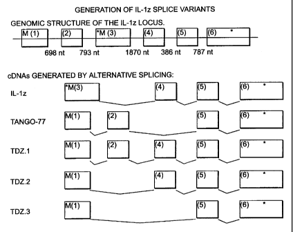
Figure 1 shows the genomic structure of the IL-1 zeta locus and the cDNAs
generated
by alternative splicing. The numbered boxes indicate individual exons 1-6 and
the
approximate size of the intervening introns is indicated at the top. The
asterisk (*)
indicates the presence of a stop codon, at the end of the coding sequence
(exon 6) or
as an in-frame stop codon (exon 3). "M" indicates a potential initiating
methionine
CA 02353483 2001-06-04
WO 00/36108 PCT/US99/29549
originating either from exon 1 or exon 3. Tango-77 is the cDNA structure
disclosed
in WO 99/06426. A significant feature of IL-1 zeta and its splice variants is
the
presence or the absence of exon 4. Exon 4 is present in IL-1 zeta, TDZ.1 and
TDZ.2
but not in Tango-77 or TDZ.3. The amino acid sequence encoded by exon 4 aligns
well with the amino acid sequences of other IL-1 family members in the first
few beta
strands of the mature peptides. By contrast, the amino acid sequences encoded
by
exons 1 and 2 of Tango-77 and exon 1 of TDZ.3 cDNAs, which replace rather than
supplement exon 4 of IL-1 zeta, TDZ.1 and TDZ.2, do not align well with other
IL-1
family members in this same region. IL-1 zeta, Tango-77, TDZ. 1, TDZ.2, and
TDZ.3
all align well with amino acid sequences of other IL-1 family members in the C-
terminal 2/3 of the mature peptide (the region encoded by exons 5 and 6 which
are
common to all of these splice isoforms). Thus, the "mature peptides" encoded
by IL 1
zeta, TDZ.1 and TDZ.2 DNAs are likely to represent functional IL-1 like
molecules.
This contrasts with the polypeptides encoded by Tango-77 or TDZ.3 DNAs which
are
less likely to represent a functional IL-1 like molecule.
It is probable that all of the splice isoforms, except TDZ.3, encode proforms
of
an IL-1 like cytokine, since in the N-terminal direction the cDNAs extend well
beyond the N-terminus of mature IL-ls. This observation predicts that IL-1
zeta,
TDZ.1 and TDZ.2 encode the same mature peptide. In connection with this
observation it is the prodomains (as well as 5' UTRs) that differ between IL-1
zeta,
TDZ.1 and TDZ.2.
Table III, which details the tissue distribution of IL-1 zeta, TDZ.1, TDZ.2,
TDZ.3 and Tango-77, shows that the expression of Tango-77 is more widespread
than
that of IL-I zeta. Table III also shows that the TDZ.1 expression is
comparable, and
almost entirely overlapping, with that of Tango-77. The tissue distribution
data
combined with the alignment information of Figure 1 show that TDZ. I is the
only
member of the splice variants that aligns well with other IL-1 family members,
and is
widespread in its expression. These observations suggest that TDZ.1 may be the
most
significant of the splice variants in terms of relevance to biological
responses.
16
CA 02353483 2001-06-04
WO 00/36108 PCT/US99/29549
Additional Sequences
Due to the known degeneracy of the genetic code, wherein more than one
codon can encode the same amino acid, a DNA sequence can vary from that shown
in
SEQ ID NOs:1, 2, 5, 6, and 7 and still encode a polypeptide having the amino
acid
sequence of SEQ ID NOs:3, 4, 8, 9, and 10, respectively. Such variant DNA
sequences can result from inadvertent mutations (e.g., occurring during PCR
amplification), or can be the product of deliberate mutagenesis of a native
sequence.
The invention thus provides isolated DNA sequences encoding polypeptides of
the invention, selected from: (a) DNA comprising the nucleotide sequences of
SEQ ID
NOs: 1, 2, 5, 6, and 7 (b) DNA encoding the polypeptides of SEQ ID NOs:3, 4,
8, 9,
and 10 (c) DNA capable of hybridization to a DNA of (a) or (b) under
conditions of
moderate stringency and which encodes polypeptides of the invention; (d) DNA
capable of hybridization to a DNA of (a) or (b) under conditions of high
stringency
and which encodes polypeptides of the invention, and (e) DNA which is
degenerate,
as a result of the genetic code, to a DNA defined in (a), (b), (c), or (d) and
which
encode polypeptides of the invention. Of course, polypeptides encoded by such
DNA
sequences are encompassed by the invention.
As used herein, conditions of moderate stringency can be readily determined
by those having ordinary skill in the art based on, for example, the length of
the DNA.
The basic conditions are set forth by Sambrook et al., Molecular Cloning: A
Laboratorv Manual, 2nd ed. Vol. 1, pp. 1.101-104, Cold Spring Harbor
Laboratory
Press, 1989, and include use of a prewashing solution for the nitrocellulose
filters 5X
SSC, 0.5% SDS, 1.0 mM EDTA (pH 8.0), hybridization conditions of about 50%
formamide, 6X SSC at about 42 C (or other similar hybridization solution, such
as
Stark's solution, in about 50% formamide at about 42'C), and washing
conditions of
about 60 C, 0.5X SSC, 0.1% SDS. Conditions of high stringency can also be
readily
determined by the skilled artisan based on, for example, the length of the
DNA.
Generally, such conditions are defined as hybridization conditions as above,
and with
washing at approximately 68 C, 0.2X SSC, 0.1% SDS. The skilled artisan will
recognize that the temperature and wash solution salt concentration can be
adjusted as
necessary according to factors such as the length of the probe.
17
CA 02353483 2001-06-04
WO 00/36108 PCT/US99/29549
Also included as an embodiment of the invention is DNA encoding
polypeptide fragments and polypeptides comprising inactivated N-glycosylation
site(s), inactivated protease processing site(s), or conservative amino acid
substitution(s), as described below.
In another embodiment, the nucleic acid molecules of the invention also
comprise nucleotide sequences that are at least 80% identical to a native
sequence.
Also contemplated are embodiments in which a nucleic acid molecule
comprises a sequence that is at least 90% identical, at least 95% identical,
at least 98%
identical, at least 99% identical, or at least 99.9% identical to a native
sequence.
The percent identity may be determined by visual inspection and mathematical
calculation. Alternatively, the percent identity of two nucleic acid sequences
can be
determined by comparing sequence information using the GAP computer program,
version 6.0 described by Devereux et al., Nucl. Acids Res. 12:387, 1984, and
available
from the University of Wisconsin Genetics Computer Group (UWGCG). The
preferred default parameters for the GAP program include: (1) a unary
comparison
matrix (containing a value of 1 for identities and 0 for non-identities) for
nucleotides,
and the weighted comparison matrix of Gribskov and Burgess, Nucl. Acids Res.
14:6745, 1986, as described by Schwartz and Dayhoff, eds., Atlas of Protein
Sequence
and Structure, pp. 353-358, National Biomedical Research Foundation, 1979; (2)
a
penalty of 3.0 for each gap and an additiona10.10 penalty for each symbol in
each
gap; and (3) no penalty for end gaps. Other programs used by one skilled in
the art of
sequence comparison may also be used.
The invention provides isolated nucleic acids useful in the production of
polypeptides. Such polypeptides may be prepared by any of a number of
conventional
techniques. A DNA sequence encoding a polypeptide of the invention, or desired
fragment thereof may be subcloned into an expression vector for production of
the
polypeptide or fragment. The DNA sequence advantageously is fused to a
sequence
encoding a suitable leader or signal peptide. Alternatively, the desired
fragment may
be chemically synthesized using known techniques. DNA fragments also may be
produced by restriction endonuclease digestion of a full length cloned DNA
sequence,
and isolated by electrophoresis on agarose gels. If necessary,
oligonucleotides that
18
CA 02353483 2001-06-04
WO 00/36108 PCT/US99/29549
reconstruct the 5' or 3' terminus to a desired point may be ligated to a DNA
fragment
generated by restriction enzyme digestion. Such oligonucleotides may
additionally
contain a restriction endonuclease cleavage site upstream of the desired
coding
sequence, and position an initiation codon (ATG) at the N-terminus of the
coding
sequence.
The well-known polymerase chain reaction (PCR) procedure also may be
employed to isolate and amplify a DNA sequence encoding a desired protein
fragment. Oligonucleotides that define the desired termini of the DNA fragment
are
employed as 5' and 3' primers. The oligonucleotides may additionally contain
recognition sites for restriction endonucleases, to facilitate insertion of
the amplified
DNA fragment into an expression vector. PCR techniques are described in Saiki
et
al., Science, 239:487, 1988; Wu et al., eds., Recombinant DNA Methodology, pp.
189-196, Academic Press, Inc., San Diego, 1989; and Innis et al., eds., PCR
Protocols: A Guide to Methods and Applications, Academic Press, Inc., 1990.
POLYPEPTIDES AND FRAGMENTS THEREOF
The invention encompasses polypeptides and fragments thereof in various
forms, including those that are naturally occurring or produced through
various
techniques such as procedures involving recombinant DNA technology. Such forms
include, but are not limited to, derivatives, variants, and oligomers, as well
as fusion
proteins or fragments thereof.
The polypeptides of the invention include full length proteins encoded by the
nucleic acid sequences set forth above. Particularly preferred polypeptides of
IL-1
zeta, TDZ. 1, TDZ.2 TDZ.3 and Xrec2 comprise the amino acid sequence of SEQ ID
NOs:3, 4, 8, 9, and 10 respectively. For IL-1 zeta, TDZ.1, TDZ.2, and TDZ-3
the N-
terminus does not encode a classical signal peptide but the extra length
relative to the
mature form of other IL-1 family members suggests that the N-terminus may act
as a
prodomain. A predicted cleavage site is the point where the conserved
structural
portion of the protein begins. Structural modeling data support this
assumption. For
IL-1 zeta, TDZ.1, and TDZ.2 the site is somewhere near the N-terminal end of
the
amino acid sequence encoded by exon 4. Thus, the polypeptide of IL-1 zeta, as
set
19
CA 02353483 2001-06-04
WO 00/36108 PCT/US99/29549
forth in SEQ ID NO:3, includes a putative prodomain that extends from amino
acids 1
to x, where x is an integer from 20 to 50. Similarly, TDZ.1 of SEQ ID NO:8
includes
a putative prodomain that extends from amino acids 1 to x' where x' is an
integer
from 40-60 and most preferably x' is about 52. TDZ.2 of SEQ ID NO:9 includes a
putative prodomain that extends from amino acids 1 to x", where x" is an
integer from
20-40 and most preferable x" is 31.
Unlike IL-1 zeta and its splice variants, the polypeptide of Xrec2, as set
forth
in SEQ ID NO:4, includes an N-terminal hydrophobic region that functions as a
signal
peptide, followed by an extracellular domain comprising amino acids 19 to 359,
a
transmembrane region comprising amino acids 360 through 378, and a C-terminal
cytoplasmic domain comprising amino acids 379 to 696. Computer analysis
predicts
that the signal peptide corresponds to residues 1 to 19 of SEQ ID NO:4
(although the
next most likely computer-predicted signal peptide cleavage sites, in
descending
order, occur after amino acids 20 and 16 of SEQ ID NO:4). Cleavage of the
signal
peptide thus would yield a mature protein comprising amino acids 19 through
696 of
SEQ ID NO:4.
The skilled artisan will recognize that the above-described boundaries of such
regions of the polypeptide are approximate. To illustrate, the boundaries of
the
transmembrane region (which may be predicted by using computer programs
available for that purpose) may differ from those described above.
The polypeptides of the invention may be membrane bound or they may be
secreted and, thus, soluble. Soluble polypeptides are capable of being
secreted from
the cells in which they are expressed. In general, soluble polypeptides may be
identified (and distinguished from non-soluble membrane-bound counterparts) by
separating intact cells which express the desired polypeptide from the culture
medium,
e.g., by centrifugation, and assaying the medium (supernatant) for the
presence of the
desired polypeptide. The presence of polypeptide in the medium indicates that
the
polypeptide was secreted from the cells and thus is a soluble form of the
protein.
In one embodiment, the soluble polypeptides and fragments thereof comprise
all or part of the extracellular domain, but lack the transmembrane region
that would
cause retention of the polypeptide on a cell membrane. A soluble polypeptide
may
CA 02353483 2001-06-04
WO 00/36108 PCTIUS99/29549
include the cytoplasmic domain, or a portion thereof, as long as the
polypeptide is
secreted from the cell in which it is produced.
In general, the use of soluble forms is advantageous for certain applications.
Purification of the polypeptides from recombinant host cells is facilitated,
since the
soluble polypeptides are secreted from the cells. Further, soluble
polypeptides are
generally more suitable for intravenous administration.
The invention also provides polypeptides and fragments of the extracellular
domain that retain a desired biological activity. Particular embodiments are
directed
to polypeptide fragments of SEQ ID NOs:3, 4, 8, 9, and 10 that retain the
ability to
bind the native cognates, substrates, or counter-structure ("binding
partner"). Such a
fragment may be a soluble polypeptide, as described above. In another
embodiment,
the polypeptides and fragments advantageously include regions that are
conserved in
the IL-1 ligand and IL-1 receptor family as described above.
Also provided herein are polypeptide fragments comprising at least 20, or at
least 30, contiguous amino acids of the sequences of SEQ ID NOs:3, 4, 8, 9,
and 10.
In one aspect, fragments derived from the cytoplasmic domain of Xrec2 of SEQ
ID
NO:4 find use in studies of signal transduction, and in regulating cellular
processes
associated with transduction of biological signals. Polypeptide fragments also
may be
employed as immunogens, in generating antibodies.
Variants
Naturally occurring variants as well as derived variants of the polypeptides
and
fragments are provided herein.
Variants may exhibit amino acid sequences that are at least 80% identical.
Also contemplated are embodiments in which a polypeptide or fragment comprises
an
amino acid sequence that is at least 90% identical, at least 95% identical, at
least 98%
identical, at least 99% identical, or at least 99.9% identical to the
preferred
polypeptide or fragment thereof. Percent identity may be determined by visual
inspection and mathematical calculation. Alternatively, the percent identity
of two
protein sequences can be determined by comparing sequence information using
the
GAP computer program, based on the algorithm of Needleman and Wunsch (J. Mol.
21
CA 02353483 2001-06-04
WO 00/36108 PCT/US99/29549
Bio. 48:443, 1970) and available from the University of Wisconsin Genetics
Computer Group (UWGCG). The preferred default parameters for the GAP program
include: (1) a scoring matrix, blosum62, as described by Henikoff et al.,
Proc. Natl.
Acad. Sci. USA, 89:10915, 1992; (2) a gap weight of 12; (3) a gap length
weight of
4; and (4) no penalty for end gaps. Other programs used by one skilled in the
art of
sequence comparison may also be used.
The variants of the invention include, for example, those that result from
alternate mRNA splicing events or from proteolytic cleavage. Alternate
splicing of
mRNA may, for example, yield a ttuncated but biologically active protein, such
as a
naturally occurring soluble form of the protein. Variations attributable to
proteolysis
include, for example, differences in the N- or C-termini upon expression in
different
types of host cells, due to proteolytic removal of one or more tenminal amino
acids
from the protein (generally from 1-5 terminal amino acids). Proteins in which
differences in amino acid sequence are attributable to genetic polymorphism
(allelic
variation among individuals producing the protein) are also contemplated
herein.
Additional variants within the scope of the invention include polypeptides
that
may be modified to create derivatives thereof by forming covalent or
aggregative
conjugates with other chemical moieties, such as glycosyl groups, lipids,
phosphate,
acetyl groups and the like. Covalent derivatives may be prepared by linking
the
chemical moieties to functional groups on amino acid side chains or at the N-
terminus
or C-terminus of a polypeptide. Conjugates comprising diagnostic (detectable)
or
therapeutic agents attached thereto are contemplated herein, as discussed in
more
detail below.
Other derivatives include covalent or aggregative conjugates of the
polypeptides with other proteins or polypeptides, such as by synthesis in
recombinant
culture as N-terminal or C-terminal fusions. Examples of fusion proteins are
discussed below in connection with oligomers. Further, fusion proteins can
comprise
peptides added to facilitate purification and identification. Such peptides
include, for
example, poly-His or the antigenic identification peptides described in U.S.
Patent No.
5,011,912 and in Hopp et al., Bio/Technology, 6:1204, 1988. One such peptide
is the
FLAG peptide, Asp-Tyr-Lys-Asp-Asp-Asp-Asp-Lys (SEQ ID NO:11), which is
22
CA 02353483 2007-12-06
72249-113
highly antigenic and provides an epitope reversibly bound by a specific
monoclonal
antibody, enabling rapid assay and facile purification of expressed
recombinant
protein. A murine hybridoma designated 4E11 produces a monoclonal antibody
that
binds the FLAG peptide in the presence of certain divalent metal cations, as
described in U.S. Patent 5,011,912. The 4E11 hybridoma cell line
has been deposited with the American Type Culture Collection
under accession no. HB 9259. Monoclonal antibodies that bind the FLAG p.;ptide
are available from Eastman Kodak Co., Scientific Imaging Systems Division, New
Haven, Connecticut.
Among the variant polypeptides provided herein are variants of native
polypeptides that retain the native biological activity or the substantial
equivalent
thereof. One example is a variant that binds with essentially the same binding
affinity
as does the native form. Binding affinity can be measured by conventional
procedures, e.g., as described in U.S. Patent No. 5,512,457 and as set forth
below.
Variants include polypeptides that are substantially homologous to the native
form, but which have an amino acid sequence different from that of the native
form
because of one or more deletions, insertions or substitutions. Particular
embodiments
include, but are not limited to, polypeptides that comprise from one to ten
deletions,
insertions or substitutions of amino acid residues, when compared to a native
sequence.
A given amino acid may be replaced, for example, by a residue having similar
physiochemical characteristics. Examples of such conservative substitutions
include
substitution of one aliphatic residue for another, such as Ile, Val, Leu, or
Ala for one
another; substitutions of one polar residue for another, such as between Lys
and Arg,
Glu and Asp, or Gln and Asn; or substitutions of one aromatic residue for
another,
such as Phe, Trp, or Tyr for one another. Other conservative substitutions,
e.g.,
involving substitutions of entire regions having similar hydrophobicity
characteristics,
are well known.
Similarly, the DNAs of the invention include variants that differ from a
native
DNA sequence because of one or more deletions, insertions or substitutions,
but that
encode a biologically active polypeptide.
23
CA 02353483 2001-06-04
WO 00/36108 PCT/US99/29549
The invention further includes polypeptides of the invention with or without
associated native-pattern glycosylation. Polypeptides expressed in yeast or
mammalian expression systems (e.g., COS- 1 or COS-7 cells) can be similar to
or
significantly different from a native polypeptide in molecular weight and
glycosylation pattern, depending upon the choice of expression system.
Expression of
polypeptides of the invention in bacterial expression systems, such as E.
coli, provides
non-glycosylated molecules. Further, a given preparation may include multiple
differentially glycosylated species of the protein. Glycosyl groups can be
removed
through conventional methods, in particular those utilizing glycopeptidase. In
general, glycosylated polypeptides of the invention can be incubated with a
molar
excess of glycopeptidase (Boehringer Mannheim).
Correspondingly, similar DNA constructs that encode various additions or
substitutions of amino acid residues or sequences, or deletions of terminal or
internal
residues or sequences are encompassed by the invention. For example, N-
glycosylation sites in the polypeptide extracellular domain can be modified to
preclude glycosylation, allowing expression of a reduced carbohydrate analog
in
mammalian and yeast expression systems. N-glycosylation sites in eukaryotic
polypeptides are characterized by an amino acid triplet Asn-X-Y, wherein X is
any
amino acid except Pro and Y is Ser or Thr. Appropriate substitutions,
additions, or
deletions to the nucleotide sequence encoding these triplets will result in
prevention of
attachment of carbohydrate residues at the Asn side chain. Alteration of a
single
nucleotide, chosen so that Asn is replaced by a different amino acid, for
example, is
sufficient to inactivate an N-glycosylation site. Alternatively, the Ser or
Thr can by
replaced with another amino acid, such as Ala. Known procedures for
inactivating N-
glycosylation sites in proteins include those described in U.S. Patent
5,071,972 and
EP 276,846, hereby incorporated by reference.
In another example of variants, sequences encoding Cys residues that are not
essential for biological activity can be altered to cause the Cys residues to
be deleted
or replaced with other amino acids, preventing formation of incorrect
intramolecular
disulfide bridges upon folding or renaturation.
Other variants are prepared by modification of adjacent dibasic amino acid
24
CA 02353483 2001-06-04
WO 00/36108 PCT/US99/29549
residues, to enhance expression in yeast systems in which KEX2 protease
activity is
present. EP 212,914 discloses the use of site-specific mutagenesis to
inactivate KEX2
protease processing sites in a protein. KEX2 protease processing sites are
inactivated
by deleting, adding or substituting residues to alter Arg-Arg, Arg-Lys, and
Lys-Arg
pairs to eliminate the occurrence of these adjacent basic residues. Lys-Lys
pairings
are considerably less susceptible to KEX2 cleavage, and conversion of Arg-Lys
or
Lys-Arg to Lys-Lys represents a conservative and preferred approach to
inactivating
KEX2 sites.
Oli og mers
Encompassed by the invention are oligomers or fusion proteins that contain
IL-1 zeta, TDZ.I, TDZ.2, TDZ.3 or Xrec2 polypeptides. When the polypeptide of
the
invention is a type I membrane protein, such as Xrec2, the fusion partner is
linked to
the C-tenninus of the type I membrane protein. Such oligomers may be in the
form of
covalently-linked or non-covalently-linked multimers, including dimers,
trimers, or
higher oligomers. As noted above, preferred polypeptides are soluble and thus
these
oligomers may comprise soluble polypeptides. In one aspect of the invention,
the
oligomers maintain the binding ability of the polypeptide components and
provide
therefor, bivalent, trivalent, etc., binding sites.
One embodiment of the invention is directed to oligomers comprising multiple
polypeptides joined via covalent or non-covalent interactions between peptide
moieties fused to the polypeptides. Such peptides may be peptide linkers
(spacers), or
peptides that have the property of promoting oligomerization. Leucine zippers
and
certain polypeptides derived from antibodies are among the peptides that can
promote
oligomerization of the polypeptides attached thereto, as described in more
detail
below.
Immunoglobulin-based Oli omers
As one alternative, an oligomer is prepared using polypeptides derived from
immunoglobulins. Preparation of fusion proteins comprising certain
heterologous
polypeptides fused to various portions of antibody-derived polypeptides
(including the
CA 02353483 2007-12-06
72249-113
Fc domain) has been described, e.g., by Ashkenazi et al., PNAS USA 88:10535,
1991;
Bym et al., Nature 344:677, 1990; and Hollenbaugh and Aruffo, "Construction of
Immunoglobutin Fusion Proteins," in Current Protocols in Immunology, Suppl. 4,
pp.
10.19.1-10.19.11,1992.
One embodiment of the present invention is directed to a dimer comprising
two fusion proteins created by fusing a polypeptide of the invention to an Fc
polypeptide derived from an antibody. A gene fusion encoding the
polypeptide/Fc
fusion protein is inserted into an appropriate expression vector.
Polypeptide/Fc fusion
proteins are expressed in host cells transformed with the recombinant
expression
vector, and allowed to assemble much like antibody molecules, whereupon
interchain
disulfide bonds form between the Fc moieties to yield divalent molecules.
The term "Fc polypeptide" as used herein includes native and mutein forms of
polypeptides made up of the Fc region of an antibody comprising any or all of
the CH
domains of the Fc region. Truncated forms of such polypeptides containing the
hinge
region that promotes dimerization are also included. Preferred polypeptides
comprise
an Fc polypeptide derived from a human IgG1 antibody.
One suitable Fc polypeptide, described in PCT application WO 93/10151,
is a single chain polypeptide extending from the N-terminal hinge
region to the native C-terminus of the Fc region of a human IgGI
antibody. Another useful Fc polypeptide is the Fc mutein described in U.S.
Patent
5,457,035 and in Baum et al., EMBO J. 13:3992-4001, 1994.
The amino acid sequence of this mutein is identical to that of the native Fc
sequence presented in WO 93/10151, except that amino acid 19 has been changed
from Leu to Ala, amino acid 20 has been changed from Leu to Glu, and amino
acid 22
has been changed from Gly to Ala. The mutein exhibits reduced affinity for Fc
receptors.
The above-described fusion proteins comprising Fc moieties (and oligomers
formed therefrom) offer the advantage of facile purification by affinity
chromatography over Protein A or Protein G columns.
In other embodiments, the polypeptides of the invention may be substituted for
the variable portion of an antibody heavy or light chain. If fusion proteins
are made
26
CA 02353483 2007-12-06
72249-113
with both heavy and light chains of an antibody, it is possible to form an
oligomer
with as many as four polypeptide extracellular regions.
Peptide-linker Based Oligomers
Alternatively, the oligomer is a fusion protein comprising multiple
polypeptides, with or without peptide linkers (spacer peptides). Among the
suitable
peptide linkers are those described in U.S. Patents 4,751,180 and 4,935,233.
A DNA sequence encoding a desired peptide linker
may be inserted between, and in the same reading frame as, the DNA sequences
of the
invention, using any suitable conventional technique. For example, a
chemically
synthesized oligonucleotide encoding the linker may be ligated between the
sequences. In particular embodiments, a fusion protein comprises from two to
four
soluble polypeptides of the invention, separated by peptide linkers.
Leucine-Zippers
Another method for preparing the oligomers of the invention involves use of a
leucine zipper. Leucine zipper domains are peptides that promote
oligomerization of
the proteins in which they are found. Leucine zippers were originally
identified in
several DNA-binding proteins (Landschulz et al., Science 240:1759, 1988), and
have
since been found in a variety of different proteins. Among the known leucine
zippers
are naturally occurring peptides and derivatives thereof that dimerize or
trimerize.
The zipper domain (also referred to herein as an oligomerizing, or oligomer-
forming, domain) comprises a repetitive heptad repeat, often with four or five
leucine
residues interspersed with other amino acids. Examples of zipper domains are
those
found in the yeast transcription factor GCN4 and a heat-stable DNA-binding
protein
found in rat liver (C/EBP; Landschulz et al., Science, 243:1681, 1989). Two
nuclear
transforming proteins,fos andjun, also exhibit zipper domains, as does the
gene
product of the murine proto-oncogene, c-myc (Landschulz et al., Science,
240:1759,
1988). The products of the nuclear oncogenesfos and jun comprise zipper
domains
that preferentially form heterodimers (O'Shea et al., Science, 245:646, 1989;
Turner et
27
CA 02353483 2001-06-04
WO 00/36108 PCT/US99/29549
al., Science, 243:1689, 1989). The zipper domain is necessary for biological
activity
(DNA binding) in these proteins.
The fusogenic proteins of several different viruses, including paramyxovirus,
coronavirus, measles virus and many retroviruses, also possess zipper domains
(Buckland et al., Nature, 338:547,1989; Britton, Nature, 353:394, 1991;
Delwart and
Mosialos, AIDS Research and Human Retroviruses, 6:703, 1990). The zipper
domains in these fusogenic viral proteins are near the transmembrane region of
the
proteins; it has been suggested that the zipper domains could contribute to
the
oligomeric structure of the fusogenic proteins. Oligomerization of fusogenic
viral
proteins is involved in fusion pore formation (Spruce et al, Proc. Natl. Acad.
Sci.
U.S.A., 88:3523, 1991). Zipper domains have also been recently reported to
play a role
in oligomerization of heat-shock transcription factors (Rabindran et al.,
Science,
259:230, 1993).
Zipper domains fold as short, parallel coiled coils. (O'Shea et al., Science,
254:539, 1991) The general architecture of the parallel coiled coil has been
well
characterized, with a "knobs-into-holes" packing as proposed by Crick in 1953
(Crick,
Acta Crystallogr. 6:689, 1953). The dimer formed by a zipper domain is
stabilized by
the heptad repeat, designated (abcdefg)õ according to the notation of
McLachlan and
Stewart, J. Mol. Biol., 98:293, 1975, in which residues a and d are generally
hydrophobic residues, with d being a leucine, which line up on the same face
of a
helix. Oppositely-charged residues commonly occur at positions g and e. Thus,
in a
parallel coiled coil formed from two helical zipper domains, the "knobs"
formed by
the hydrophobic side chains of the first helix are packed into the "holes"
formed
between the side chains of the second helix.
The residues at position d (often leucine) contribute large hydrophobic
stabilization energies, and are important for oligomer formation (Krystek et
al., Int. J.
Peptide Res., 38:229, 1991). Lovejoy et al., Science, 259:1288, 1993, recently
reported the synthesis of a triple-stranded a-helical bundle in which the
helices run
up-up-down. Their studies confirmed that hydrophobic stabilization energy
provides
the main driving force for the formation of coiled coils from helical
monomers. These
studies also indicate that electrostatic interactions contribute to the
stoichiometry and
28
CA 02353483 2007-12-06
72249-113
geometry of coiled coils. Further discussion of the structure of leucine
zippers is
found in Harbury et al., Science, 262:1401, 1993.
Examples of leucine zipper domains suitable for producing soluble oligomeric
proteins are described in PCT application WO 94/10308, and the leucine zipper
derived from lung surfactant protein D(S1'D) described in Hoppe et al., FEBS
Letters,
344:191, 1994. The use of a modified leucine
zipper that allows for stable trimerization of a heterologous protein fused
thereto is
described in Fanslow et al., Semin. Immunol., 6:267-278, 1994. Recombinant
fusion
proteins comprising a soluble polypeptide fused to a leucine zipper peptide
are
expressed in suitable host cells, and the soluble oligomer that forms is
recovered from
the culture supernatant.
Certain leucine zipper moieties preferentially form trimers. One example is a
leucine zipper derived from lung surfactant protein D (SPD), as described in
Hoppe et
al., FEBS Letters, 344:191, 1994, and in U.S. Patent 5,716,805.
This lung SPD-derived leucine zipper peptide
comprises the amino acid sequence Pro Asp Val Ala Ser Leu Arg Gln Gln Val Glu
Ala Leu Gln Gly Gin Val Gin His Leu Gin Ala Ala Phe Ser Gln Tyr (SEQ ID NO:
12).
Another example of a leucine zipper that promotes trimerization is a peptide
comprising the amino acid sequence Arg Met Lys Gln Ile Glu Asp Lys Ile Glu Glu
Ile
Leu Ser Lys Ile Tyr His Ile Glu Asn Glu Ile Ala Arg Ile Lys Lys Leu Ile Gly
Glu Arg
(SEQ ID NO:13), as described in U.S. Patent 5,716,805. In one altemative
embodiment, an N-terminal Asp residue is added; in another, the peptide lacks
the N-
tenninal Arg residue.
Fragments of the foregoing zipper peptides that retain the property of
promoting oligomerization may be employed as well. Examples of such fragments
include, but are not limited to, peptides lacking one or two of the N-terminal
or C-
terminal residues presented in the foregoing amino acid sequences. Leucine
zippers
may be derived from naturally occurring leucine zipper peptides, e.g., via
conservative
substitution(s) in the native amino acid sequence, wherein the peptide's
ability to
promote oligomerization is retained.
29
CA 02353483 2001-06-04
WO 00/36108 PCT/US99/29549
Other peptides derived from naturally occurring trimeric proteins may be
employed in preparing trimeric oligomers. Alternatively, synthetic peptides
that
promote oligomerization may be employed. In particular embodiments, leucine
residues in a leucine zipper moiety are replaced by isoleucine residues. Such
peptides comprising isoleucine may be referred to as isoleucine zippers, but
are
encompassed by the term "leucine zippers" as employed herein.
PRODUCTION OF POLYPEPTIDES AND FRAGMENTS THEREOF
Expression, isolation and purification of the polypeptides and fragments of
the
invention may be accomplished by any suitable technique, including but not
limited to
the following:
Expression Systems
The present invention also provides recombinant cloning and expression
vectors containing DNA, as well as host cell containing the recombinant
vectors.
Expression vectors comprising DNA may be used to prepare the polypeptides or
fragments of the invention encoded by the DNA. A method for producing
polypeptides comprises culturing host cells transformed with a recombinant
expression vector encoding the polypeptide, under conditions that promote
expression
of the polypeptide, then recovering the expressed polypeptides from the
culture. The
skilled artisan will recognize that the procedure for purifying the expressed
polypeptides will vary according to such factors as the type of host cells
employed,
and whether the polypeptide is membrane-bound or a soluble form that is
secreted
from the host cell.
Any suitable expression system may be employed. The vectors include a
DNA encoding a polypeptide or fragment of the invention, operably linked to
suitable
transcriptional or translational regulatory nucleotide sequences, such as
those derived
from a mammalian, microbial, viral, or insect gene. Examples of regulatory
sequences include transcriptional promoters, operators, or enhancers, an mRNA
ribosomal binding site, and appropriate sequences which control transcription
and
translation initiation and termination. Nucleotide sequences are operably
linked when
CA 02353483 2001-06-04
WO 00/36108 PCT/US99/29549
the regulatory sequence functionally relates to the DNA sequence. Thus, a
promoter
nucleotide sequence is operably linked to a DNA sequence if the promoter
nucleotide
sequence controls the transcription of the DNA sequence. An origin of
replication
that confers the ability to replicate in the desired host cells, and a
selection gene by
which transformants are identified, are generally incorporated into the
expression
vector.
In addition, a sequence encoding an appropriate signal peptide (native or
heterologous) can be incorporated into expression vectors. A DNA sequence for
a
signal peptide (secretory leader) may be fused in frame to the nucleic acid
sequence of
the invention so that the DNA is initially transcribed, and the mRNA
translated, into a
fusion protein comprising the signal peptide. A signal peptide that is
functional in the
intended host cells promotes extracellular secretion of the polypeptide. The
signal
peptide is cleaved from the polypeptide upon secretion of polypeptide from the
cell.
The skilled artisan will also recognize that the position(s) at which the
signal
peptide is cleaved may differ from that predicted by computer program, and may
vary
according to such factors as the type of host cells employed in expressing a
recombinant polypeptide. A protein preparation may include a mixture of
protein
molecules having different N-terminal amino acids, resulting from cleavage of
the
signal peptide at more than one site. Particular embodiments of mature
proteins
provided herein include, but are not limited to, proteins having the residue
at position
6, 23, 25, 26, 39, 41, or 48 of SEQ ID NO:3 and at position 1 or 19 of SEQ ID
NO:4
as the N-terminal amino acid.
Suitable host cells for expression of polypeptides include prokaryotes, yeast
or
higher eukaryotic cells. Mammalian or insect cells are generally preferred for
use as
host cells. Appropriate cloning and expression vectors for use with bacterial,
fungal,
yeast, and mammalian cellular hosts are described, for example, in Pouwels et
al.,
Cloning Vectors: A Laboratory Manual. Elsevier, New York, 1985. Cell-free
translation systems could also be employed to produce polypeptides using RNAs
derived from DNA constructs disclosed herein.
31
CA 02353483 2001-06-04
WO 00/36108 PCT/US99/29549
ProkMotic Systems
Prokaryotes include gram-negative or gram-positive organisms. Suitable
prokaryotic host cells for transformation include, for example, E. colf,
Bacillus
subtilis, Salmonella typhimurium, and various other species within the genera
Pseudomonas, Streptomyces, and Staphylococcus. In a prokaryotic host cell,
such as
E. coli, a polypeptide may include an N-terminal methionine residue to
facilitate
expression of the recombinant polypeptide in the prokaryotic host cell. The N-
terminal Met may be cleaved from the expressed recombinant polypeptide.
Expression vectors for use in prokaryotic host cells generally comprise one or
more phenotypic selectable marker genes. A phenotypic selectable marker gene
is, for
example, a gene encoding a protein that confers antibiotic resistance or that
supplies an
autotrophic requirement. Examples of useful expression vectors for prokaryotic
host
cells include those derived from commercially available plasmids such as the
cloning
vector pBR322 (ATCC 37017). pBR322 contains genes for ampicillin and
tetracycline
resistance and thus provides simple means for identifying transformed cells.
An
appropriate promoter and a DNA sequence are inserted into the pBR322 vector.
Other
commercially available vectors include, for example, pKK223-3 (Pharmacia Fine
Chemicals, Uppsala, Sweden) and pGEM 1(Promega Biotec, Madison, WI, USA).
Promoter sequences commonly used for recombinant prokaryotic host cell
expression vectors include P-lactamase (penicillinase), lactose promoter
system
(Chang et al., Nature, 275:615, 1978; and Goeddel et al., Nature, 281:544,
1979),
tryptophan (trp) promoter system (Goeddel et al., Nucl. Acids Res., 8:4057,
1980; and
EP-A-36776) and tac promoter (Maniatis, Molecular Cloning: A Laboratory
Manual,
p. 412, Cold Spring Harbor Laboratory, 1982). A particularly useful
prokaryotic host
cell expression system employs a phage XPL promoter and a c1857ts thermolabile
repressor sequence. Plasmid vectors available from the American Type Culture
Collection which incorporate derivatives of the a.PL promoter include plasmid
pHUB2
(resident in E. coli strain JMB9, ATCC 37092) and pPLc28 (resident in E. coli
RRI,
ATCC 53082).
32
CA 02353483 2001-06-04
WO 00/36108 PCT/US99/29549
Yeast Systems
Alternatively, the polypeptides may be expressed in yeast host cells,
preferably
from the Saccharomyces genus (e.g., S. cerevisiae). Other genera of yeast,
such as
Pichia or Kluyveromyces, may also be employed. Yeast vectors will often
contain an
origin of replication sequence from a 2 yeast plasmid, an autonomously
replicating
sequence (ARS), a promoter region, sequences for polyadenylation, sequences
for
transcription termination, and a selectable marker gene. Suitable promoter
sequences
for yeast vectors include, among others, promoters for metallothionein, 3-
phosphoglycerate kinase (Hitzeman et al., J. Biol. Chem., 255:2073, 1980) or
other
glycolytic enzymes (Hess et al., J. Adv. Enzyme Reg., 7:149, 1968; and Holland
et al.,
Biochem. 17:4900, 1978), such as enolase, glyceraldehyde-3-phosphate
dehydrogenase, hexokinase, pyruvate decarboxylase, phosphofructokinase,
glucose-6-
phosphate isomerase, 3-phosphoglycerate mutase, pyruvate kinase,
triosephosphate
isomerase, phospho-glucose isomerase, and glucokinase. Other suitable vectors
and
promoters for use in yeast expression are further described in Hitzeman, EPA-
73,657.
Another alternative is the glucose-repressible ADH2 promoter described by
Russell et
al., J. Biol. Chem., 258:2674, 1982; and Beier et al., Nature, 300:724, 1982.
Shuttle
vectors replicable in both yeast and E. coli may be constructed by inserting
DNA
sequences from pBR322 for selection and replication in E. coli (Amp' gene and
origin
of replication) into the above-described yeast vectors.
The yeast a-factor leader sequence may be employed to direct secretion of the
polypeptide. The a-factor leader sequence is often inserted between the
promoter
sequence and the structural gene sequence. (Kurjan et al., Cell, 30:933, 1982;
and
Bitter et al., Proc. Natl. Acad. Sci. USA, 81:5330, 1984.) Other leader
sequences
suitable for facilitating secretion of recombinant polypeptides from yeast
hosts are
known to those of skill in the art. A leader sequence may be modified near its
3' end
to contain one or more restriction sites. This will facilitate fusion of the
leader
sequence to the structural gene.
Yeast transformation protocols are known to those of skill in the art. One
such
protocol is described by Hinnen et al., Proc. Natl. Acad. Scf. USA, 75:1929,
1978.
The Hinnen et al. protocol selects for Trp+ transformants in a selective
medium,
33
CA 02353483 2001-06-04
WO 00/36108 PCT/US99/29549
wherein the selective medium consists of 0.67% yeast nitrogen base, 0.5%
casamino
acids, 2% glucose, 10 mg/ml adenine and 20 mg/mi uracil.
Yeast host cells transformed by vectors containing an ADH2 promoter
sequence may be grown for inducing expression in a "rich" medium. An example
of a
rich medium is one consisting of 1% yeast extract, 2% peptone, and 1% glucose
supplemented with 80 mg/ml adenine and 80 mg/mi uracil. Derepression of the
ADH2 promoter occurs when glucose is exhausted from the medium.
Mammalian or Insect Systems
Mammalian or insect host cell culture systems also may be employed to
express recombinant polypeptides. Bacculovirus systems for production of
heterologous proteins in insect cells are reviewed by Luckow et al.,
BiolTechnology,
6:47, 1988. Established cell lines of mammalian origin also may be employed.
Examples of suitable mammalian host cell lines include the COS-71ine of monkey
kidney cells (ATCC CRL 1651) (Gluzman et al., Cell, 23:175, 1981), L cells, C
127
cells, 3T3 cells (ATCC CCL 163), Chinese hamster ovary (CHO) cells, HeLa
cells,
and BHK (ATCC CRL 10) cell lines, and the CV 1/EBNA cell line derived from the
African green monkey kidney cell line CV 1(ATCC CCL 70) as described by
McMahan et al., EMBOJ., 10: 2821, 1991.
Established methods for introducing DNA into mammalian cells have been
described by Kaufman. R.J., Large Scale Mammalian Cell Culture, pp. 15-69,
1990.
Additional protocols using commercially available reagents, such as
Lipofectamine
lipid reagent (GibcoBRL) or Lipofectamine-Plus lipid reagent, can be used to
transfect cells (Felgner et al., Proc. Natl. Acad. Sci. USA, 84:7413-7417,
1987). In
addition, electroporation can be used to transfect mammalian cells using
conventional
procedures, such as those in Sambrook et al., Molecular Cloning: A Laboratorv
Manual, 2nd ed. Vol. 1-3, Cold Spring Harbor Laboratory Press, 1989. Selection
of
stable transformants can be performed using methods known in the art, such as,
for
example, resistance to cytotoxic drugs. Kaufman et al., Meth. in Enzymology,
185:487-511, 1990, describes several selection schemes, such as dihydrofolate
reductase (DHFR) resistance. A suitable host strain for DHFR selection can be
CHO
34
CA 02353483 2001-06-04
WO 00/36108 PCT/US99/29549
strain DX-B 11, which is deficient in DHFR (Urlaub et al., Proc. Natl. Acad.
Scf. USA,
77:4216-4220, 1980). A plasmid expressing the DHFR cDNA can be introduced into
strain DX-B 11, and only cells that contain the plasmid can grow in the
appropriate
selective media. Other examples of selectable markers that can be incorporated
into
an expression vector include cDNAs conferring resistance to antibiotics, such
as G418
and hygromycin B. Cells harboring the vector can be selected on the basis of
resistance to these compounds.
Transcriptional and translational control sequences for mammalian host cell
expression vectors can be excised from viral genomes. Commonly used promoter
sequences and enhancer sequences are derived from polyoma virus, adenovirus 2,
simian virus 40 (SV40), and human cytomegalovirus. DNA sequences derived from
the SV40 viral genome, for example, SV40 origin, early and late promoter,
enhancer,
splice, and polyadenylation sites can be used to provide other genetic
elements for
expression of a structural gene sequence in a mammalian host cell. Viral early
and
late promoters are particularly useful because both are easily obtained from a
viral
genome as a fragment, which can also contain a viral origin of replication
(Fiers et al.,
Nature, 273:113, 1978; and Kaufman, Meth. in Enzymology, 1990). Smaller or
larger
SV40 fragments can also be used, provided the approximately 250 bp sequence
extending from the Hind III site toward the Bgl I site located in the SV40
viral origin
of replication site is included.
Additional control sequences shown to improve expression of heterologous
genes from mammalian expression vectors include such elements as the
expression
augmenting sequence element (EASE) derived from CHO cells (Morris et al.,
Animal
Cell Technology, pp. 529-534, 1997; and PCT Application WO 97/25420) and the
tripartite leader (TPL) and VA gene RNAs from Adenovirus 2 (Gingeras et al.,
J.
Biol. Chem., 257:13475-13491, 1982). The internal ribosome entry site (IRES)
sequences of viral origin allows dicistronic mRNAs to be translated
efficiently (Oh et
al., Current Opinion in Genetics and Development, 3:295-300, 1993; and Ramesh
et
al., Nucleic Acids Research, 24:2697-2700, 1996). Expression of a heterologous
cDNA as part of a dicistronic mRNA followed by the gene for a selectable
marker
(e.g. DHFR) has been shown to improve transfectability of the host and
expression of
CA 02353483 2007-12-06
72249-113
the heterologous cDNA (Kaufman, Meth. in Enzymology, 1990). Exemplary
expression vectors that employ dicistronic mRNAs are pTR-DC/GFP described by
Mosser et al., Biotechniques, 22:150-161, 1997, and p2A51 described by Morris
et al.,
Animal Cell Technology, pp. 529-534, 1997.
A useful high expression vector, pCAVNOT, has been described by Mosley et
al., Cell, 59:335-348, 1989. Other expression vectors for use in marnrnalian
host cells
can be constructed as disclosed by Okayama et al., (Mol. Cell. Biol., 3:280,
1983. A
useful system for stable high level expression of mammalian cDNAs in C 127
murine
mammary epithelial cells can be constructed substantially as described by
Cosman et
al., Mol. Immunol., 23:935, 1986. A useful high expression vector, PMLSV
NI/N4,
described by Cosman et al., Nature, 312:768, 1984, has been deposited as ATCC
39890. Additional useful mammalian expression vectors are described in
EP-A-0367566, and in WO 91/18982. In yet another alternative,
the vectors can be derived from retroviruses.
Another useful expression vector, pFLAG can be used. FLAG technology is
centered on the fusion of a low molecular weight (lkD), hydrophilic, FLAG
marker
peptide to the N-terminus of a recombinant protein expressed by pFLAG
expression
vectors. pDC311 is another specialized vector used for expressing proteins in
CHO
cells. pDC311 is characterized by a bicistronic sequence containing the gene
of
interest and a dihydrofolate reductase (DHFR) gene with an internal ribosome
binding
site for DHFR translation, an expression augmenting sequence element (EASE),
the
human CMV promoter, a tripartite leader sequence, and a polyadenylation site.
Regarding signal peptides that may be employed, the native signal peptide
may be replaced by a heterologous signal peptide or leader sequence, if
desired. The
choice of signal peptide or leader may depend on factors such as the type of
host cells
in which the recombinant polypeptide is to be produced. To illustrate,
examples of
heterologous signal peptides that are functional in mammalian host cells
include the
signal sequence for interleukin-7 (IL-7) described in United States Patent
4,965,195;
the signal sequence for interleukin-2 receptor described in Cosman et al.,
Nature,
36
CA 02353483 2001-06-04
WO 00/36108 PCTIUS99/29549
312:768, 1984; the interleukin-4 receptor signal peptide described in EP
367,566; the
type I interleukin-1 receptor signal peptide described in U.S. Patent
4,968,607; and the
type II interleukin-1 receptor signal peptide described in EP 460,846.
Purification
The invention also includes methods of isolating and purifying the
polypeptides and fragments thereof.
Isolation and Purification
The "isolated" polypeptides or fragments thereof encompassed by this
invention are polypeptides or fragments that are not in an environment
identical to an
environment in which it or they can be found in nature. The "purified"
polypeptides
or fragments thereof encompassed by this invention are essentially free of
association
with other proteins or polypeptides, for example, as a purification product of
recombinant expression systems such as those described above or as a purified
product from a non-recombinant source such as naturally occurring cells and/or
tissues.
In one preferred embodiment, the purification of recombinant polypeptides or
fragments can be accomplished using fusions of polypeptides or fragments of
the
invention to another polypeptide to aid in the purification of polypeptides or
fragments of the invention. Such fusion partners can include the poly-His or
other
antigenic identification peptides described above as well as the Fc moieties
described
previously.
With respect to any type of host cell, as is known to the skilled artisan,
procedures for purifying a recombinant polypeptide or fragment will vary
according to
such factors as the type of host cells employed and whether or not the
recombinant
polypeptide or fragment is secreted into the culture medium.
In general, the recombinant polypeptide or fragment can be isolated from the
host cells if not secreted, or from the medium or supernatant if soluble and
secreted,
followed by one or more concentration, salting-out, ion exchange, hydrophobic
interaction, affinity purification or size exclusion chromatography steps. As
to
37
CA 02353483 2007-12-06
72249-113
specific ways to accomplish these steps, the culture medium first can be
concentrated
using a comrnercially available protein concentration filter, for example, an
Amicon*
or Millipore Pellicon ultrafiltration unit. Following the concentration step,
the
concentrate can be applied to a purification matrix such as a gel filtration
medium.
Alternatively, an anion exchange resin can be employed, for example, a matrix
or
substrate having pendant diethylaminoethyl (DEAE) groups. The matrices can be
acrylamide, agarose, dextran, cellulose or other types commonly employed in
protein
purification. Alternatively, a cation exchange step can be employed. Suitable
cation
exchangers include various insoluble matrices comprising sulfopropyl or
carboxymethyl groups. In addition, a chromatofocusing step can be employed.
Alternatively, a hydrophobic interaction chromatography step can be employed.
Suitable matrices can be phenyl or octyl moieties bound to resins. In
addition, affinity
chromatography with a matrix which selectively binds the recombinant protein
can be
employed. Examples of such resins employed are lectin columns, dye columns,
and
metal-chelating columns. Finally, one or more reverse-phase high performance
liquid
chromatography (RP-HPLC) steps employing hydrophobic RP-HPLC media, (e.g.,
silica gel or polymer resin having pendant methyl, octyl, octyldecyl or other
aliphatic
groups) can be employed to further purify the polypeptides. Some or all of the
foregoing purification steps, in various combinations, are well known and can
be
employed to provide an isolated and purified recombinant protein.
It is also possible to utilize an affinity column comprising a polypeptide-
binding protein of the invention, such as a monoclonal antibody generated
against
polypeptides of the invention, to affinity-purify expressed polypeptides.
These
polypeptides can be removed from an affinity column using conventional
techniques,
e.g., in a high salt elution buffer and then dialyzed into a lower salt buffer
for use or
by changing pH or other components depending on the affinity matrix utilized,
or be
competitively removed using the naturally occurring substrate of the affinity
moiety,
such as a polypeptide derived from the invention.
In this aspect of the invention, polypeptide-binding proteins, such as the
anti-
polypeptide antibodies of the invention or other proteins that may interact
with the
polypeptide of the invention, can be bound to a solid phase support such as a
column
*Trade-mark
38
CA 02353483 2001-06-04
WO 00/36108 PCT/US99/29549
chromatography matrix or a similar substrate suitable for identifying,
separating, or
purifying cells that express polypeptides of the invention on their surface.
Adherence
of polypeptide-binding proteins of the invention to a solid phase contacting
surface
can be accomplished by any means. For example, magnetic microspheres can be
coated with these polypeptide-binding proteins and held in the incubation
vessel
through a magnetic field. Suspensions of cell mixtures are contacted with the
solid
phase that has such polypeptide-binding proteins thereon. Cells having
polypeptides
of the invention on their surface bind to the fixed polypeptide-binding
protein and
unbound cells then are washed away. This affinity-binding method is useful for
purifying, screening, or separating such polypeptide-expressing cells from
solution.
Methods of releasing positively selected cells from the solid phase are known
in the
art and encompass, for example, the use of enzymes. Such enzymes are
preferably
non-toxic and non-injurious to the cells and are preferably directed to
cleaving the
cell-surface binding partner.
Alternatively, mixtures of cells suspected of containing polypeptide-
expressing cells of the invention first can be incubated with a biotinylated
polypeptide-binding protein of the invention. Incubation periods are typically
at least
one hour in duration to ensure sufficient binding to polypeptides of the
invention. The
resulting mixture then is passed through a column packed with avidin-coated
beads,
whereby the high affinity of biotin for avidin provides the binding of the
polypeptide-
binding cells to the beads. Use of avidin-coated beads is known in the art
(Berenson
et al., J. Cell. Biochem., l OD:239, 1986). Wash of unbound material and the
release
of the bound cells is performed using conventional methods.
The desired degree of purity depends on the intended use of the protein. A
relatively high degree of purity is desired when the polypeptide is to be
administered
in vivo, for example. In such a case, the polypeptides are purified such that
no protein
bands corresponding to other proteins are detectable upon analysis by SDS-
polyacrylamide gel electrophoresis (SDS-PAGE). It will be recognized by one
skilled
in the pertinent field that multiple bands corresponding to the polypeptide
may be
visualized by SDS-PAGE, due to differential glycosylation, differential post-
translational processing, and the like. Most preferably, the polypeptide of
the
39
CA 02353483 2001-06-04
WO 00/36108 PCT/US99/29549
invention is purified to substantial homogeneity, as indicated by a single
protein band
upon analysis by SDS-PAGE. The protein band may be visualized by silver
staining,
Coomassie blue staining, or (if the protein is radiolabeled) by
autoradiography.
Assays
The purified polypeptides of the invention (including proteins, polypeptides,
fragments, variants, oligomers, and other forms) may be tested for the ability
to bind
the binding partner in any suitable assay, such as a conventional binding
assay. To
illustrate, the polypeptide may be labeled with a detectable reagent (e.g., a
radionuclide, chromophore, enzyme that catalyzes a colorimetric or
fluorometric
reaction, and the like). The labeled polypeptide is contacted with cells
expressing the
binding partner. The cells then are washed to remove unbound labeled
polypeptide,
and the presence of cell-bound label is determined by a suitable technique,
chosen
according to the nature of the label.
One example of a binding assay procedure is as follows. A recombinant
expression vector containing the binding partner cDNA is constructed using
methods
well known in the art. CV 1-EBNA-1 cells in 10 cmZ dishes are transfected with
the
recombinant expression vector. CV-1/EBNA-1 cells (ATCC CRL 10478)
constitutively express EBV nuclear antigen-1 driven from the CMV immediate-
early
enhancer/promoter. CV 1-EBNA-1 was derived from the African Green Monkey
kidney cell line CV-1 (ATCC CCL 70), as described by McMahan et al. (EMBO J.
10:2821, 1991).
The transfected cells are cultured for 24 hours, and the cells in each dish
then
are split into a 24-well plate. After culturing an additional 48 hours, the
transfected
cells (about 4 x 10⁴ cells/well) are washed with BM-NFDM, which is binding
medium
(RPMI 1640 containing 25 mg/ml bovine serum albumin, 2 mg/mi sodium azide, 20
mM Hepes pH 7.2) to which 50 mg/mi nonfat dry milk has been added. The cells
then are incubated for 1 hour at 37 C with various concentrations of, for
example, a
soluble polypeptide/Fc fusion protein made as set forth above. Cells then are
washed
and incubated with a constant saturating concentration of a'25I-mouse anti-
human IgG
CA 02353483 2007-12-06
72249-113
in binding medium, with gentle agitation for 1 hour at 37 C. After extensive
washing, cells are released via trypsinization.
The mouse anti-human IgG employed above is directed against the Fc region
of human IgG and can be obtained from Jackson Immunoresearch Laboratories,
Inc.,
West Grove, PA. The antibody is radioiodinated using the standard chloramine-T
method. The antibody will bind to the Fc portion of any polypeptide/Fc protein
that
has bound to the cells. In all assays, non-specific binding of'ZSI-antibody is
assayed
in the absence of the Fc fusion protein/Fc, as well as in the presence of the
Fc fusion
protein and a 200-fold molar excess of unlabeled mouse anti-human IgG
antibody.
Cell-bound'ZSI-antibody is quantified on a Packard Autogamma counter.
Affinity calculations (Scatchard, Ann. N.Y. Acad. Sci., 51:660, 1949) are
generated on
~
RS/1 (BBN Software, Boston, MA) run on a Microvax computer.
Another type of suitable binding assay is a competitive binding assay. To
illustrate, biological activity of a variant may be determined by assaying for
the
variant's ability to compete with the native protein for binding to the
binding partner.
Competitive binding assays can be performed by conventional methodology.
Reagents that may be employed in competitive binding assays include
radiolabeled
polypeptides of the invention and intact cells expressing the binding partner
(endogenous or recombinant). For example, a radiolabeled soluble IL-1 zeta
fragment
can be used to compete with a soluble IL-1 zeta variant for binding to cell
surface IL-
1 zeta receptors. Instead of intact cells, one could substitute a soluble
binding
partner/Fc fusion protein bound to a solid phase through the interaction of
Protein A
or Protein G (on the solid phase) with the Fc moiety. Chromatography columns
that
contain Protein A and Protein G include those available from Pharmacia
Biotech, Inc.,
Piscataway, NJ.
Another type of competitive binding assay utilizes radiolabeled soluble
binding partner, such as a soluble IL-1 zeta receptor/Fc fusion or Xrec2
ligand/Fc
fusion protein, and intact cells expressing the binding partner. Qualitative
results can
be obtained by competitive autoradiographic plate binding assays, while
Scatchard
plots (Scatchard, Ann. N. Y. Acad. Sci., 51:660, 1949) may be utilized to
generate
quantitative results. *Trade-mark
41
CA 02353483 2001-06-04
WO 00/36108 PCT/US99/29549
USE OF IL-1 ZETA, TDZ.1, TDZ.2. TDZ.3 AND XREC2 NUCLEIC ACIDS OR
OLIGONUCLEOTIDES
In addition to being used to express polypeptides as described above, the
nucleic acids of the invention, including DNA, RNA, mRNA, and oligonucleotides
thereof can be used:
- as probes to identify nucleic acid encoding proteins of the IL-1
ligand and receptor families;
- to identify human chromosomes 2 and X;
- to map genes on human chromosomes 2 and X;
- to identify genes associated with certain diseases, syndromes,
or other conditions associated with human chromosomes 2 and
X;
- as single-stranded sense or antisense oligonucleotides, to inhibit
expression of polypeptides encoded by the IL-1 zeta, TDZ.1, TDZ.2,
TDZ.3 and Xrec2 genes;
- to help detect defective genes in an individual; and
- for gene therapy.
Probes
Among the uses of nucleic acids of the invention is the use of fragments as
probes or primers. Such fragments generally comprise at least about 17
contiguous
nucleotides of a DNA sequence. In other embodiments, a DNA fragment comprises
at
least 30, or at least 60, contiguous nucleotides of a DNA sequence.
Because homologs of SEQ ID NOs: 1, 2, 5, 6 and 7, from other mammalian
species, are contemplated herein, probes based on the human DNA sequences of
SEQ
ID NOs: 1, 2, 5, 6 and 7 may be used to screen cDNA libraries derived from
other
mammalian species, using conventional cross-species hybridization techniques.
Using knowledge of the genetic code in combination with the amino acid
sequences set forth above, sets of degenerate oligonucleotides can be
prepared. Such
oligonucleotides are useful as primers, e.g., in polymerase chain reactions
(PCR),
whereby DNA fragments are isolated and amplified.
42
CA 02353483 2001-06-04
WO 00/36108 PCT/US99/29549
Chromosome Mapping
All or a portion of the nucleic acids of IL-1 zeta of SEQ ID NO:1 or IL-1 zeta
splice variants of SEQ ID NOs:5, 6, and 7, including oligonucleotides, can be
used by
those skilled in the art using well-known techniques to identify human
chromosome 2,
as well as the specific locus thereof, that contains the DNA of IL-1 ligand
family
members. In addition, all or a portion of the nucleic acids of Xrec2 of SEQ ID
NO:2,
including oligonucleotides, can be used to identify human chromosome X, as
well as
the specific locus thereof that contains the DNA of IL-1 receptor family
members.
Useful techniques include, but are not limited to, using the sequence or
portions,
including oligonucleotides, as a probe in various well-known techniques such
as
radiation hybrid mapping (high resolution), in situ hybridization to
chromosome
spreads (moderate resolution), and Southern blot hybridization to hybrid cell
lines
containing individual human chromosomes (low resolution).
For example, chromosomes can be mapped by radiation hybridization.
PCR is performed using the Whitehead Institute/MIT Center for Genome Research
Genebridge4 panel of 93 radiation hybrids
(http://www-genome.wi.mit.edu/ftp/distribution/human STS
releases/july97/rhmap/g
enebridge4.html). Primers are used which lie within a putative exon of the
gene of
interest and which amplify a product from human genomic DNA, but do not
amplify
hamster genomic DNA. The results of the PCRs are converted into a data vector
that
is submitted to the Whitehead/MIT Radiation Mapping site on the internet
(http://www-seq.wi.mit.edu). The data is scored and the chromosomal assignment
and placement relative to known Sequence Tag Site (STS) markers on the
radiation
hybrid map is provided. The following web site provides additional information
about radiation hybrid mapping:
http://www-genome.wi.mit.edu/ftp/distribution/human_STS releases/july97/
07-97.INTRO.html).
Identifying Associated Diseases
As set forth below, the nucleic acids of IL-1 zeta of SEQ ID NO:1, and IL-1
zeta splice variants of SEQ ID NOs: 5, 6, and 7, have been mapped by radiation
43
CA 02353483 2001-06-04
WO 00/36108 PCT/US99/29549
hybridization and high-throughput-shotgun sequencing to the 2q11-12 region of
human chromosome 2. Human chromosome 2 is associated with specific diseases
which include but are not limited to glaucoma, ectodermal dysplasia, insulin-
dependent diabetes mellitus, wrinkly skin syndrome, T-cell leukemia/lymphoma,
and
tibial muscular dystrophy. The nucleic acids of Xrec2 of SEQ ID NO:2 have been
mapped by radiation hybridization and high-throughput-shotgun sequencing to
the
Xp22 region of human chromosome X. Human chromosome X is associated with
retinoschisis, lissencephaly, subcortical laminaiheteropia, mental
retardation,
cowchock syndrome, bazex syndrome, hypertrichosis, lymphoproliferative
syndrome,
immunodeficiency, Langer mesomelic dysplasia, and leukemia. Thus, the nucleic
acids of SEQ ID NOs: 1, 5, 6, 7, and 2 or a fragment thereof can be used by
one
skilled in the art using well-known techniques to analyze abnormalities
associated
with gene mapping to chromosomes 2 and X. This enables one to distinguish
conditions in which this marker is rearranged or deleted. In addition, nucleic
and
molecules of SEQ ID NOs: 1, 2, 5, 6, and 7 or a fragment thereof can be used
as a
positional marker to map other genes of unknown location.
The DNA may be used in developing treatments for any disorder mediated
(directly or indirectly) by defective, or insufficient amounts of, the genes
corresponding to the nucleic acids of the invention. Disclosure herein of
native
nucleotide sequences permits the detection of defective genes, and the
replacement
thereof with normal genes. Defective genes may be detected in in vitro
diagnostic
assays, and by comparison of a native nucleotide sequence disclosed herein
with that
of a gene derived from a person suspected of harboring a defect in this gene.
Sense-Antisense
Other useful fragments of the nucleic acids include antisense or sense
oligonucleotides comprising a single-stranded nucleic acid sequence (either
RNA or
DNA) capable of binding to target mRNA (sense) or DNA (antisense) sequences.
Antisense or sense oligonucleotides according to the present invention
comprise a
fragment of DNA (SEQ ID NOs: 1, 2, 5, 6 and 7). Such a fragment generally
comprises at least about 14 nucleotides, preferably from about 14 to about 30
nucleotides. The ability to derive an antisense or a sense oligonucleotide,
based upon
44
CA 02353483 2001-06-04
WO 00/36108 PCT/US99/29549
a cDNA sequence encoding a given protein is described in, for example, Stein
et al.,
Cancer Res., 48:2659, 1988; and van der Krol et al., BioTechniques, 6:958,
1988.
Binding of antisense or sense oligonucleotides to target nucleic acid
sequences
results in the formation of duplexes that block or inhibit protein expression
by one of
several means, including enhanced degradation of the mRNA by RNAseH,
inhibition
of splicing, premature termination of transcription or translation, or by
other means.
The antisense oligonucleotides thus may be used to block expression of
proteins.
Antisense or sense oligonucleotides further comprise oligonucleotides having
modified sugar-phosphodiester baokbones (or other sugar linkages, such as
those
described in W091/06629) and wherein such sugar linkages are resistant to
endogenous nucleases. Such oligonucleotides with resistant sugar linkages are
stable
in vivo (i.e., capable of resisting enzymatic degradation) but retain sequence
specificity to be able to bind to target nucleotide sequences.
Other examples of sense or antisense oligonucleotides include those
oligonucleotides which are covalently linked to organic moieties, such as
those
described in WO 90/10448, and other moieties that increases affinity of the
oligonucleotide for a target nucleic acid sequence, such as poly-(L-lysine).
Further
still, intercalating agents, such as ellipticine, and alkylating agents or
metal complexes
may be attached to sense or antisense oligonucleotides to modify binding
specificities
of the antisense or sense oligonucleotide for the target nucleotide sequence.
Antisense or sense oligonucleotides may be introduced into a cell containing
the target nucleic acid sequence by any gene transfer method, including, for
example,
lipofection, CaPO4-mediated DNA transfection, electroporation, or by using
gene
transfer vectors such as Epstein-Barr virus.
Sense or antisense oligonucleotides also may be introduced into a cell
containing the target nucleotide sequence by fonnation of a conjugate with a
ligand
binding molecule, as described in WO 91/04753. Suitable ligand binding
molecules
include, but are not limited to, cell surface receptors, growth factors, other
cytokines,
or other ligands that bind to cell surface receptors. Preferably, conjugation
of the
CA 02353483 2001-06-04
WO 00/36108 PCT/US99/29549
ligand binding molecule does not substantially interfere with the ability of
the ligand
binding molecule to bind to its corresponding molecule or receptor, or block
entry of
the sense or antisense oligonucleotide or its conjugated version into the
cell.
Alternatively, a sense or an antisense oligonucleotide may be introduced into
a
cell containing the target nucleic acid sequence by formation of an
oligonucleotide-
lipid complex, as described in WO 90/10448. The sense or antisense
oligonucleotide-
lipid complex is preferably dissociated within the cell by an endogenous
lipase.
USE OF IL-1 ZETA, TDZ.1, TDZ:2 TDZ.3 AND XREC2 POLYPEPTIDES AND
FRAGMENTED POLYPEPTIDES
Uses include, but are not limited to, the following:
- Purifying proteins and measuring activity thereof
- Delivery Agents
- Therapeutic and Research Reagents
- Controls for peptide fragmentation
- Identification of unknown proteins
- Preparation of Antibodies
Purification Reagents
Each of the polypeptides of the invention finds use as a protein purification
reagent. The polypeptides may be attached to a solid support material and used
to
purify the binding partner proteins by affinity chromatography. In particular
embodiments, a polypeptide (in any form described herein that is capable of
binding
the binding partner) is attached to a solid support by conventional
procedures. As one
example, chromatography columns containing functional groups that will react
with
functional groups on amino acid side chains of proteins are available
(Pharmacia
Biotech, Inc., Piscataway, NJ). In an alterna.tive, a polypeptide/Fc protein
(as
discussed above) is attached to Protein A- or Protein G-containing
chromatography
columns through interaction with the Fc moiety.
The polypeptide also finds use in purifying or identifying cells that express
the
binding partner on the cell surface. Polypeptides are bound to a solid phase
such as a
46
CA 02353483 2001-06-04
WO 00/36108 PCT/US99/29549
column chromatography matrix or a similar suitable substrate. For example,
magnetic
microspheres can be coated with the polypeptides and held in an incubation
vessel
through a magnetic field. Suspensions of cell mixtures containing the binding
partner
expressing cells are contacted with the solid phase having the polypeptides
thereon.
Cells expressing the binding partner on the cell surface bind to the fixed
polypeptides,
and unbound cells then are washed away.
Alternatively, the polypeptides can be conjugated to a detectable moiety, then
incubated with cells to be tested for binding partner expression. After
incubation,
unbound labeled matter is removed and the presence or absence of the
detectable
moiety on the cells is determined.
In a further alternative, mixtures of cells suspected of containing cells
expressing the binding partner are incubated with biotinylated polypeptides.
Incubation periods are typically at least one hour in duration to ensure
sufficient
binding. The resulting mixture then is passed through a column packed with
avidin-
coated beads, whereby the high affinity of biotin for avidin provides binding
of the
desired cells to the beads. Procedures for using avidin-coated beads are known
(Berenson et al., J. Cell. Biochem., 1 OD:239, 1986). Washing to remove
unbound
material, and the release of the bound cells, are performed using conventional
methods.
Measuring Activitv
Polypeptides also find use in measuring the biological activity of the binding
partner protein in terms of their binding affinity. The polypeptides thus may
be
employed by those conducting "quality assurance" studies, e.g., to monitor
shelf life
and stability of protein under different conditions. For example, the
polypeptides may
be employed in a binding affinity study to measure the biological activity of
a binding
partner protein that has been stored at different temperatures, or produced in
different
cell types. The proteins also may be used to determine whether biological
activity is
retained after modification of a binding partner protein (e.g., chemical
modification,
truncation, mutation, etc.). The binding affinity of the modified binding
partner
protein is compared to that of an unmodified binding partner protein to detect
any
47
CA 02353483 2001-06-04
WO 00/36108 PCT/US99/29549
adverse impact of the modifications on biological activity of the binding
partner. The
biological activity of a binding partner protein thus can be ascertained
before it is used
in a research study, for example.
Deliverv Agents
The polypeptides also fmd use as carriers for delivering agents attached
thereto
to cells bearing the binding partner. The polypeptides thus can be used to
deliver
diagnostic or therapeutic agents to such cells (or to other cell types found
to express
the binding partner on the cell surface) in in vitro or in vivo procedures.
Detectable (diagnostic) and therapeutic agents that may be attached to a
polypeptide include, but are not limited to, toxins, other cytotoxic agents,
drugs,
radionuclides, chromophores, enzymes that catalyze a colorimetric or
fluorometric
reaction, and the like, with the particular agent being chosen according to
the intended
application. Among the toxins are ricin, abrin, diphtheria toxin, Pseudomonas
aeruginosa exotoxin A, ribosomal inactivating proteins, mycotoxins such as
trichothecenes, and derivatives and fragments (e.g., single chains) thereof.
Radionuclides suitable for diagnostic use include, but are not limited to,
1231, 131I,
99"'Tc, I I 'In, and76Br. Examples of radionuclides suitable for therapeutic
use are 131
I,
211At, 77 Br, MRe, lsaRe, 212 pb, 212Bi, 109Pd, 64Cu, and 67Cu.
Such agents may be attached to the polypeptide by any suitable conventional
procedure. The polypeptide comprises functional groups on amino acid side
chains
that can be reacted with functional groups on a desired agent to form covalent
bonds,
for example. Alternatively, the protein or agent may be derivatized to
generate or
attach a desired reactive functional group. The derivatization may involve
attachment
of one of the bifunctional coupling reagents available for attaching various
molecules
to proteins (Pierce Chemical Company, Rockford, Illinois). A number of
techniques
for radiolabeling proteins are known. Radionuclide metals may be attached to
polypeptides by using a suitable bifunctional chelating agent, for example.
Conjugates comprising polypeptides and a suitable diagnostic or therapeutic
agent (preferably covalently linked) are thus prepared. The conjugates are
48
CA 02353483 2001-06-04
WO 00/36108 PCT/US99/29549
administered or otherwise employed in an amount appropriate for the particular
application.
Therapeutic Agents
Polypeptides of the invention may be used in developing treatments for any
disorder mediated (directly or indirectly) by defective, or insufficient
amounts of the
polypeptides. These polypeptides may be administered to a mammal afflicted
with
such a disorder.
The polypeptides may also be employed in inhibiting a biological activity of
the binding partner, in in vitro or in vivo procedures. For example, a
purified Xrec2
receptor polypeptide may be used to inhibit binding of Xrec2 ligand to
endogenous
cell surface Xrec2 receptor, or a purified IL-I zeta polypeptide, or any of
its splice
variants can be used to inhibit binding of endogenous IL-I zeta or splice
variants to its
cell surface receptor. Biological effects that result from the binding of
Xrec2 ligand to
endogenous Xrec2 receptors thus are inhibited.
Polypeptides of the invention may be administered to a mammal to treat a
binding partner-mediated disorder. Such binding partner-mediated disorders
include
conditions caused (directly or indirectly) or exacerbated by the binding
partner.
Compositions of the present invention may contain a polypeptide in any form
described herein, such as native proteins, variants, derivatives, oligomers,
and
biologically active fragments. In particular embodiments, the composition
comprises
a soluble polypeptide or an oligomer comprising soluble polypeptides of the
invention.
Particular regions of interest in the IL-1 zeta polypeptide may be derived
from
the molecular model provided in Figure 2, which depicts the secondary
structure of
the molecule. The model is based upon the crystal structures of IL-1 P and IL-
1 ra. In
the figure, P-strands are shown in yellow, with their direction indicated by
the
arrowhead. P-turns are shown in blue, and coils are shown in green. The model
demonstrates that it is possible to overlay the IL-1 zeta structure onto the
IL-1(3 and
IL-lra structure without straining the molecule. The high degree of confidence
in this
49
CA 02353483 2001-06-04
WO 00/36108 PCT/US99/29549
molecular model allows it to be used in rational drug design to generate
therapeutic
molecules derived from IL-1 zeta.
Compositions comprising an effective amount of a polypeptide of the present
invention, in combination with other components such as a physiologically
acceptable
diluent, carrier, or excipient, are provided herein. The polypeptides can be
formulated
according to known methods used to prepare pharmaceutically useful
compositions.
They can be combined in admixture, either as the sole active material or with
other
known active materials suitable for a given indication, with pharmaceutically
acceptable diluents (e.g., saline, Tris-HCI, acetate, and phosphate buffered
solutions),
preservatives (e.g., thimerosal, benzyl alcohol, parabens), emulsifiers,
solubilizers,
adjuvants and/or carriers. Suitable formulations for pharmaceutical
compositions
include those described in Reming;ton's Pharmaceutical Sciences, 16th ed.,
Mack
Publishing Company, Easton, PA, 1980.
In addition, such compositions can be complexed with polyethylene glycol
(PEG), metal ions, or incorporated into polymeric compounds such as polyacetic
acid,
polyglycolic acid, hydrogels, dextran, etc., or incorporated into liposomes,
microemulsions, micelles, unilamellar or multilamellar vesicles, erythrocyte
ghosts or
spheroblasts. Such compositions will influence the physical state, solubility,
stability,
rate of in vivo release, and rate of in vivo clearance, and are thus chosen
according to
the intended application.
The compositions of the invention can be administered in any suitable manner,
e.g., topically, parenterally, or by inhalation. The term "parenteral"
includes injection,
e.g., by subcutaneous, intravenous, or intramuscular routes, also including
localized
administration, e.g., at a site of disease or injury. Sustained release from
implants is
also contemplated. One skilled in the pertinent art will recognize that
suitable dosages
will vary, depending upon such factors as the nature of the disorder to be
treated, the
patient's body weight, age, and general condition, and the route of
administration.
Preliminary doses can be determined according to animal tests, and the scaling
of dosages for human administration is performed according to art-accepted
practices.
CA 02353483 2001-06-04
WO 00/36108 PCT/US99/29549
Compositions comprising nucleic acids in physiologically acceptable
formulations are also contemplated. DNA may be formulated for injection, for
example.
Research Agents
Another use of the polypeptide of the present invention is as a research tool
for
studying the biological effects that result from the interactions of IL-1
zeta, or any of
its splice variants, with its binding partner, and of Xrec2 with its binding
partner, or
from inhibiting these interactions, on different cell types. Polypeptides also
may be
employed in in vitro assays for detecting IL-1 zeta, Xrec2, the respective
binding
partners or the interactions thereof.
Another embodiment of the invention relates to uses of the polypeptides of the
invention to study cell signal transduction. IL-1 family ligands and receptors
play a
central role in protection against infection and immune inflammatory responses
which
includes cellular signal transduction, activating vascular endothelial cells
and
lymphocytes, induction of inflammatory cytokines, acute phase proteins,
hematopoiesis, fever, bone resorption, prostaglandins, metalloproteinases, and
adhesion molecules. With the continued increase in the number of known IL-1
family
members, a suitable classification scheme is one based on comparing
polypeptide
structure as well as function (activation and regulatory properties). Thus, IL-
1 zeta,
TDZ. 1, TDZ.2, and TDZ.3, like other IL-1 family ligands (IL-la, IL-1 P, and
IL- 18)
and Xrec2, like other IL-1 R family receptors (IL-1 RI, IL-1 RII, IL-1 Rrp 1,
and AcPL),
would likely be involved in many of the functions noted above as well as
promote
inflammatory responses and therefore perhaps be involved in the causation and
maintenance of inflammatory and/or autoimmune diseases such as rheumatoid
arthritis, inflammatory bowel disease, and psoriasis. As such, alterations in
the
expression and/or activation of the polypeptides of the invention can have
profound
effects on a plethora of cellular processes, including, but not limited to,
activation or
inhibition of cell specific responses and proliferation. Expression of cloned
IL-1 zeta,
TDZ.1, TDZ.2, TDZ.3, Xrec2, or of functionally inactive mutants thereof can be
used
to identify the role a particular protein plays in mediating specific
signaling events.
51
CA 02353483 2001-06-04
WO 00/36108 PCT/US99/29549
Cellular signaling often involves a molecular activation cascade, during which
a receptor propagates a ligand-receptor mediated signal by specifically
activating
intracellular kinases which phosphorylate target substrates. These substrates
can
themselves be kinases which become activated following phosphorylation.
Alternatively, they can be adaptor molecules that facilitate down stream
signaling
through protein-protein interaction following phosphorylation. Regardless of
the
nature of the substrate molecule(s), expressed functionally active versions of
Xrec2,
IL-1 zeta, IL-1 zeta splice variants, and their binding partners can be used
to identify
what substrate(s) were recognized and activated by the polypeptides of the
invention.
As such, these novel polypeptides can be used as reagents to identify novel
molecules
involved in signal transduction pathways.
Identification of Unknown Proteins
A polypeptide or peptide fingerprint can be entered into or compared to a
database of known proteins to assist in the identification of the unknown
protein using
mass spectrometry (W.J. Henzel et al., Proc. Natl. Acad. Sci. USA, 90:5011-
5015,
1993; Fenyo et al., Electrophoresis, 19:998-1005, 1998). A variety of computer
software programs to facilitate these comparisons are accessible via the
Internet, such
as Protein Prospector (Internet site: prospector.uscf.edu), Multildent
(Internet site:
www.expasy.ch/sprot/multiident.html), PeptideSearch (Internet
site:www.mann.embl-heiedelberg.de...deSearch/FR_PeptideSearch Form.html), and
ProFound (Internet site:www.chait-sgi.rockefeller.edu/cgi-bin/ prot-id-
frag.html).
These programs allow the user to specify the cleavage agent and the molecular
weights of the fragmented peptides within a designated tolerance. The programs
compare observed molecular weights to predicted peptide molecular weights
derived
from sequence databases to assist in detennining the identity of the unknown
protein.
In addition, a polypeptide or peptide digest can be sequenced using tandem
mass spectrometry (MS/MS) and the resulting sequence searched against
databases
(Eng et al., J. Am. Soc. Mass Spec., 5:976-989, 1994; M. Mann et al., Anal.
Chem.,
66:4390-4399, 1994; and J.A. Taylor et al., Rapid Comm. Mass Spec., 11:1067-
1075,
1997). Searching programs that can be used in this process exist on the
Internet, such
52
CA 02353483 2001-06-04
WO 00/36108 PCT/US99/29549
as Lutefisk 97 (Intemet site: www.lsbc.com:70/Lutefisk97.htm1), and the
Protein
Prospector, Peptide Search and ProFound programs described above.
Therefore, adding the sequence of a gene and its predicted protein sequence
and peptide fragments to a sequence database can aid in the identification of
unknown
proteins using mass spectrometry.
Antibodies
Antibodies that are immunoreactive with the polypeptides of the invention are
provided herein. Such antibodies specifically bind to the polypeptides via the
antigen-
binding sites of the antibody (as opposed to non-specific binding). Thus, the
polypeptides, fragments, variants, fusion proteins, etc., as set forth above
may be
employed as "immunogens" in producing antibodies immunoreactive therewith.
More
specifically, the polypeptides, fragment, variants, fusion proteins, etc.
contain
antigenic determinants or epitopes that elicit the formation of antibodies.
These antigenic determinants or epitopes can be either linear or
conformational (discontinuous). Linear epitopes are composed of a single
section of
amino acids of the polypeptide, while conformational or discontinuous epitopes
are
composed of amino acids sections from different regions of the polypeptide
chain that
are brought into close proximity upon protein folding (C. A. Janeway, Jr. and
P.
Travers, Immuno Biologv, 3:9, Garland Publishing Inc., 2nd ed., 1996). Because
folded proteins have complex surfaces, the number of epitopes available is
quite
numerous; however, due to the conformation of the protein and steric
hinderances, the
number of antibodies that actually bind to the epitopes is less than the
number of
available epitopes (C. A. Janeway, Jr. and P. Travers, Immuno Biolosv, 2:14,
Garland
Publishing Inc., 2nd ed., 1996). Epitopes may be identified by any of the
methods
known in the art.
Thus, one aspect of the present invention relates to the antigenic epitopes of
the polypeptides of the invention. Such epitopes are useful for raising
antibodies, in
particular monoclonal antibodies, as described in more detail below.
Additionally,
epitopes from the polypeptides of the invention can be used as research
reagents, in
assays, and to purify specific binding antibodies from substances such as
polyclonal
53
CA 02353483 2001-06-04
WO 00/36108 PCT/US99/29549
sera or supernatants from cultured hybridomas. Such epitopes or variants
thereof can
be produced using techniques well known in the art such as solid-phase
synthesis,
chemical or enzymatic cleavage of a polypeptide, or using recombinant DNA
technology.
As to the antibodies that can be elicited by the epitopes of the polypeptides
of
the invention, whether the epitopes have been isolated or remain part of the
polypeptides, both polyclonal and monoclonal antibodies may be prepared by
conventional techniques. See, for example, Kennet et al. (eds.), Monoclonal
Antibodies. Hvbridomas: A New Dimension in Biological Analyses, Plenum Press,
New York, 1980; and Harlow and Land (eds.), Antibodies: A Laboratorv Manual,
Cold Spring Harbor Laboratory Press, Cold Spring Harbor, NY, 1988.
Hybridoma cell lines that produce monoclonal antibodies specific for the
polypeptides of the invention are also contemplated herein. Such hybridomas
may be
produced and identified by conventional techniques. One method for producing
such
a hybridoma cell line comprises immunizing an animal with a polypeptide;
harvesting
spleen cells from the immunized animal; fusing said spleen cells to a myeloma
cell
line, thereby generating hybridoma cells; and identifying a hybridoma cell
line that
produces a monoclonal antibody that binds the polypeptide. The monoclonal
antibodies may be recovered by conventional techniques.
The monoclonal antibodies of the present invention include chimeric
antibodies, e.g., humanized versions of murine monoclonal antibodies. Such
humanized antibodies may be prepared by known techniques and offer the
advantage
of reduced immunogenicity when the antibodies are administered to humans. In
one
embodiment, a humanized monoclonal antibody comprises the variable region of a
murine antibody (or just the antigen binding site thereof) and a constant
region
derived from a human antibody. Alternatively, a humanized antibody fragment
may
comprise the antigen binding site of a murine monoclonal antibody and a
variable
region fragment (lacking the antigen-binding site) derived from a human
antibody.
Procedures for the production of chimeric and further engineered monoclonal
antibodies include those described in Riechmann et al., Nature, 332:323, 1988;
Liu et
al., PNAS, 84:3439, 1987; Larrick et al., Bio/Technology, 7:934, 1989; and
Winter et
54
CA 02353483 2007-12-06
72249-113
al., TIPS, 14:139, 1993. Procedures to generate antibodies transgenically can
be
found in GB 2,272,440, US Patent Nos. 5,569,825 and 5,545,806 and related
patents
claiming priority therefrom.
Antigen-binding fragments of the antibodies, which may be produced by
conventional techniques, are also encompassed by the present invention.
Examples of
such fragments include, but are not limited to, Fab and F(ab')2 fragments.
Antibody
fragments and derivatives produced by genetic engineering techniques are also
provided.
In one embodiment, the antibodies are specific for the polypeptides of the
present invention and do not cross-react with other proteins. Screening
procedures by
which such antibodies may be identified are well known, and may involve
immunoaffinity chromatography, for example.
Uses Thereof
The antibodies of the invention can be used in assays to detect the presence
of
the polypeptides or fragments of the invention, either in vitro or in vivo.
The
antibodies also may be employed in purifying polypeptides or fragments of the
invention by immunoaffinity chromatography.
Those antibodies that additionally can block binding of the polypeptides of
the
invention to the binding partner may be used to inhibit a biological activity
that results
from such binding. Such blocking antibodies may be identified using any
suitable
assay procedure, such as by testing antibodies for the ability to inhibit
binding of IL-1
zeta, TDZ. 1, TDZ.2, or TDZ.3 to certain cells expressing the IL-1 zeta
receptors.
Alternatively, blocking antibodies may be identified in assays for the ability
to inhibit
a biological effect that results from polypeptides of the invention binding to
their
binding partners to target cells. Antibodies may be assayed for the ability to
inhibit
IL-1 zeta-mediated, Xrec2-mediated, or binding partner-mediated cell lysis,
for
example.
Such an antibody may be employed in an in vitro procedure, or administered
in vivo to inhibit a biological activity mediated by the entity that generated
the
antibody. Disorders caused or exacerbated (directly or indirectly) by the
interaction of
CA 02353483 2001-06-04
WO 00/36108 PCT/US99/29549
the polypeptides of the invention with the binding partner thus may be
treated. A
therapeutic method involves in vivo administration of a blocking antibody to a
mammal in an amount effective in inhibiting a binding partner-mediated
biological
activity. Monoclonal antibodies are generally preferred for use in such
therapeutic
methods. In one embodiment, an antigen-binding antibody fragment is employed.
Antibodies may be screened for agonistic (i.e., ligand-mimicking) properties.
Such antibodies, upon binding to cell surface receptor, induce biological
effects (e.g.,
transduction of biological signals) similar to the biological effects induced
when IL-1
binds to cell surface IL-1 receptors. Agonistic antibodies may be used to
activate
vascular endothelial cells and lymphocytes, induce local tissue destruction
and fever
(Janeway et al., 1996), stimulate macrophages and vascular endothelial cells
to
produce IL-6, and upregulate molecules on the surface of vascular endothelial
cells.
Compositions comprising an antibody that is directed against polypeptides of
the invention, and a physiologically acceptable diluent, excipient, or
carrier, are
provided herein. Suitable components of such compositions are as described
above
for compositions containing polypeptides of the invention.
Also provided herein are conjugates comprising a detectable (e.g., diagnostic)
or therapeutic agent, attached to the antibody. Examples of such agents are
presented
above. The conjugates find use in in vitro or in vivo procedures.
The following examples are provided to further illustrate particular
embodiments of the invention, and are not to be construed as limiting the
scope of the
present invention.
EXAMPLE 1: Isolation of the IL-1 zeta and Xrec2 Nucleic Acids
Human IL-1 zeta nucleic acid sequence was obtained by sequencing EST
IMAGE clone 1628761, accession #AI014548, which encodes a partial open reading
frame (ORF). A number of cDNA libraries were screened with internal primers to
determine the expression pattern of the polypeptide. After performing PCR
using two
internal primers of human IL-1 zeta sequence, the following cDNA libraries
were
positive for IL-1 zeta sequences: bone marrow stromal, human pancreatic tumor,
and
Raji (B-cell line). IL-1 zeta clones were isolated from human genomic DNA
56
CA 02353483 2007-12-06
72249-113
sequences, bone marrow stromal and hurnan pancreatic tumor libraries, and
sequenced.
Human Xrec2 sequences were obtained by high-throughput sequencing, PCR,
and 5' RACE reactions. High-throughput shotgun sequencing of chromosome region
Xp11 yielded sequences for exons 4-6 of Xrec2 (Genbank accession numbers
AL031466 and AL031575). Similarly, sequence of chromosome region Xp22-164-
166 (Genbank accession number AC005748) yielded sequences for exons 10 - 12 of
Xrec2.
PCR (40 cycles) was performed on human brain first strand cDNA using
primers (10 picomoles/reaction) within exons 5 and 11 and Hotstar Taq
polymerase
(Qiagen, Valencia, CA), generating sequence for exons 7-9. 5' RACE reactions
were
then performed using testis cDNA and nested primers within exon 4 to obtain
exon 3
sequences which contained the predicted initiator methionine. Both PCR and the
5'
RACE reactions were performed using standard protocols.
EXAMPLE 2: Use of Purified IL-1 zeta and Xrec2 Polypeptides
Polypeptide-specific ELISA:
Serial dilutions of IL-i zeta- or Xrec2-containing samples (in 50 mM
*
NaHCO3, brought to pH 9 with NaOH) are coated onto Linbro/Titertek 96 well
flat
bottom E.I.A. microtitration plates (ICN Biomedicals Inc., Aurora, OH) at
100:1/well.
After incubation at 4 C for 16 hours, the wells are washed six times with
200:1 PBS
*
containing 0.05% Tween-20 (PBS-Tween). The wells are then incubated with
FLAG@-binding partner at 1 mg/ml in PBS-Tween with 5% fetal calf serum (FCS)
for
90 minutes (100:1 per well), followed by washing as above. Next, each well is
incubated with the anti-FLAG (monoclonal antibody M2 at I mg/ml in PBS-Tween
containing 5% FCS for 90 minutes (100:1 per well), followed by washing as
above.
Subsequently, wells are incubated with a polyclonal goat anti-mIgGl -specific
horseradish peroxidase-conjugated antibody (a 1:5000 dilution of the
commercial
stock in PBS-Tween containing 5% FCS) for 90 minutes (100 :1 per well). The
HRP-
conjugated antibody is obtained from Southern Biotechnology Associates, Inc.,
Birmingham, Alabama. Wells then are washed six times, as above.
*Trade-mark
57
CA 02353483 2001-06-04
WO 00/36108 PCT/US99/29549
For development of the ELISA, a substrate mix [100:1 per well of a 1:1 premix
of the TMB Peroxidase Substrate and Peroxidase Solution B (Kirkegaard Perry
Laboratories, Gaithersburg, Maryland)] is added to the wells. After sufficient
color
reaction, the enzymatic reaction is terminated by addition of 2 N HZSO4 (50:1
per
well). Color intensity (indicating ligand receptor binding) is determined by
measuring
extinction at 450 nm on a V Max plate reader (Molecular Devices, Sunnyvale,
CA).
EXAMPLE 3: Amino Acid Sequence
The amino acid sequence of IL-1 zeta and Xrec2 were determined by
translation of the complete nucleotide sequences of SEQ ID NOs: 1 and 2,
respectively.
EXAMPLE 4: DNA and Amino Acid Sequences
The IL-1 zeta and Xrec2 nucleic acid sequences were determined by standard
double stranded sequencing of the composite sequence of EST IMAGE clones
(accession #AI014548 (IL-1 zeta) and # AL031575 and #AC005748 (Xrec2)), and of
additional sequences obtained from PCR and 5' RACE reactions.
The nucleotide sequence of the isolated IL-1 zeta and Xrec2 DNA and the
amino acid sequence encoded thereby, are presented in SEQ ID NOs:1-4. The
sequence of the IL-1 zeta DNA fragment isolated by PCR corresponds to
nucleotides
1 to 579 of SEQ ID NO:1, which encode amino acids 1 to 192 of SEQ ID NO:3; and
the sequence of the Xrec2 DNA fragment also isolated by PCR corresponds to
nucleotides I to 2088 of SEQ ID NO:2, which encode amino acids 1 to 698 of SEQ
ID NO:4.
The amino acid sequences of SEQ ID NOs:3 and 4 bear significant homology
to other known IL-1 ligand and receptor family members, respectively.
EXAMPLE 5: Monoclonal Antibodies That Bind PoL-~peptides of the Invention
This example illustrates a method for preparing monoclonal antibodies that
bind IL-1 zeta polypeptide. The same protocol can be used to produce
monoclonal
58
CA 02353483 2001-06-04
WO 00/36108 PCT/US99/29549
antibodies that bind Xrec2 polypeptide. Suitable immunogens that may be
employed
in generating such antibodies include, but are not limited to, purified IL-1
zeta
polypeptide or an immunogenic fragment thereof such as the extracellular
domain, or
fusion proteins containing IL-I zeta (e.g., a soluble IL-1 zeta/Fc fusion
protein).
Purified IL-1 zeta polypeptide can be used to generate monoclonal antibodies
immunoreactive therewith, using conventional techniques such as those
described in
U.S. Patent 4,411,993. Briefly, mice are immunized with IL-1 zeta immunogen
emulsified in complete Freund's adjuvant, and injected in amounts ranging from
10-
100 g subcutaneously or intraperitoneally. Ten to twelve days later, the
immunized
animals are boosted with additional IL-1 zeta emulsified in incomplete Freunds
adjuvant. Mice are periodically boosted thereafter on a weekly to bi-weekly
immunization schedule. Serum samples are periodically taken by retro-orbital
bleeding or tail-tip excision to test for IL-1 zeta antibodies by dot blot
assay, ELISA
(Enzyme-Linked Immunosorbent Assay) or inhibition of IL-1 zeta receptor
binding.
Following detection of an appropriate antibody titer, positive animals are
provided
one last intravenous injection of IL-1 zeta in saline. Three to four days
later, the
animals are sacrificed, spleen cells harvested, and spleen cells are fused to
a murine
myeloma cell line, e.g., NS1 or preferably P3x63Ag8.653 (ATCC CRL 1580).
Fusions generate hybridoma cells, which are plated in multiple microtiter
plates in a
HAT (hypoxanthine, aminopterin and thymidine) selective medium to inhibit
proliferation of non-fused cells, myeloma hybrids, and spleen cell hybrids.
The hybridoma cells are screened by ELISA for reactivity against purified IL-
1 zeta by adaptations of the techniques disclosed in Engvall et al.,
Immunochem.,
8:871, 1971 and in U.S. Patent 4,703,004. A preferred screening technique is
the
antibody capture technique described in Beckmann et al., J. Immunol.,
144:4212,
1990. Positive hybridoma cells can be injected intraperitoneally into
syngeneic
BALB/c mice to produce ascites containing high concentrations of anti-IL-1
zeta
monoclonal antibodies. Alternatively, hybridoma cells can be grown in vitro in
flasks
or roller bottles by various techniques. Monoclonal antibodies produced in
mouse
ascites can be purified by ammonium sulfate precipitation, followed by gel
exclusion
chromatography. Alternatively, affinity chromatography based upon binding of
59
CA 02353483 2001-06-04
WO 00/36108 PCT/US99/29549
antibody to Protein A or Protein G can also be used, as can affinity
chromatography
based upon binding to IL-1 zeta.
EXAMPLE 6: Tissue Distribution of Xrec2 mRNA
The tissue distribution of Xrec2 mRNA was investigated by Northern blot
analysis, as follows. An aliquot of an Xrec2 riboprobe was added to two
different
multiple human tissue Northern blots (Clontech, Palo Alto, CA; Biochain, Palo
Alto,
CA). The blots were hybridized in l OX Denhardt's solution, 50mM Tris pH 7.5,
900mM NaCI, 0.1% Na pyrophosphate, 1% SDS, 200 g/mL salmon spenn DNA.
Hybridization was conducted overnight at 63 C in 50% formamide as previously
described (March et al., Nature 315:641-647, 1985). The blots then were washed
with
2X SSC, 0.1% SDS at 68 C for 30 minutes. The cells and tissues with the
highest
levels of Xrec2 mRNA were determined by comparison to control probing with a(3-
actin-specific probe.
Xrec2 was detected in human brain and heart tissues, and to a lesser extent in
ovary. In these tissues, two Xrec2 mRNAs were detected, one 7.5 kb transcript
and
one 10.0 kb transcript. An 8.0 kb Xrec2 transcript was detected in skeletal
muscle.
PCR analysis of a human cDNA tissue panel detected Xrec2 mRNA in heart, brain,
and ovary, and to a lesser extent in tonsil, fetal liver, prostate, testis,
small intestine
and colon but not in spleen, lymph node, thymus, bone marrow, leukocytes,
placenta,
lung, liver, skeletal muscle, kidney or pancreas.
Following the procedures described above, a Northern blot of RNA isolated
from tumor cells was probed with Xrec2. An 8.0 kb transcript that hybridized
weakly
to the probe was detected in SW480, a colorectal adenocarincoma cell line.
Xrec2
transcripts were not detected in HL-60 (promyeloctyic leukemia), S3 (HeLa
cell), K-
562 (chronic myelogenous leukemia), MOLT-4 (lymphoblastic leukemia), Raji
(Burkitt lymphoma), A5 49 (lung carcinoma), or G361 (melanoma) cells.
EXAMPLE 7: Binding Assay for IL-1 Zeta
Full length IL-I zeta can be expressed and tested for the ability to bind IL-1
zeta receptors. The binding assay can be conducted as follows.
CA 02353483 2007-12-06
72249-113
A fusion protein comprising a leucine zipper peptide fused to the N-terminus
of a soluble IL-1 zeta polypeptide (LZ-IL-1 zeta) is employed in the assay. An
expression construct is prepared, essentially as described for preparation of
the
FLAGO(IL-1 zeta) expression construct in Wiley et al., Immunity, 3:673-682,
1995,
except that DNA encoding the FLAG peptide was
replaced with a sequence encoding a modified leucine zipper that allows for
trimerization. The construct, in expression vector pDC409, encodes a leader
sequence
derived from human cytomegalovirus, followed by the leucine zipper moiety
fused to
the N-terminus of a soluble IL-1 zeta polypeptide. The LZ-IL-1 zeta is
expressed in
CHO cells, and purified from the culture supernatant.
The expression vector designated pDC409 is a mammalian expression vector
derived from the pDC406 vector described in McMahan et al. EMBO J., 10:2821-
2832, 1991, hereby incorporated by reference. Features added to pDC409
(compared
to pDC406) include additional unique restriction sites in the multiple cloning
site
(mcs); three stop codons (one in each reading frame) positioned downstream of
the
mcs; and a T7 polymerase promoter, downstream of the mcs, that facilitates
sequencing of DNA inserted into the mcs.
For expression of full length human IL-1 zeta protein, the entire coding
region
(i. e. , the DNA sequence presented in SEQ ID NO: 1) is amplified by
polymerase chain
reaction (PCR). The template employed in the PCR is the cDNA clone isolated
from
a (pancreatic tumor) cDNA library, as described in example 1. The isolated and
amplified DNA is inserted into the expression vector pDC409, to yield a
construct
designated pDC409-IL-1 zeta.
LZ-IL-I zeta polypeptide is employed to test the ability to bind to host cells
expressing recombinant or endogenous IL-1 zeta receptors, as discussed above.
Cells
expressing IL-1 zeta receptor are cultured in DMEM supplemented with 10% fetal
bovine serum, penicillin, streptomycin, and glutamine. Cells are incubated
with LZ-
IL-1 zeta (5 mg/ml) for about 1 hour. Following incubation, the cells are
washed to
remove unbound LZ-IL-1 zeta and incubated with a biotinylated anti-LZ
monoclonal
antibody (5 mg/ml), and phycoerythrin-conjugated streptavidin (1:400), before
61
CA 02353483 2001-06-04
WO 00/36108 PCT/US99/29549
analysis by fluorescence-activated cell scanning (FACS). The cytometric
analysis is
conducted on a FACscan (Beckton Dickinson, San Jose, CA).
The cells expressing IL-1 zeta receptors will show significantly enhanced
binding of IL-1 zeta, compared to the control cells not expressing IL-1 zeta
receptors.
EXAMPLE 8: Identification And Tissue Distribution of IL-1 Zeta
and its Splice Variants, TDZ.1. TDZ.2. and TDZ.3
Expression of IL-1 zeta in various human tissues was examined by RT PCR
with IL-1 zeta specific primers or by screening cDNA libraries with a
radiolabeled
DNA probe derived from the IL-I zeta EST described in Example 1. The results
are
shown in Table II.
In Table II, "-" indicates that in these experiments IL-1 zeta mRNA was not
detected in the particular tissue or cell line analyzed. Given the limitations
of the
assays used, it will be recognized that "-" indicates only that IL-1 zeta was
not
detected in a particular tissue in a particular experiment and does not imply
that IL-1
zeta is never expressed in that tissue. Furthermore, varying expression
patterns could
be explained by different lots of RNA source material being used to generate
the
cDNA tissue panels.
Positive results were derived as follows: "a", by PCR analysis from a panel of
first strand cDNAs (Ciontech); "b", by cDNA library screening; "c", by the
existence
of an EST; and "d", by PCR analysis of an individual RNA. "e" indicates that
expression of the gene was increased by addition of LPS to the cells. In the
source
column for tissues, "Pool" was a mixture of fetal lung, testis and B cells. In
the source
column for human cell lines, "macrophage-1" was THP-1, "macrophage-2" was
U937;
"BM stroma" was an unpublished bone marrow stromal cell line, IMTLH, derived
at
Immunex; "early hemat." was the hematopoietic precursor line HL60; and "panc.
tumor" was HPT-4.
As shown in the Table, IL-1 zeta mRNA was detected in human lymph node,
thymus, bone marrow stroma, lung, testis and placenta. In addition, IL-1 zeta
mRNA
was detected in human macrophage cell lines THP-1 and U937, hematopoietic
precursor cell line HL60, and pancreatic tumor cell line HPT-4.
62
CA 02353483 2001-06-04
WO 00/36108 PCT/US99/29549
TABLE II
SOURCE IL-1 ZETA
Human Tissue
Spleen _
Lymph Node a
Thymus a
Tonsil
Bone Marrow a, b
Fetal Liver _
Leukocyte _
Heart
Placenta a
Lung a
Liver _
Skeletal Muscle _
Kidney _
Pancreas _
Prostate _
Testis a
Ovary
Small Intestine _
Colon _
Colon tumor c
Pool c
Human cell lines
macrophage-1 d
macrophage-2 d, e
BM stroma d
Early hemat. d
Panc. tumor b
63
CA 02353483 2001-06-04
WO 00/36108 PCT/US99/29549
The tissue distribution of IL-1 zeta was also compared to the tissue
distribution of Tango-77 (WO 99/06426), an alternatively spliced form of IL-1
zeta,
using RT PCR. The primers used in the RT PCR were 5' primers specific for
either
the first exon of Tango-77 (exon (1) in Figure 1) or the first exon of IL-1
zeta (exon
(3) in Figure 1) in combination with a common 3' primer derived from the
common
terminal exon (exon (6) in Figure 1). The PCR reactions were performed using
first
strand cDNA from multiple human tissue sources purchased from Clontech, Palo
Alto, CA. PCR products of the predicted size as well as several additional PCR
products of a different size were detected. T'hree PCR products of different
sizes than
the predicted size were isolated and used to obtain sequence information from
multiple tissue cDNAs. Isolation and characterization of these PCR products
revealed
three novel IL-1 zeta splice variants. The nucleic acid sequences of these
splice
variants are represented by SEQ ID NO: 5, 6, and 7, which encode the amino
acid
sequences represented by SEQ ID NOS: 8, 9, and 10, respectively. The
organization
and relationship of these splice variants is shown in Figure 1.
The splice variants were designated TDZ. 1, TDZ.2, and TDZ.3 (Testis-
Derived Zeta variants) because they are each expressed in testis. Testis is a
common
expression tissue, however, it is not the only expression tissue. Table III
shows the
results of a tissue expression study for IL-1 zeta, Tango-77, TDZ.1, TDZ.2,
and
TDZ.3. TDZ.1 and TDZ.2 contain exons 4, 5, and 6, shown in Figure 1, which
correspond to the last three exons of IL-1 zeta and correspond to the
conserved
structural domain of the molecule. When aligned with other members of the IL-1
family, exons 4, 5, and 6 are shown to contain many conserved residues within
conserved structural motifs.
A polymorphism of Tango 77 in exon (2) of Figure 1 is noted. In the isolated
cDNAs a valine occurs in lieu of a glycine at the third residue of exon (2).
In the
Tango-77 sequence, the amino acid sequence is PAGSPLEP (SEQ ID NO: 14). In the
polymorphism the sequence is PAVSPLEP (SEQ ID NO: 15).
64
CA 02353483 2001-06-04
WO 00/36108 PCT/US99/29549
TABLE III
TISSUE DISTRIBUTION OF IL-1 ZETA AND IL-1 ZETA SPLICE VARIANTS
Tissue IL-lz Tango-77: TDZ.1 TDZ.2 TDZ.3 -
kidney - - + - -
pancreas - - - - -
skeletal - - + - -
muscle
heart - + - - -
testis + + + + +
prostate + - + - -
spleen - - - - -
ovary - + + - -
thymus - - - - -
colon + + + - -
leukocytes - - - - -
small - + + - -
intestine
liver - + + - -
brain + - - - -
placenta + + + - +
lung + + + - +
tonsil - + + - -
fetalliver + + + - -
lymph node + + + - -
bone - + + + +
marrow
EXAMPLE 9: Signaling ActivitYof Xrec2
Full length Xrec2 was cloned into the expression vector pDC304. The
resultant vector and an NFxB-driven luciferase reporter plasmid were used to
transfect
COS7 cells by the DEAE-Dextran method as described previously in Born et al.,
J.
Biol. Chem., 273: 29445-29450, 1998, hereby incorporated by reference. When
the
transfected cells were stimulated for 4 hours with IL-1 a (10 ng/ml), IL-1 0
(10 ng/ml),
or hIL-18 (40 ng/ml), no luciferase activity was detected.
Several known members of the IL-1R family are orphan receptors for which
no known cognate ligands have yet been identified. Examples of such orphan
receptors include IL-lRrp2, T1/ST2 and SIGIRR. Several of these orphan
receptors
CA 02353483 2001-06-04
WO 00/36108 PCTIUS99/29549
can, however, mediate transcriptional activation in response to IL-1, when
expressed
as chimeric molecules with the IL-1R extracellular and transmembrane domains
(IL-
1Rextm) fused to the cytoplasmic domain of the orphan receptor. For example,
an IL-
1Rextm chimera containing the cytoplasmic domain of T1/ST2 is able to mediate
transcriptional activation in response to IL-1 stimulation, as outlined in
Mitcham et
al., J. Biol. Chem., 271: 5777-5783, 1996, hereby incorporated by reference.
To test the ability of Xrec2 to mediate transcriptional activation in response
to
IL-1, the cytoplasmic domain of Xrec2 (Xrec2cyto) (amino acids 382-696 of SEQ
ID
NO:4) was expressed as a chimeric molecule with IL-1Rextm. The chimeric IL-
1 Rextm-Xrec2cyto was overexpressed in COS7 cells together with an NFxB-driven
luciferase reporter plasmid, as described above. Following IL-1 stimulation of
these
cells, no luciferase activity was detected. As a control, full length IL-1R
was
overexpressed in COS7 cells, and induction of transcriptional activity in
response to
IL-1 stimulation in these cells was observed.
The experiment was repeated using an IL-1Rextm-Xrec2cyto chimeric
receptor with a truncated cytoplasmic tail (amino acids 382-573 of SEQ ID
NO:4).
Again, the chimeric receptor was non-responsive to IL-1 in these assays,
indicating
that unlike some other members of the IL-1 R family, the cytoplasmic domain of
Xrec2 does not mediate signal transduction through NFxB.
Other members of the IL-1 receptor family function as accessory subunits,
similar to the IL-1 R AcP, which is a required signaling component of the type
I IL-
1R. To assess whether Xrec2 functions as an accessory subunit of the IL-1
receptor, a
series of chimeric receptors were created. The cytoplasmic domain of each
identified
prototypical IL-1 R family member was fused to the extracellular and
transmembrane
domains of both IL-1R and AcP, resulting in a panel of seven IL-IR chimeras
and
seven AcP chimeras. The IL-1R and AcP chimeras were co-expressed in all
possible
combinations in a murine T cell lymphoma cell line (S49.1) by electroporation,
along
with an NFKB-driven luciferase reporter plasmid. Two days after
electroporation, the
cells were stimulated with IL-1 a (10 ng/ml) or IL-1(3 (10 ng/ml) for 4 hours,
and
luciferase activity was assessed, as described previously in Born et al., J.
Biol. Chem.,
273: 29445-29450, 1998. Normally, S49.1 cells are non-responsive to IL-1.
66
CA 02353483 2007-12-06
72249-113
However, upon transient overexpression of both IL-1 R and AcP, S49.1 cells
become
IL-1 responsive. While other AcP-like molecules cooperated in this system with
other
IL-iR-like molecules to confer IL-1 responsiveness to the S49.1 cells, Xrec2
did not
do so as either an IL-1R or an AcP chimera.
67
CA 02353483 2001-06-04
WO 00/36108 PCT/US99/29549
1
SEQUENCE LISTING
<110> Sims, John E.
Born, Teresa L.
Smith, Dirk E.
<120> IL-1 ZETA, IL-1 ZETA SPLICE VARIANTS AND XREC2 DNAS AND
POLYPEPTIDES
<130> 03260.0088-00304
<140>
<141>
<150> 60/112,163
<151> 1998-12-14
<150> 60/146,675
<151> 1999-11-10
<160> 15
<170> Patentln Ver. 2.0
<210> 1
<211> 579
<212> DNA
<213> Homo sapiens
<400> 1
atgtcaggct gtgataggag ggaaacagaa accaaaggaa agaacagctt taagaagcgc 60
ttaagaggtc caaaggtgaa gaacttaaac ccgaagaaat tcagcattca tgaccaggat 120
cacaaagtac tggtcctgga ctctgggaat ctcatagcag ttccagataa aaactacata 180
cgcccagaga tcttctttgc attagcctca tccttgagct cagcctctgc ggagaaagga 240
agtccgattc tcctgggggt ctctaaaggg gagttttgtc tctactgtga caaggataaa 300
ggacaaagtc atccatccct tcagctgaag aaggagaaac tgatgaagct ggctgcccaa 360
aaggaatcag cacgccggcc cttcatcttt tatagggctc aggtgggctc ctggaacatg 420
CA 02353483 2001-06-04
WO 00/36108 PCT/US99/29549
2
ctggagtcgg cggctcaccc cggatggttc atctgcacct cctgcaattg taatgagcct 480
gttggggtga cagataaatt tgagaacagg aaacacattg aattttcatt tcaaccagtt 540
tgcaaagctg aaatgagccc cagtgaggtc agcgattag 579
<210> 2
<211> 2091
<212> DNA
<213> Homo sapiens
<400> 2
atgaaagctc cgattccaca cttgattctc ttatacgcta cttttactca gagtttgaag 60
gttgtgacca aaagaggctc cgccgatgga tgcactgact ggtctatcga tatcaagaaa 120
tatcaagttt tggtgggaga gcctgttcga atcaaatgtg cactctttta tggttatatc 180
agaacaaatt actcccttgc ccaaagtgct ggactcagtt tgatgtggta caaaagttct 240
ggtcctggag actttgaaga gccaatagcc tttgacggaa gtagaatgag caaagaagaa 300
gactccattt ggttccggcc aacattgcta caggacagtg gtctctacgc ctgtgtcatc 360
agaaactcca cttactgtat gaaagtatcc atctcactga cagtgggtga aaatgacact 420
ggactctgct ataattccaa gatgaagtat tttgaaaaag ctgaacttag caaaagcaag 480
gaaatttcat gccgtgacat agaggatttt ctactgccaa ccagagaacc tgaaatcctt 540
tggtacaagg aatgcaggac aaaaacatgg aggccaagta ttgtattcaa aagagatact 600
ctgcttataa gagaagtcag agaagatgac attggaaatt atacctgtga attaaaatat 660
ggaggctttg ttgtgagaag aactactgaa ttaactgtta cagcccctct gactgataag 720
ccacccaagc ttttgtatcc tatggaaagt aaactgacaa ttcaggagac ccagctgggt 780
gactctgcta atctaacctg cagagctttc tttgggtaca gcggagatgt cagtccttta 840
atttactgga tgaaaggaga aaaatttatt gaagatctgg atgaaaatcg agtttgggaa 900
agtgacatta gaattcttaa ggagcatctt ggggaacagg aagtttccat ctcattaatt 960
gtggactctg tggaagaagg tgacttggga aattactcct gttatgttga aaatggaaat 1020
ggacgtcgac acgccagcgt tctccttcat aaacgagagc taatgtacac agtggaactt 1080
gctggaggcc ttggtgctat actcttgctg cttgtatgtt tggtgaccat ctacaagtgt 1140
tacaagatag aaatcatgct cttctacagg aatcattttg gagctgaaga gctcgatgga 1200
gacaataaag attatgatgc atacttatca tacaccaaag tggatcctga ccagtggaat 1260
caagagactg gggaagaaga acgttttgcc cttgaaatcc tacctgatat gcttgaaaag 1320
cattatggat ataagttgtt tataccagat agagatttaa tcccaactgg aacatacatt 1380
gaagatgtgg caagatgtgt agatcaaagc aagcggctga ttattgtcat gaccccaaat 1440
tacgtagtta gaaggggctg gagcatcttt gagctggaaa ccagacttcg aaatatgctt 1500
gtgactggag aaattaaagt gattctaatt gaatgcagtg aactgagagg aattatgaac 1560
taccaggagg tggaggccct gaagcacacc atcaagctcc tgacggtcat taaatggcat 1620
CA 02353483 2001-06-04
WO 00/36108 PCT/US99/29549
3
ggaccaaaat gcaacaagtt gaactccaag ttctggaaac gtttacagta tgaaatgcct 1680
tttaagagga tagaacccat tacacatgag caggctttag atgtcagtga gcaagggcct 1740
tttggggagc tgcagactgt ctcggccatt tccatggccg cggccacctc cacagctcta 1800
gccactgccc atccagatct ccgttctacc tttcacaaca cgtaccattc acaaatgcgt 1860
cagaaacact actaccgaag ctatgagtac gacgtacctc ctaccggcac cctgcctctt 1920
acctccatag gcaatcagca tacctactgt aacatcccta tgacactcat caacgggcag 1980
cggccacaga caaaatcgag cagggagcag aatccagatg aggcccacac aaacagtgcc 2040
atcctgccgc tgttgccaag ggagaccagt atatccagtg tgatatggtg a 2091
<210> 3
<211> 192
<212> PRT
<213> Homo sapiens
<400> 3
Met Ser Gly Cys Asp Arg Arg Glu Thr Glu Thr Lys Gly Lys Asn Ser
1 5 10 15
Phe Lys Lys Arg Leu Arg Gly Pro Lys Val Lys Asn Leu Asn Pro Lys
20 25 30
Lys Phe Ser Ile His Asp Gin Asp His Lys Val Leu Val Leu Asp Ser
35 40 45
Gly Asn Leu Ile Ala Val Pro Asp Lys Asn Tyr Ile Arg Pro Glu Ile
50 55 60
Phe Phe Ala Leu Ala Ser Ser Leu Ser Ser Ala Ser Ala Glu Lys Gly
65 70 75 80
Ser Pro Ile Leu Leu Gly Val Ser Lys Gly Glu Phe Cys Leu Tyr Cys
85 90 95
Asp Lys Asp Lys Gly Gln Ser His Pro Ser Leu Gln Leu Lys Lys Glu
100 105 110
Lys Leu Met Lys Leu Ala Ala Gln Lys Glu Ser Ala Arg Arg Pro Phe
115 120 125
Ile Phe Tyr Arg Ala Gln Val Gly Ser Trp Asn Met Leu Glu Ser Ala
130 135 140
Ala His Pro Gly Trp Phe Ile Cys Thr Ser Cys Asn Cys Asn Glu Pro
145 150 155 160
Val Gly Val Thr Asp Lys Phe Glu Asn Arg Lys His Ile Glu Phe Ser
165 170 175
CA 02353483 2001-06-04
WO 00/36108 PCT/US99/29549
4
Phe Gln Pro Val Cys Lys Ala Glu Met Ser Pro Ser Glu Val Ser Asp
180 185 190
<210> 4
<211> 696
<212> PRT
<213> Homo sapiens
<400> 4
Met Lys Ala Pro Ile Pro His Leu Ile Leu Leu Tyr Ala Thr Phe Thr
1 5 10 15
Gln Ser Leu Lys Val Val Thr Lys Arg Gly Ser Ala Asp Gly Cys Thr
20 25 30
Asp Trp Ser Ile Asp Ile Lys Lys Tyr Gln Val Leu Val Gly Glu Pro
35 40 45
Val Arg Ile Lys Cys Ala Leu Phe Tyr Gly Tyr Ile Arg Thr Asn Tyr
50 55 60
Ser Leu Ala Gin Ser Ala Gly Leu Ser Leu Met Trp Tyr Lys Ser Ser
65 70 75 80
Gly Pro Gly Asp Phe Glu Glu Pro Ile Ala Phe Asp Gly Ser Arg Met
85 90 95
Ser Lys Glu Glu Asp Ser Ile Trp Phe Arg Pro Thr Leu Leu Gln Asp
100 105 110
Ser Gly Leu Tyr Ala Cys Val Ile Arg Asn Ser Thr Tyr Cys Met Lys
115 120 125
Val Ser Ile Ser Leu Thr Val Gly Glu Asn Asp Thr Gly Leu Cys Tyr
130 135 140
Asn Ser Lys Met Lys Tyr Phe Glu Lys Ala Glu Leu Ser Lys Ser Lys
145 150 155 160
Glu Ile Ser Cys Arg Asp Ile Glu Asp Phe Leu Leu Pro Thr Arg Glu
165 170 175
Pro Glu Ile Leu Trp Tyr Lys Glu Cys Arg Thr Lys Thr Trp Arg Pro
180 185 190
Ser Ile Val Phe Lys Arg Asp Thr Leu Leu Ile Arg Glu Val Arg Glu
195 200 205
Asp Asp Ile Gly Asn Tyr Thr Cys Glu Leu Lys Tyr Gly Gly Phe Val
210 215 220
CA 02353483 2001-06-04
WO 00/36108 PCT/US99/29549
Val Arg Arg Thr Thr Glu Leu Thr Val Thr Ala Pro Leu Thr Asp Lys
225 230 235 240
Pro Pro Lys Leu Leu Tyr Pro Met Glu Ser Lys Leu Thr Ile Gln Glu
245 250 255
Thr Gln Leu Gly Asp Ser Ala Asn Leu Thr Cys Arg Ala Phe Phe Gly
260 265 270
Tyr Ser Gly Asp Val Ser Pro Leu Ile Tyr Trp Met Lys Gly Glu Lys
275 280 285
Phe Ile Glu Asp Leu Asp Glu Asn Arg Val Trp Glu Ser Asp Ile Arg
290 295 300
Ile Leu Lys Glu His Leu Giy Glu Gln Glu Val Ser Ile Ser Leu Ile
305 310 315 320
Val Asp Ser Val Glu Glu Gly Asp Leu Gly Asn Tyr Ser Cys Tyr Val
325 330 335
Glu Asn Gly Asn Gly Arg Arg His Ala Ser Val Leu Leu His Lys Arg
340 345 350
Glu Leu Met Tyr Thr Val Glu Leu Ala Gly Gly Leu Gly Ala Ile Leu
355 360 365
Leu Leu Leu Val Cys Leu Val Thr Ile Tyr Lys Cys Tyr Lys Ile Glu
370 375 380
Ile Met Leu Phe Tyr Arg Asn His Phe Gly Ala Glu Glu Leu Asp Gly
385 390 395 400
Asp Asn Lys Asp Tyr Asp Ala Tyr Leu Ser Tyr Thr Lys Val Asp Pro
405 410 415
Asp Gln Trp Asn Gln Glu Thr Gly Glu Glu Glu Arg Phe Ala Leu Glu
420 425 430
Ile Leu Pro Asp Met Leu Glu Lys His Tyr Gly Tyr Lys Leu Phe Ile
435 440 445
Pro Asp Arg Asp Leu Ile Pro Thr Gly Thr Tyr Ile Glu Asp Val Ala
450 455 460
Arg Cys Val Asp Gln Ser Lys Arg Leu Ile Ile Val Met Thr Pro Asn
465 470 475 480
Tyr Val Val Arg Arg Gly Trp Ser Ile Phe Glu Leu Glu Thr Arg Leu
485 490 495
Arg Asn Met Leu Val Thr Gly Glu Ile Lys Val Ile Leu Ile Glu Cys
500 505 510
Ser Glu Leu Arg Gly Ile Met Asn Tyr Gln Glu Val Glu Ala Leu Lys
CA 02353483 2001-06-04
WO 00/36108 PCT/US99/29549
6
515 520 525
His Thr Ile Lys Leu Leu Thr Val Ile Lys Trp His Gly Pro Lys Cys
530 535 540
Asn Lys Leu Asn Ser Lys Phe Trp Lys Arg Leu Gln Tyr Glu Met Pro
545 550 555 560
Phe Lys Arg Ile Glu Pro Ile Thr His Glu Gin Ala Leu Asp Val Ser
565 570 575
Glu Gln Gly Pro Phe Gly Glu Leu Gln Thr Val Ser Ala Ile Ser Met
580 585 590
Ala Ala Ala Thr Ser Thr Ala Leu Ala Thr Ala His Pro Asp Leu Arg
595 600 605
Ser Thr Phe His Asn Thr Tyr His Ser Gln Met Arg Gln Lys His Tyr
610 615 620
Tyr Arg Ser Tyr Glu Tyr Asp Val Pro Pro Thr Gly Thr Leu Pro Leu
625 630 635 640
Thr Ser Ile Gly Asn Gln His Thr Tyr Cys Asn Ile Pro Met Thr Leu
645 650 655
Ile Asn Gly Gln Arg Pro Gln Thr Lys Ser Ser Arg Glu Gln Asn Pro
660 665 670
Asp Glu Ala His Thr Asn Ser Ala Ile Leu Pro Leu Leu Pro Arg Glu
675 680 685
Thr Ser Ile Ser Ser Val Ile Trp
690 695
<210> 5
<211> 657
<212> DNA
<213> Homo sapiens
<400> 5
atgtcctttg tgggggagaa ctcaggagtg aaaatgggct ctgaggactg ggaaaaagat 60
gaaccccagt gctgcttaga agacccggct gtaagccccc tggaaccagg cccaagcctc 120
cccaccatga attttgttca cacaagtcca aaggtgaaga acttaaaccc gaagaaattc 180
agcattcatg accaggatca caaagtactg gtcctggact ctgggaatct catagcagtt 240
ccagataaaa actacatacg cccagagatc ttctttgcat tagcctcatc cttgagctca 300
gcctctgcgg agaaaggaag tccgattctc ctgggggtct ctaaagggga gttttgtctc 360
tactgtgaca aggataaagg acaaagtcat ccatcccttc agctgaagaa ggagaaactg 420
CA 02353483 2001-06-04
WO 00/36108 PCT/US99/29549
7
atgaagctgg ctgcccaaaa ggaatcagca cgccggccct tcatctttta tagggctcag 480
gtgggctcct ggaacatgct ggagtcggcg gctcaccccg gatggttcat ctgcacctcc 540
tgcaattgta atgagcctgt tggggtgaca gataaatttg agaacaggaa acacattgaa 600
ttttcatttc aaccagtttg caaagctgaa atgagcccca gtgaggtcag cgattag 657
<210> 6
<211> 594
<212> DNA
<213> Homo sapiens
<400> 6
atgtcctttg tgggggagaa ctcaggagtg aaaatgggct ctgaggactg ggaaaaagat 60
gaaccccagt gctgcttaga aggtccaaag gtgaagaact taaacccgaa gaaattcagc 120
attcatgacc aggatcacaa agtactggtc ctggactctg ggaatctcat agcagttcca 180
gataaaaact acatacgccc agagatcttc tttgcattag cctcatcctt gagctcagcc 240
tctgcggaga aaggaagtcc gattctcctg ggggtctcta aaggggagtt ttgtctctac 300
tgtgacaagg ataaaggaca aagtcatcca tcccttcagc tgaagaagga gaaactgatg 360
aagctggctg cccaaaagga atcagcacgc cggcccttca tcttttatag ggctcaggtg 420
ggctcctgga acatgctgga gtcggcggct caccccggat ggttcatctg cacctcctgc 480
aattgtaatg agcctgttgg ggtgacagat aaatttgaga acaggaaaca cattgaattt 540
tcatttcaac cagtttgcaa agctgaaatg agccccagtg aggtcagcga ttag 594
<210> 7
<211> 474
<212> DNA
<213> Homo sapiens
<400> 7
atgtcctttg tgggggagaa ctcaggagtg aaaatgggct ctgaggactg ggaaaaagat 60
gaaccccagt gctgcttaga agagatcttc tttgcattag cctcatcctt gagctcagcc 120
tctgcggaga aaggaagtcc gattctcctg ggggtctcta aaggggagtt ttgtctctac 180
tgtgacaagg ataaaggaca aagtcatcca tcccttcagc tgaagaagga gaaactgatg 240
aagctggctg cccaaaagga atcagcacgc cggcccttca tcttttatag ggctcaggtg 300
ggctcctgga acatgctgga gtcggcggct caccccggat ggttcatctg cacctcctgc 360
aattgtaatg agcctgttgg ggtgacagat aaatttgaga acaggaaaca cattgaattt 420
tcatttcaac cagtttgcaa agctgaaatg agccccagtg aggtcagcga ttag 474
CA 02353483 2001-06-04
WO 00/36108 PCTIUS99/29549
8
<210> 8
<211> 218
<212> PRT
<213> Homo sapiens
<400> 8
Met Ser Phe Val Gly Glu Asn Ser Gly Val Lys Met Gly Ser Glu Asp
1 5 10 15
Trp Glu Lys Asp Glu Pro Gln Cys Cys Leu Glu Asp Pro Ala Val Ser
20 25 30
Pro Leu Glu Pro Gly Pro Ser Leu Pro Thr Met Asn Phe Val His Thr
35 40 45
Ser Pro Lys Val Lys Asn Leu Asn Pro Lys Lys Phe Ser Ile His Asp
50 55 60
Gln Asp His Lys Val Leu Val Leu Asp Ser Gly Asn Leu Ile Ala Val
65 70 75 80
Pro Asp Lys Asn Tyr Ile Arg Pro Glu Ile Phe Phe Ala Leu Ala Ser
85 90 95
Ser Leu Ser Ser Ala Ser Ala Glu Lys Gly Ser Pro Ile Leu Leu Gly
100 105 110
Val Ser Lys Gly Glu Phe Cys Leu Tyr Cys Asp Lys Asp Lys Gly Gln
115 120 125
Ser His Pro Ser Leu Gln Leu Lys Lys Glu Lys Leu Met Lys Leu Ala
130 135 140
Ala Gln Lys Glu Ser Ala Arg Arg Pro Phe Ile Phe Tyr Arg Ala Gln
145 150 155 160
Val Gly Ser Trp Asn Met Leu Glu Ser Ala Ala His Pro Gly Trp Phe
165 170 175
Ile Cys Thr Ser Cys Asn Cys Asn Glu Pro Val Gly Val Thr Asp Lys
180 185 190
Phe Glu Asn Arg Lys His Ile Glu Phe Ser Phe Gln Pro Val Cys Lys
195 200 205
Ala Glu Met Ser Pro Ser Glu Val Ser Asp
210 215
<210> 9
<211> 197
CA 02353483 2001-06-04
WO 00/36108 PCT/US99/29549
9
<212> PRT
<213> Homo sapiens
<400> 9
Met Ser Phe Val Gly Glu Asn Ser Gly Val Lys Met Gly Ser Glu Asp
1 5 10 15
Trp Glu Lys Asp Glu Pro Gln Cys Cys Leu Glu Gly Pro Lys Val Lys
20 25 30
Asn Leu Asn Pro Lys Lys Phe Ser Ile His Asp Gln Asp His Lys Val
35 40 45
Leu Val Leu Asp Ser Gly Asn Leu Ile Ala Val Pro Asp Lys Asn Tyr
50 55 60
Ile Arg Pro Glu Ile Phe Phe Ala Leu Ala Ser Ser Leu Ser Ser Ala
65 70 75 80
Ser Ala Glu Lys Gly Ser Pro Ile Leu Leu Gly Val Ser Lys Gly Glu
85 90 95
Phe Cys Leu Tyr Cys Asp Lys Asp Lys Gly Gln Ser His Pro Ser Leu
100 105 110
Gln Leu Lys Lys Glu Lys Leu Met Lys Leu Ala Ala Gln Lys Glu Ser
115 120 125
Ala Arg Arg Pro Phe Ile Phe Tyr Arg Ala Gln Val Gly Ser Trp Asn
130 135 140
Met Leu Glu Ser Ala Ala His Pro Gly Trp Phe Ile Cys Thr Ser Cys
145 150 155 160
Asn Cys Asn Glu Pro Val Gly Val Thr Asp Lys Phe Glu Asn Arg Lys
165 170 175
His Ile Glu Phe Ser Phe Gln Pro Val Cys Lys Ala Glu Met Ser Pro
180 185 190
Ser Glu Val Ser Asp
195
<210> 10
<211> 157
<212> PRT
<213> Homo sapiens
<400> 10
CA 02353483 2001-06-04
WO 00/36108 PCT/US99/29549
Met Ser Phe Val Gly Glu Asn Ser Gly Val Lys Met Gly Ser Glu Asp
1 5 10 15
Trp Glu Lys Asp Glu Pro Gln Cys Cys Leu Glu Glu Ile Phe Phe Ala
25 30
Leu Ala Ser Ser Leu Ser Ser Ala Ser Ala Glu Lys Gly Ser Pro Ile
35 40 45
Leu Leu Gly Val Ser Lys Gly Glu Phe Cys Leu Tyr Cys Asp Lys Asp
50 55 60
Lys Gly Gln Ser His Pro Ser Leu Gln Leu Lys Lys Glu Lys Leu Met
65 70 75 80
Lys Leu Ala Ala Gln Lys Glu Ser Ala Arg Arg Pro Phe Ile Phe Tyr
85 90 95
Arg Ala Gln Val Gly Ser Trp Asn Met Leu Glu Ser Ala Ala His Pro
100 105 110
Gly Trp Phe Ile Cys Thr Ser Cys Asn Cys Asn Glu Pro Val Gly Val
115 120 125
Thr Asp Lys Phe Glu Asn Arg Lys His Ile Glu Phe Ser Phe Gln Pro
130 135 140
Val Cys Lys Ala Glu Met Ser Pro Ser Glu Val Ser Asp
145 150 155
<210> 11
<211> 8
<212> PRT
<213> Artificial Sequence
<220>
<223> Description of Artificial Sequence: antigenic
peptide used in fusion proteins
<400> 11
Asp Tyr Lys Asp Asp Asp Asp Lys
1 5
<210> 12
<211> 27
<212> PRT
CA 02353483 2001-06-04
WO 00/36108 PCTIUS99/29549
11
<213> Artificial Sequence
<220>
<223> Description of Artificial Sequence: leucine zipper
polypeptide
<400> 12
Pro Asp Val Ala Ser Leu Arg Gln Gin Val Glu Ala Leu Gln Gly Gln
1 5 10 15
Val Gln His Leu Gin Ala Ala Phe Ser Gln Tyr
20 25
<210> 13
<211> 33
<212> PRT
<213> Artificial Sequence
<220>
<223> Description of Artificial Sequence: leucine zipper
polypeptide
<400> 13
Arg Met Lys Gln Ile Glu Asp Lys Ile Glu Glu Ile Leu Ser Lys Ile
1 5 10 15
Tyr His Ile Glu Asn Glu Ile Ala Arg Ile Lys Lys Leu Ile Gly Glu
20 25 30
Arg
<210> 14
<211> 8
<212> PRT
<213> Artificial Sequence
<220>
<223> Description of Artificial Sequence: polymorphic
sequence from exon 2 of Tango 77
CA 02353483 2001-06-04
WO 00/36108 PCT/US99/29549
12
<400> 14
Pro Ala Gly Ser Pro Leu Glu Pro
1 5
<210> 15
<211> 8
<212> PRT
<213> Artificial Sequence
<220>
<223> Description of Artificial Sequence: polymorphic
sequence from exon 2 of Tango 77
<400> 15
Pro Ala Val Ser Pro Leu Glu Pro
1 5