Note: Descriptions are shown in the official language in which they were submitted.
CA 02353701 2012-04-13
METHODFORDETECTINGTHE PRESENCEOFSTEMCELLSUTILIZINGA
DETECTABLESUBSTRATEFORALDEHYDEDEHYDROGENASE(ALDH)
TECHNICAL FIELD
The present invention relates, in general, to stem
cells, and in particular, to a method of isolating stem
cells and to reagents suitable for use in such a
method. The invention further relates to stem cell
populations isolatable in accordance with the present
method.
BACKGROUND
The most primitive hematopoietic stem cells (HSC)
will reconstitute all of the hematopoietic lineages for
an entire lifespan. These pluripotent hematopoietic
stem cells (PHSC) are the transplantable cells that are
ultimately the targets for gene delivery in stem cell-
based gene therapies. One defining characteristic for
PHSC is that they will survive most cytoablative
conditioning regimens. The mechanisms for their
resistance to these toxic agents suggest potential
strategies by which these cells can be selected in
vitro. One mechanism for drug resistance lies in the
ability to efflux toxic substances out of the cell via
the multiple drug resistance (MDR) pump. Fluorescent
substrates for the MDR pump have permitted the
isolation of PHSC based on their high capacity for dye
efflux in a variety of assay systems. Drug resistances
may also be conferred by more specific mechanisms. For
- 1 -
CA 02353701 2001-06-01
WO 00/34507
PCT/US99/28769
example, a cytosolic aldehyde dehydrogenase (ALDH)
mediates resistance to cyclophosphamide (CPA), an
alkylating agent used in cytoreductive regimens in
preparation for bone marrow transplant. Thus,
expression of ALDH can be considered a selectable
marker for true PHSC.
The therapeutic effectiveness of CPA has been
attributed largely to the ability of PHSC and
intestinal crypt cells to survive the drug regimen.
Human hematopoietic progenitors express a cytosolic
ALDH and primitive human HSC derived from mobilized
peripheral blood stem cells can be selected when placed
in culture with cyclophosphamide for 7 days. Jones et
al have demonstrated that long-term reconstituting
murine PHSC can be isolated by providing a membrane-
permeable fluorescent substrate for ALDH and by then
selecting cells with the highest levels of ALDH
activity (Jones, Blood 85:2742 (1995); Jones et al,
Blood 88:487 (1996)). In these studies, dansyl
aminoacetaldyde (DAAA) was used to stain murine bone
marrow cells prepared by countercurrent elutriation.
Preliminary studies using DAAA indicate that this
reagent is unusable on preparations of human
hematopoietic cells because the signal intensity of the
reagent is too high to resolve discrete cell
populations by flow cytometry. The present invention
provides a fluorescent ALDH substrate that is free of
the problems associated with DAAA and that can be used
in the purification of primitive human hematopoietic
cells.
- 2 -
CA 02353701 2001-06-01
WO 00/34507
PCT/US99/28769
SUMMARY OF THE INVENTION
The present invention relates to a novel reagent
and method for isolating stem cells, including human
stem cells. The reagent is a fluorescent substrate for
ALDH. The method comprises staining a cell population
that includes primitive stem cells with the substrate
in the presence of an inhibitor of MDR activity. ALDH
present in the cells converts the substrate to a
product that is trapped within the cells. Since
primitive stem cells have higher levels of ALDH
activity than other cell types, these cells stain
brighter than other cell types. The presence of the
MDR inhibitor reduces the efflux of the substrate from
the stem cells.
Objects and advantages of the present invention
will be clear from the description that follows.
BRIEF DESCRIPTION OF THE DRAWINGS
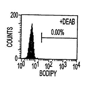
Figures 1A-1D. BAAA staining identifies cells
with high levels of ALDH activity. L1210/cpa is a
derivative of the L1210 leukemic cell line that
overexpresses ALDH. (Figs. 1A and 1B represent L1210
cells, plus DEAB and minus DEAB, respectively; Figs. 1C
and 1D represent L1210/cpa cells, plus and minus DEAB,
respectively).
Figures 2A-2D. BAAA is effluxed by an MDR pump
from hematopoietic cells, particularly primitive CD34+
cells, as evidenced by the difference between the CD34+
-3-.
CA 02353701 2001-06-01
WO 00/34507
PCT/US99/28769
cells that are BODIPYbright (Fig. 2B) in the presence and
absence of the MDR inhibitor, verapamil (Figs. 2A and
23 are at t=0, minus and plus verapamil, respectively;
Figs. 2C and 2D are at t=30', minus and plus verapamil,
respectively.)
Figures 3A-3D. ALDHbr cells (i.e., cells with low
SSC properties that stain brightly with BAAA in the
presence of an MDR inhibitor) are enriched for cells
with the primitive CD34+CD381 /- immunophenotype
traditionally associated with primitive stem cells.
Figures 4A and 43. The staining intensity with
BAAA correlates inversely with CD38 (Fig. 4A) and CD71
(Fig. 4B) expression in CD34+ cells.
Figures 5A-5C. ALDHbr cells are enriched for
early progenitors equivalent to CD34+ cells and are
more enriched for very primitive progenitors than CD34+
cells. (Fig. 5A = progenitors (HPCA), Fig. 53 = early
progenitors (5 week LTC) and Fig. 5C = primitive
progenitors (8 week LTC)).
Figure 6. Preparation of BAAA. Using an amber
vial, a solution of aminoacetaldehyde diethyl acetal
(0.019 mmol, Aldrich Chemical Co.) in dry
tetrahydrofuran (THF, 0.5 mL) was added dropwise to a
solution of BODIPY FL, SE (0.013 mmol, Molecular
Probes) in dry THF (0.5 mL). Upon complete addition,
the vial was capped and the reaction mixture was
stirred for 30 min. The THF was evaporated and the
residue was dissolved in minimal methylene chloride and
- 4 -
CA 02353701 2001-06-01
WO 00/34507
PCT/US99/28769
then chromatographed on silica gel using ethyl acetate
- hexane (1:1) as eluent. The product, BODIPY-
aminoacetaldehyde diethylacetal (BAAA) was recovered in
quantitative yield and identified by proton NMR.
DETAILED DESCRIPTION OF THE INVENTION
The present invention relates to a method of
isolating stem cells and to a reagent suitable for use
in such a method. The method comprises contacting a
population of cells comprising stem cells with a
detectable substrate for aldehyde dehydrogenase (ALDH),
which substrate is converted to a detectable product by
ALDH, that product being retained in the cells. In a
preferred embodiment, the substrate is BODIPY-
aminoacetaldehyde (BAAA) and efflux of BAAA from the
cells, particularly the stem cells present in the
population, is inhibited by the concurrent use of a
MDR-inhibitor.
Sources of cell populations that are suitable for
use in the present invention include umbilical cord
blood, bone marrow, peripheral blood and fetal liver.
Any cell population that includes stem cells can be
used regardless of tissue origin (e.g., gut, skin
muscle, nerve, etc.). While the present method can be
expected to be applicable to a variety of non-human
mammalian cell populations, it is particularly useful
. in isolating human stem cells from sources including
those referenced above.
- 5 -
CA 02353701 2001-06-01
W000/34507
PCT/US99/28769
Substrates suitable for use in the present
invention include substrates for ALDH, particularly
specific substrates for ALDH, that are detectable or
bear a detectable label and that are converted, by the
action of ALDH to products that are detectable or bear
the detectable label which products are retained in the
cells, particularly, the stem cells. In a preferred
embodiment, the substrate is a fluorescent substrate
that has a discrete fluorescence emission profile
identical to FITC. An example of such a substrate is
BAAA.
The optimum amount of substrate to be added to the
cell population can be readily be determined by one
skilled in the art (see Example). In the case of BAAA,
concentrations can vary, for example, concentrations of
about 1gM to 5gM can be used.
A concentrated solution of the substrate can be
added directly to medium comprising the cells to be
stained or harvested cells can be suspended in a
substrate-containing medium.
In order to inhibit efflux of the substrate of the
invention from the cells, concurrent use of an
inhibitor of MDR is preferred. Any of a variety of MDR
inhibitors can be used, including verapamil. The
inhibitor can be added to the cells simultaneously with
the substrate or prior to the addition of the
substrate. The optimum amount of MDR-inhibitor to be
used can be readily determined (e.g., by monitoring
loss of staining). In the case of verapamil,
- 6 -
CA 02353701 2009-07-28
WO 00/34507
PCTIUS99/28769
concentrations can vary, for example, a concentration
of about 50 M can be used.
After exposure of the cell population to the
substrate (and the MDR inhibitor) (e.g., about
30 minutes after) those cells that contain higher
concentrations of labeled product can be separated from
those that contain lower concentrations. In the case
of the use of a fluorescent label, fluorescence
activated cell sorting techniques can be used. Stem
cells can be purified from other cells of the starting
population based on low orthogonal light scattering on
a flow cytometer (identifies small cells, like
lymphocytes) and/or brightness of fluorescence. As
shown in the Example that follows, sorting the
brightest 1% of cells yielded a nearly 40-fold
enrichment for cells that initiate long term cultures.
The cell preparations that were recovered up to 65%
CD34 cells, most of which were CD38m CD71m. The
invention includes within its scope cell preparations
that are greater than 50% CD34+ cells, preferably
greater than 75% CD34+ cells, more preferably greater
than 90% CD34+ cells.
The stem cells isolated in accordance with the
present invention have application in a variety of
therapies and diagnostic regimens. They are suitable
for both transplantation and gene therapy purposes.
For example, isolation of stem cells from bone marrow
or peripheral blood of patients with cancer can provide
for the separation of stem cells from cancer cells. In
patients undergoing autologous transplantation, such
7
CA 02353701 2001-06-01
WO 00/34507
PCT/US99/28769
separation can be used to reduce the chance that cancer
cells are returned to the patient. Purified autologous
stem cells can be ex vivo expanded to hasten
neutrophil, erythroid and platelet engraftment after
autologous transplantation. Ex vivo expansion can be
effected by growth in defined cytokines, on stromal
layers and/or in bioreactors (Emerson et al, Blood
87:3082 (1996)). In addition, the incidence of graft
failure can be reduced. This is beneficial for cancer
patients undergoing autologous transplantation, for
patients suffering from auto-immune disorders, and for
patients undergoing gene therapy.
Gene therapy approaches involving the present
cells involve, in one embodiment, isolation of
autologous stem cells, exposure of the isolated cells
to a gene delivery vector and re-infusion of the
modified cells into the patient (Smith, J. Hematother.
1:155 (1992)). This approach can involve ex vivo
culture or the use of vectors capable of transferring
genes into non-dividing cells, thereby rendering ex
vivo culture unnecessary. Gene therapy can be useful
in treating, for example, congenital diseases, such as
sickle cell anemia, in which case the mutant P-globin
gene is replaced or supplemented with either the wild
type globin gene or an anti-sickling globin gene. In
the treatment of cancer, drug resistance genes can be
introduced into the stem cells to confer resistance to
cytotoxic drugs. This can reduce the incidence and
severity of myelosupporession. For the treatment of
- 8 -
_
CA 02353701 2001-06-01
WO 00/34507
PCIMS99/28769
infectious diseases, including HIV, anti-viral genes
can be introduced into the stem cells so that they are
rendered resistant to the virus (Gilboa and Smith,
Trends in Genetics 10:139 (1994))=
Isolation of stem cells results in the elimination
of T-cells that cause GvHD. This elimination can be
expected to reduce the incidence and severity of GvHD
in recipients of allogeneic transplants.
Purified allogenic stem cells can be ex vivo
expanded to hasten neutrophil, erythroid and platelet
engraftment after allogeneic transplantation. In
addition, the incidence of graft failure can be
reduced. This is likely to be particularly important
for recipients of umbilical cord blood transplants
where small cell doses limit the success of
transplantation.
Successful engraftment with stem cells can also be
expected to induce tolerance. Such would clearly
enhance solid organ transplantation.
It will be appreciated that cells of the present
invention can be used as sources of new genes (eg for
cytokines and cytokine receptors), including genes
important in growth and development.
In addition to their application in treatment and
diagnosis strategies, the stem cells of the invention
can be used in screening protocols to identify agents
that can be used, for example, to promote
differentiation or growth and/or engraftment of
hematopoietic cells. In one such protocol, stem cells
are contacted with a test compound suspected of
- 9 -
CA 02353701 2001-06-01
WO 00/34507
PCT/US99/28769
inducing differentiation and the ability of the test
compound to effect differentiation determined (using,
for example, microscopic and flow cytometric
examination). In another screening protocol, stem
cells are contacted with a test compound suspected of
inducing proliferation and/or engraftment and the
ability of the test compound to effect proliferation
and/or engraftment determined using in vitro long term
colony assays or in vivo immunodeficient mice models
(eg SCID NOD mice). (See Peault et al, Leukemia 7:s98-
101 (1993)).
In addition to the above, the substrate of the
invention can be used to identify tumors that may be
resistant to cyclophosphamide via up regulation of ALDH
activity. In accordance with this embodiment, cells of
the tumor can be contacted with the detectable
substrate, e.g., BAAA, and MDR inhibitor under
conditions such that the substrate enters the cells and
is converted therein to the detectable product. Cells
that stain brightly (e.g., with BAAA) can be expected
to be cyclophosphamide resistant.
The invention also relates to kits that can be
used to prepare the cells of the invention. The kits
can comprise reagents (e.g., ALDH substrate) that can
be used to effect isolation of the stem cells. In a
preferred embodiment, the kit includes BAAA disposed
within a container means. The kit can also include,
disposed within a container means, an MDR inhibitor,
such as verapamil.
- 10 -
CA 02353701 2001-06-01
W000/34507 PCT/US99/28769
Certain aspects of the present invention are
described in greater detail in the non-limiting
Examples that follow.
EXAMPLE 1
Experimental Procedures
Preparation of BODIPY aminoacetaldehyde.
The aldehyde dehydrogenase substrate is prepared
as BODIPY aminoacetal and lyophilized in 0.5 micromole
aliquots. These preparations are stable indefinitely
when stored at -20 C. The acetal is then solubilized
in DMSO to a final concentration of 5 mM. This
solution has been found to be stable at 4 C for up to 1
week. To convert the acetal to an acetaldehyde,
aliquots of this solution are brought to a final
concentration of 1 N HC1. Under these conditions the
acetal has a half life of 15 minutes. After 2 hours
in 1 N HC1, the vast majority of the BODIPY aminoacetal
has converted to BODIPY aminoacetaldehyde (BAAA), and
is then diluted to 200-250 mM in Dulbecco's phosphate
buffered saline (PBS). This stock is added directly to
cells prepared in Iscove's Modified Dulbecco's Medium
(IMDM) with 2% FCS at concentrations ranging from 1 to
5 M.)(See also Fig. 6.).
Antibody reagents.
Directly-conjugated fluorescent antibodies directed
against CD2 (Leu5; FITC), CD3 (Leu4; PerCP), CD5 (Leu1; PE),
CD7 (Leu9; FITC), CD10 (CALLA; FITC), CD1lb (Leu15; PE),
- 11 -
CA 02353701 2001-06-01
WO 00/34507 PCT/US99/28769
CD14 (Leu M3; PE), CD19 (Leul2; FITC), CD33 (LeuM9; PE),
CD34 (HPCA2; FITC and PE), CD38 (Leul7; PE), CD56 (Leul9;
PE) and HLA-DR (FITC) from Becton Dickinson Immunocytometry
Systems (BDIS; San Jose, CA) were used. Anti-CD7 (3A1; PE)
and anti-CD45 (KC56; PE) were purchased from Coulter
Corporation (Hialeah, FL); anti-CD3 (UCHT1; PE), anti-CD16
(3G8; PE), anti-CD19 (J4.119; PE) as well as the pooled
anti-CD34 antibodies (QBEnd10, Immu-133, Immu-134; PE) from
Immunotech, Inc. (Westbrook, ME); anti-CD3 (B-B11; FITC) and
CD38 (B-A6; FITC) from BioSource International (Camarillo,
CA); anti-CD45RA (F8-11-13; PE) from Southern Biotechnology
Associates, Inc. (Birmingham, AL); and anti-CDw90 (5E10; PE)
from PharMingen, Inc. (San Diego, CA).
Cell lines.
K562, L1210 and L1210/cpa cells (ATCC) were maintained
in suspension in RPMI 1640 media supplemented with 10% Fetal
Calf Serum (FCS) and 5 x 10⁻⁵ M P-mercaptoethanol.
Preparation of Human Umbilical Cord Blood.
Human umbilical cord blood (UCB), intended for
disposal, was collected into sterile bottles containing
anticoagulant citrate buffer. The UCB used in these studies
were processed within 24 hours of being harvested. White
cells were enriched through a preliminary red cell
agglutination where the UCB was diluted 1:2 with Dulbecco's
phosphate buffered saline (PBS) at room temperature. These
cells were then brought to a final concentration of 1%
Hespan (DuPont Pharma, Wilmington, DE) and were left to
stand undisturbed for 1 hour. Non-agglutinated white blood
- 12 -
CA 02353701 2009-07-28
W000/34507
PCT/US99/28769
cells were harvested and residual red cells were hemolysed
at 37 C in 0.17 M NH4C1 containing 10 mM Tris-HC1, pH 7.2
and 200 mM EDTA. The recovered cells were washed in IMDM
containing 2% FCS and mononuclear cells are then purified
using Ficoll-Hypaque (1.077g/m1). When held overnight, the
cells were kept on ice in a 4 C refrigerated room in IMDM
with 20% FCS.
Cell staining and fluorescence-activated cell sorting.
Mononuclear UCB cells were resuspended at 106 cells/ml
in IMDM containing 2% FCS and were labeled with 1 AM BAAA
for 30 min. When used, verapamil was included at 50 mM.
After staining, the cells were washed with ice cold staining
media and maintained on ice until their analysis and
sorting. The cells were then resuspended in staining media
with 10 mg/ml 7-aminoactinomycin D (7AAD)(Molecular Probes;
Eugene, OR). For antibody staining to permit multiparameter
analyses, the cells were resuspended in staining media (100
Oand antibodies were added directly to the cell
suspensions. The cells were incubated on ice for 20min. and
then washed again in ice cold staining media. The cells
were then analyzed or sorted on a FACStar Plus cell sorter
(BDIS) equipped with dual Coherent 1-90 argon-dye laser.
The BAAAwas excited at 488 nm and emissions were detected
using 515 DF2Ofilter in FL1.Dead and dying cells were
excluded on the basis of their high emission in the far red
wavelength due to their uptake of 7AAD.
For analyses of cell surface antigens on cells
previously sorted based on BAAA staining, the cells were
-13-
CA 02353701 2009-07-28
WO 00/34507
PCT/US99/28769
pelleted and resuspended in IMDM with 2% FCS. The
cells were then held at 37 C for 1-2 hours to permit
efflux. The cells were then pelleted and fluorescence-
conjugated antibodies were added directly to the
cells. Following incubations for 20 minutes, the cells
were washed with PBS/2% FCS and were fixed in 1%
folualdehyde in PBS/2% FCS. In all surface marker
analyses, no differences were noted between analyses
with cells stained simultaneously with BAAA
and with antibodies and those analyses performed on
FACS sorted cells that were subsequently stained with
antibodies.
Hematopoietic progenitor colony assays and long
term cultures.
ALDHbrcells were isolated directly from mononuclear
UCB cells which had been stained with BAAA. For these
assays, the ALDHbr was defined as 1% of the lymphocyte
gate of the UCB.
Hematopoietic progenitor colony assays were
performed by plating 100-200 cells in MethoCult H4431
containing agar leukocyte conditioned media and
recombinant human erythropoietin (StemCell
Technologies, Inc.). The cells
were incubated in a humidified chamber at 37 C with 5%
CO2. Hematopoietic colonies (>100 cells) were then scored
at 14 to 18 days after initiating the cultures. Long
term cultures were maintained on stromal layers of
murine MS-5 cells (provided by Dr. Tadashi Sudo of the
Kirin Pharmaceutical Research Laboratory, Gunma, Japan)
(Issaad, Blood 81:2916 (1993)). MS-5 stromal cells were
seeded into 24-well plates (Corning Costar Corp.,
Cambridge, MA) at 5 X
-14-
CA 02353701 2009-07-28
WO 00/34507
PCT/US99/28769
104 cells/well in DMEM supplemented with 10% FCS and
cultured at 37 C. When the monolayers approached 80%
confluence they were y-irradiated from a cesium source (40
Gy). After irradiation, fresh media was provided to the
cultures. For the MS-5 cells, the culture media was
replaced entirely with MEMa supplemented with 10%
FCS,10%
equine serum, P-mercaptoethanol, pyruvate. Long term
cultures were initiated with 400-2000 hematopoietic
progenitor cells/well and were maintained at 33 C with 5%
CO2. At weekly intervals half the media from each well was
removed so that the media could be replenished. Adherent
and non-adherent cells were harvested after 5or 8 weeks and
plated into HPC assays as described above. As shown in the
Example that follows, sorting the brightest 1% of cells
yields a nearly 40-fold enrichment for cells that initiate
long term cultures. The cell preparations that were
recovered were up to 65% cp34'cells, most of which were
CD34+ cells, most of which were CD38-/d1mCD71-/d1m.
Results
Synthesis of BODIPY acetal.
Due to the inherent instability of aldehydes in
aqueous solution, the reagent is prepared and stored as
an acetal. Immediately prior to its use, the acetal is
converted to an aldehyde in 1 N HCL. the aldehyde is
freely soluble in PBS and can be added directly to
cells prepared in IMDM with 2% fetal calf serum at 106
-15-
CA 02353701 2001-06-01
W000/34507
PCT/US99/28769
cells per ml. As an aminoacetaldehyde, the reagent is
membrane permeable; however, in the presence of the
aldehyde dehydrogenase (ALDH), the aldehyde moiety is
converted to a carboxylic acid that is retained in the
cell. Intracellular fluorescence can be used to select
cells.
BAAA is a Specific Substrate for ALDH.
To assay whether BAAA would permit the specific
selection of ALDH+ cells, studies initially determined
an optimal response dose for the BAAA reagent in a
murine cell line previously selected for
cylophoshamide-resistance, L1210/cpa, that is known to
be ALDH+ (Fig. 1). The parental cell line, L1210
(Figs. 1A and 1B) is cylophosphamide-sensitive and
ALDH-. This cell line exhibited essentially no
response to BAAA. In addition, a potent inhibitor of
ALDH, diethylbenzaldehyde (DEAB), was used to
demonstrate the specificity of the BAAA signal. A 10-
fold molar excess of DEAB totally blocked the
fluorescent response (Fig. 1C). Therefore, BAAA was
able to detect ALDH + cells. In these studies, the BAAA
could be used at a final concentration as low as 5 M.
This molar concentration is 10-fold lower than that
used with the dansylated reagent.
Multiple different ALDH isoenzymes exist and these
may display different abilities to convert BAAA. It
has been suggested that resistance to cyclophosphamide
is primarily mediated by a specific ALDH isoenzyme,
ALDH1. Therefore, a human cell line known to express
- 16 -
CA 02353701 2001-06-01
WO 00/34507
PCT/US99/28769
ALDH1, K562, was assayed with this novel reagent. K562
cells converted BAAA and were positive for ALDH in
these assays. This response was entirely inhibited by
DEAB. Thus, BAAA can serve as a specific substrate for
human ALDH1 and can be used to identify primary human
cells that demonstrate resistance to cyclophosphamide.
Primary UCB Cell preparations contain subsets of ALDHbr
cells.
Having demonstrated the effectiveness of this
reagent on continuous cell lines, BAAA was assayed on
primary human cells. Umbilical Cord Blood (UCB) was
chosen for its increasing promise as a source for
transplantable hematopoietic stem cells. For these
studies, the UCB was unfractionated except for having
been prepared for mononuclear cells over Ficoll-
Hypaque. This separation is significant in that two
mature ALDH+ cell types, erythrocytes and
megakaryocytes, are removed. The BAAA was tested on
UCB cells prepared in IMDM with 2% FCS at 106 cells/ml
(Fig. 2). The UCB cells were very responsive to the
BODIPY reagent, and appeared to be much more sensitive
than the continuous cell lines had been. The BAAA was
therefore titrated to an optimal concentration of 1 M.
This was the best concentration for resolving ALDHbr
subpopulations. The response was inhibited in the
presence of excess DEAB and was therefore specific for
ALDH. This molar concentration is 50-fold lower than
the concentration of dansyl aminoacetaldehyde that had
- 17 -
CA 02353701 2001-06-01
WO 00/34507
PCT/US99/28769
previously been used to detect murine pluripotent
hematopoietic stem cells.
The fluorescence emission from BAAA-stained UCB
cells exhibited a bimodal response. The brighter peak
of fluorescence emission was attributed to mature
monocytes, suggesting monocytes express a uniform level
of ALDH. Hematopoietic stem cells are small, non-
complex cells. Indeed, murine ALDH + PHSC were first
enriched using countercurrent elutriation. Therefore,
the BODIPY signal was examined only in non-complex
cells that exhibited low inherent orthogonal light
scattering (SSC1 ) (Fig. 3A). The majority of the SSC1
UCB cells were ALDHnegidirn (Fig. 1B). This was not
unexpected since the SSC1 cells are predominantly
lymphocytes, and most lymphocytes do not express ALDH.
However, a small, clearly-defined subpopulation of the
SSC1 UCB cells was ALDHbr (Fig. 3A).
BODIPY aminoacetate is a substrate for the MDR efflux
Pump.
In addition to expressing ALDH, PHSC should also
express high levels of the P-glycoprotein or multiple
drug resistance (MDR) efflux pump. Since this reagent
had never been previously characterized, the
susceptibility of BAAA to MDR efflux was assayed.
Although BODIPY aminoacetaldehyde passes through the
cell membrane without active transport, the product of
the ALDH conversion (BODIPY aminoacetate) might well be
a substrate for the MDR pump. To investigate this
possibility, UCB cells were stained with BAAA in the
- 18 -
___
CA 02353701 2001-06-01
W000/34507
PCT/US99/28769
presence of 50 M verapamil, a competitive inhibitor of
the MDR efflux pump. The verapamil-treated cells
exhibited a consistently-higher fluorescence when
compared with BAAA-stained cells that had not been
simultaneously treated with verapamil (Fig. 2). A
substantial population of ALDHdim cells were effected by
the verapamil treatment. Most importantly, the
percentage of ALDHbr cells increased by 1.8 fold in the
presence of verapamil. In verapamil-treated cells, the
ALDHbr subpopulation was equivalent to 0.8 % of the
SSC1 cells. In contrast, in cell preparations that
received no verapamil, the same fluorescence intensity
represented only 0.46 % of the SSC1 cells. This
indicated that the ALDHbr SSC1 UCB cells retain the
converted BAAA more effectively if the efflux activity
of the MDR pump is inhibited.
ALDHbr SSC1 UCB cells are highly enriched for primitive
CD34 cells.
With verapamil treatment, the ALDHbr SSC1 UCB
cells contained almost 90% CD34' cells, indicating that
at least some hematopoietic progenitors are present
(Fig. 3D). However, CD34 is expressed by a broad range
of hematopoietic progenitors that includes lineage
committed cells, as well as pluripotent progenitors.
Therefore, the developmental potential of the ALDHbr
SSC1 UCB cells was analyzed. Initially, the
immunophenotype of these cells was more carefully
defined. The immunophenotype would in no way be
conclusive; however, the primitiveness of the cell
- 19 -
CA 02353701 2001-06-01
W000/34507
PCT/US99/28769
population could be inferred by examining 2 activation
markers that are typically associated with the
differentiation of primitive cells to more lineage-
committee hematopoietic cells, CD38 and CD71. The most
primitive subsets of CD34+ cells have little to no
expression of the activation antigens CD38 or CD71. In
UCB cell preparations with BAAA and with antibodies
specific for CD34 and CD38, the ALDHbr SSC1 UCB cells
provided a single-step enrichment for essentially
purified CD34br CD38thm cells. Furthermore, when CD34+
UCB cells were examined independently, ALDH expression
was inversely proportional to the expression of both
CD38 and CD71 (Fig. 4A and 4B). Thus, the ALDHbr SSC1
UCB cells appear to contain the primitive CD34+ cells
as defined by immunophenotype.
To assay the developmental potential of the ALDHbr
SSC1 UCB cells, these cells were isolated and placed
into both short-term and long-term assays for
myeloerythroid progenitors. The short-term assay used,
the hematopoietic progenitor colony assay (HPCA),
quantifies lineage committed cells at the time of the
initial isolation. More primitive progenitors were
also assayed by maintaining the ALDHbr SSCi UCB cells
on stroma for either 5 or 8 weeks prior to performing
the HPCA (Fig. 5).
Results:
HPCA - ALDHbr SSC1 essentially equivalent to CD34+
cells.
- 20 -
CA 02353701 2001-06-01
WO 00/34507 PCT/US99/28769
LTC - 5 wk - ALDHbr SSC1 essentially equivalent to
CD34+ cells.
LTC = 8 wk - ALDHbr SSC1 outperforms CD34+ cells.
EXAMPLE 2
ALDHbr UCB cells have been shown to be
predominately CD34+CD38-/1 and highly enriched for
early myeloid progenitors. The current study was
undertaken to determine whether the ALDHbr CD34+ UCB
cells were enriched for lymphoid progenitors as well.
In 3 experiments, cultures of ALDHbr CD34+ UCB cells
were established on AFT024 stromal cells in the
presence on Kit ligand, F1t3 ligand, IL-3 (1st day
only), IL-2 and IL-7 at various dilutions. After 7-8
weeks, the cultures were analyzed for lymphocyte growth
as determined by expression of CD56, CD10, CD19 or
CD20.
Table 1
ALDIer total wells wells with lymphocyte
cells/well initiated viable cells wells
1000 6 5 5
250 16 12 12
62 48 40 40
16 48 34 34
10 24 20 17
The AFT024 cultures primarily favored the growth
of presumptive NK cells, so to more effectively test
whether ALDHbr CD34+ UCB cells contained B-lymphoid
progenitors, they were cultured on the W20 stromal cell
- 21 -
.CA 02353701 2012-04-13
line supplemented with the same cytokine combination.
Of 12 cultures established with 100 ALDHbr CD34+ cells,
all produced CD56+ and CD10 cells at nearly equivalent
proportions. 2 of the 12 wells also contained CD19+
cells.
In summary, the ALDHbr CD34+ UCB population appears
to be highly enriched for both myeloid and lymphoid
hematopoietic progenitors.
-22-