Note: Descriptions are shown in the official language in which they were submitted.
CA 02353941 2001-06-05
WO 01/37768 PCT/US00/25662
PUMPING CHAMBER FOR A LIQUEFRACTURE HANDPIECE
Background of the Invention
This invention relates generally to the field of cataract surgery and more
particularly to a pumping chamber for a handpiece for practicing the
liquefracture
s technique of cataract removal.
The human eye in its simplest terms functions to provide vision by
transmitting
light through a clear outer portion called the cornea, and focusing the image
by way of the
lens onto the retina. The quality of the focused image depends on many factors
including
the size and shape of the eye, and the transparency of the cornea and lens.
When age or disease causes the lens to become less transparent, vision
deteriorates
because of the diminished light which can be transmitted to the retina. This
deficiency in
the lens of the eye is medically known as a cataract. An accepted treatment
for this
condition is surgical removal of the lens and replacement of the lens function
by an
artificial intraocular lens (IOL).
In the United States, the majority of cataractous lenses are removed by a
surgical
technique called phacoemulsification. During this procedure, a thin
phacoemulsification
cutting tip is inserted into the diseased lens and vibrated ultrasonically.
The vibrating
cutting tip liquifies or emulsifies the lens so that the lens may be aspirated
out of the eye.
The diseased lens, once removed, is replaced by an artificial lens.
A typical ultrasonic surgical device suitable for ophthalmic procedures
consists of
an ultrasonically driven handpiece, an attached cutting tip, and irrigating
sleeve and an
electronic control console. The handpiece assembly is attached to the control
console by
an electric cable and flexible tubings. Through the electric cable, the
console varies the
power level transmitted by the handpiece to the attached cutting tip and the
flexible
tubings supply irrigation fluid to and draw aspiration fluid from the eye
through the
handpiece assembly.
The operative part of the handpiece is a centrally located, hollow resonating
bar or
horn directly attached to a set of piezoelectric crystals. The crystals supply
the required
ultrasonic vibration needed to drive both the horn and the attached cutting
tip during
phacoemulsification and are controlled by the console. The crystal/horn
assembly is
suspended within the hollow body or shell of the handpiece by flexible
mountings. The
CA 02353941 2007-08-29
2
handpiece body ten.ninates in a reduced diameter portion or nosecone at the
body's distal
end. The nosecone is externally threaded to accept the irrigation sleeve.
Likewise, the
horn bore is internally threaded at its distal end to receive the external
threads of the
cutting tip. The irrigation sleeve also has an internally threaded bore that
is screwed onto
the external threads of the nosecone. The cutting tip is adjusted so that the
tip projects
only a predetermined amount past the open end of the irrigating sleeve.
Ultrasonic
handpieces and cutting tips are more fully described in U.S. Pat. Nos.
3,589,363;
4,223,676; 4,246,902; 4,493,694; 4,515,583; 4,589,415; 4,609,368; 4,869,715;
4,922,902;
4,989,583; 5,154,694 and 5,359,996.
In use, the ends of the cutting tip and irrigating sleeve are inserted into a
small
incision of predetermined width in the cornea, sclera, or other location. The
cutting tip is
ultrasonically vibrated along its longitudinal axis within the irrigating
sleeve by the crystal-
driven ultrasonic horn, thereby emulsifying the selected tissue in situ. The
hollow bore of
the cutting tip communicates with the bore in the horn that in turn
communicates with the
aspiration line from the handpiece to the console. A reduced pressure or
vacuum source in
the console draws or aspirates the emulsified tissue from the eye through the
open end of
the cutting tip, the cutting tip and horn bores and the aspiration line and
into a collection
device. The aspiration of emulsified tissue is aided by a saline flushing
solution or irrigant
that is injected into the surgical site through the small annular gap between
the inside
surface of the irrigating sleeve and the cutting tip.
Recently, a new cataract removal technique has been developed that involves
the
injection of hot (approximately 45 C to 105 C) water or saline to liquefy or
gellate the
hard lens nucleus, thereby making it possible to aspirate the liquefied lens
from the eye.
Aspiration is conducted with the injection of the heated solution and the
injection of a
relatively cool solution, thereby quickly cooling and removing the heated
solution. This
technique is more fully described in U.S. Patent No. 5,616,120 (Andrew, et
al.).
The apparatus disclosed in the publication, however, heats the solution
separately
from the surgical handpiece. Temperature control of the heated solution can be
difficult
because the fluid tubings feeding the handpiece typically are up to two meters
long, and
the heated solution can cool considerably as it travels down the length of the
tubing.
CA 02353941 2001-06-05
WO 01/37768 PCT/US00/25662
3
Therefore, a need continues to exist for a control system for a surgical
handpiece
that can heat internally the solution used to perform the liquefracture
technique.
Brief Summarv of the Invention
The present invention improves upon the prior art by providing a surgical
handpiece having at least two lumens or tubes mounted to a body. At least one
tube is
used for aspiration and at least one other tube is used to inject heated
surgical fluid for
liquefying a cataractous lens. A portion of the second tube is enlarged to
form a pumping
chamber. The pumping chamber works by boiling a small volume of the surgical
fluid.
As the fluid boils, it expands rapidly, thereby propelling the liquid
downstream of the
io pumping chamber out of the second tube. The pumping chamber may use a pair
of
electrodes, at least one of the electrodes containing a countersink.
Accordingly, one objective of the present invention is to provide a surgical
handpiece having at least two tubes.
Another objective of the present invention is to provide a surgical handpiece
having
1s a pumping chamber with two electrodes, at least one electrode containing a
countersink..
Another objective of the present invention is to provide a surgical handpiece
having
a device for delivering the surgical fluid through the handpiece in pulses.
These and other advantages and objectives of the present invention will become
apparent from the detailed description and claims that follow.
20 Brief Description of the Drawings
FIG. I is a front, upper left perspective view of the handpiece of the present
invention.
FIG. 2 is a rear, upper right perspective view of the handpiece of the present
invention.
25 FIG. 3 is a cross-sectional view of the handpiece of the present invention
taken
along a plane passing through the irrigation channel.
CA 02353941 2001-06-05
WO 01/37768 PCT/US00/25662
4
FIG. 4 is a cross-sectional view of the handpiece of the present invention
taken
along a plane passing through the aspiration channel.
FIG. 5 is an enlarged partial cross-sectional view of the handpiece of the
present
invention taken at circle 5 in FIG. 4.
FIG. 6 is an enlarged partial cross-sectional view of the handpiece of the
present
invention taken at circle 6 in FIG. 3.
FIG. 7 is an enlarged cross-sectional view of the handpiece of the present
invention
taken at circle 7 in FIGS. 3 and 4, and showing a resistive boiler pump.
FIG. 8 is an exploded, partial cross-section view of one embodiment of the
handpiece of the present invention.
FIG. 9 is a block diagram of a control system that can be used with the
handpiece
of the present invention.
Detailed Description of the Invention
Handpiece 10 of the present invention generally includes handpiece body 12 and
operative tip 16. Body 12 generally includes external irrigation tube 18 and
aspiration
fitting 20. Body 12 is similar in construction to well-known in the art
phacoemulsification
handpieces and may be made from plastic, titanium or stainless steel. As best
seen in
FIG. 6, operative tip 16 includes tip/cap sleeve 26, needle 28 and tube 30.
Sleeve 26 may
be any suitable commercially available phacoemulsification tip/cap sleeve or
sleeve 26
may be incorporated into other tubes as a multi-lumen tube. Needle 28 may be
any
commercially available hollow phacoemulsification cutting tip, such as the
TURBOSONICS tip available from Alcon Laboratories, Inc., Fort Worth, Texas.
Tube 30
may be any suitably sized tube to fit within needle 28, for example 29 gauge
hypodennic
needle tubing.
As best seen in FIG. 5, tube 30 is free on the distal end and connected to
pumping
chamber 42 on the proximal end. Tube 30 and pumping chamber 42 may be sealed
fluid
tight by any suitable means having a relatively high melting point, such as a
silicone
gasket, glass frit or silver solder. Fitting 44 holds tube 30 within bore 48
of aspiration
horn 46. Bore 48 communicates with fitting 20, which is journaled into horn 46
and
sealed with 0-ring seal 50 to form an aspiration pathway through horn 46 and
out fitting
CA 02353941 2001-06-05
WO 01/37768 PCT/US00/25662
20. Horn 46 is held within body 12 by 0-ring seal 56 to form irrigation tube
52 which
communicates with irrigation tube 18 at port 54.
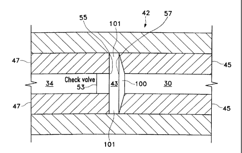
As best seen in FIG. 7, in a first embodiment of the present invention,
pumping
chamber 42 contains a relatively large pumping reservoir 43 that is sealed on
both ends by
s electrodes 45 and 47. Electrical power is supplied to electrodes 45 and 47
by insulated
wires, not shown. In use, surgical fluid (e.g. saline irrigating solution)
enters reservoir 43
through port 55, tube 34 and check valve 53, check valves 53 being well-known
in the art.
Electrical current (preferably Radio Frequency Alternating Current or RFAC) is
delivered
to and across electrodes 45 and 47 because of the conductive nature of the
surgical fluid.
As the current flows through the surgical fluid, the surgical fluid boils. As
the surgical
fluid boils, it expands rapidly out of pumping chamber 42 through port 57 and
into tube
30 (check valve 53 prevents the expanding fluid from entering tube 34). The
expanding
gas bubble pushes the surgical fluid in tube 30 downstream of pumping chamber
42
forward. Subsequent pulses of electrical current form sequential gas bubbles
that move
surgical fluid down tube 30. The size and pressure of the fluid pulse obtained
by pumping
chamber 42 can be varied by varying the length, timing and/or power of the
electrical
pulse sent to electrodes 45 and 47 and by varying the dimensions of reservoir
43. In
addition, the surgical fluid may be preheated prior to entering pumping
chamber 42.
Preheating the surgical fluid will decrease the power required by pumping
chamber 42
and/or increase the speed at which pressure pulses can be generated.
Preferably, electrode 45 contains small depression or countersink 100 having
any
suitable depth but approximately 0.003 inches being preferred. Pumping
reservoir 43 is
narrowest at periphery 101 (on the order of 0.1 mm) and as a result, fluid in
pumping
reservoir 43 boils first at periphery 101 and the steam wave front travels
down countersink
100 toward the central axis of tube 30. The surgical fluid conducts
electricity much better
in the liquid state than in the vapor state. Consequently, current flow
diminishes greatly at
periphery 101 where boiling occurs first.
While several embodiments of the handpiece of the present invention are
disclosed,
any handpiece producing adequate pressure pulse force, rise time and frequency
may also
be used. For example, any suitable handpiece producing a pressure pulse force
of between
0.03 grams and 50.0 grams (between 1 gram and 50.0 grams being preferred),
with a rise
time of between 1 gram/second and 50,000 grams/second (with between 500
grams/second
_ _ _. __...~.. . _,___..__ ._.,~~..,.....~..._..... _ .,.
CA 02353941 2001-06-05
WO 01/37768 PCT/US00/25662
6
and 50,000 grams/second being preferred) and a frequency of between 1 Hz and
200 Hz
may be used, with between 10 Iiz and 100 Hz being most preferred. The pressure
pulse
force and frequency may be varied with the hardness of the material being
removed. For
example, the inventors have found that a lower frequency with a higher pulse
force is
s more efficient at debulking and removing the relatively hard nuclear
material, with a
higher frequency and lower pulse force being useful in removing softer
epinuclear and
cortical material. Infusion pressure, aspiration flow rate and vacuum limit
are similar to
current phacoemulsification techniques.
As seen in FIG. 9, one embodiment of control system 300 for use in operating
io handpiece 310 includes control module 347, RF amplifier 312 and function
generator 314.
Power is supplied to RF amplifier 312 by DC power supply 316, which preferably
is an
isolated DC power supply operating at 200 volts. Control module 347 may be
any
suitable microprocessor, and may receive input from operator input device 318.
Function
generator 314 provides the electric wave form to amplifier 312 and preferably
operates at
15 450 KHz to help minimize corrosion.
In use, control module 347 receives input from surgical console 320. Console
320
may be any commercially available surgical control console such as the LEGACY
SERIES TWENTY THOUSAND surgical system available from Alcon Laboratories,
Inc., Fort Worth, Texas. Console 320 is connected to handpiece 310 through
irrigation
20 line 322 and aspiration line 324, and the flow through lines 322 and 324 is
controlled by
the user via footswitch 326. Irrigation and aspiration flow rate information
in handpiece
310 is provided to control module 347 by console 320 via interface 328, which
may be
connected to the ultrasound handpiece control port on console 320 or to any
other output
port. Control module 347 uses footswitch 326 information provided by console
320 and
25 operator input from input device 318 to generate two control signals 330
and 332. Signal
332 is used to operate pinch valve 334, which controls the surgical fluid
flowing from
fluid source 336 to handpiece 310. Fluid from fluid source 336 is heated in
the manner
described herein. Signal 330 is used to control function generator 314. Based
on signal
330, function generator 314 provides a wave form at the operator selected
frequency and
30 amplitude detenmined by the position of footswitch 326 to RF amplifier 312
which is
amplified to advance the powered wave form to handpiece 310 to create heated,
pressurized pulses of surgical fluid.
CA 02353941 2001-06-05
WO 01/37768 PCT/US00/25662
7
As best seen in FIGS. 3, 4 and 7, surgical fluid may be supplied to pumping
chamber 43 through tube 34 or, as seen in FIG. 8, surgical fluid may be
supplied to
pumping chamber 243 through irrigation fluid tube 234 which branches off main
irrigation
tube 235 supplying cool surgical fluid to the operative site. As seen in FIG.
8, aspiration
tube 237 may be contained internally to handpiece 10.
Any of a number of inethods can be employed to order limit the amount of heat
introduced into the eye. For example, the pulse train duty cycle of the heated
solution can
be varied so that the total amount of heated solution introduced into the eye
does not vary
with the pulse frequency. Alternatively, the aspiration flow rate can be
varied as a
function of pulse frequency so that as pulse frequency increases aspiration
flow rate
increases proportionally.
This description is given for purposes of illustration and explanation. It
will be
apparent to those skilled in the relevant art that changes and modifications
may be made to
the invention described above without departing from its scope or spirit. For
example, it
1s will be recognized by those skilled in the art that the present invention
may be combined
with ultrasonic and/or rotating cutting tips to enhance performance.