Note: Descriptions are shown in the official language in which they were submitted.
CA 02354202 2001-07-26
Doc. No. 51-20 CA Patent
Stone Cell Determination using Fluorescence
Field of the Invention:
This invention relates to a novel method and apparatus for determining stone
cells
(sclereids) in paper or pulp. Specifically this invention uses the
fluorescence
characteristics of stone cells to enhance the contrast between the stone cell
and the
surrounding fibers.
Background of the Invention:
A sclereid is a type of thick walled highly lignified cell found in many
plants. Sclereids
found in trees are often called stone cells. Stone cells can be found in the
cortex, phloem
and pith of several species of either hardwood or softwood trees. They are
most
commonly found in the bark of the tree. When pulp and paper mills use tree
species
which contain stone cells and use the whole tree with no debarking, or when
debarking is
not efficient (as in the winter), or when species are used which contain stone
cells in the
heart of the tree, the stone cells will appear in the finished product. This
is true for both
chemical and mechanical pulps. Further, even when additional measures are
taken to
remove the stone cells there will still be some remaining in the final
product.
When stone cells are present in the papermakers furnish often they cause
difficulties. A
stone cell on a calendared sheet of paper causes an opaque spot known as a
fish eye. The
fish eye will shed ink causing problems for the printer as non-inked areas
show up as
flaws in print. Stone cells also cause difficulties when making paper, for
example, in
mills where high-speed paper machines are employed the stone cell can cause a
weak
area on the forming sheet, which can cause breaks in the paper they are
making. Breaks
on the paper results in down time and loss of production. As such, the number
of stone
cells is a quality issue in pulp and, therefore pulp is commonly sold with
maximum stone
cell count specifications.
Currently there is no universal method for determining and for quantifying
stone cells.
The methods currently in use are time consuming and operator dependent.
Further, many
mills have adopted their own in house method for determining stone cells.
CA 02354202 2001-07-26
Doc. No. 51-20 CA Patent
A first prior art method uses the hardness of the stone cell as a way
separating a stone cell
from its matrix. In a dark room a light source is placed at an angle to a non-
calendared
hand sheet made using either standard method TAPPI T 205 or CPPA C.4. Where a
shadow appears the bump causing the shadow is checked to see if the bump is
solid by
rubbing it with a pencil. A hard bump is considered to be a stone cell. This
method is
very time consuming and operator dependant.
A second prior art method relies upon the transformation of the stone cells
into fish eyes.
It involves making the same standard hand sheet as first method and then
calendaring the
sheet between two hardened steel rollers under several hundred pounds of
hydraulic
pressure. The hand sheet is then put onto a light box and the places where the
paper has
circular opaque spots are counted as stone cells. The difficulty in this
method is that the
fish eyes can be quite small and poor hand sheet preparation can make them
impossible
to see. This method is also very operator dependant.
A newer piece of equipment developed by Optest Equipment Inc. uses a hand
sheet of a
known weight, thickness, and diameter that is put over a light. A camera
mounted above
the sample looks for subtle changes in colour in a magnified and therefore
small portion
of this hand sheet. The small sample size is a major drawback of this method.
Stone
cells are of a similar color to the fiber so there are also difficulties to
determing stone
cells on their color. This colour change must be calibrated before analysis
and all
parameters must remain constant from sample to sample for the instrument to
stay in
calibration. The particles having this colour are counted as stone cells.
Thair, B. W. and Corcoran, P. J have disclose a rapid method wherein a sample
of a
bleached kraft pulp stock is stained, spread in a thin layer, and examined in
transmitted
light. Differences in color, size and opacity make the stone cells Baser to
identify.
Unfortunately, both the pulp stock and the stone cells take up the stain, and
as such the
differences in colour and opacity may not be easily discernable using
transmitted light.
' 2
CA 02354202 2001-07-26
Doc. No. 51-20 CA Patent
Brief Description of the Drawings
Exemplary embodiments of the invention will now be described in conjunction
with the following drawings, in which:
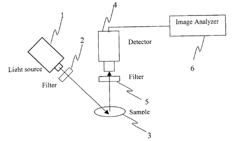
Figure 1 is a schematic block diagram of an apparatus according to a first
preferred
embodiment of the present invention;
Figure 2 is a schematic block diagram of an apparatus according to a second
preferred
embodiment of the present invention;
Figure 3 shows a gray value histogram from a Confocal Microscope Image.
Detailed description of Invention
The determination of a stone cell within the pulp matrix is made possible by
the fact the
stone cell fluoresces strongly while the surrounding pulp does not. High
contrast images
are obtained under these conditions in which the stone cells appear as bright
spots whose
dimension and number can then be quantified using one of automated and manual
means
for quantification. The instant invention is useful for determining stone
cells within the
finished product, such as to determine the quality of paper produced from a
particular
batch of pulp. Advantageously, the instant invention is also useful for
determining stone
cells within pulp samples spread out on a non-reflecting background, such as
to
determine the efficiency of centrifugal cleaners in the pulp processing plant.
Referring to Figure l, shown is an apparatus in accordance with a first
preferred
embodiment of the instant invention. The apparatus comprises a light source 1
for
irradiating a portion of a sample 3 with light at within a predetermined range
of
wavelengths or at a predetermined wavelength. For example, the light source 1
is
selected from a group comprising: a mercury vapor lamp; a deuterium lamp; a
tungsten
filament lamp; and, a laser. Optionally, more than one light source is
provided in order to
cover a wider wavelength range. A filter 2 is provided to filter the incident
light, for
instance the filter 2 excludes any incident wavelengths above 550 nm. A second
other
filter 5 disposed between the sample 3 and the detector 4 to prevent incident
light from
CA 02354202 2001-07-26
Doc. No. S l-20 CA Patent
reaching the detector 4. Filtering the incident light enhances the contrast
between the
stone cells and the surrounding pulp or paper matrix. The detector 4 includes
means for
producing an image of the portion of the sample 3. For instance, the detector
4 is a
charge coupled device (CCD) camera for providing a digitized image comprising
a
plurality of individual pixels, each pixel variable across a grayscale having,
for example,
256 possible discrete values. Of course other than 256 possible discrete
values could be
supported. Further, detectors other than a CCD camera are suitable for use
with the
instant invention.
The image thus obtained by the detector 4 is provided to an image analyzer 6.
The image
analyzer comprises a processor (not shown) for executing code to automatically
analyze
the image and determine areas of strong fluorescence, for instance areas of
the sample
represented by individual pixels having a value of approximately 256. Of
course, the
actual value that is used to define a bright spot may be less than 256 and may
include a
range of allowable values.
Optionally, the sample 2 is mounted on an x-y table (not shown) for moving the
sample
in a controlled manner such that different portions of the sample can be
irradiated, for
instance in a raster fashion. Further optionally, the x-y table is under the
control of a
processor such that the sample is scanned automatically according to a
predetermined
pattern. This allows other detectors, such as for example a photodiode
detector, to be
used to scan the sample for the occurrence of stone cells.
Referring now to Figure 2, shown is an apparatus in accordance with a second
preferred
embodiment of the instant invention. Drawing elements identical to those
described with
reference to Figure 1 and having identical function have been assigned like
numbers. In
the apparatus according to the second preferred embodiment, the detector 4 is
replaced by
one of a lens and a system of lenses 7. A user observes the filtered light
directly, and
quantification of the stone cells in the sample 2 is performed manually, for
instance by
counting an absolute number of stone cells per unit area. Of course, in the
second
embodiment the user performs the function of the image analyzer 6, which was
described
with reference to Figure 1.
4
CA 02354202 2001-07-26
Doc. No. 51-20 CA Patent
Optionally, the sample 2 is mounted on an x-y table (not shown) for moving the
sample
in a controlled manner such that different portions of the sample can be
irradiated, for
instance in a raster fashion. Further optionally, the x-y table is under the
control of a
processor such that the sample is scartrted automatically according to a
predetermined
pattern. This allows other detectors, such as for example a photodiode
detector, to be
used to scan the sample for the occurrence of stone cells.
In a method according to the instant invention, a sample containing stone
cells is placed
under a radiation source that emits light preferably in the 200 to 550 nm
range.
Optionally, a radiation source that emits light other than in the 200 to 550
nrrt range is
used. A filter is disposed between the light source and the surface of the
sample under
investigation to exclude any incident wavelengths above 550 nm from reaching
the
sample. The stone cells in the sample will begin to fluoresce and emit light
that can be
detected by a detector. The wavelength of the light that is emitted by
fluorescence is
greater than the wavelength of the incident light, and as such a filter is
disposed between
the surface of the sample under investigation and the detector to exclude all
incident
light. The stone cells are observed as bright spots. The detector can be a
camera whose
pictures are sent to an image analysis system where the stone cells can be
counted and
quantified. Preferably the image is obtained absent magnification such that a
maximum
field of view is captured by each image. Optionally, the image is magnified.
Optionally, the detector is an eyepiece or an ocular device fitted with the
same filter and
the stone cells are counted manually absent an image analysis system. Further
optionally
the image is magnified.
When testing for stone cells, it is necessary to look for a few stone cells
being carried
within a much larger sample matrix of either paper or pulp. The prior art
methods, in
which portions of the sample under investigation are inspected manually for
the
occurrence of stone cells, are not well suited for the analysis of samples
having large a
surface area. Advantageously, the present method and apparatus can accommodate
larger
sample sizes that will give better, and more statistically meaningful, data.
For instance,
using automated image analysis to analyze a plurality of separate images from
different
CA 02354202 2001-07-26
Doc. No. 51-20 CA Patent
portions of the sample allows a stone cell size-distribution-plot to be
obtained. Further
advantageously, information about the relative number of stone cells of
different sizes is
available, which is useful when determining the overall quality of the wood
pulp or
paper. Still further advantageously, using fluorescence will make the
determination of
what is and what is not a stone cell easier.
Preliminary testing shows a strong fluorescence signal for the stone cell and
a small
response from the surrounding fiber. The propensity of the stone cells to
fluorescence
was determined using a confocal microscope where the sample was excited at
488nm and
detected using a camera. The images were analyzed for their brightness levels.
When
the average gray level is high it means the fluorescence intensity is great
indicating a
bright object or one that fluoresces. Low gray levels mean the picture is dark
and
fluorescence intensity is low.
Table 1: Average Gray Levels from Confocal Microscope Images.
Paper and stonePaper Stone cells Pulp
cell
Average gray 106 75 106 19
level
The paper or pulp and the stone cell have very different gray values making it
easy to
distinguish between them. Referring to Figure 3, the gray value histogram
shows this to a
greater extent where for this analysis each of the pixels in an image has a
value of gray
from black at 1 to white at 256. The number of pixels at each gray level is
counted and
the histogram is plotted. The large counts at 256 show an intense "bright
spot", a stone
cell fluorescing. The counts at lower gray levels are from the paper. The
stone cell
strongly fluoresces and will appear as a bright spot that can be counted and
quantified.
Numerous other embodiments may be envisaged without departing from the
spirit and scope of the invention.
6