Note: Descriptions are shown in the official language in which they were submitted.
CA 02354395 2001-06-08
WO 00/33733 PCT/EP99/09699
METHOD AND APPARATUS FOR DETERMINING ALVEOLAR
OPENING AND CLOSING
The invention refers to a method for the determination of the
alveolar opening and alveolar closing of the lung depending
on the pressure respiration. In particular, the invention
enables to a regional determination of the alveolar opening
and alveolar closing.
It is known that to measure the lung mechanics, pressure and
volume should be recorded and superimposed. If one increases
the pressure continuously, as from a certain pressure the
first alveoli (terminal lung units or air sacks) begin to
change over from the state of collapse into the state of
openness. If the pressure is increased further, more and more
of the closed alveoli are opened. The maximum number of state
changes takes place finally at the turning point of the
pressure / volume curve. Thereafter, the opening ebbs away on
a further increase in pressure and changes over into
saturation, wherein ideally all the alveoli are opened.
One problem in the measurement of the lung mechanics is that
the distribution of this opening phenomenon is not
homogeneous over the entirety of the lung. For example, the
lung is made heavier by the oedema formation, i.e. because of
increased accumulation of liquid in the case of
inflammations. Thereby a gravity dependent gradient results
from the sternum to the spinal column. Thereby above all the
lowest parts of the lung are compressed and collapse.
In the case of a traditional pressure-volume measurement,
however, one does not receive any information concerning the
regional pressure-volume relationship, but one only receives
average information on the pressure-volume relationship of
the entire lung.
For the regional measurement of the pressure-volume
relationship the so-called electrical impedance tomography is
CA 02354395 2001-06-08
11-01-2001 EP 009909699
2
PCT/EP99/09699 80768 q/q7/ubr
Bohm, Stephan et al. January 12, 2001
known. In this process, a number of electrodes are placed
around the thorax, wherein an alternating current with e.g.
50 kHz at 5 nA peak to peak amplitude is applied to
respectively adjacent electrodes. The other electrodes
respectively are used with the alternating current to carry
out the measurement of impedance against a defined reference
potential. As soon as all the electrodes, one after another,
have served as current conducting electrodes, a cycle for
data detection is concluded. In order to eliminate
statistical disturbances, as a rule a plurality of data
detection cycles is averaged, in order to obtain a
corresponding picture. The maximal impedance changes in the
zone of the thorax are caused by the breathing in and out of
air. In this context it can be observed that the impedance
change which is measured by electrodes is a measure of the
change of volume in the lung. Therefore according to the
process of electrical. impedance tomography, measurements can
also be carried out wi,:h respect to the pressure-volume
relationship in the lung. However, the special feature of
electrical impedance tomography is that on the basis of a
computer-based evaluation of the signals at the electrodes, a
two-dimensional or even three-dimensional image of the
impedance changes can be compiled.
From Dijkstra A. M. et al.: "Review Clinical Applications of
Electrical Impedance Tomography", Journal of medical
Engineering & Technology, GB, Basingstoke, Hants, no. 3, May
1993 (1993-05), pages 89-98 a general review of clinical
applications of electrical impedance tomography is known. It
is shown that besides respiratory applications also
AMENDED SHEET
CA 02354395 2001-06-08
11-01-2001 EP 009909699
2a
applications for the central nervous, cardiovascular and
digestive systems are possible.
From Eung Je Woo et al.: "Measuring Lung Resistivity Using
Electrical Impedance Tomography", IEEE Transactions on
Biomedical Engineering, US, IEEE Inc. New York, vol. 39, no.
7, 1 July 1992 (1992-07-01), pages 756-760 a method for
measuring the lung resistivity using electrical impedance
tomography is known. It is proposed to use the electrical
impedance tomography imaging techniques in the measurement of
lung resistivity for the detection and monitoring of apnea
and edema.
The artificial respiration of a sick lung, wherein oedemas
have formed, is a special problem, because it cannot be
exactly controlled whether the lung has already closed and/or
collapsed in certain parts. Then it was found that the
mortality rate can be reduced substantially when a
predetermined pressure is artificially maintained in the
23 lung, which just makes possible keeping open all the alveoli
(terminal lung units, air sacks). However, this pressure is
not known in the case of artificial respiration, because the
alveolar opening and/or closing of the lung could not yet be
regionally determined.
2 !5
Therefore the object of the invention is to make available a
method for the determination of the alveolar opening and
AMENDED SHEET
CA 02354395 2009-03-06
3
alveolar closing of the lung, depending on the respiration
pressure.
This object is solved by a method for determining whether
alveolii of a lung are in a state of opening or a state of
closing ventilated by an artificial ventilator with inspiratory
and expiratory airway pressures, comprising the steps of:
measuring an impedance signal in a lung zone by means of an
electrical impedance tomograph,
changing at least one of the inspiratory and expiratory
airway pressures;
observing a resulting course of the measured impedance
signal; and
determining from the resulting course of the measured
impedance signal a first respiration pressure value at which
alveolar closing in said lung zone occurs and/or a second
respiration pressure value at which alveolar opening in said
lung zone occurs.
In another embodiment, there is provided an apparatus for
determining whether alveolii of a lung are in a state of opening
or a state of closing ventilated by an artificial ventilator with
inspiratory and expiratory airway pressures, comprising:
an electrical impedance tomograph for measuring an impedance
signal in a lung zone,
a control unit which is connected to the artificial
ventilator for changing at least one of the inspiratory and
expiratory airway pressures, and
a processing unit for observing a resulting course of the
measured impedance signal,
wherein the processing unit determines from the resulting
course of the measured impedance signal a first respiration
pressure value at which alveolar closing in said lung zone
occurs and/or a second respiration pressure value at which
alveolar opening in said lung zone occurs.
CA 02354395 2009-03-06
3a
The method according to the invention is based on the cognition
that the alveolar opening and/or closing can be determined from
an impedance signal gained with the method of electrical
impedance tomography. Thereby at least two important values can
be determined, namely a first respiration pressure value which
corresponds to the alveolar closing of the corresponding lung
zone and a second respiration pressure value which corresponds to
the alveolar opening of the corresponding lung zone.
Accordingly, the apparatus according to the invention comprises a
means for measuring according to the method of electrical
impedance tomography an impedance signal (AU) in at least one
lung zone depending on the respiration pressure, a means for
determining from the impedance signal a first respiration
pressure value which corresponds to the alveolar closing of the
corresponding lung zone, and a means for determining from the
impedance signal a second respiration pressure value which
corresponds to the alveolar opening of the corresponding lung
zone.
In contrast to computer tomography and magnetic resonance
tomography, the process according to the invention can also be
carried out at the bed of the patient, because no costly
instruments are necessary. In this case there is no radiation
stress either for the patient or for the staff. In the case of
critical patients constant supervision of the state and degree of
openness of the lung can therefore be carried out.
The first effect of the process according to the invention is
that the impedance signal is influenced by the breathing
movements of the patient. In each breathing movement the lung
volume rises and falls. Using the regional impedance curves of
electrical impedance tomography it can be observed that
CA 02354395 2001-06-08
WO 00/33733 PCT/EP99/09699
4
the average change of the impedance signal, due to breathing
movements, is conspicuously greater in zones wherein the
lung has not yet collapsed, whereas in zones wherein the lung
has already collapsed, only minor changes in the impedance
signal are caused. For example the change in the impedance
signal due to breathing movements can be determined on the
basis of the unaveraged root mean square of the impedance
signal over a plurality of breaths. The change in the
impedance signal on the basis of breathing movements is
:L0 therefore determined from the signal energy of the high
frequency portions of the impedance signal, which are based
on the breathing movements. But it is equally possible that
the change in the impedance signal based on breathing
movements can be determined on the basis of an average peak
:L5 to peak value of the impedance signal over a plurality of
breaths.
The alveolar closing and/or opening of the lung or the first
and second respiration pressure value respectively is
20 determined on the basis of the change in the impedance signal
due to breathing movements, in that the change in the
impedance signal based on breathing movements is compared
with predetermined breathing movement comparative values. In
doing so, it must be taken into account that with respect to
25 the two comparative values, as a rule a certain hysteresis is
found. This means that the opening of the pulmonary cells
does not take place at the same pressure as the closing of
the alveoli (terminal lung units), but that both comparative
values fall away from each other. In this context it must in
30 addition be taken into consideration in which direction the
respective comparative value passes in order to be able to
precisely identify the hysteresis.
With respect to the comparative values it is conceivable that
35 fixed comparative values are predetermined. However, in this
case disturbance factors, e.g. based on offset changes, enter
fully into the measurement. Therefore it is expedient to
determine the breathing movement comparative values
dynamically from the average change in the impedance signal
CA 02354395 2001-06-08
WO 00/33733 PCT/EP99/09699
on the basis of breathing movements of another zone of the
lung. Preferably the lung is divided into a plurality of
zone planes perpendicularly to the gravity vector, wherein
the other lung zone is a zone which is in the direction of
5 the gravity vector above the lung zone which is concerned. In
this case use is made of the fact that as a rule the lung
part which is lowest in the direction of the gravity vector
is more strongly affected by the pathological appearance of
the collapse of the alveoli (terminal lung units) than the
correspondingly higher part of the lung zone. Alveolar
closing of a lung zone, for example, can be determined as
soon as the breathing movement comparative value of the lower
lung zone is less by a predetermined factor than the
breathing movement comparative value of the lower zone.
A further effect which is suitable to determine the alveolar
opening or closing of the lung or the first and second
respiration pressure value respectively is the change in the
impedance signal due to the collapse of the alveoli. In the
case of a pathological lung or an unphysiological condition
such as i.e. anaesthesia it is observed that even with
constant pressure the lung zone collapses, i.e. the pulmonary
units therefore collapse spontaneously. This collapse takes
place all the more strongly as the respiration pressure
falls, wherein the effect in addition is reinforced like an
avalanche over time. Consequently according to the invention
alveolar closing of the lung zone or the first respiration
pressure value respectively is determined as soon as the
average change in the impedance signal due to the collapse of
the alveoli falls below a collapse comparative value.
Accordingly alveolar opening of a lung zone or the second
respiration pressure value respectively is found as soon as
the average change in the impedance signal based on the
opening of the alveoli is above an opening comparative value.
The average change in. the impedance signal due to the
collapse of the alveoli, for example, can be determined on
the basis of the mean increase in the impedance signal
depending on time with a predetermined respiration pressure.
CA 02354395 2001-06-08
WO 00/33733 PCT/EP99/09699
6
The average increase, for example, can be determined by the
Gaui compensation computation, in that a straight line is
placed in a segment of the impedance signal depending on time
at constant pressure. The collapse comparative value and/or
the opening comparative value can be prescribed as fixed
values, or however they can be determined from a dynamic
comparative value determination. The dynamic determination of
the comparative value is carried out expediently on the basis
of an impedance signal in a different lung zone. Preferably
the lung is divided, as was described above, into a plurality
of zone planes in the direction of the gravity vector,
wherein the comparative value is derived from the lung zone
which is above the lung zone concerned in the direction of
the gravity vector.
A further effect caused by the alveolar opening or closing of
a lung zone is the average change of the impedance signal on
the basis of respiration pressure changes. As soon as a
sudden respiration pressure change is applied to the lung,
the impedance signal for this pressure change does not follow
at once, but respectively with a certain delay.
Accordingly, alveolar closing or the first respiration
pressure value respectively of a lung zone is determined, as
soon as the average change in the impedance signal based on
respiration pressure changes falls below a first respiration
pressure comparative value, and wherein an alveolar opening
or the second respiration pressure value respectively of a
lung zone is determined as soon as the average change of the
impedance signal based on respiration pressure changes moves
above a fixed second respiration change comparative value. In
this context use is made of the observation that the lung
mechanics responds with a certain inertia to changes in
pressure. This inertia is larger in the sick zones than in
the healthy zones of the lung, because the sick zones. only
open as from a higher pressure, so that the sick zones can be
localised according to the invention.
CA 02354395 2001-06-08
WO 00/33733 PCT/EP99/09699
7
The change in the impedance signal due to respiration
pressure changes, for example, can be determined on the
basis of the average initial rise in the impedance signal
after a sudden increase in respiration pressure. The initial
rise is all the smaller, the more the lung zone which is
concerned tends on the basis of pathological changes to a
collapse of the terminal lung units or alveoli. Another
possibility is that the change of the impedance signal on the
basis of respiration pressure changes is determined based on
the time constant of the impedance signal, with which the
impedance signal follows a change in the respiration
pressure. The first respiration pressure comparative value
and/or the second respiration pressure comparative value can
be prescribed or, however, can be determined dynamically, as
was described already above for the other processes. In the
case of dynamic determination of the first respiration
pressure comparative value and/or of the second respiration
pressure comparative value, the determination is carried out
on the basis of the average change of the impedance signal
due to respiration pressure changes in another lung zone. The
other lung zone is again preferably a zone which is in the
direction of the gravity vector above the lung zone
concerned. In this process the lung is subdivided for the
measurement into a plurality of zone planes in the direction
of the gravity vector.
According to a preferred embodiment it is provided that
setting out from a respiration pressure wherein the lung
alveoli are opened in almost all the lung zones, the
respiration pressure is reduced step by step, until an
alveolar closing of a lung zone is found in one lung zone.
Apart from the division of the lung into zones in the
direction of the gravity vector, it is also conceivable that
the lung is divided into a plurality of radial sectors,
wherein the centre point axis of the sectors is in the
direction of the gravity vector.
CA 02354395 2001-06-08
WO 00/33733 PCT/EP99/09699
8
A device for carrying out the method according to the
invention consists of a plurality of electrodes which are
applied around the thorax, of an electrical impedance
tomograph for the control of individual electrodes and for
the evaluation of the impedance signals at the uncontrolled
electrodes, in order to obtain a regional impedance signal in
the thorax, and of a processing unit to evaluate the regional
impedance signals for determining the first respiration
pressure value and the second respiration pressure value.
Falsification of the signals is to be determined in this
context, in particular, due to breathing movements, because
on each intake or outlet of breath, the positions of the
electrodes in relation to each other alter. In order to
eliminate the resultant signal falsifications at the
electrodes, a sensor is provided to measure the changing
periphery of the thorax caused by the breathing movements. In
addition, the electric impedance tomograph comprises a
correction unit, wherein the change of impedance signals of
the electrodes caused, by breathing movements is corrected by
including the sensor signal.
An important aspect of the apparatus according to the
invention is to control an artificial respiration unit. This
can be particularly useful for a sick lung because it cannot
be exactly controlled whether the lung has already closed
and/or collapsed in certain parts. However, according to the
invention it was found that the mortality rate can be reduced
substantially when a predetermined pressure is artificially
maintained in the lung, which just makes it possible to keep
open all the alveoli. This can be done by providing a control
unit which is connected to the artificial respiration unit
and the processing unit, whereby the first respiration
pressure value and the second respiration pressure value is
fed from the processing unit to the control unit to control
the artificial respiration.
The signals obtained by regional impedance tomography can be
used to determine an optimal therapeutic level of the so-
called positive end-expiratory pressure (PEEP). It is
CA 02354395 2001-06-08
WO 00/33733 PCT/EP99/09699
9
important to find an optimal biological compromise between
treating alveolar overdistension in one part of the lung and
atelectasis in another. As a priority, PEEP levels must be
set high enough to prevent as much as possible the collapse
of alveoli at the end of expiration in the most dependent
parts of the lung; at the same time the over-stretching of
the non-dependent upper parts on the lungs must be avoided.
Both these pathological conditions -alveolar collapse and
alveolar overdistension - can be recognized as a reduced
amplitude of the ventilation-induced impedance changes in a
regions of interest. An optimal level of PEEP, however, leads
to an even distribution of ventilation (and thus impedance
changes) throughout the entire lung.
In addition, an optimal level of PEEP prevents the collapse
of airways. If airways are kept open during the entire
respiratory cycle, the respiratory gases are exchanged
efficiently. These parts are thus ventilated and the
impedance signals follow this ventilation. If, however, the
conducting airways are collapsed during the entire
respiratory cycle, the terminal lung units -in particular the
alveoli- are cut off from the supply of fresh gas. Gas
exchange suffers and no ventilation-induced change in the
impedance signal can be detected. These lung areas become
silent on the impedance tomographic image. The oxygen within
the cut-off alveoli is absorbed and with the progressive
decrease in their gas content, the absolute impedance of such
a lung unit is reduced. In a scenario where PEEP levels are
not high enough to prevent the expiratory collapse of airways
and terminal lung units (alveoli) but where pressures are
sufficiently high to open collapsed airways during
inspiration, ventilation of these lung units takes place only
during this period of the respiratory cycle. The changes in
the impedance signals of such a lung region can be amplified
compared to an area of normal ventilation since these
collapsed lung units start from a low expiratory air content
but are filled rapidly to approximately normal volumes during
inspiration. During expiration they collapse, again and the
process of tidal recruitment/collapse begins anew.
CA 02354395 2001-06-08
WO 00/33733 PCT/EP99/09699
Observing the signals from regional impedance tomography it
is possible to determine the points of airway/alveolar
opening and closing by systematically titration inspiratory
5 and expiratory airway pressures.
In accordance with a further aspect of the present invention,
the apparatus comprises a monitoring unit for monitoring the
first respiration pressure value and the second respiration
10 pressure value. By monitoring these values the patient can be
observed by a monitoring device gaining important pieces of
information with regard to the lung functioning. All the
direct and derived impedance signals and/or images discussed
above should be calculated continuously and should be
available for on-line display. Any single one of them or a
combination of them can be used for the automatic or semi-
automatic control of a therapeutic device, such as a
mechanical ventilator. The information obtained by electrical
impedance tomography can be used to guide specific clinical
maneuvers aiming at optimal lung recruitment and at keeping
most alveoli open or at finding the best biological
compromise between alveolar over-distension and alveolar
collapse.
Furthermore, regional pressure-volume curves generated by
electrical impedance tomography can be used to define
pressure points of specific clinical relevance. These points
are the alveolar opening and closing pressure of a specific
lung region, the lower and the upper inflection point of the
inspiratory and the expiratory pressure-volume curve.
Additional information on lung behavior can be obtained by
analyzing the shape and the area the pressure-volume-curve.
Further details and advantages of the invention will be
explained in more detail on the basis of the example of an
embodiment shown in the drawing. It shows:
Fig. 1 pressure-impedance curves in four different zones
of the lung,
CA 02354395 2001-06-08
WO 00/33733 PCT/EP99/09699
11
Fig. 2a an impedance signal depending on time for the
entire lung,
Fig. 2b an impedance signal depending on time for the upper
zone of the lung,
Fig. 2c an impedance signal depending on time for the lower
lung zone with the relevant pressure curve for
figures 2a, 2b and 2c,
Fig. 3a an impedance signal depending on time for the
entire lung zone,
Fig. 3b an impedance signal depending on time for the upper
lung zone, and
Fig. 3c an impedance signal depending on time for the lower
lung zone with the relevant pressure signal for
figures 3a, 3b and 3c,
Fig. 4 a superimposition of a pressure-impedance and a
pressure-volume curve of an entire lung during
inflation and deflation,
Fig. 5 three curves indicating the changes of impedance
during mechanical ventilation as a function of
time,
Fig. 6 impedance signals of the upper and the lower parts
of the lung together with the signal of the total
lung during a slow insuflation at a constant flow
of oxygen,
Fig. 7 independent inflation-deflation pressure-impedance
curves of the upper and the lower part of the lung,
CA 02354395 2001-06-08
WO 00/33733 PCT/EP99/09699
12
Fig. 8 impedance curves of the upper and lower parts of
the lung at decreasing levels of positive end-
expiratory pressures (PEEP),
Fig. 9 impedance curves of the upper and the lower lung of
a patient suffering from severe lung failure, and
Fig. 10 impedance curves according to Fig. 9 together with
an arterial. oxygenation index,
Fig. 11 an external, electrodes set up,
Fig. 12 an internal electrodes set up,
Fig. 13 an electrical impedance tomography internal and
external electrodes set up,
Fig. 14 shows a electrical impedance tomography set up with
internal electrodes using an intratracheal
catheter, an esophageal catheter, a pulmonary
artery catheter and a superior vena cava catheter,
Fig. 15 shows a superior vena cavae internal electrode set
up,
Fig. 16 shows a pulmonary artery (swan-ganz) internal
electrode set up,
Fig. 17 shows an intra-tracheal tube internal electrode set
up and
Fig. 18 shows an esophageal tube internal electrode set up.
Figure 1 shows pressure-impedance curves according to
electrical impedance tomography in four different zones of
the lung. In comparison with the known pressure-volume
curves, the corresponding pressure-impedance curves show a
similar course. As from a certain pressure point, the first
alveoli (terminal lung units or air sacks) change over from
CA 02354395 2001-06-08
WO 00/33733 PCT/EP99/09699
13
the state of collapse to the state of opening. When the
pressure is further increased, more and more closed alveoli
are opened until the opening finally ebbs away and at higher
pressures forms the flat part of the impedance signal.
Comparison of the individual curves over the various zones of
the lung shows that the opening phenomenon is not
homogeneously distributed over the entire lung in this case.
The measurements are carried out according to the method of
electrical impedance tomography, wherein the zones 1 to 4 in
the direction of the gravity vector subdivide the lung into
planes which are perpendicular thereto. In the uppermost zone
of the lung, the expected pressure-impedance distribution
appears, whereas in the regions 2 to 4, increasingly
pathological manifestations of the closing phenomenon are
seen to be recognized. For example, pathological changes in
the lung may be caused by oedema formation (increased
accumulation of liquid in the case of inflammation), whereby
the lung is heavier in the direction of the gravity vector.
Inter alia, above all the lowest parts of the lung are
compressed thereby and therefore can only open at a later
point in time or at higher pressures.
Figures 2a, 2b and 2c show impedance signals depending on
time for different zones of the lung, wherein as the pressure
signal, the pressure signal marked in fig. 2c respectively
forms the basis. After one half of the paths of the curve,
there is respectively a change in the scale, wherein in the
second half of the figures, the path of the curve is
correspondingly compressed. Figure 2a shows an impedance
curve for the total zone of the lung, whereas the path of the
curve according to fig. 2b concerns the upper zone and the
path of the curve according to fig. 2c refers to the lower
zone of the lung. In fig. 2c the underlying pressure signal
is marked, which refers to all three figures. Accordingly the
respiration pressure is suddenly increased after a certain
initial time and then it is reduced step by step, until
another pulse follows. The lower zone of the lung is in its
turn pathologically altered. According to the invention, this
CA 02354395 2001-06-08
WO 00/33733 PCT/EP99/09699
14
pathological alteration can be discerned in the curves which
are shown, in particular, on the basis of two processes:
On the one hand it is possible to evaluate the change in the
impedance signal due to breathing movements. They are
expressed in the impedance signal in high frequency
oscillations, the sinusoid course of which is to be discerned
in the first half of the signals. When one compares the
changes in the impedance signal on the basis of breathing
movements according to the parameters Al and Al' it is
noticeable that the breathing movements in the upper zone of
the lung cause larger impedance changes than in the lower
zone of the lung. In. addition it is striking that this
phenomenon is dependent on respiration pressure, as a
comparison of the magnitudes A2 and A2' shows.
Another process according to the invention for the regional
determination of alveolar opening and closing of the lung
consists of the evaluation of the mean change in the
impedance signal based on the collapse of the alveoli. This
effect is marked in figures 2b and 2c by the magnitudes B
and/or B'. The impedance signal according to fig. 2b
fluctuates at constant pressure around a constant offset,
whereas in the impedance signal according to fig. 2c, a drop
in the impedance signal is also to be seen at constant
pressure. Consequently the ascending gradient B and/or B'
makes a statement as to whether collapse of the lung is
taking place.
Figures 3a, 3b and 3c show an impedance signal as the
response to a pulse-shaped pressure increase, which is shown
in fig. 3c. In the lower zone of the lung according to fig.
3c, the pulse signal responds thereto with a delayed
response, whereas the impedance signal according to fig. 3b
follows the pressure increase without delay. Therefore a
method for regional determination of the alveolar opening and
closing of the lung can be derived from the change in the
impedance signal on the basis of respiration pressure
CA 02354395 2001-06-08
WO 00/33733 PCT/EP99/09699
changes. For example, this change can be inferred from the
initial gradient of the impedance signal on pressure
changes.
5 Another possibility is to analyse the phase difference during
conventional tidal breaths between different lung zones.
Having two wave forms of tidal breaths of the impedance
signal, one from the upper level and one from the bottom
level, the change in the impedance signal on the basis of
10 respiration pressure changes can be calculated from the phase
difference between these two sinusoidal-like curves. This
kind of analysis showed also very consistent results.
In summary, there are at least three possibilities to
15 determine the alveolar opening and the alveolar closing of
the lung from the impedance signal: Firstly, regional
amplitudes detected as the distance between peaks and valleys
during tidal breaths or just as the standard deviation of the
signal during a certain period of time can be analysed,
either for one region or as a comparative method for
different regions. Secondly, knowledge-based methods can be
introduced as shown e.g. according to Fig. 2c where the
impedance curve shows a behaviour which differs from the
expected behaviour of a healthy lung. Furthermore, it can be
use of temporal delays of inflation of the impedance signal,
either in one region or among different regions.
Figures 4 to 10 show additional impedance curves of a patient
with a sick lung. As described above, the lung is made
heavier by the oedema formation, i.e. because of increased
accumulation of liquid in the case of inflammations. Thereby
a gravity depend gradient results from the sternum to the
spinal column. Thereby above all the lowest parts of the lung
are compressed and collapse.
Fig. 4 shows a superimposition of a pressure-impedance and a
pressure-volume curve of an entire lung during inflation and
deflation.
CA 02354395 2001-06-08
WO 00/33733 PCT/EP99/09699
16
Fig. 5 shows three curves indicating the changes of
impedance during mechanical ventilation as a function of
time. The uppermost curve represents the upper, the lowest
curve the lower part of the lung. The middle curve represents
the impedance changes of the entire lung (upper and lower
parts together). After an initial phase of steady state,
ventilation is stopped. The lungs collapse immediately (they
de-recruit). Then, the lungs are inflated with a constant
flow of breathing gas. Note the delay in time (indicated by
the arrow) before the impedance of the lower part of the lung
begins to show a positive change in its impedance signal.
Thus, a considerable time lag in the recruitment of alveoli
in the lower, most dependent part of the lung is noticed.
After the successful recruitment manoeuvre, a new steady
state of ventilation is reached. Now, the amplitude of the
signal and the mean level of impedance in the lower part have
both increased.
Fig. 6 shows impedance signals of the upper and the lower
parts of the lung together with the signal of the total lung
during a slow insufflation at a constant flow of oxygen. The
upward convexity of the upper curve indicates a distension of
open alveoli as lung volume increases. The upward concavity
of the curve representing the lower lung areas indicates a
delayed (arrow) opening of collapsed lung units. The steep
slope of the curve beyond 90seconds shows that the
recruitment process is still going on without ever reaching a
saturation as in the upper lung. As can be expected from the
experimental set up the curve of the total lung is almost a
straight line; it represents the change in the air content of
the total lung. It increased linearly with time.
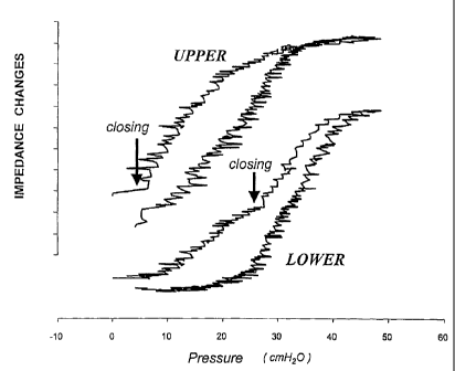
Fig. 7 shows independent inflation-deflation pressure-
impedance curves of the upper and the lower part of the lung.
Compared to the upper curve the lower curve is shifted
towards the right, indicating a delayed opening of dependent
alveoli. As opposed to the upper lung, the lower one does not
show a saturation behaviour of its impedance changes at high
away pressures. Thus, in the dependent lung zones the
CA 02354395 2001-06-08
WO 00/33733 PCT/EP99/09699
17
recruitment of collapsed alveoli still incomplete even at
airway pressures as high as 50 cmH2O. On the deflation limb,
when airway pressures are reduced, collapse of the lower lung
regions occurs earlier than in the respective upper lung
zones (arrows indicate alveolar closing).
Fig. 8 shows impedance curves of the upper and lower parts of
the lung at decreasing levels of positive end-expiratory
pressures (PEEP). The impedance amplitude of the upper lung
(U) is divided by the amplitude of the lower (L) lung. The
U/L-ratio is given in the top line. With decreases in PEEP
the mean impedance of these lung units decreases, too. At
high PEEP levels the upper lung zones are distended (small
amplitude) and ventilation is shifted to the lower lung zones
(large amplitude). The U/L ratio remains below 1. Once
overdistension is relieved, ventilation is distributed more
evenly (U/R - 1). Once PEEP becomes too low to keep all lung
units open, alveoli start to collapse. The amplitude of the
impedance signal of the lower lung decreases and shifts to
the upper lung regions. The U/L ratio exceeds 1. Finally,
hardly any ventilation-induced impedance change can be seen
in the lower curve.
Fig. 9 shows impedance curves of the upper and the lower lung
of a patient suffering from severe lung failure (adult
respiratory distress syndrome, ARDS) on day one on mechanical
ventilation. PEEP is stepwise decreased from 12 to 0 cmH2O.
Initially, as distension is overcome, the amplitude of the
impedance in the upper lung zones increases at the expense of
the ventilation of the respective lower lung zones. Finally,
at a PEEP level below 4 cmH2O a decrease in the impedance
amplitude indicates that alveolar collapse has also occurred
in the upper lung zones. When, after this collapse, the PEEP
level is returned to its original level (100%), the lung
zones do not reach their original state of inflation, again.
Despite the same distending pressure, the upper part achieves
80%, the lower lung only 42% of its original impedance (thus
volume).
CA 02354395 2001-06-08
WO 00/33733 PCT/EP99/09699
18
Fig. 10 shows two curves which are the same as before. In
addition, arterial oxygenation index (Pa02/FiO2) is shown in
the lower line. The open lung is characterized by a Pa02/FiO2
> 500 mmHg. As PEEP is decreased, the lower lung units start
to collapse and ventilation is shifted towards the upper lung
zones. This way, the loss of gas exchanging alveoli in the
lower part of the lung is at least partially compensated.
Oxygenation index decreased only slowly. Once, however, the
PEEP is no longer high enough to stabilize the upper lung
zones, their collapse is indicated by a steep drop in
oxygenation curve. Even when setting the PEEP back to the
original value, the loss of functional lung units is not
reversed. Only 52% of the baseline oxygenation can be
achieved.
As already mentioned above, the invention can make use of an
electrical impedance tomography apparatus. However, it has to
be observed that several adoptions and variations of the
conventional electrical impedance tomography apparatus are
possible to optimise the measurement according to the
invention. This optimisations are described in the following
with reference to the figures 11 to 18.
Fig. 11 shows an optimised external electrodes set up
according to the invention. In order to overcome the known
contact problems of conventional skin electrodes (high
resistance to electrical currents, poor contact between skin
and electrode, displacement and electrical noise with motion
and breathing, etc) electrical bobbins to generate and detect
magnetic field could be used. These could be arranged on
circular band around the thorax or on catheters within the
body. Alternatively the bobbins could be mounted on a fixed
frame that encompasses the thorax. This frame could then be
moved relative to the longitudinal direction of the body to
obtain tomographic or spiral images of different segments of
the thorax.
Furthermore, it should be noted that the number of electrodes
can be increased from 16 to 32 or more electrodes in order to
CA 02354395 2001-06-08
WO 00/33733 PCT/EP99/09699
19
improve the resolution of the signal obtained by regional
electrical impedance tomography even more.
Fig. 12 shows an internal electrodes set up according to the
invention. Generally speaking, the set up according to Fig.
12 is based on the cognition that the distance between the
electrodes should be reduced. It is conceivable that
electrodes or bobbins could be mounted on tubes and catheters
that are placed within the body. Since both the trachea and
the esophagus are located in the approximate centre of the
thorax endotracheal and/or naso-gastric tubes could be used
as electrical centres for the generation of regional
electrical impedance tomographic images. Furthermore,
catheters brought into the blood stream, such as central
venous or pulmonary artery catheters could serve a similar
purpose. Bobbins or electrodes could be placed on one single
or on multiple locations along the tubes and/or catheters in
order to obtain images at different locations within the
chest. It could be feasible to use one or more of these tubes
and/or catheters at the same time. Depending on the clinical
situation of the patient, tomographic images of the
electrical impedance of the chest can thus be generated by
using external electrodes/bobbins around the thorax alone or
by combining them with internal electrodes/bobbins as
'25 described above. Any one of the catheters or tubes has to be
designed according to the needs defined by its general
clinical purpose and by its specific function within the
impedance tomography setting.
Fig. 13 and 14 show a set up in which all electrodes of the
internal set up according to Fig. 12 are used for electrical
impedance tomography measurements. As it becomes from Fig.
14, the distances between the electrodes can be reduced
significantly.
Images and signals from regional electrical impedance
tomography can be used to detect clinically important and
dangerous situations instantaneously. If the endotracheal
tube is placed in the correct anatomical position within the
CA 02354395 2001-06-08
WO 00/33733 PCT/EP99/09699
trachea, both lungs are ventilated evenly. If, however, the
tube is advanced too far only one of the two main bronchi is
intubated; thus only this one lung is ventilated. The EIT-
signal for the non-ventilated lung will be electrically
5 silent whereas the other half of the lung shows a normal or
an increased intensity.
To detect this condition, the regional impedance signal of a
representative part of each lung has to be determined. If the
10 ventilation-induced impedance change falls below an expected
reference value a high suspicion for the presence of an
incorrect intubation is generated. In the presence of such a
suspicion the magnitude of the local impedance change of the
right has to be compared with that of the left lung. If the
15 difference exceeds a certain threshold, a one-sided
intubation can be diagnosed with certainty.
If -for whatever reason- lung tissue is disrupted and free
air gets into the space between the lung and the rib cage
20 (pneumothorax) or in a spaces within the lung (bulla), this
pathological accumulation of air will, after an initial
increase in local impedance, show a markedly reduced or no
further change in its impedance. This region will become
11 silent'' on the EIT-image. The cyclic ventilation of the
surrounding lung tissue demarcates the pneumothorax or bulla.
A similar but opposite change in the impedance properties (a
reduction) can be seen if fluid accumulated in the space
between the lung and the rib cage (pleural effusion). Again
the ventilated lung tissue demarcates the pathological fluid
accumulation.
Fig. 15 shows a set up where only the superior vena cavae is
used for an internal electrode set up. Accordingly, Fig. 16
shows a pulmonary artery (swan-ganz) internal electrode set
up. Furthermore, according to Fig. 17, the intra-tracheal
tube is used for an internal electrode set up. Eventually,
according to Fig. 18, the esophageal is used for an internal
electrode set up. Intrapulmonary, intra-abdominal and
esophageal pressures can be measured by the appropriate tubes
CA 02354395 2001-06-08
WO 00/33733 PCT/EP99/09699
21
or catheters (i.e. endotracheal, esophageal or gastric
tubes, urine or intra-abdominal catheters) . Each one of
these pressures, a combination of them or a difference
between them can be plotted against the signal from regional
impedance tomography to obtain information about the regional
pressure impedance relationship. During mechanical
ventilation this information could be used to titrate the
appropriate levels of airway pressure (i.e. peak or mean
airway pressure or positive end-expiratory pressure) with
respect to regional of global lung expansion and ambient,
intra-abddominal, intra-thoracic or other pressures. Pressure
and impedance signals should be fed into the same device.
In the following, several measures for the improvement of the
signal quality will be described. The improvements in the
efficiency and performance of the electrodes and the signal
transmission will ameliorate the EIT image acquisition in
terms of speed and reliability. This will allow obtaining the
EIT data in synchrony with the respiratory cycle. The
synchronization can be achieved using external ventilator
signals, automated plethysmograph signals or with the
system's own impedance signals. This is of physiological
importance, as it will provide information about the regional
lung changes along the respiratory cycle especially at end
inspiration and expiration. This way tidal recruitment and
de-recruitment of terminal lung (alveoli) within one
respiratory cycle can be detected.
Furthermore the EIT image acquisition can also be triggered
by or synchronized with the cardiac cycle using the signal
from simple ECG electrodes. Regional changes in pulmonary
perfusion can thus be analyzed. Furthermore the
synchronization with the cardiac cycle will help reduce or
eliminate cardiac disturbances of impedance images of the
lung; the resolution of respiratory imaging will thus
increase.
Today, electrical impedance signals of the thorax are
relative signals (they reflect changes but no absolute
CA 02354395 2001-06-08
WO 00/33733 PCT/EP99/09699
22
values) and it has been difficult to convert them into
absolute numbers. Using the above mentioned catheters and/or
tubes within the thorax it is conceivable that internal
reference signals for electrical impedance (i.e. a tissue
calibration factor) could be generated by currents that are
injected and/or received between two or more of these
catheters or tubes.
The circumference of the thorax and therefore the distance
between adjacent electrodes changes with breathing. These
changes can easily be measured by conventional methods or
detected automatically by plethysmographic means. Data
reflecting these changes in circumference can be used within
the algorithms for image reconstruction, thereby enhancing
the quality of the impedance tomographic images. These data
can either be inputted continuously or at discrete time
intervals.
The quality of the images obtained by impedance tomography
alone can be enhanced further if the data from morphometric
measurements or anatomical images are superimposed. Ideally,
measurements or pictures from computed tomography or magnetic
resonance imaging are projected (mathematically,
geometrically or literally) on top of the images obtained
from impedance measurements. Areas with a certain electrical
behavior can thus be seen in relation to their underlying
anatomical structures. This way the size of "gray" zones with
undetermined morphology and functionality can be reduced
(i.e. areas of collapsed lung tissue could be distinguished
from the rib cage, from intrapleural fluid or from bone,
muscle or fat) . Alternatively simple body measurements, (i.e.
weight, height, body mass index, circumferences or others)
could be used to normalize the mathematical algorithms for
impedance image reconstruction.
In the following, an appropriate use of the regional
impedance tomography is described to optimize airway pressure
application in chronic obstructive pulmonary desease (COPD).
In COPD the lung tissue looses its elastic recoil and
CA 02354395 2001-06-08
WO 00/33733 PCT/EP99/09699
23
intrinsic stability. During expiration, small airways
collapse if the pressure within them gets lower than a
certain threshold pressure. Gas is thus trapped within the
lungs. If inspiratory pressures are higher than the pressures
required to re-expanding these collapsed airways, gas can
move into the terminal parts of the lung and the alveoli. If
the inspired amount of gas is larger than the amount that
leaves the lung during expiration the lung is gradually
expanded until a new steady state at high lung volumes is
reached. The way the diseased lug tissue is easily
overdistended and is rendered incapable of gas exchange.
In COPD the collapse of airways can be found in one part of
the lung and the overdistension of lung units in another.
Thus both these pathological situations can found at the same
time.
At times, patients with COPD require support of their
ventilation by the application of positive (or more
infrequently negative) pressure ventilators. If the absolute
amount of airway pressure is too high, lung tissue gets
overdistended and dysfunctional for gas exchange. If,
however, the applied pressures are too low to prevent the
collapse of small airways, gas is trapped within the lung
without being efficiently exchanged. Often, airway collapse
and overdistension coexist within the same lung at a chosen
pressure. For an optimal therapeutic result, the best
compromise between these two conflicting lung conditions has
to be found. Traditional lung mechanics give only a rough
estimate of such a compromise. Information about the regional
expansion and movement of air is required to approach this
comprise.
Regional electrical impedance tomography provides data and
images of regional lung ventilation. With increases in airway
pressures the gradual emptying of trapped gas can be detected
in one area of the lung, whereas other parts of the lung get
progressively distended until in the truly overdistended
stage no changes in impedance can be detected. By comparing
CA 02354395 2001-06-08
WO 00/33733 PCT/EP99/09699
24
and integrating the quantities of overdistension and
emptying of the various portions of the lung at changing
airway pressures a best therapeutic "compromise pressure" can
be found that reflects optimal lung expansion at minimal
S pressures.
Furthermore, not only electrodes can be used on the
catheters, but only the pressure measurements of the
catheters can be used for optimising the accuracy of the
regional pressure impedance curves.