Note: Descriptions are shown in the official language in which they were submitted.
CA 02356309 2001-06-26
WO 00/41629 PCTlIB00100009
1
SAFETY MECHANISM AND METHODS FOR ROTATING IMAGING DEVICE
BACKGROUND OF THE INVENTTON
The invention relates generally to the field of
ultrasonic imaging, and in particular to the imaging of body
lumens or cavities. More specifically, the invention relates
to the use of imaging devices that are. rotated at high speeds
to produce an image of a body lumen or cavity.
The use of rotatable imaging devices to produce an
image of a body lumen is well known. For example, one
pioneering effort is described in U.S. Patent No. 4,794,931,
the complete disclosure of which is herein incorporated by
reference. In U.S. Patent No. 4,794,931, a drive cable having
an imaging element at a distal end is rotated within a
catheter to product an image of a diseased region prior to
therapy.
Recently, there has been an advancement in the field
of rotatable imaging devices where the size of the imaging
devices has been substantially reduced. For instance, one
such imaging device is described generally in copending U.S.
Application Serial No. 09/017,576, filed February 3, 1998, the
complete disclosure of which is herein incorporated by
reference. Such an imaging device is small enough to operate
within traditional guide wire lumens of therapeutic catheters,
such as angioplasty balloon catheters.
,,One potential problem that may arise when operating
imaging devices within catheter lumens having a distal exit
port, such as within guide wire lumens of therapeutic
catheters, is that the rotating imaging device may
accidentally be advanced beyond the distal exit port and into
the body lumen, thereby posing a risk of damage to the luminal
wall.
CONFIRMATION COPS
CA 02356309 2001-06-26
WO 00/41629 PCT/IB00/00009
2
Hence, it would be desirable to provide a way to
prevent the unwanted advancement of a rotating imaging device
beyond a distal exit port of a catheter. Such a safeguard
should be reliable and easy to use to~maximize its acceptance
in the industry.
SUNll~IARY OF THE INVENTION
The invention provides exemplary techniques for
preventing the unwanted advancement of a rotating imaging
device beyond an exit port of a catheter and into a body
lumen. In one exemplary embodiment, a catheter is provided
which comprises a catheter body having a proximal end, a
distal end and a lumen, such as a guide wire lumen, which
terminates in an exit port at the distal end. An
ultrasonically recognizable pattern is disposed proximally to
or at the exit port. The recognizable pattern is provided to
produce a unique image when imaged with an imaging element of
an imaging device which is rotated within the lumen.
The catheter is preferably included as part of a
system which includes a controller having a motor to rotate
the imaging device. The controller is configured to stop
rotation of the imaging device upon receipt of a signal from
the imaging device indicating that the presence of the
recognizable pattern has been detected. In this way, once the
imaging device has been advanced through the lumen and up to
the recognizable pattern, the presence of the pattern will be
detected by the controller which will stop rotation of the
imaging device. As such, if the imaging device is advanced
beyond the exit port, the imaging device will not be rotating,
thus substantially reducing the chances of damaging the
luminal or cavity wall. Alternatively, if the motor is
employed to also translate the imaging device, the signal may
be employed to stop translation of the imaging device so that
the rotating imaging device will not be advanced distally
beyond the exit port.
A wide variety of recognizable patterns may be
provided to indicate when the imaging device has advanced too
far within the lumen. For example, the pattern may comprise a
CA 02356309 2001-06-26
WO 00/41629 PCT/IB00/00009
3
tubular reflective member which is crimped or otherwise
attached about the tubular body. Other patterns which may be
employed include ultrasonically reflective materials having a
variety of shapes and sizes which may be attached to or
integrally formed within the catheter body, echogenic
coatings, changes in the diameter of the catheter body, the
distal end of the catheter body, and the like. Preferably,
the pattern is fashioned to have a shape or configuration
which allows it to be differentiated from the rest of the
image. For example, the pattern may include a plurality of
elongate apertures which will appear as voids in the resulting
image, thus differentiating the pattern from a stent. As the
controller recognizes the voids, rotation of the imaging
device is stopped.
The catheter is preferably a therapeutic catheter
having a therapeutic element for treating a region of the body
lumen. For example, the therapeutic element may comprise an
angioplasty balloon. As another example, the therapeutic
element may comprise a stent delivery system. As a further
examples, the therapeutic element may comprise a laser or a
rotatable cutting element.
In another aspect, the lumen preferably extends the
length of the catheter body. In this way, the catheter may be
inserted into the body lumen over a guide wire in an over-the-
wire manner. Typically, the lumen will have a diameter in the
range from about 0.25 mm to about 5 mm, and from about 0.25 mm
to about 0.5 mm for applications within the coronary arteries.
The imaging device preferably has a diameter in the range from
about 0.20 mm to about 2 mm.
The invention further provides an exemplary
attachment for a catheter that has a lumen terminating in an
exit port at a distal end of the catheter. The attachment
comprises a tubular member which may be coupled about the
catheter proximal to the exit port. The tubular member is
constructed of an ultrasonically reflective material and has a
unique shape that will produce a unique image when imaged with
an ultrasonic imaging element which is rotated within the
lumen. In this way, a catheter may be conveniently modified
CA 02356309 2001-06-26
WO 00/41629 PCT/IB00/00009
4
so that it may be used with a safety system that will stop
rotation of an imaging element upon detection of the tubular
member.
In one exemplary method of 'the invention, a body
lumen is visualized by introducing a catheter into the body
lumen. The catheter comprises a catheter body having a lumen
which terminates in an exit port and an ultrasonically
recognizable pattern disposed at or near the exit port. An
imaging device is introduced through the lumen and positioned
so that an imaging element is at a location that is to be
imaged. The imaging device is rotated while the imaging
element is actuated to produce an image of the body lumen.
Rotation of the imaging device is stopped if an image of the
pattern is detected so that advancement of the rotating
imaging device beyond the exit port is prevented.
Alternatively, translation of the imaging device may be
stopped so that the rotating imaging device will not moved
distally beyond the exit port.
In one aspect, the catheter is introduced into the
body lumen by advancing the catheter over a guide wire. Once
properly positioned, the guide wire is withdrawn and the
imaging device is introduced into the guide wire lumen.
In another aspect, a therapeutic element is deployed
while the imaging device is rotating to produce an image of
the therapeutic element. In this way, the body lumen may be
visualized throughout the therapeutic procedure. For example,
visualization may occur while a balloon is being inflated or a
stent is being deployed. The pattern preferably has a unique
shape to allow it to be easily differentiated from the
therapeutic element. In this way, once the unique shape is
detected, rotation of the imaging device may be stopped.
BRIEF DESCRIPTION OF THE DRAWINGS
Fig. 1 is a schematic view of an exemplary imaging
system according to the invention.
Fig. 2 is a cross-sectional side view of a catheter
having a tubular reflective member disposed near a distal end
according to the invention.
CA 02356309 2001-06-26
WO 00/41629 PCT/IB00/00009
Fig. 2A is a cross-sectional view of the catheter of
Fig. 2 taken along lines A-A.
Fig. 3 is a perspective view of an exemplary tubular
reflective member according the invention.
5 Fig. 4 is a partially cut-away side view of a distal
end of a catheter having the tubular reflective member of Fig.
3.
Fig. 5 illustrates an alternative embodiment of a
tubular reflective member disposed about a distal end of a
catheter according to the invention.
Fig. 6 illustrates the catheter of Fig. 4 having an
imaging element of a rotating imaging device disposed within
the tubular reflective member according to the invention.
Fig. 7 illustrates a reflected image that is
detected by the imaging element of Fig. 6 when disposed within
the tubular reflective member according to the invention.
Fig. 8 is a schematic view of a distal end of a
catheter showing an electrical circuit which is opened when an
imaging device is advanced beyond a distal end of the catheter
according to the invention.
Fig. 9. illustrates a cross-sectional end view of a
distal end of a catheter having a plurality of radiopaque
markers disposed within the catheter body according to the
invention.
DETAILED DESCRIPTION OF THE SPECIFIC EMBODIMENTS
The invention provides various systems and
techniques to prevent the advancement of a rotating imaging
device beyond a distal exit port of a catheter. In this way,
if the imaging device is inadvertently advanced beyond the
exit port and into a body lumen or cavity, the techniques
provided by the invention will stop rotation of the imaging
device to substantially reduce or eliminate the risk of
perforating the wall of the body lumen.
The invention may be'used with essentially any
rotatable imaging device which is rotated within a lumen or
cavity of a catheter body to produce an image. Such imaging
. devices typically comprise an elongate drive cable having an
CA 02356309 2001-06-26
WO 00/41629 PCT/IB00/00009
6
ultrasonic imaging element or transducer disposed at a distal
end. Such imaging devices include, among others, imaging
cores, imaging wires, imaging guide wires, and the like.
Merely by way of example, rotatable imaging devices which may
be used with the invention are described in U.S. Patent No.
4,794,931, previously incorporated by reference, and in
copending U.S. Application Serial Nos. 09/017,578, filed
February 3, 1998 and 60/059,718, filed September 22, 1997, the
disclosures of which are herein incorporated by reference.
The rotatable imaging devices of the invention may
have a wide range of outer dimensions, including outer
diameters in the range from about 0.20 mm to about to about
0.5 mm for applications within the coronary arteries. Such a
range of diameters allows the imaging devices to be used
within conventional guide wire lumens.
Catheters which may be used-with the invention
preferably comprise a catheter body having a proximal end, a
distal end and at least one lumen which terminates at an exit
port at the distal end. In many cases, the lumen will
comprise a guide wire lumen which is employed to introduce the
catheter into a body lumen in an over-the-wire manner.
Following insertion, the guide wire lumen serves as an imaging
lumen to receive the imaging device so that an image may be
produced. Because the lumen terminates at the distal end, the
2S invention provides techniques for stopping rotation of the
imaging device either before or upon the exit of the imaging
device from the exit port. Exemplary catheters having a lumen
which terminates in an exit port at the distal end include
PTCA catheters, PCA catheters, various other balloon
catheters, atherectomy catheters, "common lumen" catheters as
described generally in U.S. Patent No. 5,314,408, the complete
disclosure of which is herein incorporated by reference, and
the like. The imaging lumens of such catheters have a
diameter which is large enough to receive the rotatable
imaging device so that an image may be produced.
The invention may be used in connection with a
variety of diagnostic and therapeutic procedures which involve
the use of a rotatable imaging device. Such procedures can
CA 02356309 2001-06-26
WO 00/41629 PCT/IB00/00009
7
include, for example, imaging in real time stent deployment
and placement, imaging during placement of radiation devices
in radiation procedures, imaging during directional coronary
atherectomy procedures (DCA), imaging'during placement of a
balloon during balloon ai~gioplasty procedures, imaging while
placing stent grafts, imaging during neurology procedures,
imaging during urology procedures, imaging during gastro-
intestinal procedures, imaging intracardiac structures during
ablation, and the like.
Advancement of the rotating imaging device beyond
the distal exit port is preferably accomplished by providing
an ultrasonically recognizable pattern at or proximal to the
distal end of the catheter body. During imaging, the imaging
element captures a reflected signal which is sent to a
Z5 controller to produce an image. When the imaging element
reaches'the pattern; a signal is reflected indicating the
presence of the pattern. Once the pattern is detected by the
controller, rotation of the imaging device is stopped so that
the imaging device will not be rotating when advanced beyond
the distal end of the catheter body. In one alternative, a
motor which is employed to translate the imaging device
through the catheter body may be stopped when the pattern is
detected. In this way, the imaging device will be prevented
from distally advancing beyond the exit port.'
A wide variety of ultrasonically reflective patterns
may be employed to assist in stopping rotation of the imaging
device upon detection of the pattern. For example, the
pattern may comprise a tubular reflective element that is
disposed about the catheter body. In this way, an existing
catheter may easily be modified to include an ultrasonically
reflective pattern that may be detected to stop rotation of
the imaging device.
Other ultrasonically reflective patterns include
echogenic coatings, such as Echo-CoatT", that provide an
acoustically reflective interface between the catheter and the
coating. Such coatings may be blended in the catheter body or
applied to an external surface of the catheter body.
Preferably, the remainder of the catheter body is constructed
CA 02356309 2001-06-26
WO 00/41629 PCT/IB00/00009
8
of an echo translucent or an acoustically transparent
polymers) so that the remainder of the catheter body is not
displayed in the image produced on the monitor. Such coatings
may be applied circumferentially or in any type of pattern
S that may be recognized and detected.
Other possible ultrasonically recognizable patterns
include the use of ultrasonically reflective polymers which
are disposed at or near the distal end of the catheter body.
Such polymers may be formed as part of a co-extrusion or as a
blended material within the catheter body. Such polymers are
preferably fashioned in a unique shape or composition to
facilitate discernment of the pattern. As another
alternative, radiopaque markers. may be disposed on or within
the catheter body and may be constructed from materials such
as gold, tantalum, platinum, palladium, and the like. Such
markers may be placed at known distances from each other. The
controller may be configured to detect these distances in the
resulting image to stop rotation of the imaging device. Still
further alternatives include the use of holes disposed in the
catheter body or a change in the diameter of the imaging lumen
of the catheter. As still another alternative, the distal end
of the catheter body may be detected to stop rotation of the
imaging device. In summary, the ultrasonically reflective
pattern may comprise any detectable pattern that may be
differentiated from the rest of the image produced to allow
system software to stop the imaging element from rotating
and/or translating.
In one alternative embodiment, spring-loaded
contacts may be provided at the distal end of the catheter.
In this way, when the imaging device passes through the distal
end, the contacts are opened causing a break in an electrical
circuit.
Referring now to Fig. 1, an exemplary embodiment of
an ultrasonic imaging system 10 will be described. System 10
comprises a controller 12 which is coupled to a monitor 14.
Controller 12 is also coupled to a motor 16 which is employed
to rotate a drive cable 18 of an imaging device. Controller
12 includes circuitry and software which is configured to
CA 02356309 2001-06-26
WO 00/41629 PCT/IB00/00009
9
receive a reflected signal from an ultrasonic imaging element
and to produce an image based on the reflective signal on
monitor 14. An exemplary controller which may be used with
the invention is a Clear View Ultra'" intraluminal ultrasound
system, commercially available from Boston Scientific
Corporation. A variety of commercially available motors may
be employed to rotate drive cable 18.
Referring now to Fig. 2, one type of catheter 20
which may be utilized with system 10 will be described, it
being appreciated that a wide variety of catheters may be
employed with the invention as previously described. Catheter
is representative of a conventional PTCA catheter which
comprises a catheter body 22 having a proximal end 24 and a
distal end 26. Coupled to proximal end 24 is a hub 28 having
15 a balloon inflation port 3o and a guide wire or imaging device
entry port 32. As best shown in Fig. 2A, disposed within
catheter body 22 is a sheath 34 having a central lumen 36.
Disposed within lumen 36 is a guide wire 38. As illustrated
in Fig. 2, guide wire 38 extends between entry port 32 and
20 distal end 26. As is known in the art, catheter 20 may be
inserted through a body lumen by first inserting guide wire 38
into the lumen and then advancing catheter 20 over guide wire
38 in an over-the-wire manner.
Catheter 20 further includes a balloon 39 which is
inflated by introducing a fluid through balloon inflation port
30. As best illustrated in Fig. 2A, a balloon inflation lumen
40 is provided between catheter body 22 and sheath 34 to
deliver the fluid from port 3o to balloon 39.
Also disposed at distal end 26 is a tubular
reflective member 42. Reflective member 42 is constructed of
an ultrasonically reflective material, such as stainless
steel, and is placed about catheter body 22. In this way,
when guide wire 38 is withdrawn from lumen 36 and an imaging
device is inserted through lumen 36 and rotated to produce an
image, reflective member 42 will be visualized by the imaging
device if advanced up to reflective member 42. The placement
of reflective member 42 just distal to balloon 39 is
advantageous since the imaging element will often be employed
CA 02356309 2001-06-26
WO 00/41629 PC'T/IB00/00009
to visualize proper placement of balloon 39. In the event
that the imaging element is advanced beyond balloon 39, the
presence of reflective member 42 will~~be detected so that
rotation and/or translation of the imaging device may be
5 stopped.
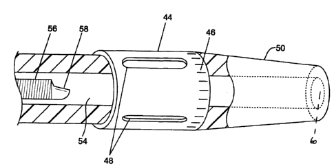
Referring now to Fig. 3, an exemplary embodiment of
a tubular reflective member 44 will be described. Reflective
member 44 comprises a tubular body 46 that is constructed of
an ultrasonically reflective or opaque material, such as
10 stainless steel. Tubular body 46 includes three elongate
apertures 48. The placement of apertures 48 is advantageous
in that the resulting image produced by the imaging device is
essentially the inverse image of that produced by a stent as
described in greater detail hereinafter. In this way,
reflective member 44 may easily be distinguished from a stent.
Although shown with three elongate apertures, it will be
appreciated that the number, size and geometry of the
apertures may be varied. Use of a tubular body is
particularly advantageous in that it may easily be crimped or
otherwise attached about an existing catheter to provide the
catheter with an ultrasonically reflective pattern that may be
detected to stop rotation of the imaging device. As one
example, tubular body 46 may be crimped at one end to secure
tubular body 46 to a catheter body 50 as illustrated in Fig.
2S 4. Alternatively, as illustrated in Fig. 5, tubular body 46
may include a longitudinal slit 52 to facilitate the crimping
of tubular body 46 about catheter body S0.
As illustrated in both Figs. 4 and 5, catheter body
50 includes a lumen 54 into which a rotatable imaging device
56 is received. Imaging device 56 includes an imaging element
58 which is rotated within lumen 54 to produce an image of the
area surrounding catheter body 50 as is known in the art.
Catheter body 50 further includes an exit port 60. To prevent
the advancement of imaging device 56 through exit port 60
while imaging device 56 is rotating, tubular reflective member
44 is placed just proximal to or at exit port 60. As
illustrated in Fig. 6, when imaging device 56 is distally
advanced within lumen 54, imaging element 58 will eventually
CA 02356309 2001-06-26
WO 00/41629 PCT/IB00/00009
11
reach reflective member 44. The resulting image that is
detected by reflective member 44 is produced on a monitor
screen as illustrated in Fig. 7. In image 61, three echos 62
are illustrated and represent the metallic areas of reflective
member 44. Three voids 64 exist which are representative of
apertures 48 in tubular body 46. When the controller detects
the pattern of voids 64, it knows that imaging device 56 has
been advanced up to tubular reflective member 44. As such,
the controller will send a signal to stop rotation of imaging
device S6 so that if it is advanced beyond exit port 60 it
will not be rotating, thereby posing no risk of danger to the
luminal wall of the patient.
Referring back now to Fig. 1, a description of one
exemplary algorithm employed by controller 12 to stop rotation
15. _of motor 16 when the presence of an ultrasonically reflective
pattern is detected will be described. Such an algorithm is
particularly useful when employing a tubular member with three
equidistantly spaced slots which are parallel to the axis of
the tubular member as illustrated in Fig. 3. Such slots
provide a distinct ultrasonically detectable signature that
does not occur naturally within human vessel, or as a
byproduct of transcatheter or surgical interventions.
In this embodiment, controller 12 preferably
acquires data in Polar (R-B) format. The data acquired
includes a series of individual frames (one complete 360
degree rotation of the imaging device) of sample points.
Controller 12 preferably acquires 256 equally spaced 8-bit
samples along a vector, with 256 vectors per frame (a frame
being one complete 360 degree rotation of the imaging device).
One frame, or data set, is therefore a 256 by 256 array of 8-
bit sample values. The distance from the transducer face (R),
of a given data point, is determined by multiplying the sample
spacing (propagation speed of ultrasound times the sample
period) by the sample number (depth) along a given vector.
The angle (B)in degrees relative to the beginning of the frame
is determined by dividing 360 degrees by the number of vectors
within a frame times the vector number (360 degrees/256*vector
number). Hence, the 8-bit sample at array position [0,0] is
CA 02356309 2001-06-26
WO 00/41629 PCT/IB00/00009
12
acquired at the face of the transducer, when the imaging
device is directed at the 12 O'clock position [255,127] is the
last sample on the vector pointed at the 6 O'clock position
(middle of the frame 180 degrees).
The face of the transducer on the imaging device is
located a fixed depth within the catheter body. At the
beginning of a vector, acoustic energy is transmitted from the
transducer. The receiver begins sampling the vector, and an
acoustic near field artifact is generated that typically
settles to a sample value less than 50w (1/5 full-scale) by the
outer edge of the catheter body. When the imaging device is
advanced into the tubular member, the reflective material of
which the tubular member is constructed generates a return
echo at the catheter body that will have a sample value of at
least 200 (4/S full-scale). This high value is present on
all vectors directed at the tubular member. When the imaging
device is pointed at the slot within the tubular member, the
sample values will return to less than 50, and remain at this
low value until the tubular member is again encountered. A
frame of data sampled from within the tubular member contains
a pattern of three long (approximately 64 vectors) highs, and
three short (approximately 21 vectors) lows (depending on the
thickness of the slots) at the outer edge of the catheter
body. This pattern is present when the sample values are
wrapped around so that the beginning and end of the frame do
not form separate highs and lows. A digital signal processor
or other suitable device is employed to continuously monitor
the sample values searching for this pattern. When such a
pattern is encountered, and repeated over a fixed number of
frames, then the digital signal processor commands the motor
rotating the imaging device to stop.
Referring now to Fig. 8, an alternative embodiment
of a catheter body 66 will be described. For convenience of
discussion, only a distal end of catheter body 66 will be
described. Disposed in catheter body 66 is an electrical
circuit 68 having a pair of contacts 70 which are biased
together by a pair of springs 72. When contacts 70 are
adjacent to each other, the circuit is closed. If, however,
CA 02356309 2001-06-26
WO 00/41629 PCT/IB00/00009
13
an imaging device is passed through an eXit port 74, contacts
70 will move apart from each other and cause the circuit to
open, providing that the imaging device tip is non-conductive.
The opening of circuit 68 may then be'detected by the
controller to stop rotation of the imaging device in a manner
similar to that described with previous embodiments.
Referring to Fig. 9, another alternative embodiment
of a catheter body 76 will be described. Catheter body 76 is
shown in cross-section and includes four equally spaced
radiopaque markers 78. Markers 78 are disposed at a distal
end of catheter body 76 and are spaced at known angles
relative to each other so that the image produced by the
imaging device may be detected by the controller to stop
rotation of the imaging device. Although markers 78 are shown
15' within catheter body 76, it will be appreciated that markers
78 may be disposed externally on catheter body 76. Further,
the shape, size, geometry and configuration of markers 78 may
be varied to produce a distinct recognizable image that may be
employed to stop rotation of the imaging device.
In another alternative of the invention, two or more
ultrasonically distinct patterns may be positioned such that a
region of interest is defined. For example, such patterns may
be placed at two ends of a balloon or stent. These patterns
may be employed to produce a start pattern and a stop pattern
?5 that is recognized by the controller. In this way, the
imaging device may automatically be moved back and forth
within the region of interest to provide multiple views of the
region of interest.
Although the foregoing invention has been described
in detail for purposes of clarity of understanding, it will be
appreciated that certain modifications may be practiced within
the scope of the appended claims.