Note: Descriptions are shown in the official language in which they were submitted.
CA 02356867 2001-06-27
ADV-6973
- 1 -
DESCRIPTION
MICROORGANISM IDENTIFICATION SYSTEM
TECHNICAL FIELD
The present invention relates to a system for
identifying a causative microorganism of an infection by
determining various types of inflammation markers present
in biological specimens, particularly blood, urine,
saliva, nasal mucous, phlegm, exudate from diseased
areas, etc. and analyzing the reaction patterns thereof.
Upon infection by a microorganism, inflammation markers
are produced in the body according to its type and appear
in the above biological specimens. By determining the
types, amounts of production, molecular structures, etc.
of the inflammation markers, it is possible to
immediately identify the causative microorganism.
BACKGROUND ART
In the past, bacteria, mycetes, viruses infection
microorganisms such as bacteria, mycetes, viruses have
been identified and diagnosed by the method of culturing
the blood etc. of the patient and making a determination
from the result and the method of detecting an antigen or
antibody specific to the causative microorganism from the
blood serum etc.
The culturing method includes a method of culturing
for several days or more by a medium selecting
microorganism and then separating and identifying it and
the method of identification from biochemical symptoms
based on the metabolism of the microorganisms. Normally,
several types of microorganisms are involved in
infection, and therefore, in order to confirm the
presence of the several types of causative microorganisms
suspected, it is necessary to use a number of selective
media or biochemical property test kits. Accordingly, a
relatively large amount of time is required until the
determination. Further, it is sometimes difficult to
CA 02356867 2001-06-27
- 2 -
detect a causative microorganism from the blood. This
becomes a major problem in the case of an infectious
disease where prompt appropriate determination has to be
taken.
As a method of diagnosis of blood serum, there is a
method of detecting the microbial antigens, anti-
microbial antibodies, microbial components, microbial
metabolites, etc., followed by identifying the causative
microorganisms. This method of diagnosis of blood serum
has the advantage of enabling identification and
diagnosis in a shorter time, compared with the blood
culturing method, but suffers from numerous problems such
as the problem in the specificity to the antigens or
components of the microorganism, the fact that microbial
metabolites are small in amount, the detection
sensitivity is low, and the types identified by test kits
are limited, the fact that several days are taken before
the appearance of antibodies due to infection in the case
of anti-microbial antibodies, and the fact that sometimes
antibody-positive responses are given due to existing
opportunistic infections.
On the other hand, as the method for determining
inflammation markers, there is capillary precipitation,
single immunodiffusion, latex turbidity,
immunofiltration, immunoturbidity, enzyme immunoassay,
etc. These assay methods are being actively used when
diagnosing the activity, gravity, and progress of various
diseases causing inflammation or tissue disorders.
However the above method are not used as the method for
identification of the causative microorganisms in
infectious diseases.
Infectious diseases are sometimes acute conditions,
where life and death hangs in the balance, and therefore,
it is necessary to determine the causative microorganisms
as fast as possible and apply quick treatment by
administration of the appropriate antibiotics. However,
as explained above, the culturing method is problematic
CA 02356867 2001-06-27
- 3 -
in the speed of identification of the causative
microorganisms at present, while the serum diagnosis
method is problematic in the detection sensitivity and
specificity and in the range of application etc.
Accordingly, the practice is to administer several
antibiotics, while guessing the causative microorganisms.
Accordingly, various side effects arise and
microorganisms tolerant to antibiotics are created.
DISCLOSURE OF THE INVENTION
The object of the present invention is to solve the
above problem by providing a system for immediately and
easily identifying all of the causative microorganisms of
an infection by determining the state of inflammation
markers generated from an infection.
In accordance with the present invention, there is
provided a microorganism identification system for
identifying a causative microorganism comprising
determining inflammation markers in the body generated
from microbial infection.
BRIEF DESCRIPTION OF THE DRAWINGS
The present invention will be explained further
below with reference to the drawing:
Figure 1 is a view for explaining the present
invention.
BEST MODE FOR CARRYING OUT THE INVENTION
To immediately identify a causative microorganism,
the method of examining the white blood cells, which
change along with infection, has been devised in addition
to the method of detecting the substances relating to
infecting microorganisms by applying the antigen-antibody
method. However, these methods have problems in terms of
convenience since a large amount of biological specimens
is required and an equipment not suitable for carrying
around is used.
The present inventor found that the types and
amounts of production of various inflammation markers
appearing in the body and their molecular structures etc.
CA 02356867 2001-06-27
- 4 -
differ depending upon the causative microorganisms of
infections. Inflammation markers appear due to
inflammation due to major invasive surgery, post-surgical
infection, invasion of pathogenic bacteria, etc. However,
the pattern of expression of inflammation markers, which
is correlated with the fluctuations in the state of the
disease, differs depending upon the causes of the
inflammation. Further, changes are seen in the molecular
structures of the various proteins of the inflammation
markers due to the differences in the causative
microorganisms.
That is, the present invention determines the types
of inflammation markers, the ratio of the amounts
produced, the state of change in the molecular
structures, etc. and analyzes the pattern of expression
to determine whether or not there is infection and, when
there is infection, the causative microorganisms is
determined.
Inflammation markers are proteins, and therefore, it
is possible to prepare antibodies of the inflammation
markers. Accordingly, when a plurality of antibodies
prepared using, as antigens, the inflammation markers
expressed in the body due to infection by various types
of microorganisms etc. are provided on a microorganism
identification chip and, when biological specimens
obtained for a specific purpose are reacted, an antigen-
antibody pattern depending upon the causative
microorganisms will appear. Note that, when the amount of
antigens is very small, the reaction sensitivity can be
raised by combining a chemiluminescence system (for
example, acridinium is used, as a luminescent substance,
or luminol is used, as a chemiluminescent material).
Then, the antigen-antibody reaction pattern of the
microorganism identification chip is optically read and
analyzed by a simple device programmed with the reaction
patterns of various microorganisms in advance, it is
possible to accurately identify the causative
CA 02356867 2001-06-27
- 5 -
microorganisms.
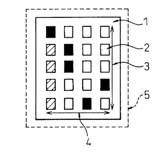
Figure 1 will now be explained. Figure 1 shows an
example of the configuration of the present invention.
Reference numeral 1 indicates a test chip body made
of polyester film, etc.
The overall size is, for example, 25 to 250,000 mm2,
preferably 100 to 400 mm2, with a thickness of 0.5 to 5
mm, but it may be suitably adjusted to another size
depending upon the number of amount of the inflammation
markers to be determined.
In the Figure, reference numeral 2 indicates one
location where an inflammation marker antibody is
arranged. The size of one location is, for example, 0.025
to 25 mm2 (preferably 1 to 4 mm2). In Figure 1, 20
inflammation marker antibodies are arranged. The distance
of arrangement is set to a suitable distance for
optically measuring the surface, but a distance of about
0.5 to 2 mm in the left and right and up and down
directions may be preferably illustrated.
Each vertical five-location array 3 includes
different inflammation marker antibodies, while each
horizontal array 4 includes the same inflammation marker
changed in the molecular structure, such as molecular
weight, isoelectric points, i.e., the array of so-called
"isomers".
This square or rectangular uniform array is an
example. It is also possible to use a triangular shaped
array, diamond shaped array, or other various arrays or
even uneven patterns.
The four patterns of isomers and the five types of
antibodies are preferable numbers, but more or less of
these are also possible depending upon the extent to
which the microorganisms to be determined can be
confirmed.
Figure 1 shows an example of a pattern of color
formation in the case of an actual reaction. The pattern
can be read by an optical measurement apparatus (e.g., a
CA 02356867 2001-06-27
- 6 -
CCD camera, infrared or X-ray camera, fluorescent
scanner, or PC scanner).
EXAMPLES
The present invention will now be further explained
in detail, but is by no means limited to, the following
Examples.
Example 1
The blood and urine of eight cancer patients
undergoing major invasive surgery were taken before and
directly after surgery and on the second, third, fifth,
seventh, 10th, 14th, and 21st days from the day after
surgery. Further, each of the patients was administered
albumin (ALB) from during surgery to the third day after
surgery and was administered a fatty emulsion from the
fourth day. The blood and urine thus obtained were
separated and purified using cellulose acetate membrane
electrophoresis and sodium dodecyl sulfate (SDS)
polyacrylamide gel electrophoresis, gel filtration,
various types of chromatography, etc. depending upon the
objective in order to obtain the al-acidic glycoprotein,
al-antitrypsin, haptoglobin, CRP, lactoferrin,
transferrin, ceruloplasmin, lysozyme, granular elastase,
myoperoxidase, IL6, IL8, and other protein-based
inflammation markers. The inflammation markers thus
obtained were classified into those of the noninfected
patients and infected patients. Further, the inflammation
markers of the infected patients were classified by
causative microorganisms identified by conventional
methods.
Next, the inflammation markers were injected, as
antigens, into rabbits together with adjuvants. Anti-
human inflammation marker rabbit antibodies were prepared
by an ordinary method from the blood serum. The various
types of antibodies thus obtained were placed in porous
tissue in the case of the wet type and were arranged on
filter paper in the case of the dry type and then dried
CA 02356867 2001-06-27
- 7 -
to prepare the test chip.
Example 2
In order to prepare a test solution for reaction
with the inflammation marker antibodies placed on the
test chip, inflammation marker antibodies were injected,
as antigens, into goats to prepare anti-rabbit
inflammation marker goat antibodies. After preparing the
goat antibodies, labeling substances were bonded for the
color forming operation. Luminol or a acridinium ester
derivative was used as the label according to an ordinary
method to raise the luminescence sensitivity.
This enabled measurement by chemiluminescence and,
as a result, a method of raising the sensitivity to 10 to
12 mol was able to be obtained.
Example 3
The blood and urine of eight cancer patients
undergoing major invasive surgery obtained before and
directly after surgery and on the second, third, fifth,
seventh, 10th, 14th, and 21st days from the day after
surgery were dropped on the test chip as biological
specimens, then reactions were caused with the test
solutions and the luminescence patterns were optically
measured.
Further, for confirmation, the total protein was
measured by the burette method, the albumin by the BCG
method, the C-reactive protein (CRP) by the
immunoturbidity, the granular elastase by latex
agglutination, and the al-acidic glycoprotein, al-
antitrypsin, and haptoglobin by a COBASMIRA (Trade mark)
automatic system (made by Roche).
The present invention will now be explained using
the results of Comparative measurement or identification
using the present identification system and a
conventional identification system.
Example 4
The arterial and venous blood and urine of seven
cancer patients undergoing major invasive surgery and
CA 02356867 2001-06-27
-
then not showing complications were obtained before and
directly after surgery and on the second, third, fifth,
seventh, 10th, 14th, and 21st days from the day after
surgery. The degree of invasiveness of the surgery was
investigated by the fluctuations in the proteins of the
inflammation markers .
Further, all the patients were administered albumin
(ALB) from during surgery to the third day after surgery
and were administered a fatty emulsion from the fourth
day. The arterial and venous blood and urine thus
obtained were measured comparatively by the present
identification method and the conventional method. As a
result, a high correlation was observed in the reaction
pattern of the inflammation markers and the usefulness of
the present identification method was confirmed.
The results of the measurement by the present
identification system are given below. The total protein
dropped from immediately after surgery to the second and
third day, then returned to the presurgical value from
the seventh to 14th day. The albumin fell immediately
after surgery, then increased from the first to third
days after surgery. The effect of administration of
albumin was observed.
The CRP sharply rose immediately after surgery and
fluctuated the most wildly. The granular elastase rose
rapidly from the first to second day after surgery. The
al-antitrypsin fell directly after surgery, increased
from the first day after surgery, and peaked from the
fifth to seventh day. The haptoglobin fluctuated in the
same way as the al-antitrypsin, but peaked from the
fifth to the 10th day. The (32-microglobulin fell from
immediately after surgery to the second day, then
increased, peaked from the 10th day to the 14th day, then
fell again.
In all inflammation markers, the trends in
fluctuation of the arterial blood and venous blood were
CA 02356867 2001-06-27
- g _
similar. In diagnosing the condition of the body due to
invasive surgery when there are no complications such as
infections, the fluctuations in the CRP and granular
elastase exhibited the highest correlation with the
degree of invasiveness. The other inflammation markers
fell immediately after surgery, but it was learned that
the peaks of fluctuation became later in the order of
al-antitrypsin, al-acidic glycoprotein, haptoglobin, and
~2-microglobulin.
It could be confirmed from the above results that
the present identification method is useful in diagnosing
the condition of the body after surgery when there are no
complications.
Example 5
The blood serum and urine of eight cancer patients
undergoing major invasive surgery and then showing
infectious diseases obtained before and directly after
surgery and on the second, third, fifth, seventh, 10th,
14th, and 21st days from the day after surgery were
dropped on test chips of the present identification
method and reactions of the test solutions were caused,
then the chemiluminescence patterns were optically
measured. As a result, with post-surgical infection, a
high correlation was shown between the degrees of
fluctuation of al-acidic glycoprotein, al-antitrypsin,
and haptoglobin and the state of the infectious disease.
For confirmation of this correlation, the inflammation
markers in various biological specimens were measured by
a conventional method. A Sepharex SP membrane was used
for electrophoresis. The amount coated was made 0.8 ~1
for detection of proteins of the inflammation markers per
cm of coating length and 2.4 ~l for detection of
glycoprotein. The dyeing after electrophoresis was
conducted using a Ponso 3R dye for protein dyeing and
Schiff's reagent for glycoprotein dyeing. The
CA 02356867 2001-06-27
- 10 -
densitometry was performed by a Densitometer EDC (Helena
Institute) at a wavelength of 525 nm for the protein
fraction and 570 nm for the glycoprotein fraction.
As a result, since the al-globulin fraction and the
a2-globulin fraction exhibited similar fluctuations as
al-acidic glycoprotein, al-antitrypsin, and haptoglobin
and were correlated with the state of fluctuation of the
condition of the infectious disease, it could be
confirmed that the present identification method is
useful for the diagnosis of the presence of an infectious
disease and the degree of recovery.
Example 6
The blood serum and urine of three cancer patients
undergoing major invasive surgery and then showing
infectious diseases due to Pseudomonas aeruginosa
obtained before and directly after surgery and on the
second, third, fifth, seventh, 10th, 14th, and 21st days
from the day after surgery were dropped on test chips of
the present identification method, reactions were caused
with the test solutions, then the chemiluminescence
patterns were optically measured.
In this Example, test chips provided with antibodies
of molecular isomers differing in sugar chains in
parallel with the normal inflammation markers were used.
As a result, the locations of the inflammation markers
differing in sugar chains also strongly reacted in
addition to the normal inflammation markers set on the
test chips.
In particular, al-acidic glycoprotein, one of the
inflammation markers, exhibited the highest reaction at
the locations of the molecular weights of 51 KDa and 46
KDa from immediately after surgery to the third day after
surgery. This is believed to be due to the difference
caused in the sugar chains of the al-acidic glycoprotein
due to the infection by Pseudomonas aeruginosa. Further,
CA 02356867 2001-06-27
- 11 -
the patterns of fluctuation of lactoferrin and
transferrin exhibited high correlations with the state of
the infectious diseases due to Pseudomonas aeruginosa. To
confirm the result, the same specimens were fractionated
and identified by isoelectric electrophoresis and SDS-
polyacrylamide gel electrophoresis. In the case of
Pseudomonas aeruginosa, known as a causative
microorganism for hospital infections, more change
appeared in the isoelectric point rather than the
molecular size.
That is, a band of a different pH was seen due to
the appearance of the al-acidic glycoprotein different
from the normal case. Further, the lactoferrin and
transferrin were also detected and disappeared along with
the recovery of the infectious diseases, but the patterns
of fluctuation exhibited a high correlation similar to
the present identification method.
It was able to be confirmed from the above results
that the present identification method is useful as an
immediate identification method for Pseudomonas
aeruginosa.
Example 7
The blood serum and urine of three cancer patients
undergoing major invasive surgery and then showing
infectious diseases due to MRS (methicillin- resistant
Staphylococcus), becoming a problem as a causative
microorganism of hospital infections, obtained before and
directly after surgery and on the second, third, fifth,
seventh, 10th, 14th, and 21st days from the day after
surgery were dropped on test chips of the present
identification method, the test solutions were made to
react, then the chemiluminescence patterns were optically
measured.
In this Example, test chips provided with isomers of
antibodies of different molecular weights in parallel
with the normal inflammation markers were used. As a
result, the transferrin and lactoferrin antibody
CA 02356867 2001-06-27
- 12 -
locations arranged on the test chip reacted strongly with
not only the normal inflammation markers, but also the
same inflammation markers of different molecular weights
to give patterns distinctive to MRS infection.
To confirm the results, the same specimens were used
for separation and identification of various inflammation
markers by their differences in molecular weight by SDS-
polyacrylamide gel electrophoresis. In the case of MRS
infection, changes were observed (appearance around
82,000 of molecular weight) in the size of the molecules
of the proteins transporting the iron of transferrin
(molecular weight 80,000) and lactoferrin (molecular
weight 80,000) among the inflammation markers. This
matched the results of measurement of the present
identification method.
It was able to be confirmed from the above results
that the present identification method is useful as an
immediate identification method for MRS.
INDUSTRIAL APPLICABILITY
As explained above, the present invention provides a
simple system for immediate identification of a causative
microorganism by determination and analysis of
inflammation markers generated from the cause of onset of
infectious diseases.
According to this system, it is possible to
administer suitable antibiotics etc. and take other
action earlier, and therefore, the side-effects are
reduced and the patients can vacate their beds earlier,
whereby the medical costs are greatly reduced arising due
to long hospitalization, keeping down the appearance of
bacteria having tolerance to antibiotics, etc. Therefore
the present invention extremely greatly contributes to
medicals.