Note: Descriptions are shown in the official language in which they were submitted.
1
PROSTHESIS FOR THE SURGICAL TREATMENT OF HERNIAS
The subject of this invention is a prosthesis for the surgical treatment of
heinias.
A hernia is a defect in the abdominal wall into which the peritoneum and the
intra-
abdominal viscera thrust themselves. It is most often located at the groin and
the navel.
There are also hernias called ruptures located at incisions made duririg a
surgical
operation on the abdomen.
Surgical repair of hernias has two goals, first, to assure the solidity of the
wall
definitively so there will be no recurrence and, second, to do it with as
little
inconvenience as possible, particularly with little pain in order to permit
rapid resumption
of activity.
We note that where inguinal hernia in men is concerned, the repair work is
more
complicated than the simple closing of an orifice because the inguinal cord
which
contains the testicle ducts and the vas deferens must be preserved.
The surgical treatment of hernias may be carried out by sutures pulling
together the edges
of the hernial orifice or by putting in place a prosthesis in synthetic mesh
to seal the
orifice without bringing together the edges. With a prosthesis, the absence of
tension
alleviates the pain and reduces the risk of recurrence.
CA 02357020 2001-01-23
2
There are several types of prostheses, all made of a supple mesh of synthetic
material,
notably of material like dacron, polyethylene, PTFE, etc.
Existing prostheses are offered in several shapes. The most common have the
shape of a
rectangle or a square of supple tissue that can be applied as is or cut as
desired.
Some are precut, usually in oval shape adapted to the area of weakness of the
inguinal
hernia with a slit for passage of the inguinal cord. Others are molded with a
certain
convexity adapted to the shape of the abdominal wall.
There is, also, a prosthesis called "plug" which consists of a sort of
conically shaped cork,
intended to be introduced into the hernial orifice to obstruct it.
The setting in place of prostheses may be done in various ways, in particular
by the
inguinal, retroperitoneal route or by laparoscopy.
The retroperitoneal method or Stoppa procedure necessitates rnaking a large
median
abdominal incision in order to access the retroperitoneal space: and the
bottom surface of
the muscular system. Admittedly this technique permits the expansive spreading
out of a
supple prosthesis on the bottom surface of the muscular wall, so that
abdominal pressure
holds the prosthesis against the wall around the hernial orifice, giving it
great solidity.
CA 02357020 2001-01-23
3
However, you will see that the retroperitoneal method has the disadvantages of
requiring
a debilitating and painful incision and, moreover, cannot be done under local
anesthetic.
Laparascopy permits placing the prosthesis in the retroperitoneal space, while
avoiding
the making of a large incision. However, this technique is proving difficult
to perform
and requires great expertise on the part of the surgeon, not to niention that
it cannot be
done under local anesthetic. In addition, this technique is likely to expose
the patient to
complications, some of which may be serious.
The inguinal route consists of cutting directly into the inguinal region and
then, after
dissection of the anatomic elements, putting the prosthesis in place, either
in the
retroperitoneal space (Rives procedure), or on the surface wall of the musculo-
aponeurotic system (Lichtenstein procedure).
This technique has the advantage of being simple, easily reproduced and doable
under
local anesthesia. However, we see that with this technique it is, particularly
difficult to set
a prosthesis in place in the retroperitoneal space, guaranteeing optimal
solidity. In fact,
due to the narrowness of the passage, spreading out prostheses; which are at
present
supple, proves difficult and they have a tendency to wrinkle. The absence of
perfect
spreading on the bottom surface of the muscular wall brings a risk of
engagement of the
peritoneal sac and increases the possibilities of a relapse.
CA 02357020 2001-01-23
4
To make up for these disadvantages, various devices facilitating the setting
in place and
spreading out of prostheses in the retroperitoneal space have been proposed.
So, through documents EP-0.557.964 and WO-92.06639, we know of apparatus
consisting of a device that is intended to make deployment of the prosthesis
in the
retroperitoneal space easier. In fact, these pieces of apparatus consist of a
tubular device
completed by a sheath and a button permitting introduction of the prosthesis
through a
laparoscopy trocar and obtaining its deployment through that trocar.
We note that these devices are, in fact, mainly intended for putting in place
prostheses by
the laparoscopic method, but are not in any way intended to be used for the
inguinal
method in traditional surgery.
We also know from document WO-96.09795 of a prosthesis constituted by two
superimposed layers of mesh surrounded by a peripheral frame intended to give
it
sufficient rigidity to facilitate setting it in place and spreading it out in
the retroperitoneal
space.
You will notice that this prosthesis is made up of several thicknesses of mesh
in a non-
resorbent material of a synthetic type and that the multiplication of these
thicknesses
leads to an increase of risks of intolerance by the organism, natably in case
of infection.
The framework, also, is made up of a non-resorbent material and is presented
in the fonn
of a relatively thick and rigid ring
CA 02357020 2001-01-23
CA 02357020 2006-09-27
64371-370
with no interruption. This ring then rests against the
femoral veins which, over time, may traumatize them and
bring about complications. Moreover, the circumference of
this prosthesis has rough patches due to cutting the free
5 edge and intended to facilitate anchoring said prosthesis in
the tissues of patients. These rough patches are also
likely to traumatize the tissues, particularly the femoral
veins and the vas deferens. In addition, this flat, rigid
prosthesis does not fit properly the convex shape of the
visceral sac and abdominal wall.
Through the document US 5,368,602, we also know a surgical
mesh for covering an opening made in a body tissue, intended
to cover the opening and allow the tissue to grow inside the
surgical tissue and which comprises a thin adhesive
consisting of a flexible mesh defined by a peripheral edge
which has the same flexibility as that of the said mesh.
This surgical mesh is intended to be positioned by
laparoscopy, thus, it is suitable for rolling in a trocar,
then unrolled after extraction from said trocar. To
facilitate the rolling and unrolling, the flexible mesh
comprises, on its peripheral edge, at least one semi-rigid
element having an elongated, narrow configuration which is
thicker than the mesh. This surgical mesh, aside from the
fact that it's positioning is carried out by laparoscopy,
has the disadvantage that it is difficult to spread out.
Finally, through document WO-97.22310, we know about a
prosthesis composed of a supple sheet associated with a
self-opening structural device intended to facilitate
SENT BY:900-55 METCALFE 9- 4- 1; 8=28AM 6132328440- 815 853 9538;# 2/ 5
5a
the deployment of the prosthesis in the retroperitoneal space
when it is set in place through the inquinal orifice or by a
laparoscopy trocar. Z'his device can take on a curved shape,
facilitating, solely, the expansion and setting in place of one
of the ends of the prosthesis, but not resolving in any way the
difficulties in spreading out the other end. This device can,
also, take the shape of a ring whose circumference necessarily
rests on the femoral veins with the risks of traumatism to them
mentioned above. Moreover, the non-reZsorbent nature of the
material used for the creation of ttie ring of this prosthesis
once again exposes the patient to the risk of intolerance.
Finally, the flat shape of this prosthesis is incapable of
adapting properly to the convexity of the peri.toneal sac and the
viscera it contains.
AMENDED PAGE
CA 02357020 2001-01-23
6
The aim of this invention is to offer a prosthesis for the surgical treatment
of hernias,
implantable by the inguinal route under local or loco-regional anesthesia and
that
remedies the previously mentioned disadvantages.
The prosthesis that is the subject of this invention is characterized
essentially by the fact
that it is composed of two parts, that is, a synthetic non-resorbent mesh and
a ring fixed to
the peripheral edge of said synthetic mesh, said ring being made of a flexible
resorbent
material, permitting it to bend out of shape and then resume its initial form;
and by the
fact that said ring offers an interruption intended to be positioned over the
femoral veins.
According to an additional characteristic of the device of the invention, the
association of
the said mesh and the said ring is realized in such a way that said mesh
inside the said
ring maintains a certain laxity, permitting it to take on a convex shape. This
permits a
perfect fit of the mesh to the convexity of the peritoneal sac and to the
concavity of the
bottom abdominal wall.
According to another additional characteristic of the prosthesis of the
invention, at least
one divider positioned diametrically is fastened by its ends to the ring and
said divider,
made of the same material as the said ring, is curved in shape and holds the
mesh in a
convex form.
According to another additional characteristic of the prosthesis of the
invention, each of
the end parts of the
CA 02357020 2001-01-23
CA 02357020 2006-09-27
64371-370
7
ring, on both sides of the interruption, presents, near the
extreme edge, a zone of lesser resistance, permitting the
said interruption to expand. This permits easy cutting of
the ring.
According to another additional characteristic of
the prosthesis of the invention, the mesh has, at each of
the edge ends of the ring, a radial slit, creating a tongue
intended to be applied over the femoral veins. This avoids,
at the free edge of the prosthesis, any pressure whatsoever
being applied on the said femoral veins.
According to a particular method of creation of
the prosthesis of the invention, the latter is round in
shape and comprised concentrically in the peripheral ring of
an empty ring inside the mesh, linked to the said peripheral
ring by means of spokes and presenting an interruption with
regard to the interruption of said peripheral ring, the edge
ends of the rings being linked in pairs by two of said
spokes between which there is no mesh, while a cord is
threaded peripherally close to the said peripheral ring;
said cord permits, by traction on both of its ends, shaping
the prosthesis into a frustum, presenting a space laterally.
According to an additional characteristic of the
prosthesis of the invention, the ring or rings, together
with the possible dividers or spokes, are made up of fine
strips of round or flattened sections.
According to yet a further aspect of the present
invention, there is provided a prosthesis for repairing a
hernia, the prosthesis comprising: an implantable flexible
patch; and at least one hoop extending continuously about
and substantially surrounding a portion of the patch to urge
the portion of the patch into a spread out configuration
that is constructed and arranged to overlie the hernia, the
CA 02357020 2006-09-27
64371-370
7a
hoop including opposing ends that are spaced apart to form
an interruption, the hoop having a resiliency that allows
the hoop to deform from an initial shape and then return to
the initial shape to return the portion of the patch to the
spread out configuration.
According to still a further aspect of the present
invention, there is provided a prosthesis for the surgical
treatment of a hernia, the prosthesis comprising: a
synthetic, non-absorbable patch and at least one hoop
extending continuously about and substantially surrounding
the patch to urge the patch into a spread out configuration
that is constructed and arranged to overlie the hernia, the
hoop having an interruption intended to be positioned
opposite the femoral vessels, wherein the hoop is made of an
absorbable material and has a flexibility that allows the
hoop to deform from an initial shape and then go back to the
initial shape to return the patch to the spread out
configuration.
8
According to an additional characteristic of the prosthesis of the invention,
the ring or
rings, together with the possible dividers or spokes, are made of a resorbent
material,
notably like polyglycolic acid.
The advantages and the characteristics of the device of the invention will
emerge more
clearly from the description which follows and which refers to the annexed
diagram,
which shows several non-limiting methods of production.
In the annexed diagram:
- Figure 1 shows a surface view of a first method of productiori of the
prosthesis
according to the invention.
- Figure 2 shows a profile view of the same prosthesis.
- Figure 3 shows a surface view of a variation of the same prosthesis.
- Figure 4 shows an angle view of another variation of the sarr.ie prosthesis.
- Figure 5 shows a surface view of a second method of production of the
prosthesis
according to the invention.
- Figure 6 shows an angle view of the same prosthesis in its configuration for
setting in
place.
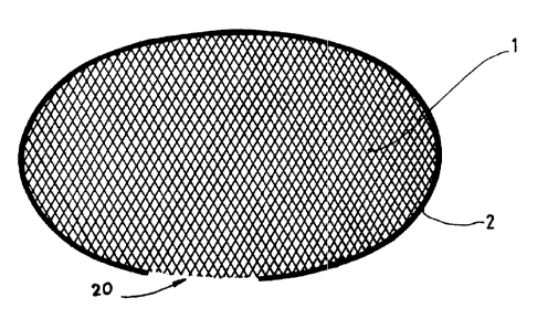
With reference to Figures 1 and 2, we can see that according to a first method
of
production, the prosthesis according to the invention consists of a mesh 1 in
oval shape
bordered by a
CA 02357020 2001-01-23
9
ring 2. The mesh 1 is fastened to the ring 2 in such a way as to not be under
tension, that
is, conserving a certain laxity that permits it to take on a convex. shape as
is visible in
Figure 2.
The mesh 1 is made of a synthetic non-resorbent material of the polypropylene
type,
while the ring 2 is made of a resorbent material of polyglycolic acid type.
The ring is intended to recall its shape to the mesh when it is set in place
by the inguinal
route. It is sufficiently supple to be bendable without breaking at the moment
of its
introduction and rigid enough to resume its initial shape and to restore
tension to the
mesh 1 in the retroperitoneal space.
The mesh 1 is thus completely spread out and has no folds, because its
original convex
shape allows it to fit into the visceral sac and the concave shape, of the
bottom surface of
the abdominal wall. In connection with this, we note that, according to a
particular
method of production, the convexity of the mesh 1 may be given to it when it
is
manufactured, specifically by molding.
The prosthesis may be of several shapes, oval, round to go on top of the
roughly rounded
subsistence discharges in the case of an umbilical hernia or a nzpture, or
pear-shaped, that
is, more or less oval with one narrower end. They can also have varying
dimensions in
order to be
CA 02357020 2001-01-23
10
applicable to different types of hernias or ruptures.
While on this subject, and in the case of an oval-shaped prosthesis, the
dimensions of the
latter are 8 to 14 centimeters, preferably 12 centimeters for the :large axis
and 6 to 10
centimeters, preferably 8 centimeters for the small axis.
We can also see in Figure 1 that the ring 2 shows an interruption 20 which is
intended to
be positioned at the femoral veins so as not to traumatize them. In this
conformation, the
surgeon can slit the mesh 1 with scissors over several centimeters to create a
tongue
which is applied, without tension, over the femoral veins.
Referring now to Figure 3, we can see that in one variation each of the end
parts 21 of the
ring 2 at the interruption 20, have, close to the end 22 of the ring 2, an
area 23 of less
resistance permitting the ring 2 to be broken so that the interruption 20 may
be enlarged if
that is necessary.
On the other hand, the mesh 1 has two more or less radial slits 10 one at each
of the ends
22 of the ring 2, permitting the creation of a tongue 11 to be applied over
the femoral
veins to avoid their traumatization by the free edge of the mesh 1 which,
without the slits
10, would be under tension.
CA 02357020 2001-01-23
11
We note that the presence of the slits 10 can be independent of t:he presence
of the areas
of less resistance 23.
If we look now at Figure 4, we can see that according to one variation of the
prosthesis
according to the invention, the ring 2 is connected to two diametrical
dividers 3 crossing
over each other in an approximate right angle and made of the same resorbent
material as
the ring 2.
The dividers 3 are fastened by their ends 30 to the ring 2 and their lengths
are chosen so
that they can take on a curved shape, permitting the convexity of the mesh 1
to be
maintained.
In this variation, the dividers 3 are preferably trivo in number, but it is of
course possible
that a prosthesis according to the invention may consist of either a single
divider or more
than two dividers. In this conformation, the position of the interruption 20
of the ring 2
must be different depending on whether it is a matter of the right side or the
left side.
If we refer now to Figure 5, we can see that according to a second method of
production,
the prosthesis is round in form, the ring 2 is doubled by an internal
concentric ring 4, with
no mesh inside it and linked to the ring 2 by means of spokes 5.
Concerning this, we see that such a prosthesis has dimensions on the order of
4 to 7
centimeters, preferably 5 centimeters, for the external diameter of the
CA 02357020 2001-01-23
12
ring 2 while the internal ring 4 has a diameter of 1 to 2 centimeters.
With regard to the interruption 20 of the ring 2, the ring 4 has an
interruption 40, the end
edges 22 of the ring 2 being linked to the free edges 41 of the rir.ig 4 by
two spokes 5
linking the interruptions 20 and 40 defining a space 50.
According to a first method of production represented by figure 5, between the
spokes 5
defining said space 50, there is no mesh. However, and according to another
method of
creation not shown, at least one of the spokes 5 defining said space 50 is
provided with a
tongue of mesh, notably of a mobile type. Such a tongue extends to the
interior of said
space 50 and is intended to be placed over the femoral veins.
A cord 6, preferably of a resorbent material, is threaded peripherally through
the mesh 1
close to the ring 2 and this cord, through a traction on its two ends 60 which
emerge at
the interruption 20, permits forming the prosthesis into a frustum as is shown
in Figure 6.
The prosthesis thus shaped constitutes an umbrella prosthesis intended for the
treatment
of indirect inguinal hernias.
In this configuration, the prosthesis may be set in place by being introduced
into the
inguinal orifice, small diameter first and the space 50 defined by the two
spokes 5
CA 02357020 2001-01-23
13
linking the interruptions 20 and 40 and being destined for passage of the
inguinal cord.
After introduction of the prosthesis, the cord 6 is removed, pernnitting the
prosthesis to
spread out like an umbrella, due to the elastic effect of the two rings 2 and
4 and the
spokes 5.
Now it is appropriate to describe briefly the technique for setting in place
such a
prosthesis.
So, it is advisable, after local or loco-regional anesthesia, to make an
inguinal incision
and to open the inguinal canal by making an incision in the aporieurosis. Next
a series of
incisions and/or dissections is made adapted to the type, direct or indirect,
of the hernia
treated.
In the case of setting in place a prosthesis like that illustrated in Figures
1 to 4, the
dissection of the retroperitoneal space is then assured before intr=oducing
said prosthesis.
This latter is flattened transversely between the fingers of one hand and is
slid into the slit
by its first end. The prosthesis, if necessary, is then subjected to a slight
bending to assure
introduction of the second end. It is then spread out in the retroperitoneal
space, the ring 2
permitting it to resume its initial form. The position of the prostllesis is
adjusted so that
the femoral veins are facing the interruption 20 of the ring 2, the mesh 1
being possibly
slit, specifically with scissors in order not to exert pressure on the said
femoral veins. The
prosthesis can then be
CA 02357020 2001-01-23
14
anchored, specifically by suture, before closing the incisions.
If it is a matter of setting in place a prosthesis like the one illustrated in
Figures 5 and 6,
after opening the inguinal canal, a dissection is made in the preperitoneal
space so as to
create a small receptacle destined to receive the prosthesis. The latter is,
then formed in a
frustum, placed around the free edge of the inguinal canal and introduced into
the
inguinal orifice, small diameter first. The cord 6 is then severed, permitting
the prosthesis
to spread out, possibly aided digitally, before assuring the position of the
latter and, if
necessary, its fixation before closing the incisions.
The result of this is that whatever the method of creation of the prosthesis
according to
the invention, its placement is easy and quick and it can be done under local
or loco-
regional anesthesia.
The mesh 1 always remains deployed and is applied perfectly without folds on
the bottom
surface of the muscolo-aponeurosis.
CA 02357020 2001-01-23