Note: Descriptions are shown in the official language in which they were submitted.
CA 02358285 2001-07-05
WO 00/40966 PCT/US00/00457
METHOD AND RIT FOR EXTRACTING PRION PROTEIN
BACKGROUND OF THE INVENTION
FIELD OF THE INVENTION
The present invention relates to a method for extracting
prion protein from a biological material, such as, for example,
an animal tissue or a biological fluid. The extraction process
permits testing for the presence of abnormal prion protein, e.g.,
for diagnosis of transmissible spongiform encephalopathies.
BACKGROUND OF THE INVENTION
Prion diseases or transmissible spongiform encephalopathies
(TSEs) cause progressive degenerative disorders of the central
nervous system resulting in death (Prusiner, Med. Res. Rev.
16:487, 1996; Weissman, FEBS Letters 289:3, 1996). Scrapie, a TSE
in sheep, was first described over 200 years ago (Pattison, Vet.
Rec. 123:661, 1988), and is the prototype of these diseases.
There are no known treatments for these diseases and no known
antemortem tests for the presence of the disease in an animal.
Prion diseases are caused by a conformational change of the normal
host prion protein to an abnormal structure that forms aggregates.
Because of the recent outbreak of bovine spongiform encephalopathy
in the United Kingdom and the connection between this TSE and the
new variant, Creutzfeld-Jakob (Bruce et al., Nature 389:498,
1997), a human TSE, there is a need for new methods that are both
sensitive and accurate to diagnose TSEs. Ideally, this diagnosis
could be used to test animals before they show clinical signs and
before they enter the human food chain or into pharmaceuticals
prepared for human use.
- 1 -
CA 02358285 2001-07-05
WO 00/40966 PCT/US00/00457
Description of the Prior Art
Most of the methods used to prepare and purify the disease-
causing agents of TSEs involve a complex sequence of enzyme and
detergent treatments and centrifugations (Bolton et al., J. Virol.
53:596, 1985). Abnormal prion protein is poorly soluble in the
typical biological buffers. One method for obtaining purified
abnormal prion protein is hydrophilic interaction chromatography
(HILIC) (Alpert, J., Chromatogr. 499:177, 1990), which is the
inverse of reversed-phase chromatography. Typically, one starts
with 70-85% organic solvent and runs a decreasing organic
gradient. Elution is in the order of least to most polar. The
mostly organic mobile phases of HILIC are compatible with proteins
not normally occurring free in aqueous solution, such as membrane
proteins (Jeno et al., Anal. Biochem. 215:292, 1993), (3-amyloid
peptide (1-43) (Alpert et al., Eighth Symposium of the Protein
Society, July 1994, San Diego, CA), and histones (Lindner et al.,
J. Chromatogr. A. 782:55, 1997). Surfactants and other
denaturants elute in or near the void volume, while proteins and
peptides are generally well-retained.
After HILIC purification, the prion protein can be detected
using capillary electrophoresis immunoassay (Schmerr and Jenny,
Electrophoresis 19:409, 1998) or by capillary isoelectric focusing
(Schmerr et al., J. Chromatogr. A. 802:135, 1998).
As noted above, present analytical methods to detect abnormal
prion protein generally are used post mortem, thus there is a need
for an antemortem assay for abnormal prion protein. In addition,
a method is required for isolation of abnormal prion protein
without ultracentrifugation steps, which require instrumentation
that is not readily available to veterinary diagnostic
laboratories. Centrifugation requires the presence of abnormal
prion protein as aggregates, whose large size facilitates pellet
- 2 -
CA 02358285 2001-07-05
WO 00/40966 PCT/US00/00457
formation in the centrifuge tubes. Such aggregates are difficult
to dissolve and detect in subsequent steps. The use of
centrifugation also jeopardizes the possibility of detecting
monomeric abnormal prion protein, potentially decreasing the
sensitivity of any assay. There is an even more pressing need for
a fast, reliable field assay, such as a qualitative immunoassay,
to test livestock for infection with a TSE. Thus, there is a need
in the art for an efficient, simple method for extracting abnormal
prion protein.
Additionally, the antibodies that have been produced detect
abnormal prion protein in its monomeric form, with the exception
of the antibody produced to the native abnormal prion protein
(Korth et al., Nature 390:74, 1997). As a result, abnormal prion
protein must be deaggregated with strong detergents or
denaturants; these denaturants must then be removed before
performing most immunoassays. Thus, there is a need in the art
for a rapid, simple method to extract prion protein free of
detergents or denaturants for immunoassay analysis.
The present invention provides a new method for the
extraction of all sizes of the abnormal prion protein, whether in
aggregated or monomeric form. The invention makes it possible to
test for abnormal prion protein in samples from a live animal,
e.g., using immunoassays. For example, diagnosis can be based on
blood samples, which will allow for the testing of live animals
and facilitate the removal of infected animals from flocks and
herds, and prevent possible contamination of products for
consumption.
SUI~iARY OF THE INVENTION
The invention provides a method for extracting abnormal prion
protein from a biological material suspected of containing
- 3 -
CA 02358285 2001-07-05
WO 00/40966 PCT/US00/00457
abnormal prion protein. The method comprises incubating a mixture
of extraction solvent and an isotonic or hypotonic aqueous
preparation of the biological material under conditions effective
to extract abnormal prion protein from the biological material
into the extraction solvent. The extraction solvent is a polar
organic solvent in which the abnormal prion protein is soluble,
and it is miscible with a hypotonic or isotonic aqueous solution
but immiscible with a lyotropic aqueous solution. Lyotropic
activity of the mixture is increased so that the extraction
solvent separates as a distinct phase from the aqueous preparation
of the biological material to yield extraction solvent containing
any abnormal prion protein from the biological material.
The invention further provides a method for detecting the
presence of abnormal prion protein in an animal, comprising
assaying a separated extraction solvent prepared as described
above for abnormal prion protein.
Also provided is a kit for isolating abnormal prion protein
from a biological sample. The kit comprises an extraction
solvent, which has the characteristics set forth above, and a
lyotropic salt or aqueous lyotropic salt solution to add to an
aqueous preparation of a biological sample so that the organic
solvent becomes immiscible with the aqueous preparation. In
another embodiment of the invention, the kit includes a prion
protein detection assay, preferably an assay for an abnormal prion
protein.
Thus, it is an object of the invention to provide a rapid
method for isolating abnormal prion protein from a biological
sample.
It is also an obj ect of the invention to provide an early
detection method for organisms infected with abnormal prion
protein.
- 4 -
CA 02358285 2001-07-05
WO 00/40966 PCT/US00/00457
It is a further object of the invention to provide a solvent
extraction technique for isolating abnormal prion protein from a
biological sample.
Still another object of the invention is to simplify
analytical testing of a biological material from an animal or
human for the presence of abnormal prion protein.
Yet another object of the invention is to provide an extract
containing abnormal prion protein for further testing or
purification.
These and other objects of the invention are presented in
greater detail in the accompanying Drawings and Detailed
Description of the Invention.
BRIEF DESCRIPTION OF THE DRAWINGS
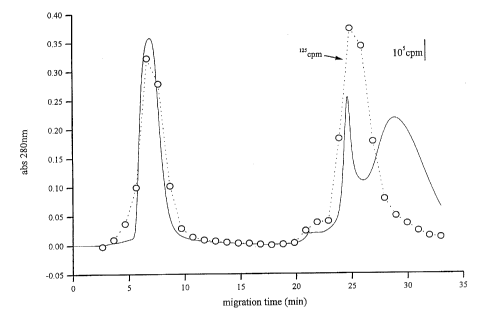
FIG. 1. Chromatogram from HILIC of l2sl_prion protein, as
detected by radioactivity (open circles) and by absorbance at 280
nm (solid line) .
FIG. 2. Antibody binding of HILIC factions of lzSI-prion
protein.
DETAILED DESCRIPTION OF THE INVENTION
The present invention provides a method for extracting
abnormal prion protein from biological material. The extracted
product can be tested by an immunoassay for the abnormal prion
protein. This extraction of abnormal prion protein, in
conjunction with an immunoassay, can be used to diagnose living
organisms for infection with a TSE, and will be of worldwide use
for testing for TSE infected animals and humans. Thus, the
present invention advantageously permits testing for prion protein
in clinics and veterinary labs that lack expensive centrifuges,
and further permits testing in the field.
- 5 -
CA 02358285 2001-07-05
WO 00/40966 PCT/US00/00457
An aqueous preparation of the biological material is combined
with an extraction solvent to form a mixture. The extraction
solvent is a polar organic solvent in which abnormal prion protein
is soluble, and miscible with a non-lyotropic isotonic aqueous
solution but immiscible with a lyotropic aqueous solution. In a
specific preferred embodiment, the extraction solvent is
hexafluoro-2-propanol (also termed hexafluoroisopropanol or HFIP).
Although in the examples, infra, the volumes of extraction buffer
and aqueous preparation of biological material are about equal,
any ratio can be used that yields two distinct phases, as
described below.
In a preferred embodiment, the mixture is incubated at a
temperature ranging from about 20QC to about 100pC. Nevertheless,
any temperature at which both the extraction solvent and aqueous
preparation of biological material are in a liquid phase, i.e.,
between the freezing point and the boiling point, can be used.
The extraction solvent-biological material mixture is
incubated under conditions effective to extract abnormal prion
protein from the biological material into the extraction solvent.
After incubation, the lyotropic activity of the mixture is
increased so that the extraction solvent separates from the
aqueous preparation. The extraction solvent containing prion
protein is removed from the aqueous preparation of biological
material.
According to the invention, the lyotropic activity of the
extraction solvent-aqueous preparation can be increased by adding
a lyotropic salt. The salt can be added as a solid directly to
the mixture, or as a concentrated aqueous solution. Preferred
examples of lyotropic salts include sodium sulfate and ammonium
sulfate. In a specific embodiment, exemplified infra, the ionic
- 6 -
CA 02358285 2001-07-05
WO 00/40966 PCT/US00/00457
strength is increased by adding about a 1:1 ratio (vol/vol) of 0.5
M sodium sulfate to the mixture.
The invention is particularly advantageous because it does
not require obtaining material from autopsy or necropsy.
According to the invention, the biological material can be a
sample from a living animal. The method of the invention provides
for extraction of abnormal prion protein from a biological fluid
or organ biopsy for analytical testing. Alternatively, the
biological material can be obtained from an autopsy or necropsy,
e.g., of animal products to be used for food, pharmaceutical,
cosmetic, or other products for use by humans or with other
animals. In a further embodiment, the method of the invention can
be used to remove prion protein from such materials to ensure that
infectious prions are not transmitted.
The extraction method of the invention can be used to extract
both normal prion protein and abnormal prion protein. By
pretreating the biological sample with proteinase-K, the normal
prion protein can be digested. Thus, where only abnormal prion
protein is desired, the aqueous preparation of biological material
can be pre-treated with proteinase-K prior to mixing it with the
extraction solvent.
The extraction solution containing any prion protein can be
dried to yield an extractant pellet. Prion protein in the
solution or extractant pellet can be further purified, e.g., by
hydrophilic interaction chromatography.
Abnormal prion protein in an animal can be detected by
assaying material in the separated extractant pellet for its
presence. Preferably, the assay method is an immunoassay for
abnormal prion protein. The sample can be treated with
proteinase-K prior to isolation of the prion protein so that no
normal prion protein is isolated.
CA 02358285 2001-07-05
WO 00/40966 PCT/US00/00457
In another aspect, the invention provides kits for isolating
abnormal prion protein from a biological sample. In one
embodiment, a kit comprises extraction solvent, e.g., hexafluoro-
2-propanol. The kit further comprises a lyotropic salt or aqueous
lyotropic salt solution to add to an aqueous preparation of a
biological sample so that the extraction solvent becomes
immiscible with the aqueous preparation.
The invention further provides a kit for detecting the
presence of abnormal prion protein from a biological sample. In
addition to the kit components described above, the detection kit
includes a detection assay for abnormal prion protein. The
preferred detection assay for the presence of abnormal prion
protein is an immunoassay.
Although this extraction method and kit have been developed
primarily for the diagnosis of scrapie (TSE in sheep), they are
useful for diagnosis of other TSEs in other vertebrates,
particularly in mammalian animals and avians, such as humans,
bovines, swine, elk, deer, poultry, and rodents. Abnormal prion
protein can be detected in tissue of animals and humans as early
as two weeks after infection. A significant application of the
process of this invention is detection of abnormal prion protein
in human blood. This technique can also be used for extraction
of the abnormal prion protein from process material used to
produce human pharmaceuticals or other products intended for human
use, including food supplements.
As used herein, the term ~~about~~ or ~~approximately~~ means
within 20%, preferably within 10%, and more preferably within 5%
of a given value or range.
_ g _
CA 02358285 2001-07-05
WO 00/40966 PCT/US00/00457
Biological Materials
The present invention permits the extraction of prion protein
from a biological material. Generally, prion proteins are found
in vertebrates, as discussed above. Therefore, under most
circumstances, the biological material will be from an animal or
human. Prion protein can also be produced during fermentation
processes with eukaryotic cells. It may be expressed as a
recombinant prion protein. Of greater concern is the possibility
of incidental expression of endogenous prion protein by cells that
have been recombinantly modified to express another protein. This
possibility is more likely if the cells are of neural origin, such
as PC12 cells . In this case, the biological material may be a
fermentation product, e.g., recombinant protein.
Examples of biological materials from animals include, but
are by no means limited to, tissues, such as brain, muscle
(including heart), liver, appendix, pancreas, gastrointestinal
tract organs, skin, and lymphoid tissue, such as thymus, spleen,
tonsil, lymph nodes, etc. Alternatively, the biological material
may be a biological fluid. The term biological fluid refers to
cerebrospinal fluid, blood, serum, plasma, milk, urine, saliva,
tears, mucous secretions, sweat, semen and bodily fluids
comprising these components. It also refers to culture fluid (or
culture medium) used in the production of recombinant proteins or
containing cells in suspension prior to transplantation. Also
encompassed by the term "biological materials" are products made
from animal organs or tissues, including serum proteins (such as
albumin and immunoglobulin), hormones, food and processed food
products, nutritional supplements, bone meal, animal feed,
extracellular matrix proteins, gelatin, and other animal by-
products used in manufacturing or final goods.
_ g _
CA 02358285 2001-07-05
WO 00/40966 PCT/US00/00457
Where the biological material is a solid tissue or product,
it must first be dissolved or suspended in an aqueous solution so
that it will be suitable for the extraction process. For example,
brain tissue may be suspended in sucrose solution (e.g., 0.32 M
sucrose) at 10% weight to volume. Other hypotonic or isotonic
solutions include 5% dextrose, phosphate buffered saline, tri-
buffered saline, HEPES-buffered saline, or any of the foregoing
buffers. The biological material in the aqueous solution can also
be homogenized, ground, or otherwise disrupted to maximize contact
between the extraction solvent and the biological material.
However, if a biological fluid is the biological material,
addition of liquid is not likely to be necessary, unless to dilute
the ionic strength of the biological fluid to permit miscibility
of the extraction solvent.
Prion Protein
The term "prion protein" as used herein refers to a native
protein expressed in neural tissue, particularly the brain and at
lower levels in lymphoid tissues and all other tissues. Under
some circumstances, prion protein adopts a pathogenic
conformation, which is termed herein abnormal prion protein.
Certain mutations of the prion gene in some individuals appear to
predispose prion protein to adopt the pathogenic conformation.
Exposure of an organism to a transmissible infectious agent, the
prion, can also induce the conformational change leading to the
pathology.
Abnormal prion protein is much less susceptible to
proteolysis than normal prion protein. Treatment of a biological
material with a proteinase, particularly proteinase-K, digests
normal prion protein, but not abnormal prion protein.
- 10 -
CA 02358285 2001-07-05
WO 00/40966 PCT/US00/00457
In specific examples, infra, sheep abnormal prion protein
(PrPa°) is extracted by the method of the invention. However,
other prion proteins from other species, particularly those
mentioned above, can also be extracted using the method of the
invention.
Included in the category of abnormal prion protein are human
prion proteins found in the neurodegenerative diseases Kuru,
Creutzfeld-Jakob Disease (CJD), Gerstmann-Straussler Syndrome
(GSS), and fatal familial insomnia. Some case of CJD and GSS are
associated with known mutations of the prion gene. CJD is also
associated with exposure to TSEs. For example, as noted above,
CJD has been associated with bovine spongiform encephalopathy. The
present invention permits, for the first time, extraction of
abnormal prion protein in patients prior to autopsy. Detection
of extracted abnormal prion protein can be used in the diagnosis
of any of these diseases.
Scrapie (sheep, goats) and bovine spongiform encephalopathy
(cows) are abnormal prion diseases of animals. Prion proteins
have also been isolated in chicken, mink, pigs, mouse, hamster,
and guinea pig. Furthermore, mouse, hamster, and guinea pig can
develop a spongiform encephalopathy by exposure to prions from
human or other animal sources. Prion protein from any of these
sources can be detected or extracted by the method of the
invention.
Extraction Solvent and Conditions
An extraction solvent for use in the present invention must
be capable of separating as a distinct phase from water or an
aqueous solution under lyotropic conditions. At the same time,
prion protein must be soluble in the extraction solvent . Some
polar organic solvents meet these criteria. The preferred polar
- 11 -
CA 02358285 2001-07-05
WO 00/40966 PCT/US00/00457
organic solvent is hexafluoro-2-propanol. Other solvents that can
be used include isopropanol; 1,1,1-trifluoro-2-propanol (TFIP);
2,2,3,3-tetrafluoro-1-propanol (tetFlP); perfluoro-t-butyl
alcohol (PFtBA); 1,1,1,3,3,3-hexafluoroacetone (HFA);
trifluoroacetic acid (TFA); 2,2,2-trifluoro-1-ethanol (TFE);
2,2,3,3,4,4,4-heptafluoro-2-propanol (HFB); 1,1,1,3,3,4,4,4-
octafluoro-2-butanol (OFIB); 1-methyl-2-pyrrolidinone (NMP); see
Wille et al., J. Mol. Biol., 259:608, 1996. Other possible
solvents that can be evaluated include DMSO; tetrahydrofuran; and
the like. Alternatively, a solvent that is not miscible with
water, but in which prion protein is soluble, could be used.
In a specific embodiment, the solvent is miscible with water,
e.g., at physiological ionic strength (isotonic aqueous solution)
or lower than physiological ionic strength (hypotonic aqueous
solution). However, when the buffer comprises lyotropic salts
present at a concentration (or ionic strength) above a threshold
value, the extraction solvent is not soluble in the aqueous
solution: the two phases separate into an extraction solvent layer
and an aqueous solution layer. These are referred to herein as
"lyotropic conditions". The value for ionic strength of a
lyotropic salt of the aqueous solution which achieves lyotropic
conditions can vary depending on the extraction solvent selected
and the salts) used. It can be readily determined by titration
or other systematic variation of lyotropic salt concentration,
with testing for miscibility or immiscibility of the extraction
solvent with the aqueous solution. As used herein, an aqueous
solution with an ionic strength of a lyotropic salt at which the
extraction solvent separates from water is referred to as a
lyotropic aqueous solution. Aqueous solutions containing lower
concentrations of lyotropic salts or physiological salts, such as
isotonic buffers, are considered non-lyotropic solutions.
- 12 -
CA 02358285 2001-07-05
WO 00/40966 PCT/US00/00457
Generally, about equal volumes of an extraction solvent and
the aqueous preparation of a biological material are used in a
solvent extraction process. However, the ratio of extraction
solvent to aqueous preparation can range from about 5:1 to about
1:5, preferably from about 3:1 to about 1:3.
After mixing the extraction solvent with the aqueous
preparation, the mixture can be incubated for some period of time
and at a particular temperature to enhance extraction of prion
protein into the extraction buffer. The incubation time can vary
from 1 minute to hours, and can be determined by analyzing the
extracted material for the presence of prion protein. After the
amount of prion protein in the extraction material versus time
reaches a plateau, which can be tested using chromatographic or
immunoassay techniques, or both, as described in the Examples,
additional incubation will have no effect on the prion protein
yield. In a specific embodiment, the incubation time is 5
minutes.
In addition, the temperature of incubation can be adjusted
to increase the efficiency of extraction, provided that the
extraction solvent and aqueous solution are both liquids at the
selected temperature. Warmer temperatures, i.e., above room
temperature, are preferred, since they increase the solubility of
prion protein in the extraction solvent. Particularly useful are
temperatures within the range of about 50°C-60°C. As with other
variables, such as the ionic strength of the aqueous preparation
and time of incubation, an optimal temperature can be determined
by routine experimentation and testing.
Phase Separation
Various lyotropic salts can be used to increase the ionic
strength of the aqueous solution, thereby inducing phase
- 13 -
CA 02358285 2001-07-05
WO 00/40966 PCT/US00/00457
separation. Among the preferred salts are sodium sulfate and
ammonium sulfate, which are lyotropic. Both are used at a
concentration well below that which precipitates proteins. For
example, in a specific embodiment, a 0.5 M solution of sodium
sulfate is added to an equal volume of the extraction
solvent/aqueous preparation mixture, resulting in a final
concentration of 0.25 M sodium sulfate. This concentration is
sufficient to induce phase separation of HFIP and water. Other
salts can also be used, provided they achieve the requisite
lyotropic activity at a concentration at which they are soluble.
The term "lyotropic activity" is used herein to refer to the
structure-forming properties of a lyotropic salt solution. The
lyotropic activity is achieved by achieving a sufficient
concentration of lyotropic salt to induce phase separation of the
organic and aqueous phases. Protein precipitating concentrations
of the lyotropic salt are avoided.
"Lyotropic" or "structure-forming" salts, also known as
kosmotropes, promote the ordering of the aqueous solution, thereby
excluding organic solutes. If the organic solute is the
extraction solvent, phase separation occurs. If it is a protein,
the protein is salted out of solution. Good structure-forming
salts include sodium or ammonium sulfate, phosphates, citrates,
etc. (Washabaugh and Collins, J. Biol. Chem., 261:12477, 1986)
(Structure-breaking salts, or chaotropes, include guanidinium
hydrochloride, sodium perchlorate, sodium bromide, etc. These
have the opposite effect; they drive organic solutes into aqueous
solution.) The ionic strength of an aqueous solution is a
function of the total number of ions in solution, regardless of
whether they are structure-forming or structure-breaking ions. The
term "ionic strength" relates to the concentration of a salt.
- 14 -
CA 02358285 2001-07-05
WO 00/40966 PCT/US00/00457
As discussed above, the lyotropic salt can be added as a
solid or concentrated liquid, provided that the final lyotropic
salt concentration is effective to induce the phase separation.
Preferably, the final ratio of extraction solvent to aqueous phase
(which includes the aqueous preparation of biological material and
any salt solution) after increasing the lyotropic activity of the
mixture is about 1:10 to about 10:1, preferably (and as
exemplified infra) about 1:3 to about 3:1, provided that at the
lower ratio of extraction solvent to aqueous phase, the final salt
concentration is still high enough to induce phase separation.
After increasing the lyotropic activity, the extraction
solvent, which now contains any prion protein that was present in
the biological material, separates from the aqueous preparation.
The separation process takes a few minutes, and is complete when
both phases are clear and discrete separation is observed between
them. Once the two phases are completely separated, the
extraction solvent, now containing any prion protein, can be
removed or withdrawn, e.g., by drawing off with a pipette or
syringe, or with a separation flask.
The extraction solvent containing any prion protein (termed
herein "extract") can be dried, e.g., by evaporation, by
lyophilization, or vacuum centrifugation to yield highly
concentrated or dry extract. The extract may contain other
components, including cellular lipids, lipid membrane-binding
proteins, and other more hydrophobic cellular components. If
desired, prion protein can be isolated or purified away from these
components, e.g., by hydrophilic interaction chromatography, as
exemplified infra, or other chromatographic techniques (cation
exchange chromagraphy, gel permeation chromatography, reverse-
phase chromatography, and affinity chromatography, e.g., on an
antibody column).
- 15 -
CA 02358285 2001-07-05
WO 00/40966 PCT/US00/00457
Alternatively, the concentrated or dried extractant material
can be analyzed directly, as described infra, to detect abnormal
prion protein.
Prion Protein Detector; Immunoassays
Various prion protein detection assays, including assays for
selectively detecting abnormal prion protein, are known in the art
to be an effective tool for analyzing prion protein. Capillary
gel electrophoresis has proven to be an effective analytical tool
for abnormal prion protein (Schmerr and Jenny, Electrophoresis,
19:409, 1998). A preferred method is immunoassay, e.g., as
described in Schmerr and Jenny, supra. Antiserum described in
this reference is specific for abnormal prion protein, as it was
found to react in Western blotting with scrapie-infected brain,
but not normal brain. Other antisera reactive with prion protein
are well known in the art. A preferred immunoassay is a plate
ELISA (for example, Grathwohl et al., J. Virol. Methods, 64:205,
1997) .
Thus, in some cases, detection of the presence of prion
protein, and particularly abnormal prion protein, is based on the
biophysical and chemical characteristics of prion protein. These
include proteinase resistance (particularly to proteinase K) and
digestion profile (whether with proteolytic enzymes, glucolytic
enzymes, chemicals, heat, denaturants, etc.). The effects of such
treatments on apparent molecular weight and isoelectric point, and
various binding assays, can be evaluated. Proteinase resistance
and digestion profile can be detected by chromatography, gel
electrophoresis, and other molecular weight-sensitive techniques.
Isoelectric point can be measured using capillary isoelectric
focusing (IEF) or gel isoelectric focusing, although capillary IEF
is able to measure the prion pI of 3 more effectively than most
- 16 -
CA 02358285 2001-07-05
WO 00/40966 PCT/US00/00457
gels. Furthermore, qualitative determination of overall charge
(acidity or basicity) can be determined by ion exchange
chromatography. Other biophysical techniques known in the art can
also be used to identify prion protein.
Examples of assays for detection of prion protein include
apparent molecular weight and isoelectric point of the protein,
including after heating, cyanogen bromide cleavage, neuraminidase
treatment, etc. (Bolton et al., J. Virol., 53:596, 1985);
glycosidase treatment and lectin binding (Somerville and Ritchie,
J. Gen. Virol., 71:883, 1990); proteinase-K resistance (Race et
al., Am. J. Vet. Res., 53:883, 1992); and immunoassay (Farquhar
et al., J. Virol. Methods, 24:215, 1989).
Alternatively, sequencing or microsequencing of the
extracted, and preferably purified, prion protein permits one to
unambiguously confirm its identity.
Immunoassays for prion protein can be accomplished by
techniques known in the art, e.g., radioimmunoassay, ELISA
(enzyme-linked immunosorbant assay), "sandwich" immunoassays,
immunoradiometric assays, gel diffusion precipitation reactions,
immunodiffusion assays, in situ immunoassays (using colloidal
gold, enzyme or radioisotope labels, for example), Western blots,
precipitation reactions, agglutination assays (e.g., gel
agglutination assays, hemagglutination assays), complement
fixation assays, immunofluorescence assays, protein A and protein
G assays, immunoelectrophoresis assays, measuring levels thereof
in appropriate physiological samples, etc. In one embodiment,
antibody binding is detected by detecting a label on the primary
antibody. In another embodiment, the primary antibody is detected
by detecting binding of a secondary antibody or reagent to the
primary antibody. o
- 17 -
CA 02358285 2001-07-05
WO 00/40966 PCT/IJS00/00457
The extraction method of the invention provides an
inexpensive source of prion protein, which can be used to generate
additional antibodies. Moreover, because the extraction
conditions of the invention differ greatly from conventional
extraction conditions, prion protein extracted in accordance with
the invention may have a different conformation and elicit a
different population of antibodies if used for immunization.
This method shortens the extraction time to 1 to 2 hours .
Moreover, because of its simplicity, it can be automated. The
method extracts prion protein of all molecular sizes, so it is not
limited. It also solubilizes the abnormal prion protein so that
most immunoassays can be used to detect it. Furthermore, and not
insignificantly, it reduces the infectivity of the abnormal prion
protein, making the process safer.
Kits
The components for practicing the present invention can be
conveniently provided in a kit form. In its simplest embodiment,
a kit of the invention provides extraction solvent, preferably
HFIP, and lyotropic salt (or a concentrated lyotropic salt
solution) for increasing the lyotropic activity of the extraction
solvent-aqueous preparation mixture. The amounts of each
component can be pre-measured to provide a specified number of
assays. In a further embodiment, the kit will include a sample
container, preferably of plastic or a material treated to avoid
non-specific binding of prion protein.
As used herein, the term container has its broadest meaning,
i.e., any receptacle for holding material or reagent. It can be
fabricated from glass, plastic, ceramic, metal, or any other
material typically employed to hold reagents. However, an
- 18 -
CA 02358285 2001-07-05
WO 00/40966 PCT/US00/00457
acceptable material will not be reactive with the contents it is
intended to hold.
The kit can also include proteinase-K for digesting normal
priori protein in the biological sample.
In a further embodiment, the kit includes a sample container
with a volume indicator for the aqueous preparation of the
biological sample. In this embodiment, the polar organic solvent
and the lyotropic salt are optimally provided in pre-measured
units for use in conjunction with the sample container. The
biological sample preparation can be placed in the sample
container. The pre-measured unit of extraction solvent can be
added, followed by mixing. Then, the pre-measured unit of
lyotropic salt can be added to induce phase separation of the
extraction solvent and the water. In still a further embodiment,
proteinase-K for treating the aqueous preparation of the
biological sample is provided in the kit, preferably in a pre-
measured unit.
A kit for extracting priori protein from a tissue sample may
include a dilution buffer, such as a 0.32 M sucrose solution or
phosphate buffered saline, for homogenization of the tissue for
the aqueous preparation of the biological material.
In a further embodiment, in which the kit is a kit for
detecting the presence of abnormal priori protein in a biological
material or sample, the kit provides an abnormal priori protein
detector or assay, as described above. Immunoassays, as described
above, are preferred for detection of abnormal priori protein
extracted in accordance with the invention.
In still a further embodiment, the kit includes an
immunochromatographic membrane or support. The extraction solvent
containing any priori protein can be applied to the support
directly, or the dried extract can be applied, e.g., after
- 19 -
CA 02358285 2001-07-05
WO 00/40966 PCT/US00/00457
resolubilization. Under appropriate conditions, prion protein can
flow through the support. It may be captured, e.g., by
immobilized anti-prion antibody, and immobilized prion protein
detected. Numerous methods and devices known in the art for
immunochromatographic assays can be employed in the invention.
Immunochromatographic assays are particularly useful under field
conditions, where laboratory equipment is not available. Examples
of such assays are provided in U. S . Patents No . 5 , 248 , 619 , No .
5,451,504, No. 5,500,375, No. 5,624,809, and No. 5,658,801.
A kit of the invention preferably includes packaging and
instructions for its use, e.g., on the packaging or package
insert.
The present invention may be better understood by reference
to the following non-limiting Examples, which are provided as
exemplary of the invention.
Example 1
Analysis of Abnormal Prion Protein Extracted From Infected Sheep
Brain and Lymph Nodes.
Brain or lymph node tissue from each of two scrapie infected
sheep was homogenized in 10% sarcosyl and treated with proteinase
K to digest the normal host prion protein but not the altered
abnormal form of prion protein. Equal volumes (0.5 mM) of
homogenate and HFIP were mixed and incubated for five minutes at
56°C. To this mixture, 0.5 mM of 0.5 M NaZS04 were added, and the
mixture was incubated an additional five minutes. Under these
conditions, the HFIP layer separated from the aqueous layer. The
HFIP layer was drawn off and dried in a centrifuge. The dried
samples were resuspended in 25 ~1 of distilled water and mixed.
Ten ~1 of the sample was mixed with 5 ~1 of 20% SDS buffer ando
boiled for five minutes at 100°C.
- 20 -
CA 02358285 2001-07-05
WO 00/40966 PCT/US00/00457
Western blot analysis was performed on a 10% to 15% gradient
polyacrylamide gel. The protein was transferred from the
polyacrylamide gel to nitrocellulose under standard conditions.
The nitrocellulose was blocked by incubation with a solution of
5% fish gelatin, and washed with a Tris-Tween buffer. The
nitrocellulose was incubated overnight with a rabbit anti-prion
protein (antibody raised against whole prion protein diluted 1 to
2,500; Kascak et al., Immunol. Invest., 26:259, 1997; see also,
Miller et al., J. Vet. Diagn. Invest. 5:309; Kascak et al., J.
Virol., 59:676, 1986). After incubating with the anti-prion
antibody, the nitrocellulose was washed with Tris-Tween and then
reacted with an anti-rabbit IgG-HRP (horseradish peroxidase)
conjugate and incubated for one hour. The nitrocellulose was then
washed extensively and developed with a chemiluminescent reagent
(Pierce UltraSuperSignal~). Peroxidase activity was detected
using a chemiluminescent imager (Chemi-Imager-4000; Alpha,
Innotech) .
Extracts from both scrapie infected sheep containing 2.75 ~1
of material produced bands indicative of abnormal prion protein
for both brain tissue and lymph node tissue. A brain tissue
extract containing 1.5 ~1 of material from one of the scrapie
infected sheep also produced a band indicative of abnormal prion
protein. A Western blot of similar extracts from normal
(noninfected) sheep did not produce any bands indicative of
abnormal prion protein.
Example 2
Analysis of Abnormal Prion Protein Extracted and Purified From
Infected Sheep Brain and Lymph Nodes:
This example shows further purification and analysis ofo.
abnormal (scrapie) prion protein (PrPsc) using hydrophilic
- 21 -
CA 02358285 2001-07-05
WO 00/40966 PCT/US00/00457
interaction chromatography (HILIC). Tissue samples including
sheep brain and lymph nodes were processed with detergent and
proteinase K as previously described. The resulting extracts were
applied to a HILIC column and eluted with a decreasing gradient
of acetonitrile in 0.1% trifluoroacetic acid and 50 mM hexafluoro-
2-propanol. Recovery from the column was approximately 75% as
determined with a radioiodinated priors protein. After drying, the
collected peak fractions were resuspended in water and assayed
with antibodies specific for the priors protein. The method
permitted efficient purification of the priors protein as well as
testing by immunoassay, since interfering detergents were removed.
Example 3
Analysis of Abnormal Priors Protein Extracted From Infected Sheep
Brain.
Preparation of sheep brain material.
Scrapie infected sheep brains were obtained from field cases
that were positive for the abnormal priors by Western blot (Race
et al . , Am. J. Vet . Res . 53 : 883 , 1992 ) . A pool was made of 3
positive brains. The same pool was used for all the experiments
presented here. Normal brains came from sheep from a scrapie-free
flock and were negative for abnormal priors protein by Western
blot. The brain material was prepared for chromatography by a
modification of the method of Bolton et al. (J. Virol. 53:596,
1985). Briefly, the brain stems were dissected out, weighed and
placed in 0.32 M sucrose (10% w/v). The material was then
homogenized for 60 s with a Brinkman Polytron (Kinematica AG,
Lucerne Switzerland) using a 0.7 cm stainless steel generator at
the highest speed. The homogenate was centrifuged at 10,000 g for
20 min to remove particulates, and the resultant supernatant fluid
was centrifuged at 230,000 g for 1 h. This pellet was then
- 22 -
CA 02358285 2001-07-05
WO 00/40966 PCT/US00/00457
subjected to a series of washes and ultracentrifugations as above.
The sample was treated with 10 mM Tris pH 7.4 containing 10%
sodium lauryl sulfate and proteinaae K (50 ~g/ml). After the
final ultracentrifugation, the sample was resuspended in 10 mM
Tris pH 7.4 (200 ~tl/g of the initial brain sample) .
Hydrophilic interaction chromatography (HILIC).
The sample was solubilized in 0.01 M Tris HC1, pH 8.00
containing 2 mM EDTA, 5% SDS and 10% hexafluoro-2-propanol at
100°C for 10 min. After SDS treatment, the sample was placed in
a solvent consisting of 100% acetonitrile containing 0.1% TFA acid
and 50 mM hexafluoro-2-propanol (buffer A) and applied to a
hydrophilic interaction column. All columns that were used were
from PolyLC, Inc. (Columbia, MD, USA) with the dimensions 200 x
4.6-mm; 5 ~.m; 300-A. Three packings were evaluated, PolyWAX LPTM
(an anion-exchange material), PolyHYDROXYETHYL ATM (a neutral
material) and PolySULFOETHYL AT"~ (a strong cation-exchange
material) (all three trademarks are the property of PolyLC, Inc.).
The flow rate was 0.5 ml/min. The conditions for eluting prpsc
were 100% A for 8 min and then a linear gradient to 100% water
containing 0.1% trifluoroacetic acid and 50 mM hexafluoro-2-
propanol (buffer B) in 15 min, then 100% B for 10 min. Peak
fractions were collected and dried in a vacuum centrifuge (Savant
Instruments, Inc, Farmingdale, NY, USA). Fractions were
resuspended in 10 ~1 of deionized HZO and the fraction that tested
positive by immunoblot for PrPS~ was used in a capillary
electrophoresis assay.
Labeling prion protein with Izsl.
PrPs° was labeled with lasl using IODOGENT"~ (Pierce, Rockford;v
IL, USA). The labeled protein was separated from the free lzsl by
- 23 -
CA 02358285 2001-07-05
WO 00/40966 PCT/US00/00457
passing it through a solid phase extraction cartridge containing
PolyWAX LPT""(PolyLC, Inc.) that had been equilibrated with buffer
A. The labeled PrPS~ was eluted from the cartridge using buffer
B. The unbound 125I was retained on the cartridge, which could be
discarded as solid radioactive waste. The fractions containing
the labeled PrPS~ were dried in a vacuum centrifuge, dissolved in
water, diluted 1/10 in buffer A, and loaded onto the HPLC column.
Dot blots.
One-~1 aliquots of peak fractions from HILIC chromatography
were applied to nitrocellulose paper, dried, and then incubated
in 20 mM Tris, pH 7.5, containing 500 mM NaCl, 0.05% Tween 20
(TTBS) and 5% fish gelatin for 1 h. The blot was washed 2x with
TTBS and then incubated with a dilution of 1/500 of antibodies
made to peptides of the prion protein for 3 h at 25°C (rabbit
antibodies to peptide 142-154). After incubation, the blot was
washed 2x with TTBS and then incubated with biotinylated protein
G (Bio-Rad Laboratories, Hercules, CA, USA) for 1 h. Again the
blot was washed as above. Horseradish peroxidase coupled to
NeutrAvidinTM (Pierce, Rockford, IL, USA) was added to the blot
and incubated for 1 h at 25°C. After incubation, the blot was
washed 6x with TTBS. After washing, the blot was incubated in the
SuperSignalo Substrate(Pierce) system for 10 min and then exposed
to Kodak X-OMAT AR (Eastman Kodak Company, Rochester, NY, USA) X-
ray film for 15 sec.
Binding assay for lzsIPrPBC.
Fractions containing lzsl from the PolyWAX LP column were
assayed for binding activity to an antibody that had been produced
to the peptide corresponding to residues 142-154 of the prim;
protein. The tubes containing radioactivity were dried in a
- 24 -
CA 02358285 2001-07-05
WO 00/40966 PCT/US00/00457
Savant vacuum centrifuge at 42°C and resuspended in 10 ~,1 of HZO
and then diluted with buffer-containing salts in 0.1% BSA. A PVC
plate was coated with the antibody in 0.1 M NazC03, pH 9Ø After
washing with the above buffer, 100 ~,1 of lzsl -Prpsc was incubated
on the plates at 37°C for 2 h and then overnight at 4°C. The
plate was washed and cut into individual wells and counted.
Background cpm were subtracted from the cpm in the wells.
Capillary electrophoresis conditions.
Free zone capillary electrophoresis (Schmerr and Jenny,
Electrophoresis 19:409, 1998) was performed on a Beckman PACE
5500 (Beckman Instruments, Fullerton, CA, USA). Laser-induced
fluorescence (LIF) detection was done using an air-cooled argon
laser (Beckman Instruments) with excitation at 488 nm and emission
at 520 nm. Unmodified capillaries were obtained from Beckman
Instruments. A 20 cm (length to the detector) x 201 ~m I.D.
capillary was used with 200 mM Tricine, pH 8Ø This buffer
contained 0.1% N-octylglucoside (Boehringer Mannheim GmbH,
Indianapolis, IN, USA) and 0.1% BSA (Sigma Chemical Co., St.
Louis, MO, USA). In preparation for the separation, the capillary
was rinsed for 1 min with 0.25 M NaOH, rinsed for 2 min with H20,
and then rinsed 2 min with buffer. The separating conditions were
30KV for 3 min at 20°C. The current was about 20 E.tA. The sample
was injected for 15 sec followed by a 5 sec injection of running
buffer. The sample volume was about 0.95 nl. Rinses were carried
out under high pressure and sample injection carried out under low
pressure.
Immune complex and prior binding assays.
Fifteen microliters of fluorescein-labeled peptide containing.' -
about 2 pmoles of the fluorescent labeled peptide was mixed with
- 25 -
CA 02358285 2001-07-05
WO 00/40966 PCT/US00/00457
affinity-purified rabbit IgG to demonstrate binding of antibody
to the fluorescein-labeled peptide. One ~1 of peak fractions from
the HILIC chromatography was added to the assay. After mixing the
components, the samples were incubated at 25°C for the 10 min.
Resul is .
The chromatogram of PrPs~ after purification and iodination
is shown in FIG. 1. Radioactivity (cpm) from the 1251-labeled
prion protein coincides with the absorbance at 280 nm, except for
the last peak detected by absorbance. The yield of abnormal
protein, based on recovery oflasl cpm loaded onto the column, was
about 76%. The peaks of cpm and A280 absorbance coincide with the
peaks showing antibody activity in the binding assay (FIG. 2) .
The main peak in the binding assay at about 25 min coincides with
peaks for 125I-PrP and A280 absorbance at 25 min in FIG 1. Similar
results were obtained for a chromatogram (not shown) from an
extraction (unpurified) of scrapie infected sheep brains.
A wide range of pI values have been reported in the
literature (Schmerr and Jenny, supra; Safar et al., Proc. Natl.
Acad. Sci. USA 87:6373, 1990; Somerville et al., J. Gen. Virol.
70:25, 1989) for abnormal prion protein. The pI of this protein
would affect the binding of this protein to column packings.
There was no great difference between the retention times on the
positively-charged PolyWAX LP column and the neutral
PolyHYDROXYETHYL A column. This suggested that the protein might
be acidic. Abnormal prion protein samples containing SDS eluted
from the negatively-charged PolySULFOETHYL A column in a broad
envelope. Accordingly, the abnormal prion protein purified on the
PolyWAX LP column was re-run on the PolySULFOETHYL A column. Its
elution in or near the void volume indicates that it is indeed.
acidic. This was confirmed by both gel isoelectric focusing and
- 26 -
CA 02358285 2001-07-05
WO 00/40966 PCT/US00/00457
capillary electric focusing (Schmerr et al., Chromatogr. A.
802:135, 1998). pI values ranged from 3-6 with a major species
at 3.00. These results suggest that in hydrophilic interaction
chromatography, it is necessary to use a neutral or an anion
exchange material.
In capillary immunoelectrophoresis using HILIC purified
samples; resultant electropherograms (not shown) indicate that
samples from infected sheep did react, whereas samples from normal
(noninfected) sheep did not react. Since SDS inhibits typical
immunoassays including capillary electrophoresis assays, it is
necessary to remove SDS in order to perform such assays. A
competition assay using capillary electrophoresis could be
performed on samples after HILIC chromatography, since SDS elutes
in or near the void volume (Jeno et al., Anal. Biochem., 215:292,
1993 ) .
Example 4
Analysis of Abnormal Prion Protein Extracted From Infected Sheep
Blood.
Buffy coat centrifuge fractions from blood samples from TSE-
infected sheep were diluted with Tris buffered saline (10%
tissue:90% buffer). The samples were then treated with proteinase
K to digest the normal host prion protein but not the altered
abnormal form of prion protein. After digestion, the treated
sample was mixed with an equal volume of hexafluoro-2-propanol
(HFIP) and incubated at 56°C for five minutes. An equal volume
of 0.5 M sodium sulfate was added and the phases were allowed to
separate. The layer containing HFIP was removed and the sample
dried in a vacuum centrifuge.
The pellet was resuspended in water and the suspension was
put in an organic chromatography mobile phase containing 95%
- 27 -
CA 02358285 2001-07-05
WO 00/40966 PCT/US00/00457
acetonitrile, 5% water, 0.1% trifluoroacetic acid and 50 mM HFIP.
The mobile phase was applied to a solid phase extraction cartridge
of PolyHYDROXYETHYL AspartamideTM (PolyLC, Inc.). Abnormal prion
protein was eluted from this support with 100% water, 0.1%
trifluoroacetic acid and 50 mM HFIP, and then dried and
resuspended in water.
The presence of abnormal prion protein was detected by
capillary immunoelectrophoresis. Abnormal prion protein was not
detected in electropherograms for control samples of blood that
was not infected with TSE.
Example 5
Analysis of Abnormal Prion Protein Extracted From Infected Mule
Deer Blood.
The procedure of Example 4 was repeated with blood samples
from TSE-infected mule deer. The presence of abnormal prion
protein was detected by capillary immunoelectrophoresis. Abnormal
prion protein was not detected in electropherograms for control
samples of blood that was not infected with TSE.
Example 6
Analysis of Abnormal Prion Protein Extracted From Infected Elk
Blood.
The procedure of Example 4 was repeated with blood samples
from TSE-infected elk. The presence of abnormal prion protein
was detected by capillary immunoelectrophoresis. Abnormal prion
protein was not detected in electropherograms for control samples
of blood that was not infected with TSE.
- 28 -
CA 02358285 2001-07-05
WO 00/40966 PCT/US00/00457
All patents, patent applications, test protocols, and
publications cited herein are hereby incorporated by reference in
their entireties.
The present invention is not to be limited in scope by the
specific embodiments described herein. Incteect, various
modifications of the invention in addition to those described
herein will become apparent to those skilled in the art from the
foregoing description and the accompanying figures. Such
modifications are intended to fall within the scope of the
appended claims.
- 29 -