Note: Descriptions are shown in the official language in which they were submitted.
CA 02358510 2001-07-12
WO 00/42435 PCT/US00/00652
1
DESCRIPTION
Methods For Detecting An Analyte Of Interest
Using Tyramide Coating Technoloey
Introduction
The present invention relates to methods of using tyramide coated live cells
for
flow cytometry, preferably using catalyzed reporter deposition and
amplification staining.
Background Of The Invention
The following information is presented solely to assist the understanding of
the
reader, and none of the information is admitted to describe or constitute
prior art to the
claims of the present invention.
Flow cytometry is a sensitive and quantitative method for measuring the
fluorescence or light scatter of particles or cells. This method has been
widely used to
study cellular physiology, especially as it relates to the immune system and
control of the
cell cycle. Nolan et al, "The Emergence of Flow Cytometry for Sensitive, Real-
time
Measurements of Molecular Interactions", Nature Biotechnolo~y, Vol. 16,
(1998), which
is incorporated by reference herein in its entirety including any drawings,
describe recent
flow cytometry developments for fields as diverse as ligand binding and enzyme
kinetics,
drug screening, diagnostics and detection of soluble agents, and DNA sequence
detection
or analysis. They describe developments such as advances in automated sample
handling,
molecular approaches for incorporating affinity tags or fluorescent probes
into proteins
and the availability of microsphere reagents that enable multiplexing.
Flow cytometric analysis of cell surface molecules is a technology used in
both
medical diagnostic laboratories and biomedical research laboratories. In
clinical practice
flow cytometry is used for samples derived from patients infected with human
immunodeficiency virus type l, patients with leukemias and lymphomas, and
patients with
primary immunodeficiences.
Lollini et al., "Flow Cytometry on Intracellular Antigens After Tyramide
Signal
Amplification", Immunological Blackboard: Bulletin of the Gruppo Di
Cooperazione in
Immunolo~ia, Vol. l, Number 2 (1998), which is incorporated herein by
reference in its
totality, including any drawings, describes tyramide signal amplification
(TSA) for
detection of intracellular antigens by flow cytometry and indicates that TSA
is not superior
to conventional techniques for detecting surface antigens on live cells.
Lollini et al. states
CA 02358510 2001-07-12
WO 00/42435 PCT/US00/00652
2
on page 5, "(t)he main problem appeared to be a high level of spontaneous
activation and
non-specific binding of the fluorescent substrate to live cell membranes".
Various methods have been described for assaying biological samples with
amplified reporter systems. Bobrow et al., U.S. Patent 5,196,306, U.S. Patent
5,583,001
and U.S. Patent 5,731,158, which are all herein incorporated by reference in
their totality
including any drawings, describe methods for detecting or quantitating
analytes using an
analyte dependent enzyme activation system as well as catalyzed reporter
deposition
methods. Specifically, Bobrow et al. describe colorimetric and fluorometric
solid phase
enzyme immunoassays which are enhanced by amplification of the reporter
molecules.
Chao et al., "Immunofluorescence Signal Amplification By The Enzyme
Catalyzed Deposition Of A Fluorescent Reporter Substrate (CARD)", Cytometry
23:48-53
(1996), describe a CARD system that uses horseradish peroxidase substrate
Cy3.29
tyramide to deposit fluorogen molecules onto fixed tissues and cells as well
as proteins
bound to nitrocellulose membranes, with up to a 15 fold increase over standard
indirect
immunofluorescence methods.
Malisius et al., "Constant Detection of CD2, CD3, CD4, And CDS In Fixed and
Paraffin-Embedded Tissue Using The Peroxidase-Mediated Deposition Of Biotin
Tyramide", The Journal of Histochemistry and Cytochemistr~, Vol. 45(12):1665-
1672,
( 1997), describe a method for enhancing detection of leukocyte antigens in
formalin-fixed
tissue samples.
Summary Of The Invention
This invention features methods for enhancing the detection and/or
quantitation of
an analyte of interest on a live cell in flow cytometric analysis. The
invention provides a
method for tyramide coating live cells for flow cytometry, wherein live cells
are
preferably exposed to a catalyzed reporter deposition system which results in
specific
tyramide coating of cells which contain or express an analyte of interest. The
invention,
however, features flow cytometric detection of tyramide coated live cells
regardless of
how the cell is coated with tyramide and encompasses the use of any such cells
which can
be prepared using various techniques known by those skilled in the art. Thus,
the present
invention allows for increased detection of an analyte of interest in a sample
of live cells
by flow cytometric methods. Furthermore, the present invention allows for
detection of
analytes which are present in low copy number in a live cell sample.
The term "low copy number" means that the analyte of interest is present on or
in
the cell but is not represented in an easily detectable amount. An aspect of
the present
invention is that rare, hard to detect analytes may be readily detected by the
increase in the
CA 02358510 2001-07-12
WO 00/42435 PCT/US00/00652
3
staining of the cell caused by the amplification of the labeling molecule.
Hence, a low
copy number analyte, such as the Fas ligand, would not have to be over-
expressed in order
to be detected by flow cytometry. The low copy number is preferably less than
20,000
molecules/cellular surface, more preferably less than 10,000
molecules/cellular surface
and most preferably less than 5000 molecules/cellular surface.
In a first aspect, the present invention features a method of flow cytometry
which
involves coating live cells with tyramide and analyzing the cells with a flow
cytometric
device.
By "tyramide coating" or "coating live cells with tyramide" is meant to relate
to
any process which results in cell surfaces being coated with tyramide, such as
the enzyme
dependent deposition of tyramide on the surface of cells containing the
analyte of interest.
In the presence of oxygen radicals, short lived tyramide radicals are formed
which form
covalent linkages with aromatic molecules such as certain amino acids
(tyrosine and
tryptophan for example) found in most proteins. Since cell surfaces have an
abundance of
proteins the tyramide radicals bind to the surface of the cell to which it is
in closest
proximity. The generation of oxygen radicals, by the catalytic activity of the
enzymatic
portion of the second binding partner and the appropriate substrate, over a
period of time,
produces tyramide radicals that coat the surface of the cell. The live cells
preferably are
not fixed before contacting with the binding partner specific for the analyte
of interest, and
have not been treated with a conventional fixation procedure such as methanol
fixation.
See, Lollini et al., supra, page 2. However the cells may be fixed by
procedures known in
the art after contacting with the binding partner which is specific for the
analyte of
interest.
What is meant by "live cells" is that the cells to be assayed for an analyte
of
interest are viable when contacted with the binding partner for the analyte of
interest. In
certain embodiments the cells are viable during flow cytometric analysis. The
cells are
preferably viable during and after flow cytometric analysis to allow for
selection and/or
sorting of cells which have or do not have the analyte of interest, if
desired, and used for
therapeutic and/or research methods. It is known by those of skill in the art
that the cells
may also be manipulated to remain in a certain stage of the cell cycle during
analysis. It is
also understood that the cells may be fixed for analysis after contact with
the binding
partner specific for the analyte of interest.
By "viable" is meant that the cells are capable of being grown, cultured, or
further
propagated at the time at which contact with the binding partner for the
analyte of interest
occurs. Essentially, viable cells are alive and capable of mitotic or meiotic
division and
further growth after contact with the binding partner specific for the analyte
of interest. In
CA 02358510 2001-07-12
WO 00/42435 PCT/US00/00652
4
a preferred embodiment of the invention, the cells are capable of being grown,
cultured, or
further propagated after being analyzed by flow cytometry.
By "cells" is meant the smallest unit of living structure capable of either
aided or
un-aided existence, composed of a membrane-enclosed interior which may contain
a
nucleus or nucleoid, free compact DNA, and/or other organelles such as
mitochondria, the
golgi apparatus, centrioles, endoplasmic reticulum, vacuoles, microsomes,
lysosomes,
ribosomes and the like. The cells can be bacterial cells as well as eukaryotic
cells such as
plant cells, yeast or fungal cells or mammalian cells. In a preferred
embodiment, the live
cells are mammalian cells. Examples of various cells available for flow
cytometric
analysis exist throughout the art. Cell types can include but are not limited
to basal,
epithelial, erythrocytes, platelets, lymphocyte, T-cells, B-cells, natural
killer cells,
granulocytes, monocytes, mast cells, Jurkat, neurocyte, neuroblast,
cytomegalic, dendritic,
macrophage, blastomere, endothelial, HeLa, tumor, interstitial, Kupffer,
Langerhans',
Langhans, littoral, tissue cells such as muscle cells, adipose cells, CHO
cells, KFL9,
K562, enucleated cells and the like as well as cells readily prepared and sold
by
immunological and microbiological resources currently.
By "aided existence" is meant adding components to the buffer or medium
containing the cells which allows the cell to remain viable.
In a preferred embodiment, the present invention features a method for
tyramide
coating live cells for flow cytometric analysis by contacting the live cells
with one or more
of the following; a first binding partner specific for the analyte of
interest, a second
binding partner with enzymatic activity and which specifically binds to the
first binding
partner, a substrate for the enzymatic activity of the second binding partner,
and a labeling
molecule containing tyramide. The tyramide containing labeling molecule is
coated on the
cells possessing the analyte of interest as a result of the product of the
enzymatic activity
of the second binding partner and the substrate reacting with the tyramide. A
detectable
marker may be added after tyramide coating to facilitate flow cytometric
analysis. The
detectable marker can be a fluorochrome molecule which is attached to a
binding partner
specific for the tyramide containing molecule. In a preferred embodiment the
labeling
molecule is tyramide attached to a fluorochrome. In a further embodiment, the
tyramide
containing molecule is comprised of tyramide attached to a fluorochrome and a
binding
partner specific for the binding partner which is bound to the analyte of
interest.
The term "binding partner" refers to biochemical or chemical molecules such as
polypeptides, glycoproteins, glycolipids, lipids, or nucleic acids which bind
to the analyte
of interest or to a first binding partner which specifically binds to the
analyte of interest.
Binding partners may be attached naturally through contacting a molecule with
a receptor
CA 02358510 2001-07-12
WO 00/42435 PCT/iJS00/00652
for such a molecule. The polypeptides can be conjugated proteins, antibodies
and the like.
Hence, a binding partner may consist of an antibody bound to a label or an
enzyme bound
to a binding partner, or an antibody bound to a binding partner. Pairs of
binding partners
can be but are not limited to, (i) streptavidin and biotin, (ii) an antibody
and an epitope,
5 (iii) an antibody and a protein, (iv) a protein and a receptor molecule or
receptor protein,
(v) a nucleic acid and a nucleic acid, (vi) a nucleic acid and a protein,
(vii) a hormone and
a hormone receptor, (viii) a cytokine and a cytokine receptor. The nucleic
acids can be
DNA, RNA, mixed oligonucleotides, peptide nucleic acids (PNA), Locked Nucleic
Acids
(LNA) as described in Koshkin, et al., Tetrahedron Letters 1998 39:4381-4384,
which is
incorporated herein by reference in its entirety including any drawings, and
the like. In a
preferred embodiment the binding partner with specificity to a first binding
partner which
has bound the cellular analyte of interest, has enzymatic activity. It would
be clear to one
of skill in the art that various combinations of binding partners which are
capable of
binding by either covalent or non-covalent means can be used in the invention
to tyramide
coat live cells.
By "contacting" is meant bringing the live cells into close proximity with the
binding partners in a manner which allows the cellular analytes of interest to
interact with
and bind to binding partner. "Contacting" preferably refers to bringing the
live cells into
close proximity with the binding partners in a manner which allows previously
bound
partners to interact with unbound partners and thereby bind. "Contacting" may
also refer
to bringing the live cells into close proximity with an enzyme substrate in a
manner which
allows any previously bound partners which posses enzymatic activity to
interact with the
substrate for the enzyme.
By "analyte of interest" is meant a molecule in or on the surface of a cell.
The
molecule can be a protein, glycoprotein, glycolipid, lipid, a nucleic acid, or
a biochemical
or chemical molecule as defined above. In preferred embodiments the molecule
is a cell
surface expressed molecule such as but not limited to cell surface ligands
such as Fas
ligand (which binds CD95) and the ligands for CD 1 through CD 166, CD 1
through CD 166
as disclosed in "Leukocyte Typing VI: White Cell Differentiation Antigens"
Edited by
Kishimoto et al., Garland Publishing, Inc. New York 1997, which is
incorporated herein
by reference in its entirety including any drawings, hormone receptor
molecules, cytokine
receptor molecules, MHC class I, MHC class II, cell receptors for IgG, and
IgE, cell
receptors for complement components such as receptors for C3a, CSa, CR1 and
CR3, T-
Cell or B-Cell receptor molecules, viral antigens, tumor antigens,
histocompatibility
antigens, differentiation antigens, T-cell antigen, Ly antigen, Ly-6 (Classon
et al., Dev.
Immunol. Vol. 6(1-2):149-156, 1998, Kato et al., Otolarynaol Head Neck Surg.
Vol.
CA 02358510 2001-07-12
WO 00/42435 PCT/US00/00652
6
119(4):408-411, 1998, IgD, IgM and the like. Also included are cell surface
molecules
within families of molecules such as those disclosed above.
In another embodiment, the cell is transformed to express a surface molecule
that
is not a natural component of the cell. These transformed cells may express
molecules
such as bacterial antigens, viral proteins or cellular proteins normally
expressed intra-
cellularly and engineered for secretion and expression on the surface of the
cell. This type
of transformation is common and routinely preformed by those in the art and
generally
involves the insertion of exogenous DNA or RNA constructs composed of a
sequence
specific for the molecule of interest wherein the construct is configured and
arranged in a
manner suitable for expression when inside of the cell. In addition the
analyte of interest
can be a molecule which has been inserted into the cell by experimental
methods. This
molecule may be a dye or a chemical molecule which the cell can internalize or
bind on
it's surface.
The term "enzymatic activity" refers to the ability of the binding partner to
act as a
catalyst to induce chemical changes in other substances. In one embodiment the
enzymatic activity catalyzes the dehydrogenation (oxidation) of various
substances in the
presence of hydrogen peroxide. In a preferred embodiment the enzymatic
activity refers
to the reaction between the horseradish peroxidase portion of a binding
partner and a
peroxide substrate. The enzymatic activity could also be the result of the
reaction between
enzymes such as, but not limited to, oxidases, phosphatases, esterases and
glycosidases
and their respective substrates.
By "labeling molecule" is meant that substance which ultimately binds to the
cell
or binding partner attached to the cell that leads to the deposition/coating
of tyramide on
the surface of the cell. The labeling molecule can be tyramide alone or
tyramide
conjugated to a binding partner for either the analyte of interest or a first
binding partner,
or tyramide conjugated with a binding partner and a fluorochrome. In one
embodiment
the labeling molecule comprises a phenol group and is capable of being
conjugated with a
molecule such as biotin, a fluorochrome or a binding partner. In a preferred
embodiment
the labeling molecule is tyramide conjugated with biotin. The labeling
molecule generally
brings tyramide into close proximity with the cell. Once bound to the cell the
biotin-
tyramide conjugate is available for binding to a detectable marker such as a
streptavidin-
fluorochrome conjugate.
By "detectable marker" is meant that substance or molecule which is attached
to
the binding partner or added to tyramide labeled cells, and which can be
detected by flow
cytometric analysis. Such markers are generally fluorochromes and include but
are not
CA 02358510 2001-07-12
WO 00/42435 PCT/US00/00652
7
limited to fluorescein, phycoerythrin, CYS, allophycocyanine, Texas Red,
peridenin
chlorophyll, cyanine, tandem conjugates such as phycoerythrin-CYS, and the
like.
In preferred embodiments of the invention the method for tyramide coating live
cells for flow cytometric analysis results in increased detection by flow
cytometry. The
increase is preferably 4 to 5 fold in fluorescent signal with respect to
standard flow
cytometry, more preferably 40 to 65 fold, most preferably at least 50 fold and
up to 61 fold
greater with respect to standard flow cytometric measurements.
By "standard flow cytometry" is meant analysis of cell samples by commercially
available devices such as those provided by Becton-Dickenson or Beckman-
Coulter for
flow cytometric analysis of cell samples or such other devices currently known
or which
can be produced based on currently available technology. Standard flow
cytometry can
encompass multiparametric DNA analysis, platelet studies, reticulocyte
enumeration, cell
biology/functional studies, innovative research in immunobiology, cell
physiology,
molecular biology, genetics, microbiology, water quality and plant cell
analysis as well as
a broad range of research applications. Current flow cytometers are
manufactured with
the ability to measure more than one and up to four separate fluorochrome
colors. Under
standard methods for flow cytometric analysis a specific labeled antibody is
added to live
cells expressing a given analyte. The antibody is labeled with the appropriate
fluorochrome which allows for detection. The analysis may involve quantitation
and/ or
detection of the analyte and may involve sorting or harvesting the cells
possessing the
analyte of interest.
In an additional embodiment of the invention the binding partner which is
specific
for the analyte of interest is chemically attached to biotin, or biotinylated
by methods
which are routine and well known in the art. In another embodiment the binding
partner is
a biotinylated antibody. In a further embodiment the binding partner which is
specific for
the analyte of interest is a biotinylated construct combining a protein or
nucleic acid
molecule with biotin. Linking the respective binding partners to the biotin
molecule
prepares the binding partner to be readily available to binding partners which
have been
chemically attached to the glycoprotein streptavidin which has high affinity
for binding
the biotin molecule. Those in the art would readily recognize that other
proteins which
specifically bind molecules with similar characteristics as biotin and
streptavidin and
which are readily attached to antibodies or cellular analytes are within the
scope of the
present invention.
In another embodiment of the invention the binding partner which possesses
enzymatic activity is a streptavidin-enzyme conjugate. Streptavidin is a
60,000 Dalton
extracellular protein of Streptomyces avidinii with four high-affinity biotin
binding sites.
CA 02358510 2001-07-12
WO 00/42435 PCT/US00/00652
8
Analogues of Streptavidin or recombinant proteins of Streptavidin are within
the scope of
the present invention. Streptavidin is readily conjugated with other proteins
and such
conjugates can be but are not limited to streptavidin-peroxidase, streptavidin-
hydrolase,
streptavidin-oxidase, streptavidin-glycosidase and streptavidin-phosphatase.
In a preferred
embodiment the streptavidin-enzyme conjugate is streptavidin-horseradish
peroxidase.
The binding partner which possesses enzymatic activity is also called the
enzyme in
different embodiments of the invention.
An additional embodiment of the present invention features a method for
tyramide
coating live cells for multiparameter flow cytometric analysis by contacting
the live cells
with the following; a first binding partner specific for a first analyte of
interest, a second
binding partner with enzymatic activity and which specifically binds to the
first binding
partner, a substrate for the enzymatic activity of the second binding partner,
and a labeling
molecule containing tyramide. After tyramide coating and the addition of a
detectable
marker, the live cells are contacted with a third binding partner specific for
a second
analyte of interest, a fourth binding partner with enzymatic activity and
which specifically
binds to the third binding partner, a substrate for the enzymatic activity of
the fourth
binding partner, and a labeling molecule containing tyramide and specific for
the third or
fourth binding partners. The tyramide containing labeling molecules are coated
on the
cells possessing the analytes of interest as a result of the enzymatic
activities of the second
and fourth binding partners causing tyramide deposition. Detectable markers
are added
after tyramide coating to facilitate flow cytometric analysis. The first and
second
detectable markers can be the same fluorochrome molecule which is attached to
a binding
partner specific for the tyramide containing molecules and would be detected
by an
increase in fluorescence with respect to single fluorochrome bound cells. In
another
embodiment the first and second detectable markers are different fluorochrome
molecules
which are selected based on the wavelength at which they fluoresce. The flow
cytometric
analysis would comprise analyzing the cells at the various wavelengths to
determine the
presence or absence of both bound fluorochromes.
By "multiparameter flow cytometric analysis" is meant detecting more than one
analyte of interest in a sample of cells, or on cells within a population of
heterogeneous
cells at a given time by flow cytometry.
It is readily recognizable that more than 2 fluorochromes may be selected for
the
preceding embodiment and the restriction to 4 fluorochromes is presently based
on
available flow cytometric devices. Hence, at the present up to 4 different
molecules may
be analyzed by flow cytometric methods. However, within the scope of the
invention, any
improvements to such devices which allow for additional wavelengths or
fluorochromes to
CA 02358510 2001-07-12
WO 00/42435 PCT/US00/00652
9
be distinguished and therefore it would be reasonable to select additional
fluorochromes to
detect more than 4 analytes.
In another embodiment the present invention provides a method for tyramide
coating live cells for double label analysis for flow cytometry by contacting
the live cells
with the following; a first binding partner specific for a first analyte of
interest, a second
binding partner with enzymatic activity and which specifically binds to the
first binding
partner, a substrate for the enzymatic activity of the second binding partner,
and a labeling
molecule containing tyramide. In one embodiment of the present invention,
after tyramide
coating and the addition of a detectable marker, the live cells are contacted
with a third
binding partner specific for a second analyte of interest. The third binding
partner is
preferably conjugated to a detectable marker. In a further embodiment, the
third binding
partner is added with the addition of the first binding partner. In an even
further
embodiment of the present invention, the third binding partner is added at
anytime during
double label analysis as this third binding partner, which is preferably
conjugated to a
detectable marker, is not directly associated with the amplification of
tyramide coating
associated with the first and second binding partners.
By "double label" is meant labeling the live cells by tyramide coating for
flow
cytometry and further labeling the live cells by standard flow cytometric
methods.
In another embodiment the present invention provides a method for tyramide
coating live cells for flow cytometry using serial amplification by contacting
the live cells
with the following; a first binding partner specific for a first analyte of
interest, a second
binding partner with enzymatic activity and which specifically binds to the
first binding
partner, a substrate for the enzymatic activity of the second binding partner,
and a labeling
molecule containing tyramide which is attached to and/or contains a binding
partner which
enables the conjugated tyramide-binding partner labeling molecule to bind to
the second
binding partner with enzymatic activity. After this initial tyramide coating,
the cells are
further contacted with additional second binding partner, additional substrate
for said
second binding partner, and additional labeling molecule containing tyramide.
The
sequential addition of both the labeling molecule (i.e. tyramide or another
detectably
labeled phenol attached to biotin) that can bind to the second binding partner
with
enzymatic activity and the second binding partner, can be repeated as many
times as
necessary to achieve the desired level of deposited labeling molecule,
detectable label, or
signal. This novel procedure results in the amplification of labeling
molecules deposited
on the cell surface. After the desired number of amplifications or sequential
additions, the
presence of the labeling molecule containing tyramide is detected by the
addition of a
detectable marker which binds to the labeling molecule, and, is capable of
either directly
CA 02358510 2001-07-12
WO 00/42435 PCT/US00/00652
or indirectly generating a signal. This novel process can be repeated as many
times as
necessary and results in further tyramide coating of the live cells and an
enhanced
detection of low copy number analytes.
By "serial amplification" is meant contacting the live cells or an analyte of
interest
5 which is bound to a solid phase with repeated coatings of tyramide by
additionally
contacting the cells with labeling molecules and enzyme-substrate binding
pairs.
Hence, an additional aspect of the present invention provides a method for
detecting an analyte of interest which is bound to a solid phase by tyramide
coating using
a serial amplification procedure, by contacting the bound analyte with the
following; a
10 first binding partner specific for the analyte of interest, a second
binding partner with
enzymatic activity and which specifically binds to the first binding partner,
a substrate for
the enzymatic activity of the second binding partner, and a labeling molecule
containing
tyramide which is attached to and/or contains a binding partner which enables
the
conjugated tyramide-binding partner labeling molecule to bind to the second
binding
1 S partner with enzymatic activity. After this initial tyramide coating, the
analyte is further
contacted with additional second binding partner, additional substrate for
said second
binding partner, and additional labeling molecule containing tyramide.
The sequential addition of the labeling molecule (i.e. tyramide or another
detectably labeled phenol attached to bioting) that can bind to the second
binding partner
with enzymatic activity and the second binding partner can be repeated as many
times as
necessary to achieve the desired level of deposited labeling molecule,
detectable label, or
signal. This novel procedure results in the serial amplification of labeling
molecules
deposited on the solid phase. After the desired number of serial
amplifications or
sequential additions, the presence of the labeling molecule containing
tyramide is detected
by the addition of a detectable marker which binds to the labeling molecule
and is capable
of either directly or indirectly generating a signal. This novel process can
be repeated as
many times as necessary and results in further tyramide coating of the solid
phase and
enhanced detection of low copy number analytes.
By "solid phase" is meant supports as used in assays, which are well known by
those of skill in the art, which include but are not limited to synthetic
polymer supports,
such as polystyrene, polypropylene, substituted polystyrene, e.g., laminated
or
carboxylated polystyrene; polyacrylamides; polyamides; polyvinylchloride, and
the like;
glass beads; agarose; nitrocellulose; nylon; polyvinylidenedifluoride; surface-
modified
nylon and the like. Preferably the solid phase is chosen or configured so that
it contains an
excess of proteins that do not bind to the binding partner which is specific
for the analyte
of interest.
CA 02358510 2001-07-12
WO 00/42435 PCT/US00/00652
11
In another embodiment the present invention provides a diagnostic method for
tyramide coating live cells for flow cytometry by removing cells from a
patient and
contacting the cells with the following, a first binding partner specific for
the analyte of
interest, a second binding partner with enzymatic activity and which
specifically binds to
the first binding partner, a substrate for the enzymatic activity of the
second binding
partner, and a labeling molecule containing tyramide, and a detectable marker.
By "diagnostic method" is meant the determination of the nature of a disease.
Preferably the disease is caused by a cell, or a changed cell, such as a
cancerous cell or a
virally infected cell, or a mutated cell, which has a known cell surface
analyte. Examples
of such methods include but are not limited to determining the phenotype of a
lymphoma
or leukemia, determining the immunological status of a patient with AIDS or
with a
primary immunodeficiency syndrome such as severe combined immunodeficiency
disease.
In an additional aspect, the present invention provides a method for selecting
cells
for therapeutic purposes by tyramide coating live cells which possess an
analyte of
interest, and selecting the live cells for therapeutic purposes.
By "therapeutic purposes" is meant the selection of cells from a sample of
heterogeneous cells taken from a patient for use in the treatment of abnormal
conditions.
As an example, cells selected by using methods of the invention are useful in
patients requiring bone morrow transplantation. Bone marrow transplantation
has
involved two procedures that utilize the selection of cells based on surface
analyte
composition for diagnostic and purposes. A first example procedure which
involves
selection of live cells positive for the cell surface analyte CD34 using
antibodies to
identify the cells has been used for reconstitution of bone marrow function
after marrow
ablative chemo-radiotherapy. See Rowley et al., "Isolation of CD34+ cells from
blood
stem cell components using the Baxter Isolex system" Bone Marrow
Transplantation Vol.
21:1253-1262 (1998), which is incorporated herein by reference in its entirety
including
any drawings. The use of tyramide coating for identifying live CD34 positive
cells would
be advantageous because the technique gives greater separation between
positive and
negative cells as exemplified by the increase in flow cytometric detection.
Furthermore,
malignant cells have been purged from bone marrow for autologous
transplantation.
Purging has used many different technologies including antibody mediated
identification
of the malignant cells. see Pichert et al., "Selection and Immunogenetic
Purging of
Peripheral Blood CD34 Positive Cells for Autologous Transplantation in B-cell
Non-
Hodgkin's Lymphomas" Ann. Oncol. Vol. 9:51-54. (1998). For identification of
malignant cells in blood or bone marrow using antibodies, the amplification
staining
CA 02358510 2001-07-12
WO 00/42435 PCT/US00/00652
12
procedure would be advantageous because it would give a greater separation
between
positive and negative subpopulations.
By "patient" is meant an organism which is a donor or recipient of explanted
cells
or the cells themselves. Preferably, a patient is a mammal or mammalian cells.
More
preferably, a patient is a human or human cells.
In another embodiment, the present invention provides an antibody-binding
partner
conjugate configured and arranged for use with methods for tyramide coating
live cells for
flow cytometry.
In additional embodiment, the present invention provides a device for flow
cytometry comprising tyramide coated cells.
In another embodiment, the invention is a method of flow cytometry wherein the
improvement comprises coating live cells with tyramide.
In a further embodiment the present invention provides a kit for use with a
method
of tyramide coating live cells for flow cytometry. The kit includes materials
for tyramide
coating live cells and/or detecting such cells by flow cytometry. The kit
preferably
contains components such as, but not limited to, premade buffers,
amplification reagents,
and a detailed protocol. The premade buffers of the kit of the invention are
physiological
mediums of a pH which supports the viability of the cells. In one aspect the
premade
buffers are Ficoll/Hypaque with 0.01 % hydrogen peroxide, isotonic buffered
saline and
0.005% sodium azide at a pH of between 7.3 and 7.5, and Bovine Serum Albumin
at 1%.
In further embodiments the kit contains isotonic buffered saline with .005%
azide at a pH
of between 7.3 and 7.5, Ficoll/Hypaque, streptavidin-horseradish peroxidase,
peroxide,
biotin-tyramide, and detailed protocol.
The amplification reagents include the components of the invention which are
responsible for generating tyramide radicals and hence the subsequent coating
of the cell
which contains or displays the analyte of interest. These amplification
reagents can
include but are not limited to peroxide, conjugated-peroxidase, tyramide, and
conjugated
tyramide. In another embodiment of the invention the amplification reagents
include a
conjugated antibody-enzyme component such as an antibody-horseradish
peroxidase
conjugate. In yet another embodiment of the invention the kit contains an
analyte specific
antibody conjugate in it's own buffer which is to be used in the assay. Such
an antibody
conjugate can be, but is not limited to, an antibody-biotin conjugate or an
antibody-
horseradish peroxidase conjugate. The antibody may be specific for, but not
limited to the
following cellular analytes, cell surface ligands such as Fas ligand (which
binds CD95)
and ligands for CD 1 through CD 166, surface antigens CD 1 through CD 166 as
disclosed in
"Leukocyte Typing VI: White Cell Differentiation Antigens" Edited by Kishimoto
et al.,
CA 02358510 2001-07-12
WO 00/42435 PCT/US00/00652
13
Garland Publishing, Inc. New York 1997, supra, hormone receptor molecules,
cytokine
receptor molecules, MHC class I, MHC class II, viral antigens, tumor antigens,
cell
receptors for IgG, and IgE, cell receptors for complement components such as
receptors
for C3a, CSa, CR1 and CR3, T-Cell or B-Cell receptor molecules, T-cell
antigen, Ly
antigen, Ly-6 (Classon et al., Dev. Immunol. Vol. 6(1-2):149-156, 1998, Kato
et al.,
Otolaryngol Head Neck Surg. Vol. 119(4):408-411, 1998, IgD, IgM and the like.
Also
included are cell surface molecules within families of molecules such as those
disclosed
above.
In a further aspect, the invention features a device for serial amplification
and/or
multiparameter analysis of a sample. Preferably such a device is configured
and arranged
to repeat the addition of a second binding partner with enzymatic activity and
a labeling
molecule as described above. One of skill in the art would recognize that a
device of this
manufacture would be configured to incorporate the addition of a sample
believed to
possess an analyte of interest, the addition of the binding partners of the
method, as
described above, and would include instrumentation which incorporates
intermediate
washing steps which are necessary for immunoassays such as flow cytometry,
ELISA,
radio-immunoassays, analyte dependent enzyme activation system (ADEAS) assays,
catalyzed reporter deposition amplification assays, and the like, or other
immunohistochemical staining methods.
Another aspect of the present features a method for detecting or quantitating
an
analyte in an assay wherein said method comprises using an analyte dependent
enzyme
activation system, wherein the method is an improvement which comprises
repeatedly
adding enzyme, substrate and labeling molecule, and wherein repeatedly added
labeling
molecule is deposited on the cell or a solid phase and can either directly or
indirectly
generate a signal which can be detected or quantitated.
By "repeatedly added" is meant the enzyme, substrate and labeling molecule are
further added after they are initially introduced to the sample. Such an
addition can be
considered a cycle, where the first addition represents one, or the first,
tyramide
(detectably labeled phenol) coating event, and subsequent "repeated additions"
represent
further cycles of tyramide coating. In a preferred embodiment the enzyme,
substrate and
labeling molecule are repeatedly added more than once. Hence, in a preferred
embodiment at least two cycles of tyramide coating are performed.
What is meant by "analyte dependent enzyme activation system" is a labeling
method which incorporates a first binding partner specific for an analyte of
interest, a
second binding partner with enzymatic activity, a substrate for said activity,
and a
detectably labeled phenol, such as a tyramide-biotin conjugate. The detectably
labeled
CA 02358510 2001-07-12
WO 00/42435 PCT/US00/00652
14
phenol is capable of being activated by the reaction between the enzyme and
the substrate
in a manner which results in it's being deposited on the surface to which the
first binding
partner has bound to the analyte of interest. Further examples of analyte
dependent
enzyme activation systems are discussed in Bobrow et al., U.S. Patent Number
5,583,001
(1996) and U.S. Patent Number 5,196,306 (1993), which are herein incorporated
by
reference in their entirety including any drawings or figures.
The summary of the invention described above is not limiting and other
features
and advantages of the invention will be apparent from the following detailed
description of
the invention and from the claims. One of skill in the art would readily
recognize that in
certain aspects of the invention additional steps may be added, such as
washing steps,
which are practiced regularly when performing assays which require addition of
multiple
binding partners or detectable molecules. Such procedures are described herein
in the
following examples and have been described in the art. Furthermore, the
methods
described herein have been disclosed, in some instances, in a sequential
manner which one
of skill in the art would readily recognize as convenient, but not necessary.
Hence, in
certain aspects of the invention, the binding partners may be added in a
sequential manner,
simultaneously or in an arbitrary manner which can still result in the binding
of an analyte
of interest to a binding partner resulting in the tyramide coating of live
cells for flow
cytometry or the serial amplification of tyramide coating cell surfaces or
solid phases for
the detection of an analyte of interest.
Brief Description Of The Drawings
The drawings will herein briefly be described.
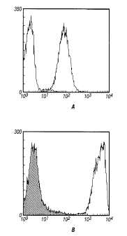
Figures 1 a and 2b shows the comparison of flow cytometric detection of human
class I MHC molecule on Jurkat cells that have been prepared for flow
cytometric analysis
by (Figure 1 a) standard staining methods or (Figure 1 b) by tyramide coating.
Figure 2 shows the results of flow cytometric assays of cells expressing MHC
class
I molecule which have been exposed to increasing concentrations of anti-MHC
class I
antibody and have been stained by either standard staining methods or by
tyramide
coating. The y axis represents the Fluorescence Index in which an index of 1
indicates no
specific staining. The x axis represents the increase in concentration of the
monoclonal
antibody.
Figure 3 shows the results of flow cytometric assays of cells expressing CD3
which have been exposed to increasing concentrations of anti-Fas ligand
antibody
(beginning with sub-optimal conditions) and have been stained by either
standard staining
methods or by tyramide coating. The y axis represents the Fluorescence Index
in which an
CA 02358510 2001-07-12
WO 00/42435 PCT/US00/00652
index of 1 indicates no specific staining. The x axis represents the increase
in
concentration of the monoclonal antibody.
Figure 4 shows the flow cytometric results for detecting CD45 on Jurkat cells
by
tyramide coating cells using a horseradish-anti CD45 antibody conjugate vs. an
antibody
5 control conjugate. Figure 4a represents the results obtained using control
antibody
molecules conjugated to horseradish peroxidase. Figure 4b represents the
results obtained
using antibody molecules directly conjugated to horseradish peroxidase
specific for CD45.
Figure 5 shows the results of experiments to eliminate "bystander staining".
Figure
Sa shows the results of standard flow cytometric detection of the analyte CDS
in a
10 population of CDS positive and CDS negative cells. Figure Sb shows the
results of flow
cytometric detection of the analyte CDS in a tyramide coated population of CDS
positive
and CDS negative cells, where the tyramide coating procedure was performed
incorporating methods to eliminate bystander staining. Figure Sc shows the
results of flow
cytometric detection of the analyte CDS in a tyramide coated population of CDS
positive
15 and CDS negative cells, without incorporating methods to eliminate
bystander staining.
Figure 6 shows the results of experiments in which peripheral blood
mononuclear
cells have been treated with phytohemagglutinin and interleukin 2, washed and
cultured
for 2 days with phorbol myristic acetate and ionomycin. Figure 6a shows the
results of
tyramide coating the cells for flow cytometry. One peak in the histogram
represents the
tyramide coating a control sample of cells, while the other peak represents
cells which
have been tyramide coated to detect Fas ligand using a specific binding
partner
(monoclonal antibody specific for Fas ligand. Figure 6b shows the results of a
serial
amplification tyramide coating procedure to detect the presence of Fas ligand.
Figure 7 shows three histogram peaks. The tallest peak (histogram 1)
represents
K562 cells which have been tyramide coated by single cycle serial
amplification to
determine the presence of Fas ligand. Histogram 2 represents the results of
tyramide
coating by single cycle serial amplification to determine the presence of Fas
ligand on the
surface of KFL9 cells which express the ligand. Histogram 3 represents the
result of
tyramide coating by 2 cycle serial amplification to determine the presence of
Fas ligand on
the surface of KFL9 cells which express the ligand.
The drawings are not necessarily to scale, and certain features of the
invention may
be exaggerated in scale and shown in schematic form in the interest of clarity
and
conciseness.
CA 02358510 2001-07-12
WO 00/42435 PCT/US00/00652
16
Detailed Description Of The Preferred Embodiments
The method of tyramide coating live cells for flow cytometric analysis
represents a
new method for detecting and/or quantitating cellular analytes. The present
invention
offers a method which allows for better detection of cell surface molecules
and moreover
allows for detection of cellular analytes by flow cytometry by using lower
concentrations
of antibodies (see figures) 2 and 3), antibodies of lower affinity and will
allow the
detection and analysis of molecules previously incapable of being detected by
less
sensitive flow cytometric methods. Hence, the present invention offers a more
sensitive
method for flow cytometric analysis of cellular analytes in samples of live
cells.
Standard Flow Cytometry
Flow cytometry permits sensitive detection and rapid quantification of some
features of single cells, such as relative size complexity, and endogenous
fluorescence, as
well as the quantitative analysis of any cellular compound that can be labeled
with a
fluorochrome. General technical basis and a review of applications of flow
cytometry can
be found in Melamed, M.R. et al., "Flow Cytometry and Cell Sorting", 2"a ed.
Wiley-Liss,
New York ( 1990) which is incorporated herein by reference in its entirety
including any
drawings. One of the main achievements of flow cytometry is the rapid
quantification of
analytes on a large number of, single particles or cells.
The flow cytometer is an instrument that analyses cells one at a time by
producing
a stream of fluid containing the cells. This stream is focused so that it
passes through a
laser beam of a defined wavelength. Generally, the fluorochromes selected for
use as
detectable markers are selected based on the ability of the fluorochrome to
fluoresce when
excited by light with the wavelength used by the laser. When the fluorochrome
is excited
by the laser beam, it emits light which is then assessed by the
photomultiplier tubes of the
flow cytometer. This technique is capable of analyzing 10,000 cells within 1
to 2 minutes.
Furthermore, as discussed above, currently available flow cytometers have
filters to detect
the emittance from various fluorochromes which fluoresce at different
wavelengths, and
allow for up to four different fluorochromes to be used as detectable markers
which means
currently at least up to 4 different molecules may be detected simultaneously.
One limitation of standard or standard flow cytometric analysis has been the
sensitivity of the technique. Cells to be assessed by flow cytometry are
reacted in the cold
with antibodies specific for defined cell surface molecules. The antibodies
are generally
labeled with a fluorescent molecule, although a second reaction with a
molecule which
possesses a fluorescent label that can bind bound antibody can also be used as
a detectable
marker. After labeling the cells with cell surface molecule specific
antibodies and after
CA 02358510 2001-07-12
WO 00/42435 PCT/US00/00652
17
washing the cells to remove any unbound antibodies, the cells are placed into
a flow
cytometer. Using this method the analyte of interest would have to be
represented on the
cell surface in multiple copies, or multiple antibodies would have to be
prepared for
different epitopes of the analyte, in order to detect the amount of
fluorescent marker that
has bound via antibody to the cell surface antigen.
Although flow cytometry has been used successfully for many different
molecules,
it is considerably less sensitive than many other procedures for the detection
of cell
surface molecules. As an example, cells that have been transfected to express
the
cytotoxic molecule Fas ligand on their surface are capable of being detected
by using a
cytotoxic assay. However, even though detection of expression of Fas ligand is
possible
by analysis of the transfected cells capability of killing target cells
sensitive to Fas ligand,
we were unable to detect the expression of Fas ligand by conventional flow
cytometric
analysis.
Amplification Staining
Amplification staining has been found to be of importance in the detection of
cellular analytes for various immunological and immunogenetic procedures. For
methods
of immunohistochemistry (analysis of slide fixed tissues or cell samples by
fluorescent
microscopy) the use of enzyme based amplification staining methods has led to
enhanced
sensitivity.
The Catalyzed Reporter Deposition (CARD) method described by Bobrow et al
"Catalyzed Reporter Deposition, A Novel Method Of Signal Amplification",
Journal of
Immunoloaical Methods, 125: 279-285 (1989) and 137: 103-112 (1991) is an
amplification staining method used for both immunohistochemical methods,
microplate
immunoassays (such as ELISAs) as well as membrane immunoassays. Both the CARD
method or the analyte dependent enzyme activation system refer to an enzyme
system
where an enzyme is coupled to a member of a specific binding pair, the enzyme
then
catalyzes the formation of an activated conjugate which is deposited wherever
a receptor
for the activated conjugate is immobilized. This system has led to methods for
maximizing the sensitivity of methods aimed at the cellular localization of
proteins and
nucleic acids, especially in cases where target levels are known or suspected
to be low.
These methods have evolved to improve the sensitivity of both
immunohistochemistry and
in situ hybridization techniques.
CA 02358510 2001-07-12
WO 00/42435 PCT/US00/00652
18
Tmramide Coating Live Cells for Flow Cytometr~
In order to enhance the sensitivity of flow cytometric analysis, we have
provided a
system of amplified reporter deposition. As described in Example I, the
current method
preferably employs the use of biotinylated antibodies specific for cell
surface molecules, a
steptavidin-horseradish peroxidase and the substrate peroxide and a reporter
molecule
such as tyramide. The enzyme reacts with its' substrate to produce oxygen
radicals which
interact with the phenolic group of tyramide to create a short lived radical
activated
phenolic substrate. It is believed that the radical activated phenolic
substrate binds with
electron rich moieties such as tyrosine and tryptophan present in the proteins
found on
most cell surfaces. It is for this reason that in a preferred embodiment
tyramide may be
replaced with a phenolic molecule which can be attached to a binding partner.
Tyramide
can be readily attached to fluorescein, biotin or rhodamine as described in
Anton H.N. et
al., "Rapid Synthesis of Biotin-, digoxigenin-, Trinitrophenyl-, and
Fluorochrome-labeled
Tyramides and Their Application for In Situ Hybridization using CARD
Amplification",
The Journal of Histochemistry and Cytochemistry, Vol. 46(6): 771-777, (1998),
which is
herein incorporated by reference in its entirety including any drawings.
We have investigated the potential of enzymatic amplification to enhance
signals
in flow cytometry. KFL9 and K562 cells labeled with Anti-Fas ligand monoclonal
antibody when incubated with the enzymatic incubation steps as described in
Example I,
step 3, produced a 4 to 5 fold increase in fluorescent signal when compared to
cells
incubated without the enzymatic amplification step. The enhancement in the
signal
indicates that the use of this technology will allow more sensitivity in the
detection of cell
surface molecules which will be advantageous for both diagnostic and research
applications. We have used amplification staining with flow cytometry to
enhance the
specific fluorescence signal up to 61 fold greater than in standard flow
cytometric staining
in assessing the expression of cell surface molecules (Figure 1 ).
A phenomenon of an aspect of amplification staining has been termed "bystander
staining". Bystander staining refers to the staining of negative cells in test
tubes that
include both analyte positive and negative cells. When analyte negative cells
are
amplification stained for flow cytometry using control antibodies which should
not bind
the cell there is no detectable staining of the cells other than background
staining which is
commensurate with that found in standard staining procedures. However, if
there are both
negative and positive analyte cells in the same test tube, the amplification
staining
procedure may stain both the positive and negative cells. The elimination of
bystander
staining occurs in the amplification step of the method (step 4 of Example 1).
By
resuspending the cells in a low volume 25 to 100 microliters of
Ficoll/Hypaque, pH 8.5
CA 02358510 2001-07-12
WO 00/42435 PCT/US00/00652
19
with or without the addition of exogenous protein such as milk protein the
bystander
staining effect is reduced and almost eliminated (Figure 5). This step
strengthens the
overall specificity of the method, however it is still possible to
differentiate analyte
positive cells from analyte negative cells based on their respective
fluorescence without
eliminating bystander staining.
Cell surface analytes can be present on a cell surfaces in amounts which are
not
easily detectable by current methods. For example, peripheral blood
mononuclear cells
treated for 3 days in culture medium with phytohemagglutinin and interleukin 2
have
stimulated FAS ligand activity which can be measured in a cytotoxicity assay
and is
indicative of the presence of FAS ligand. Cells which have been exposed to
phytohemaggluting an interleukin 2 in such a manner are positive for FAS
ligand activity
in a cytotoxicity assay. However detecting FAS ligand on these cells by flow
cytometric
analysis is inconclusive for the presence of the cell surface molecule using
standard flow
cytometric means (Figure 6a). Surprisingly, FAS ligand can be detected by
tyramide
coating the cell surface using serial amplification. Figure 6b, shows the
detection of FAS
ligand on cells which were positive when analyze by cytotoxic activity but
were
inconclusive for FAS ligand when analyzed by standard flow cytometric methods.
These
cells when analyzed by flow cytometry using serial amplification methods as
described
herein are positive for the presence of the cell surface molecule FAS ligand.
Furthermore, we have shown that additional cycles of serial amplification for
tyramide coating results in improved detection of cell surface analytes. K562
cells do not
normally express Fas ligand, as shown in Figure 7. When K562 cells are
analyzed for the
presence of FAs ligand by tyramide coating using amplification staining and
flow
cytometry the results are negative (Figure 7). KFL9 cells express Fas ligand
on their
surface. Figure 7 illustrates the improved staining which results from
additional cycles of
serial amplification staining.
Those in the art will appreciate that the method of the invention can be used
for a
variety of flow cytometric analysis methods for analyte detection in live cell
samples.
Also, those in the art would recognize that more than one fluorochrome may be
used
depending on the quality of the flow cytometer used for analysis. In addition,
with the
inventive teachings described herein, incorporation of more than one
fluorochrome onto
cells assayed for more than one cell surface molecule could provide a method
for the rapid
detection of more than one analyte of interest in a sample of live cells.
CA 02358510 2001-07-12
WO 00/42435 PCT/US00/00652
Examples
The following examples serve to illustrate the method for amplification
staining
live cells for flow cytometry of the invention. These examples are in no way
intended to
limit the scope of the invention.
5 Example 1 Tyramide Coating Live Cells for Flow Cytometric Analysis
1. A sample of live cells expressing or presumed to be expressing a cell
surface analyte of interest are added to a test tube.
2. To this sample, antibody (or antibody conjugate) specific for the analyte
is
added in a physiological buffer, such as phosphate buffered isotonic saline
with 0.005%
10 azide and 1 % bovine serum albumin, at room temperature. The cells are
washed once in
with the same medium without antibody.
3. Add a substance with enzymatic activity (streptavidin-horseradish
peroxidase) that will bind to antibody or antibody conjugate. Cells are
incubated in a
physiological buffer (as above) at room temperature. The cells are washed in
phosphate
15 buffered isotonic saline.
4. Tyramide-biotin molecules and peroxide are added to the cells in a solution
of Ficoll/Hypaque, pH 8.5 in a small or low volume 25 to 100 microliters in
the presence
or absence of exogenous proteins (such as milk protein). Cells are incubated
at room
temperature in a physiological buffer that does not contain azide. The cells
are washed
20 with phosphate buffered saline and once with phosphate buffered saline with
added
sodium azide and bovine serum albumin.
5. Streptavidin-fluorochrome molecules are added to the cells in phosphate
buffered saline with added sodium azide and bovine serum albumin. The cells
are washed
and the analyzed by flow cytometry
(A washing step may be included between each of steps 1, 2, and 3. Steps 2 and
3 may be
combined with the use of an antibody conjugated to an enzyme as shown below).
Example 2 Detection of CD45 with Coniu~ated Monoclonal Antibody
1. A sample of Jurkat cells are added to two test tubes, a control tube and
experimental tube.
2. To the control sample control antibody conjugated with horseradish
peroxidase is added and to the experimental sample antibody specific for CD45
conjugated with horseradish peroxidase. Both samples are suspended in a
physiological
buffer, such as isotonic saline with 0.005% sodium azide, at room temperature.
CA 02358510 2001-07-12
WO 00/42435 PCT/US00/00652
21
3. After the cells are washed, tyramide-biotin molecules and hydrogen
peroxide are added to the samples in a solution of Ficoll/Hypaque, pH 8.5 in a
small or
low volume (25 to 100 microliters) in the presence or absence of exogenous
proteins (such
as milk protein). Cells are incubated at room temperature in a physiological
buffer that
does not contain azide.
4. After cells are washed, streptavidin-fluorochrome molecules are added to
the cells, the samples are washed and the analyzed by flow cytometry
The results demonstrate that antibodies directly conjugated to horseradish
peroxidase work well in the amplification procedure. (see Figure 4)
Example 3 Elimination of Bystander Staining
1. A sample of Jurkat (CDS positive) and K562 (CDS negative) cells of
approximately equal numbers is added to a test tube.
2. To this sample, antibody (or antibody conjugate) specific for the analyte,
CDS, is added in a physiological buffer, such as phosphate buffered isotonic
saline with
0.005% azide and 1% bovine serum albumin, at room temperature. The cells are
washed
once in with the same medium without antibody.
3. Add a substance with enzymatic activity (streptavidin-horseradish
peroxidase) that will bind to antibody or antibody conjugate. Cells are
incubated in a
physiological buffer (as above) at room temperature. The cells are washed in
phosphate
buffered isotonic saline.
4. Tyramide-biotin molecules and peroxide are added to the cells in a solution
of Ficoll/Hypaque, pH 8.5 in a small or low volume 25 to 100 microliters in
the presence
or absence of exogenous proteins (such as milk protein). Cells are incubated
at room
temperature in a physiological buffer that does not contain azide. The cells
are washed
with phosphate buffered saline and once with phosphate buffered saline with
added
sodium azide and bovine serum albumin.
5. Streptavidin-fluorochrome molecules are added to the cells in phosphate
buffered saline with added sodium azide and bovine serum albumin. The cells
are washed
and the analyzed by flow cytometry.
Samples contained CDS positive and CDS negative populations of approximately
equal
numbers.
CA 02358510 2001-07-12
WO 00/42435 PCT/US00/00652
22
Example 4 Amplification Staining Of Live Cells For Flow C ometry
1. Place half a million cells in 12 x 75 mm squared round bottom tubes.
2. Wash the cells once with phosphate buffered isotonic saline (pH 7.3) with
1% bovine serum albumin and 0.005% sodium azide (referred to as staining
buffer).
3. Add to the cells biotinylated antibody (0.5 -1 microgram) specific for
analyte in 50 microliters of staining buffer and incubate for 10 minutes at
room
temperature in the dark.
4. Wash the cells one or two times with staining buffer.
5. Add to the cells streptavidin conjugated with horseradish peroxidase in 50
microliters of staining buffer and incubate for 10 minutes at room temperature
in the dark.
6. Wash the cells one time with phosphate buffered isotonic saline, pH 7.3.
7. Wash the cells one time with phosphate buffered isotonic saline, pH 7.3
containing 0.01 % hydrogen peroxide.
8. Add to the cells biotinylated tyramide (1-1.5 mg/ml) in 50 microliters of
Histopaque (Sigma brand name of Ficoll/Hypaque, density 1.077) containing 0.01
hydrogen peroxide. Incubate the suspension for 10 minutes at room temperature
in the
dark.
9. Wash the cells one time with phosphate buffered isotonic saline, pH 7.3
and one time with staining buffer.
10. Add to the cells 0.5 microgram streptavidin conjugated with a
fluorochrome such as phycoerythrin-CYS in 50 microliters of staining buffer.
Incubate for
10 minutes at room temperature in the dark.
11. Wash the cells one or two times with staining buffer and resuspend in 0.5
milliliters of staining buffer.
' 12. Analyze the cells on a FACScan II flow cytometer with the following
photomultiplier tube voltage settings: Forward scatter channel, E00; side
scatter channel,
507; FLl, 620; FL2, 603; and FL3, 650. The amplification gain settings are:
forward
scatter channel, 1.5; and side scatter channel, 1Ø
Example 5 Serial Amplification Staining Method
1. Place half a million cells in 12 x 75 mm squared round bottom tubes.
2. Wash the cells once with phosphate buffered isotonic saline (pH 7.3) with
1 % bovine serum albumin and 0.005% sodium azide (referred to as staining
buffer).
3. Add to the cells biotinylated antibody (0.5 -1 microgram) specific for Fas
ligand analyte in SO microliters of staining buffer and incubate for 10
minutes at room
temperature in the dark.
CA 02358510 2001-07-12
WO 00/42435 PCT/US00/00652
23
4. Wash the cells one or two times with staining buffer.
5. Add to the cells streptavidin conjugated with horseradish peroxidase in 50
microliters of staining buffer and incubate for 10 minutes at room temperature
in the dark.
6. Wash the cells one time with phosphate buffered isotonic saline, pH 7.3.
7. Wash the cells one time with phosphate buffered isotonic saline, pH 7.3
containing 0.01 % hydrogen peroxide.
8. Add to the cells biotinylated tyramide (1-1.5 mg/ml) in 50 microliters of
Histopaque (Sigma brand name of Ficoll/Hypaque, density 1.077) containing 0.01
hydrogen peroxide. Incubate the suspension for 10 minutes at room temperature
in the
dark.
9. Wash the cells one time with phosphate buffered isotonic saline, pH 7.3
and one time with staining buffer.
10. Repeat steps 5-9 at least once.
11. Add to the cells 0.5 microgram streptavidin conjugated with a
fluorochrome such as phycoerythrin-CYS in 50 microliters of staining buffer.
Incubate for
10 minutes at room temperature in the dark.
12. Wash the cells one or two times with staining buffer and resuspend in 0.5
milliliters of staining buffer.
13. Analyze the cells on a FACScan II flow cytometer with the following
photomultiplier tube voltage settings: Forward scatter channel, E00; side
scatter channel,
507; FL1, 620; FL2, 603; and FL3, 650. The amplification gain settings are:
forward
scatter channel, 1.5; and side scatter channel, 1Ø
All patents and publications mentioned in the specification are indicative of
the
levels of skill of those skilled in the art to which the invention pertains.
All references
cited in this disclosure are incorporated by reference to the same extent as
if each
reference had been incorporated by reference in its entirety individually.
One skilled in the art would readily appreciate that the present invention is
well
adapted to carry out the objects and obtain the ends and advantages mentioned,
as well as
those inherent therein. The methods, substituents (such as buffers,
fluorochromes,
enzymes and substrates), and target materials described herein as presently
representative
of preferred embodiments are exemplary and are not intended as limitations on
the scope
of the invention. Changes therein and other uses will occur to those skilled
in the art,
which are encompassed within the spirit of the invention, are defined by the
scope of the
claims.
CA 02358510 2001-07-12
WO 00/42435 PCT/US00/00652
24
It will be readily apparent to one skilled in the art that varying
substitutions and
modifications may be made to the invention disclosed herein without departing
from the
scope and spirit of the invention. For example, those skilled in the art will
readily
recognize that the present methods can incorporate a variety of different cell
types,
physiological buffers, enzyme-substrate systems, and different target
materials. Thus,
such additional embodiments are within the scope of the present invention and
the
following claims.
The invention illustratively described herein suitably may be practiced in the
absence of any element or elements, limitation or limitations, which is not
specifically
disclosed herein. Thus, for example, in each instance herein any of the terms
"comprising", "consisting essentially of and "consisting of may be replaced
with either
of the other two terms. The terms and expressions which have been employed are
used as
terms of description and not of limitation, and there is no intention that in
the use of such
terms and expressions of excluding any equivalents of the features shown and
described or
portions thereof, but it is recognized that various modifications are possible
within the
scope of the invention claimed. Thus, it should be understood that although
the present
invention has been specifically disclosed by preferred embodiments and
optional features,
modification and variation of the concepts herein disclosed may be resorted to
by those
skilled in the art, and that such modifications and variations are considered
to be within
the scope of this invention as defined by the appended claims.
In addition, where features or aspects of the invention are described in terms
of
Markush groups or other grouping of alternatives, those skilled in the art
will recognize
that the invention is also thereby described in terms of any individual member
or subgroup
of members of the Markush group or other group.
Thus, additional embodiments are within the scope of the invention and within
the
following claims.