Note: Descriptions are shown in the official language in which they were submitted.
CA 02359496 2001-10-19
COMPUTER ASSISTED RADIOTHERAPY DOSIMETER SYSTEM AND
METHOD
DESCRIPTION
TECHNICAL FIELD:
The invention relates to radiotherapy dosimeter systems and methods,
especially of the kind which use a plurality of dosimeter sensors distributed
in a
region to be irradiated and means for monitoring radiation levels detected by
the
sensors.
BACKGROUND ART:
Radiotherapy treatment of cancer patients involves the use of machines
which produce high energy X-rays or high energy electrons. It is common
practice
to verify the radiation dose delivered to the patient with a dosimetry system
such
as the Thomson & Nielsen Patient Dose Verification System.
There are three different types of dosimetry system used in radiotherapy.
These are based on (a) film or thermal luminescent dosimeters (TLD), (b)
diodes
and (c) MOSFETs. Diode and MOSFET systems use electronic dosimeter sensors
together with electronic reading systems, whereas film or TLD use chemical or
thermal methods of reading the detectors into an electronic reading system.
Since diode and MOSFET based dosimetry systems have the convenience
of direct electronic reading of the dosimeters, they also have the potential
advantage of direct data communication with computer systems. The person using
a patient dosimetry system (usually a medical physicist, dosimetrist or
therapist)
requires the radiation dose information from the system to be in a format that
is
suitable for good quality assurance records.
The state of the art with patient dose verification systems is for the dose
data
to be presented in one of three formats - (a) on a display on the reading
instrument, (b) on a print-out from the electronic reader or (c) on a computer
screen. In the latter case, the information presented on the computer screen
is in
the form of numbers and, in some cases, graphs.
Thomson & Nielsen MOSFET dosimetry systems use ExcelT"" spreadsheets
for this purpose. Sun NucIearT"" and ScanditronixT"" have diode-based systems
which use WindowsT"" - based systems with numerical tables and graphs of data.
A disadvantage of these known systems is that it is not easy to confirm that
the
dose values measured were taken at the proper locations on the body of the
patient.
CA 02359496 2001-10-19
2
SUMMARY OF INVENTION:
An object of the present invention is to at least mitigate this disadvantage
and to this end, there is provided a dosimetry system having means for
displaying
a representation of the body, e.g., a patient, to be irradiated, showing
specific
locations of radiation sensors in relation to the body.
According to one aspect of the present invention, there is provided a
dosimetry system in which a plurality of sensors for disposition on, in or
near a body
to be irradiated, for example a patient, are connected, in use, to a sensor
reading
instrument which is interfaced with a display system, for example a personal
computer, which is arranged to display, in use, one or more representations,
for
example drawings or photographs, of the body to be irradiated, along with the
graphics artefacts representing locations of the dosimeter sensors in relation
to the
body.
Preferably, the display system is arranged to display the representations,
prior to irradiation, with the sensor location artefacts and sensor
identifiers and,
after irradiation, with the measured doses associated with each sensor.
Preferably, the display system provides for adjustment of the sensor location
artefacts prior to the irradiation, to select desired locations, and then may
provide
for printing of the representations, showing the sensor location artefacts,
prior to
irradiation, thus allowing the print-out to be used by an operator as a guide
when
positioning the sensors.
According to a second aspect of the invention, a method of using a
dosimetry system of the first aspect to monitor radiation doses at various
locations
on a body comprises the steps of:
(i) displaying one or more representations of the body to be irradiated and
graphics artefacts, e.g., points or icons, representing locations of a
plurality of
dosimeter sensors, and
(ii) adjusting the display to position the sensor location artefacts at
preselected
sites on, in or near the body at which radiation doses are to be measured.
Preferably, the method further comprises the steps of:
(iii) irradiating the body and obtaining data of radiation measured at each of
the
sensors,
(iv) displaying the data for each sensor in the same display as the one or
more
representations of the body with the sensor location artefacts at said
preselected
positions.
In preferred embodiments of either aspect of the invention, in the display,
the
graphics artefacts representing the dosimeter sensors comprise points or icons
associated with respective identifiers, conveniently interconnected in the
display
CA 02359496 2001-10-19
3
by, for example, lead lines. The positions of the points or icons may be
adjusted
relative to the representation of the body to locate them at positions on the
image
which correspond to the actual locations at which the sensors are (to be)
located.
Following irradiation, each of the identifiers then is associated,
conveniently in a
table, with the corresponding dose data.
Embodiments of the invention advantageously enable the physicist to plan
the locations where dose measurements are required, ensure that the dosimeters
are placed according to plan, and confirm that the body (patient) has received
the
correct dose to the correct location according to the plan.
Yet another advantageous feature is that the one or more representations
of the body, together with the preselected dosimeter sensor locations, may be
printed prior to patient treatment so as to facilitate correct positioning of
the
dosimeter sensors in the correct anatomical positions by the medical personnel
performing the radiotherapy procedure.
Advantageously, embodiments of the present invention may provide real-
time display of data from the dosimetry system reader.
Another advantageous feature is that the patient's treatment information may
be readily recorded (e.g. patient's name, identification of radiotherapy
machine
used, energy of the machine).
The one or more representations used to indicate the positions of the
dosimeter sensors on the body, e.g. on the patient's anatomy, may comprise
standard line drawings or custom images, such as scanned photographs or
digital
camera images. In the latter cases, the use of actual images of the body
facilitates
proper location of the sensors.
Another advantageous feature of embodiments of the present invention
which use a computer display is that the software may calculate the radiation
dose
using the data input from the reading instrument and any calibration or
correction
factors previously input by the physicist, typically following a previous
calibration
of the dosimetry system in a known manner. The software then may compare the
dose calculations with predetermined target doses and indicate, conveniently
by
highlighting in the display, any deviation for corrective action.
A further feature of embodiments of the present invention is the capability
to view, print or electronically save the final report with all the relevant
dosimetry
data collected during the patient's treatment.
According to a third aspect of the invention there is provided software for
intertacing a plurality of dosimeter sensors and a reader to a microcomputer
or
personal computer to provide for the display of an image or other
representation of
CA 02359496 2001-10-19
4
the body/patient and the locations of the sensors in relation to the body, in
a system
according to the first aspect.
BRIEF DESCRIPTION OF THE DRAWINGS:
A dosimetry system in accordance with the invention will now be described,
by way of example, with reference to the accompanying drawings, in which: -
Figure 1 illustrates, partially and schematically, a dosimetry system for
irradiating a person;
Figure 2 illustrates a portion of a display of the system;
Figure 3A illustrates a representation displayed during assignment of sensor
positions; Figure 3B illustrates a representation subsequently displayed
during
assignment of sensor positions;
Figure 4 illustrates a report provided by the system;
Figure 5 is a flowchart depicting operation of the system;
Figures 6A to 6F and Figures 7A to 7F show display screens displayed
during operation of the system.
DETAILED DESCRIPTION OF A PREFERRED EMBODIMENT:
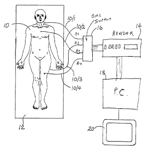
A dosimetry system for monitoring the amount of radiation to which a patient
is subjected will be described with reference to Figure 1 which illustrates a
patient
10 who is to receive radiation therapy while lying on a table 12. The therapy
entails irradiating the patient 10 by means of a radiation therapy machine,
which
might be an X-ray machine, a CT scanner, or other machine having means (not
shown) for irradiating the patient. The dosimetry system comprises a set of
MOSFET radiation sensors A1...A4 positioned at predetermined locations on the
patient's body and connected by leads 10/1...10/4, respectively, to a reader
14 (e.g.
Thomson & Neilsen's reader, Model No. 50 [TN-RD-50]) by way of a bias supply
unit 16. The reader 14 is connected to a personal computer 18 which controls a
display device 20. The sensors A1-A4, bias supply 16, reader 14 and computer
18
may be of known construction and so will not be described in detail. The
personal
computer 18 is equipped with the system software, such as Visual BasicT"", or
the
like, suitably configured, as will be described hereafter. The sensors A1-A4
and,
when applicable, other parts of the dosimetry system, have been previously
calibrated using known techniques.
Operation of the dosimetry system involves two main phases, namely (i)
assignment of graphics artefacts representing the sensors to selected
positions on
the representations, and (ii) measurement and display of the measured doses.
These two phases need not be performed at the same time. For the first phase,
the
CA 02359496 2001-10-19
patient need not be present and, in fact, the first phase could be carried out
remotely from the radiation therapy machine. For convenience, however, both
phases will be described as if carried out together.
Figure 2 illustrates a portion of the display 20 controlled by the computer 18
5 and showing representations of the patient 10; specifically, in outline,
front 10F
and rear 10R views of the patient 10 and positions of graphics artefacts
representing the four dosimeter sensors A1, A2, A3, and A4. The display also
shows a table 22 listing the sensors A1-A4 and associated data. When the
irradiation process has been carried out, the data will include the dose
measured
by each sensor.
Referring now to Figure 3A, which illustrates the type of graphic
representation first shown to the user on the computer screen 20, when the
sensor
artefacts have not been assigned, but merely grouped to the right of the front
images 1 OF. The sensors A1, A2, A3 and A4 are represented by graphics
artefacts
comprising respective sensor dots connected by lead lines to respective labels
(identifiers) A1 - A4.
Initially, the computer prompts the user to assign dosimeter sensors to
various parts of the anatomy, which the user does by "dragging and dropping"
the
dots and identifiers. Once this task has been completed, the display screen
shown
to the user is as illustrated in Figure 3B. In the example shown, the user has
dragged and dropped both the dots and labels of the dosimeter sensors (e.g.,
A1,
A2 etc.) so that the dots are located at the required sites on the images and
the
identification labels are conveniently placed nearby. A description of each
site,
e.g., "rear of neck", is optionally recorded in a database.
Having completed this task of assigning sensors to desired locations, the
user may print out the diagram or photo of the patient with dosimeter
locations so
that the medical personnel may then use the photo as a guide when placing the
dosimeters in the desired locations on the patient.
Following irradiation, the dose information from the dosimeter sensors is
read into the computer by operating the dosimetry system connected as in
Figure
1. (The dosimeters may be removed from the patient for this part of the
procedure).
The dose measurements are stored in the computer and displayed on a final
report, along with the patient and treatment information. Figure 4 shows the
format
of the final report with the dosimeter sensor position information, as well as
the
dose measurements, target doses and the deviation information.
The software used by the system may be developed using Visual Basic T""
or any other software program suitably configured, to carry out the above
process.
The software program catalogs its functions into the following sections:
CA 02359496 2001-10-19
6
(i) System setup
(ii) Pre-irradiation
(Iii) Treatment Information
(iv) Measuring Dose
(v) Viewing & Printing reports
Figure 5 shows a flow chart of the system software program. The main tasks
the software needs to perform include: i) recording information sent by the
Reader
ii) organizing this information on the computer screen iii) recording
treatment
information, indicating dosimeters' position and iv) printing out measurement
reports.
To start with, the user has the option of deciding if he wants to just view a
report that is already existing (by clicking on the 'Report File' icon on
screen) or to
run the program for new readings. In the first instance, the user may view
only the
existing reports. In the latter case, the program is started by clicking the
"Program"
icon on the computer screen (step 50). A Start ~ Program menu is displayed on
the
screen. The program then checks if the system is set up (step 52) by checking
all
the initial set up parameters, e.g., if an appropriate port has been selected,
if the
password is correct etc. If the system is not set up, the user is prompted to
click on
the "TN-Dose Reporter 2.31 " entry of the computer's "Start I Programs" menu
to run
the setup program and the "Setting Up the System" panel (Figure 6A) is
displayed
on the computer screen (step 54). At this stage the user is prompted to input
data
like a password, Institution's name, the patient's name, selection of the
communication port etc. Once the user appropriately inputs all the values
required
to set up the system, the program moves to the next step of Pre-Irradiation
(step
56). The Pre-Irradiation display is shown on the monitor and in this step the
user
may modify calibration parameters, modify system settings etc (Figure 6B) by
entering desired data into the computer to be displayed on the screen. Once
this
step is completed the program moves to the step of Treatment Information (step
58). This can be carried out without picture (Figure 6C) or with picture
(Figure 6D).
A table is shown where the user may type in the appropriate information e.g.,
Patient- ID, Radiation setting and Dosimeter-Assignments. In the previous case
the
user may describe the dosimeter sensors' locations with words (e.g., 'chest',
'stomach' etc. ) and type words in the corresponding cells of the dosimeter
Assigning table (Figure 6C). To do the latter, the user may click upon the
"Show
Picture" icon whereupon an image representing a human body will be displayed
on the screen.
CA 02359496 2001-10-19
7
The user is prompted with an option to use the same image displayed on
screen or select another image stored in the memory of the computer (step 60).
If the user decides to select another image, the computer then instructs the
user to
assign dosimeter sensors to various parts of the anatomy and the user has to
indicate the sensors locations on the newly selected image (step 62). There is
also
an option of taking an actual photograph of the patient using a digital camera
and
using that image on the screen instead of using previously stored images. The
photograph thus taken may be displayed on screen by the program to be selected
by the user.
The selected image is now provided in an on-screen picture box which
accommodates the image as background and some labels, red dots and lines for
linking a label with a dot, as foreground (Figure 6D). Each label and dot may
be
"dragged and dropped" to appropriate positions on the image representing the
human body to indicate the dosimeters' locations graphically. In the table
provided
on the screen, corresponding to each label or identifier representing a
dosimeter
sensor, a target dose of radiation may be entered.
Once labelling of the irradiation locations on the image corresponding to the
patients body is successfully completed, the program performs the step of
Making
Measurement (step 64) and the next screen titled Making Measurement appears.
The screen now displays a table where all the labels or identifiers
representing the
dosimeter sensors are shown. Dose data from the actual sensors placed on the
patient's body is read by the reader 14 and is inputted to the computer and
the data
read is placed in the corresponding row in the table next to the identifiers
which
also represent the same dosimeter sensors identifiers marked on the image
(Figures 6E).
In the next step, the program extracts information and creates a report. The
user is prompted for viewing/printing and saving the final reports. Once this
option
is selected, the dose measurements are stored in the computer and displayed on
a final report (Figure 6F) along with the patient and treatment information
(step 68).
Next in a display the program asks the user if another measurement needs
to be performed (step 70). If the answer is "No" the program exits. If the
answer is
"Yes", i.e., if the user decides to perform another measurement, the program
goes
back to step 54 and starts the Pre-Irradiation procedure again.
The software is generally composed of a) Visual Components, b) Main
Module, c) Supporting Modules.
a) Visual components include the functional display panels and some supporting
windows.
CA 02359496 2001-10-19
b) Main Module provides the entry point to run the software and is named as
Lib_main. When the program starts to run, the main() subroutine in this module
is
called first followed by the other subroutines, e.g., main tryPort(), main
tryScreen()
etc.
c) Supporting Modules consists of subroutines for performing various functions
including:
Lib StepO: stores the subroutines needed for the panel "Setting up the system"
Lib Step1: provides subroutines needed for the panel"Pre-Irradiation"
Lib Step2: consists of subroutines needed for the panel "Treatment
Information"
Lib Step3: stores the subroutines needed for the panel "Measuring Dose"
Lib_Step4: provides subroutines needed for the panel "Viewing/Printing
Reports"
Lib MyTypes: for defining some custom data types
Globals: for defining global variables
Lib_util: consists of general purpose service subroutines
Lib comm: stores subroutines for communication with the Reader and subroutines
for message analysis.
The following is a detail description of the steps the software carries out in
order to proceed from System Setup to Viewing/Printing Reports.
1. System Setup
Prior to use, the system is set up by selecting the communication port of the
computer to be used for reading the data from the reader, setting up the title
of the
measurement reports, setting or changing the password and determining its
protection scope, inputting the lists of radiation machines and TN-RD-50
Readers.
The user clicks on the "TN-Dose Reporter 2.31" entry of the computer's "Start
~
Programs" menu to run the program. The "Set Up the System" panel is shown
(Figure 6A) and the user is required to input some information or make some
decisions, which include:
(1 ) Choosing a serial port to communicate with the TN-RD-50 Reader.
(2) Inputting the Institution Name and the Report Title. They will be printed
on
the measurement reports. The default Report Title is "DOSIMETRY REPORT".
(3) Building up the list of radiation machines types.
(4) Building up the list of radiation machines' SIN.
(5) Building up the list of TN-RD-50 Readers' S/N.
(6) Setting or changing the user's password and determining the password-
protection's scope.
CA 02359496 2001-10-19
9
Once the system is set up, the "Set Up The System" panel will not be shown
when the program is run later. To view or change system settings, the user can
select the action of "Modify System Settings" from panel.
When the program is started, it checks the computer's hardware resources
and lists all available serial ports in the pull-down list. If there is no
port available
(for example, in case all ports being used by other applications), the program
will
give out a message and automatically show the panel of "Viewing/Printing
Reports".
After setup, a new folder (for example: "c:\TN-Dosimetry") is established in
the computer. This folder holds a file for history of messages (e.g.
"MessageHistory.txt") and two sub folders ("Libs" and "Reports"). These
folders
may not be renamed.
2. Pre-Irradiation
Once the set up process is completed, the computer will display one or more
representations of the body to be irradiated and points or icons representing
a
plurality of dosimeter sensors in the panel of "Pre-Irradiation" (Figure 6B).
In this
step, the user can modify Calibration Factors (CFs) and Correction Factors
(CRs),
check dosimeter sensors, modify system settings, or view existing reports.
The Reader can be set to read in radiation units (cGy or R) using
Calibration Factors determined by the user for each dosimeter. The Reader can
also be set to read the MOSFET voltage in mV. In order to give the user more
flexibility, this Dose Reporter program allows the user to store the CFs in
the
program when the mV mode is used. The program also enables the user to specify
Correction Factors (CRs) to be used in the dose calculation.
If the Reader is set to output radiation units (cGy or R), then the CFs and
CRs in the program are inoperable. If the user sets the output of the Reader
to mV,
then CFs and CRs must be set, because they will be used to calculate the doses
according to the formula "Dose = CR * (Voltage / CF)". The user can get a hard
copy of CFs and CRs by clicking the "Print" button.
[Note: An example of the use of a CR would be if the user wanted to determine
Dmas but was measuring doses with less than full build-up.]
The allowed CF range is 0.1 mV/cGy to 99.99 mVIcGy. If the user enters a
too large or too small value, it will be trimmed into this range. The allowed
CR
range is 0.100 to 9.000. If the user enters a value beyond this range, it will
be
trimmed into this range.
When the user has finished modifying CFs or CRs, the user can set them as
defaults. Otherwise, the default CF and CR is 1.OOmVIcGy and 1.000
respectively.
If the user does not like other users changing CFs or CRs (or both), the user
can
CA 02359496 2001-10-19
set up a password (in "Setting Up the System" Panel) and put CFs or CRs (or
both)
into the protection scope, then restart this program. A realistic example of
this panel
is shown in Figures 7B and 7C.
A Message Window is used to display the messages from the TN-RD-50
5 Reader. The user can view all messages (in the current measurement
procedure)
or just view recent messages. Every message displayed here is also saved into
a
file "c:\TN-Dosimetry\MessageHistory.txt" simultaneously.
3. Treatment Information:
10 In this step, the user may adjust the display to position the sensor
artefacts
(points or icons) at preselected locations on, in or near the body at which
radiation
doses are to be measured by dragging the artefacts to various locations of the
picture representing a human body on the screen (Figure 6D). Optionally, this
can
be done without the image as well (Figure 6C).
The user determines the number of patients in the current treatment, and,
for each patient, selects the position on the screen to type in the
appropriate
information e.g. Patient's ID, Treatment Plan Reference and Radiation
Settings.
There is an on-screen picture-box (See Figure 2) which accommodates an
image as background and some labels, lines and red dots as foreground. The
user
can select the background image from the software's built-in images, or use
any
image that has been stored in the computer's hard disk in BITMAP, JPEG or GIF
format. For every assigned dosimeter sensor , the picture-box shows on the
foreground a label, a red dot, and a line to link the label and dot. Every
label and
dot can be dragged to appropriate positions to indicate the dosimeters' sites
graphically. Thus the user assigns dosimeter sensors to various locations on a
patient's body through an on-screen table, and types in words to describe the
locations and target doses of each dosimeter sensor.
Figure 2 is an example of a picture that appears on the screen to let the user
input the Patient Information, Treatment Plan Reference, and Radiation
Settings
(the user can set them by importing treatment information from an existing
measurement report by clicking "Import Existing Treatment Info"). The user
also
needs to assign dosimeter sensors to the patient(s).
When the user assigns dosimeters to the current patient, the corresponding
Site Pointer and Dosimeter Label will appear on the image area. To indicate
the
dosimeter sensor's site, the user may simply drag the Site Pointer and
Dosimeter
Label to the appropriate place on the image. (The user can drag the Pointer
and
Label to the same place, and the pointer will disappear.)
CA 02359496 2001-10-19
11
The user can describe the dosimeter sensors' locations with words or with
pictures. To do the former, the user may type words in the corresponding cells
of
the dosimeter-Assigning Table (Figure 6C). To do the latter, the user may
click
"Show Picture", whereupon a human body image will be displayed on screen, as
Figure 6D.
The software uses a table to store treatment information in this step. For
every patient, the software creates an instance of this table that
accommodates
fields to keep Patient's ID, Treatment Plan Reference, Radiation Settings,
Dosimeters' Positions and Target Doses. It also includes a field to keep a
reference
to the selected background image, and some fields to keep the relative
coordination
of every foreground label, dot and line.
Clicking the "Print" button on the picture's bottom-right corner can print out
the picture. (If that button is not enabled, the user may click the "Apply"
button.)
The user can change the human body image. For example, 5 optional
images, called "Standard Images", are generally provided. They are
#0, Unisex Body
#1, Female Chest
#2, Male Head
#3, Female Head
#4, Female Body
Besides the standard images, the user can use their own images,
conveniently called "Custom Images", such as those from a digital camera photo
or a scanned photo. Any BITMAP (*.bmp), JPEG (*.jpg) and GIF (*.gif) images
can
be used as a custom image. If the image to be used has been stored in another
format, some tools (such as Paint or PhotoShop) may be used to open them and
save them in BITMAP or JPEG format. There is no special requirement on the
images' size.
To change the image, the current image is double-clicked, or right-clicked
to pop up a menu and in the menu "change image" in the menu is selected. An
image-selection window, as in Figure 7D, will be displayed on screen.
To select a standard image, its preview window is clicked. To select a
custom image, the user should click on the corresponding item in the library
of
custom images to preview it, then, click on the preview window.
When the program is run for the first time, the library of custom images is
empty. To populate it, the user may click the "Add new Custom Image" button,
then
select an image file from the open-file dialog box. That image will be copied
to the
library and can be used as a custom image by the program.
CA 02359496 2001-10-19
12
4. Measuring Dose
This step involves irradiating the body and obtaining data of radiation
measured at each of the sensors. Dose data from the patient's body is read by
the
reader 14 through preassigned sensors (marked as e.g., A1, A2, A3 and A4)
connected to the reader. Output from the Reader 14 is transmitted through a
cable
connected to the computer (by an RS-232 cable for example) and placed in the
corresponding row in the table of recorded data on the screen. The user can
activate the "Recording" procedure to allow the input data to overwrite the
existing
data, or freeze this procedure to prevent the recorded from being changed.
The panel of this step is shown in Figure 6E. In this step, the user is
required to perform 3 actions:
(1 ) Zero MOSFETS: press the Reader's START (or ZERO) button for 1 second
to initiate the procedure.
(2) Place MOSFETS ON PATIENT(s) body. (To do it correctly, it is suggested
that the user print out the dosimeter-site diagram as a reference.)
(3) Read MOSFETs: click the "Record" button on the screen, then follow the
prompt.
In the measurement procedure, if "NIA" appears in the "Voltage" column, it
means that the voltage is Not Available since the Reader has been set up to
output
doses in the radiation units cGy or R. Voltages are only shown in this column
when
the user is using the Reader in the "mV" mode and applying Calibration Factors
(CFs) and/or Correction Factors (CRs) to translate m/v to radiation units. A
realistic
example of actual measurement is shown in Figure 7E.
5. Viewing/Printing Reports
This step involves displaying and printing the data for each sensor in the
same display as the one or more representations of the body with the sensor
points
or icons at said preselected positions. The software extracts the information,
that
is necessary to create a measurement report, from the inputted data in step 58
and
recorded data in step 64. This information is stored into a special array.
Then, from
this array, a report summary is composed and the corresponding image (see
Figure
4) is drawn. If the user needs to save this report, the software will save all
fields of
this array to the hard disk of computer 18 (next time, they can be read into
the array
if needed). The data in this array is also used to print out the report. It
may also be
saved to a floppy disk or other removable storage medium or transmitted via a
network or modem connection.
In this last step, the user can review the information in the report summary
before printing and saving (Figure 6F). All report files have a filename with
CA 02359496 2001-10-19
13
extension ".dsrpt". The default file name may be "Patient First Name + Patient
Last
Name + Date + .dsrpt". For example, if John Smith was treated on May 10, 2000,
then the default file name would be
JohnSmith 2000May10.dsrpt
The default folder for saving reports is "c:ITN-Dosimetry\Reports", but the
user can
save the reports in any folder.
When the user wants to print out the reports, there are two styles available.
Style #1 accommodates a picture to indicate dosimeters' sites graphically.
Style
#2 doesn't print out the picture, but uses a table to provide more information
about
the treatment. In this step, the user can also type in comments.
It has been stated that an existing custom-image may be used to indicate
dosimeters' locations. But, in fact, that image need not have to exist before
the
user runs the program. The user can use the REAL photos of the patients) in
current treatment using a digital camera.
It should be appreciated that the software enabling implementation of the
invention could be used with various kinds of hardware. Hence, the invention
also
embraces software per se, conveniently carried by a suitable storage medium,
for
operating a dosimetry system as described hereinbefore.