Note: Descriptions are shown in the official language in which they were submitted.
CA 02359574 2008-06-25
SURGICAL INSTRUMENT HAVING A
FASTENER DELIVERY MECHANISM
Field of Invention
The present invention relates, in general, to a surgical instrument and, more
particularly,
to a surgical instrument having a feeding mechanism for feeding at least one
surgical fastener
from a surgical instrument to attach a prosthetic in the 'repair of a defect
in tissue such as an
inguinal hernia.
Background of the Invention
An inguinal hernia is a condition where a small loop of bowel or intestine
protrudes
through a weak place or defect within the lower abdominal muscle wall or groin
of a patient.
This condition commonly occurs in humans, particularly males. Hernias of this
type can be a
congenital defect wherein the patient is born with this problem, or can be
caused by straining or
lifting heavy objects. Heavy lifting is known to create a large amount of
stress upon the
abdominal wall and can cause a rupture or tearing at a weak point of the
abdominal muscle to
create the defect or opening. In any case, the patient can be left with an
unsightly bulge of
intestinal tissue protruding through the defect, pain, reduced lifting
abilities, and in some cases,
impaction of the bowel, or possibly other complications if the flow of blood
is cut off to the
protruding tissue.
A common solution to this problem is surgery. In the surgical procedure, the
defect is
accessed and carefully examined, either through an open incision or
CA 02359574 2001-10-17
endoscopically through an access port such as a trocar. In either case, the
careful
examination can be well appreciated, as a network of vessels and nerves exist
in
the area of a typical defect, which requires a surgeon to conduct a hernia
repair
with great skill and caution. Within this area are found vascular structures
such as
gastric vessels, the external iliac vessels, and the inferior epigastric
vessels, and
reproductive vessels such as the vas deferens extending through the inguinal
floor.
Once the surgeon is familiar with the anatomy of a patient, the surgeon
carefully pushes the bowel back into the patient's abdomen through the defect.
Repairing the defect can involve closure of the defect with sutures or
fasteners but
generally involves placing a surgical prosthetic such as a mesh patch over the
open
defect, and attaching the mesh patch to the inguinal floor with conventional
suture
or with surgical fasteners. The mesh patch acts as a barrier and prevents
expulsion
of bowel through the defect. Suturing of the mesh patch to the inguinal floor
is
well suited to open procedures but much more difficult and time consuming with
endoscopic procedures. With the adoption of endoscopic surgery, endoscopic
surgical instruments that apply surgical fasteners are falling more and more
into
use. However, the tissue of the inguinal floor offers special challenges to
the
surgeon when a needle or fastener is used to penetrate structures such as
Cooper's
ligament.
At present, there are a variety of surgical instruments and fasteners
available for the surgeon to use in an endoscopic or open procedure to attach
the
mesh patch to the inguinal floor. One of the earliest types of endoscopic
surgical
instruments used is a surgical stapler. A plurality or stack of these unformed
staples are generally contained within a stapling cartridge in a serial
fashion, and
are sequentially advanced or fed within the instrument by a spring mechanism.
A
secondary valving or feeding mechanism is employed to separate the distal most
staple from the stack, to hold the remainder of the spring loaded stack, and
to feed
the distal most stapler into the staple forming mechanism. Feeding mechanisms
of
this type are found in U.S. Patent No. 5,470,010 by Robert Rothfuss et al. and
in
U.S. Patent No. 5,582,616, also by Robert Rothfuss et al.
-2-
CA 02359574 2001-10-17
Another hernia mesh attachment instrument uses a helical wire fastener that
resembles a small section of spring. Multiple helical wire fasteners are
stored
serially within the 5mm shaft, and are corkscrewed or rotated into tissue. A
load
spring is used to bias or feed the plurality of helical fasteners distally
within the
shaft. A protrusion extends into the shaft to prevent the ejection of the
stack of
fasteners by the load spring and permits passage of a rotating fastener.
Instruments and fasteners of these types are found in U.S. Patent No.
5,582,616
by Lee Bolduc et al., U.S. Patent No. 5,810,882 by Lee Bolduc et al., and in
U.S. Patent No. 5,830,221 by Jeffrey Stein et al.
Whereas the above surgical instruments are used for hernia fastening
applications, they use a spring mechanism to feed a plurality of fasteners
through
the surgical instrument. Spring mechanisms typically use a long soft coil
spring to
push a stack of fasteners through a guide or track within the shaft of the
surgical
instrument. These types of feeding mechanisms are generally simple and
reliable,
but require an additional secondary valving mechanism or protrusion to
separate
and feed one fastener from the stack, while preventing the remainder of the
stack
of fasteners from shooting out of the instrument.
Other surgical fasteners are used for hernia mesh attachment but utilize
either a reloadable single shot instrument or a rotary magazine that holds a
small
number of fasteners. These types of surgical fastening instruments can be
found in
U.S. Patent No. 5,203,864 and U.S. Patent No. 5,290,297, both by Edward
Phillips. These instruments have not gained acceptance by the surgical
conununity, possibly due to their single shot capabilities and the large size
of the
rotary magazine, which can restrict such an instrument to an open procedure.
Whereas all the above surgical instruments are used for hernia fastening
applications, they either use a spring mechanism to feed the plurality of
fasteners
through the surgical instrument, or a rotary magazine in lieu of a feeding
mechanism. Other types of surgical fasteners are available, such as surgical
clips,
and they can utilize feeding mechanisms that do not require the use of a
spring to
-3-
CA 02359574 2001-10-17
feed the clips distally. A reciprocating feeding mechanism is described in
U.S.
Patent Nos., 5,601,573 U.S. Patent No. 5,833,700, and U.S. Patent 5,921,997 by
Fogelberg et al. Fogelberg et al. teaches a clip applier with a feeding
mechanism
that utilizes a reciprocating feed bar to feed a serial stack of clips. A
feeder shoe
operably engages with and moves with the distally moving feed bar and
slidingly
engages with the proximally moving feed bar. Thus, the feeder shoe indexes or
pushes the stack of clips distally with the distally moving feed bar and
remains
stationary relative to the proximally moving feed bar. A valving mechanism is
also required to separate the distal most clip from the stack and to hold the
stack
stationary as the distal most clip is applied onto a vessel. Whereas Fogelberg
et
al. teaches a reciprocating feeding mechanism with a single reciprocating
member,
he does not teach the use of the clip applier in the attachment of hernia
mesh, nor
does he teach the individual driving or feeding of each clip by a moving
member.
Another fastener feeding mechanism that uses a reciprocation is that
disclosed in U.S. Patent No. 4,325,376 by Klieman et al. A clip applier that
stores a plurality of clips in a serial fashion within a clip magazine is
disclosed.
The clips are in a stack wherein the proximal most clip is pushed or fed
distally by
a pawl that is ratcheted or indexed distally by a reciprocating member or
ratchet
blade with each actuation of the instrument. As the pawl indexes distally, it
pushes the stack of clips distally. A secondary valving mechanism is also
described. Thus, the feeding mechanism of Klieman et al. teaches the use a
single
reciprocating member and pawl to push or feed the stack of clips distally, and
requires a secondary valving mechanism to feed the distal most clip.
Additionally,
Klieman et al. and does not teach the use of the clips for the attachment of
hernia
mesh on tissue.
U.S. Patent No. 3,740,994 by DeCarlo Jr. describes a novel reciprocating
feeding mechanism that indexes a plurality of staples or clips, and readies
them for
discharge by reciprocating one of a pair of opposing leaf spring assemblies.
The
staples reside serially within a guide rail with a fixed leaf spring assembly
extending into the plane of the guide rail. A reciprocating leaf spring
assembly
-4-
CA 02359574 2001-10-17
opposedly extends inwardly towards the fixed leaf spring assembly. As the a
reciprocating leaf spring assembly moves distally, each of individual leaf
springs
of the assembly engage a staple and move it distally. The distally moving
plurality
of staples deflect the local individual leaf springs of the fixed leaf spring
assembly,
and the deflected leaf springs return to the un-deflected position after
passage of
the staple. As the moving leaf spring assembly moves proximally, the leaf
springs
of the fixed leaf spring assembly hold the staples stationary and prevent
distal
movement thereof. A secondary guide rail and valving mechanism is provided to
separate a single staple from the stack for forming and to hold the stack of
staples
stationary as the single clip is formed.
Additionally, similar feeding mechanisms are disclosed in U.S. Patent No.
4,478,220 by Di Giovanni et al. and U.S. Patent No. 4,471,780 by Menges et al.
Both of these related patents teach a reciprocating feeding mechanism that
uses one
fixed member and one reciprocating member to feed or index a plurality of
clips
distally. Angled flexible fmgers are hingedly attached to the reciprocating
member and operatively engage the clips when moving distally, and slidingly
engage with the clips when moving proximally. The angled flexible fmgers
within
the fixed member deflect out of the way when the clips move distally and
spring
up to stop proximal movement of the clip after the clip has passed. A
secondary
valving mechanism is also disclosed.
Thus, the feeding mechanism of DeCarlo et al., Di Giovanni et al., and
Menges et al. operatively engage and individually move each clip distally
between
a single reciprocating member and a fixed member. However each instrument
requires a secondary valving mechanism for the feeding and forming of the
distal
most clip. Additionally, the surgical instruments are not indicated for use in
the
attachment of a prosthetic over a hernia.
Unfortunately, the majority of the feeding mechanisms described above require
two feeding mechanisms; a primary feeding mechanism to feed a plurality of
clips
distally, and a secondary valving or feeding mechanism to separate and feed
the
-5-
CA 02359574 2001-10-17
distal most ' fastener while preventing the distal movement of the remaining
fasteners. Such additional mechanisms are costly and increase the size or
diameter
of the instrument size. Likewise, the single shot or rotary magazines were
found
to have limitations. What is needed is an improved reciprocating feeding
mechanism that does not require the use of a secondary valving mechanism, and
can simultaneously engage with and independently drive each fastener distally.
Such a mechanism would have two reciprocating members and would provide
superior advantages such as lower cost, reduced complexity, and a smaller
diameter shaft.
SgpMM of the Invention
A delivery device for delivering a plurality of individual surgical
fasteners is disclosed. The delivery device has a drive mechanism having a
distal
and a proximal end, and a first and a second opposing member. The members are
moveable proximally and distally with respect to the delivery device, and
individually with respect to each other. The device further includes at least
one
surgical fastener located between the first and the second members. Each of
the at
least one surgical fasteners has a proximal end and a distal end. The surgical
fasteners are preferably made from a superelastic nickel titanium alloy.
Additionally an actuator having at least three sequential positions is
included. The
first position of the actuator is for moving the drive mechanism distally. The
second position is for moving the first member proximally to partially deploy
the
distal end of the fastener. The third position of the actuator is for moving
the
second member proximally, to fully deploy the distal end of the fastener.
Brief Description of the Drawings
The novel features of the invention are set forth with particularity in the
appended claims. The invention itself, however, both as to organization and
methods of operation, together with further objects and advantages thereof,
may
-6-
CA 02359574 2001-10-17
best be understood by reference to the following description, taken in
conjunction
with the accompanying drawings in which:
FIG. 1 is an isometric view of a surgical instrument wherein a left handle
half is removed to show the elements within and a trigger is in an open
position;
FIG. 2 is an isometric view of the surgical instrument of FIG. 1 wherein
the trigger is moved from the open position of FIG. 1 to a closed position as
shown, and an end effector is extended from the surgical instrument;
FIG. 2B is an exploded isometric view of some of the internal elements of
the surgical instrument of FIG. 1, with some elements removed for clarity;
FIG. 3 is a side view, in cross section, of a first side of the surgical
instrument of FIG. 1 with the left handle half removed, wherein all of the
internal
elements are shown assembled and the trigger is in an open position;
FIG. 4 is a side view of a second side of the surgical instrument of FIG. 3
with the left handle half in place and with the right handle half removed,
showing
all of the internal elements therein and the trigger in an open position;
FIG. 5 is a side view of the first side of the surgical instrument of FIG. 3
wherein the trigger is moved to a partially closed position to extend the end
effector from the surgical instrument;
FIG. 6 is a side view of the second side of the surgical instrument of FIG.
5, wherein the trigger is moved to a partially closed position to extend the
end
effector from the surgical instrument;
FIG. 7 is a side view of the first side of the surgical instrument of FIG. 5
wherein the trigger is moved to a fully closed position to retract a first
portion of
-7-
CA 02359574 2001-10-17
the end effector into the surgical instrument, and to expose a portion of a
fastener
at the end effector;
FIG. 8 is the view of the second side of the surgical instrument of FIG. 7,
wherein the trigger is moved to a fully closed position to retract an upper
portion
of the end effector into the surgical instrument, and to expose a portion of a
fastener at the end effector;
FIG. 9 is an isometric view of a fastener of the preferred invention wherein
the fastener of the preferred invention has a pair of distal barbs and a pair
of
longer proximal arms, the fastener of the preferred invention is shown in an
unconstrained state;
FIG. 10 is a side-view of FIG. 9 wherein the fastener of the preferred
invention is shown in an unconstrained state;
FIG. 11 is an isometric view of the fastener of FIG. 9 wherein the fastener
of the preferred invention is shown in a constrained state as found within the
surgical instrument of FIG. 1;
FIG. 12 is a side-view of FIG. 11 wherein the fastener of the preferred
invention is shown in a constrained state;
FIG. 13 is a bottom-view of FIG. 12 wherein the fastener of the preferred
invention is shown in a constrained state;
FIG. 14 is a cross-sectional side view of a distal end of a shaft of the
surgical instrument of the present invention showing the end effector normally
retracted therein and a plurality of surgical fasteners of the preferred
invention
contained therein;
-8-
CA 02359574 2001-10-17
FIG. 15 is a cross-sectional view 10-10 of the shaft and the end effector of
FIG. 9 and showing a passageway and a fastener of the preferred invention
contained therein;
FIG. 16 is a fragmentary perspective view of a surgical grasper instrument
placing a mesh patch over a defect or hernia in the inguinal floor of the
lower
abdomen, particularly the left inguinal anatomy;
FIG. 17 is a cross-sectional side view of the inguinal floor of the lower
abdomen of FIG. 16 illustrating the placement of the mesh patch above the
tissue
in preparation for repair of the defect, according to the present invention;
FIG. 18 is a cross-sectional side view of the inguinal floor of the lower
abdomen wherein the distal end of the shaft of FIG. 14 is pushing the mesh
patch
downward onto the inguinal floor, and the end effector is moving downwardly
within the shaft with a fastener contained therein;
FIG. 19 is a cross-sectional side view of the inguinal floor and instrument
of FIG. 18 wherein the end effector of the present invention is extended from
the
shaft and into the inguinal floor, the end effector containing a fastener of
the
preferred invention therein;
FIG. 20 is a cross-sectional side view of the inguinal floor and instrument
of FIG. 19 wherein a first portion of the end effector is partially retracted
into the
shaft to deploy a first barb of the fastener of the preferred invention
contained
therein and to engage the first barb with the inguinal floor;
FIG. 21 is the cross-sectional side view of FIG. 20 wherein the first
portion of the end effector of the present invention is fully retracted into
the shaft,
the full retraction releasing the arms of the fastener of the preferred
invention into
the portion of the shaft previously occupied by the first portion of the end
effector;
-9-
CA 02359574 2001-10-17
FIG. 22 is the cross-sectional side view of FIG. 21 wherein a second
portion of the end effector of the present invention is fully retracted into
the shaft,
the full retraction engaging a second barb of the fastener of the preferred
invention
with the inguinal floor and both arms with the shaft;
FIG. 23 is a cross sectional side view of FIG. 22 wherein the shaft of the
surgical instrument of FIG. 22 has moved upwardly to release the arms of the
fastener of the preferred invention, the released arms attaching the surgical
mesh
to the inguinal floor;
FIG. 24 is a is a fragmentary side-view of a trigger lockout mechanism of
the present invention of FIG. 1 with a lockout arm fixably attached to the
pivotable trigger, and operably coupled with a lockout wheel;
FIG. 25 is a fragmentary cross-section view of the lockout mechanism of
the present invention showing the lockout wheel in an initial position and
engaged
with a wheel detent, wherein the lockout arm is moving upwardly from a start
position (dashed lines) to a second position (cross section) adjacent to the
lockout
wheel;
FIG. 26 is a fragmentary cross-section view of FIG. 25 showing the
upwardly moving lockout arm engaging with a first tooth of the lockout wheel,
wherein the engagement has rotated the locking wheel one tooth
counterclockwise
and the locking arm is preparing to return to the initial position (dashed
lines);
io-
CA 02359574 2008-06-25
FIG. 27 is a fragmentary cross-section view of FIG. 26 showing the upwardly
moving lockout arm engaging with a final tooth of the lockout wheel, wherein
the
repeated firing of the trigger has rotated the lockout wheel to the final
booth, and a locking
tab is positioned just below the upwardly moving locking arm (cross section);
FIG. 28 is a fragmentary cross-section view of FIG. 27 showing the upwardly
moving lockout arm further engaging with a final tooth of the lockout wheel,
wherein the
lockout wheel has rotated counterclockwise to position the locking tab below
the lockout
arm;
FIG. 29 is a fragmentary cross-section view of FIG. 28 showing the detent arm
preventing further rotation of the locking wheel and the lockout arm attached
to the trigger
captured between a tooth and the locking arm of the locking wheel.
Detailed Description of the Invention
The present invention relates, in general, to a surgical instrument and, more
particularly, to a surgical instrument having a feeding mechanism for serially
feeding at
least one surgical fastener from a surgical instrument to attach a prosthetic
in place in the
repair of a defect in tissue such as an inguinal hernia.
By way of example, the present invention is illustrated and described in
conjunction with a repair of an inguinal hernia. However, it should be
understood that the
present invention is applicable to various other surgical procedures that
require the repair
of defects in tissue.
-11-
CA 02359574 2001-10-17
The Surgical Instrument
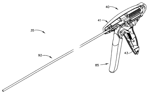
As best shown in FIGS. I and 2, the surgical instrument or fastener
delivery device of the present invention is a hand held surgical instrument 35
containing a plurality of surgical fasteners or surgical elements that are
generally
used for the attachment of a prosthetic to tissue, or as a tissue marker. The
surgical fasteners 105 of the present invention are formed from a superelastic
nickel titanium alloy, are stored within the surgical instrument in a
compressed or
collapsed state, and expand to an unconstrained state upon release from the
surgical instrument. Actuation of the instrument simultaneously releases a
fastener
105 of the present invention from a distal end of the instrument and indexes
the
plurality of fasteners 105 within the instrument.
Surgical instrument 35 of the present invention has a handle 40, an
elongated shaft 92 extending distally from the handle 40, and a trigger 85
extending downwardly from the handle 40. Handle 40 has a right half 41 and a
left half 42 that are generally mirror images of each other and, in FIGS. 1
and 2,
the left half 42 is omitted. Elongated shaft 92 is fixedly attached to the
handle 40,
and is formed from a rigid hollow material such as stainless steel tubing. A
grip
43 is fixedly attached to and extends downwardly from a proximal end of handle
40 and adjacent to the trigger 85. Trigger 85 pivotably mounts within handle
40
and is moveable from an open position as shown in FIG. 1 to a closed position
adjacent to the grip 43 as shown in FIG. 2. Movement of the trigger 85 to the
closed position extends an end effector 95 from a distal end of the shaft 92
(FIG.
2) for the placement and release of a fastener.
FIG. 2B is an isometric exploded view of the majority of the elements
found within the surgical instrument 35. The exploded view is provided to
familiarize the reader with the key elements contained therein, and the method
of
assembly used to form the surgical instrument 35. For clarity, a number of
elements such as the left handle half 42 are removed. Some of the elements of
FIG. 2B are complex in shape and the reader is advised to return to this
figure for
-12-
CA 02359574 2001-10-17
identification or comprehension of features referenced below. The elements of
the
surgical instrument 35 are contained within the right and left handle halves
41,42
which can be formed from an engineering thermoplastic such as styrene,
polycarbonate, or any one of a number of suitable materials. A shaft slot 44
is
located at the distal end of the upper portion of the handle halves 41,42 for
the
reception and retention of the shaft 92 therein.
A latch slot 45 is located proximally to and below the shaft slot 44 within
the right handle half 41. Latch slot 45 is right-angled in shape and is
provided for
the reception of a latch 55 therein. Latch 55 has a rigid latch post 57 at a
distal
end and a right-angled beam 56 extending distally therefrom. Beam 56 is formed
from a resilient spring material such as stainless steel. A distal end of beam
56 is
captured and held within the latch slot 45 with a significant amount of the
beam 56
cantilevering therefrom. The cantilever portion of the beam 56 enables the
latch
post 57 to move freely up and down as the beam 56 deflects. The significance
of
the latch 55 will be described later.
A first and a second slider 60, 70 are opposing members that extend
generally proximally and distally throughout the shaft 92 and handle 40 of the
surgical instrument 35 and form a drive mechanism for the fasteners 105. First
and second sliders 60, 70 are moveable proximally and distally with respect to
the
surgical instrument 35 and individually with respect to each other, and are
slidably
retained within a pair of guide slots 46 located within each of the handle
halves
41, 42. In FIG. 2B, the first and second sliders 60, 70 have a proximal and a
distal end and are shown spaced apart prior to assembly to show a plurality of
fasteners 105 that are stored therebetween. Fasteners 105 extend along the
entire
length of the first and second sliders 60, 70. First and second sliders 60, 70
have
distal first and second feed members 61, 71 that slidably mount within the
shaft
92, and a larger proximal first and second sequencing member 62, 72 that
slidably
mount within the handle halves 41, 42. First and second feed members 61, 71
are
semi-circular in cross section and have a first and second outer surface 64,
74. A
pair of first and second stab posts 64a, 74a extends outwardly from a distal
end of
13-
CA 02359574 2001-10-17
each first and second outer surface 64, 74 respectively. A first and second
contact
surface 63, 73 completes the semi-circular cross section of the first and
second
feed members 61, 71 respectively. First and second contact surfaces 63, 73
opposably face each other along the entire length of the first and second
sliders 60,
70 and have a first and second fastener channel 65, 75 extending therein. When
assembled, first and second sliders 60, 70 make sliding contact along the
entire
length of first and second contact surfaces 63, 73 and first and second
fastener
channels 65, 75 form a hollow rectangular channel for the holding and feeding
of
fasteners 105 serially therethrough (FIG. 15).
The fastener channels 65, 75 of the first and second sliders 60, 70 are "U"
shaped for the reception of the fasteners 105 therein and have a pair of
opposed
inner surfaces or channel floors for engaging with the fasteners 105. The
inner
surfaces have a plurality of projections or fastener drive features spaced
thereon
for engagement with the fasteners 105. As best shown in the enlarged FIG. 14,
these projections or sawteeth 120, extend proximally to distally along the
entire
length of the floors of the first and second fastener channels 65, 75 and are
equally
spaced a longitudinal distance "D" apart. The distance "D" is between 8 inches
and .005 inches. The spacing "D" of the present invention is .475 inches. The
spacing "D" can space the fasteners apart from one another so that the
fasteners do
not engage or touch as they are fed within the surgical instrument 35. Each
sawtooth 120 has a proximal incline 122 and a distal step 121 as shown. The
role
of the sawteeth 120 in the feeding of the fasteners 105 will be discussed in
detail
later.
At the distal end of the first and second fastener channels 65, 75 are a first
and a second fastener guide 66, 76 respectively which are a tapered lead-in at
the
proximal end of fastener channels 65, 75 to assist in the loading of the
fasteners
105 therein. These fastener guides 66, 76 are generally mirror images of each
other. In FIG. 2B, the first fastener guide 66 is hidden.
-14-
CA 02359574 2001-10-17
The larger proximal portions of the first and second sliders 60, 70 are the
first and second sequencing members 62, 72, which control the timing and
sequencing of a fastener feeding mechanism that releases a fastener from the
distal
end of the instrument, and indexes or feeds the plurality of fasteners
distally within
the instrument. The first sequencing member 62 has a pair of guide ribs 68
extending laterally outwardly from either side and a first spring stop 67
extending
upwardly at a proximal end. Guide ribs 68 mount within the guide slots 46 of
the
right and left handle halves 41, 42 and slidably secure the assembled sliders
60, 70
within the handle 40. A pair of "C" shaped guide channels 69 are located
underneath and extend longitudinally along the proximal half of the first
sequencing member 62. The second sequencing member 72 has second spring
stop 77 located at a proximal end of second sequencing member 72 and a forked
stop 78 extending upwardly at a distal end. A cam plate 79 extends outwardly
from the far side of the second sequencing member 72 towards the right handle
half 41. A pair of slider ribs 83 extends laterally outward along the proximal
half
of the second sequencing member 72. First and second sliders 60, 70 can be
formed as a single piece from an engineering thermoplastic such as a liquid
crystal
polymer, a polycarbonate, nylon, a styrene or the like.
The first and second sliders 60,70 are slidably interlocked together by
inserting the pair of slider ribs 83 located on the second sequencing member
72
into the pair of guide channels 69 of the first sequencing member 62. First
and
second sliders 60,70 are made sharp by the attachment of penetrating members
or
first and second stab plates 96, 97 thereon. First and second stab plates 96,
97 are
then attached to the first and second sliders 60, 70 by placing first and
second stab
plates 96, 97 over first and second stab posts 64a, 74a and then placing the
assembled stab plates 96, 97 and first and second sliders 60, 70 into the
hollow
shaft 92 to form a shaft sub-assembly. This method of stab plate retention is
best
shown in FIG. 14. Stab plates 96, 97 are used to pierce tissue during the
placement of a fastener 105 into tissue and can be made from a rigid material
such
as stainless steel.
-15-
CA 02359574 2001-10-17
Next, the shaft sub-assembly is placed into an fastener feeding station (not
shown) and the fastener 105 are fed one at a time into the first and second
fastener
guides 66, 76 and into the hollow channel formed from fastener channels 65,
75.
The fastener 105 is inserted until the fastener 105 engages with the feeding
mechanism, which will be described later. Once the fastener 105 is in place,
the
first and second sliders 60, 70 are reciprocated proximally and distally
relative to
one another to feed or index the fastener 105 further into the shaft sub-
assembly.
This process is repeated for each new fastener 105 until the first and second
sliders
60, 70 are fully loaded with a plurality of fasteners 105 in a serial fashion.
The
plurality of fasteners 105 are equally spaced along the entire length of the
first and
second sliders 50, 60. The shaft sub-assembly containing the fastener 105 is
then
placed into the right handle half 41. Shaft 92 is received in shaft slot 44
and the
guide ribs 68 of the first slider 60 are slidably placed into the guide slot
46. Next,
a lockout wheel 100 is placed into a wheel receptacle 48 located within the
right
handle half 41 at a position proximal to the pivot bore 47.
A trigger assembly is constructed by placing a trigger plate 87 and a
lockout arm 88 over a pivot 86 that extends laterally on either side of
trigger 85
and fixably attaching them to trigger 85 with a pair of pins 89. A drive arm
90
extends upwardly from the trigger plate 87 and a spring post 91 extends from
the
far side of the trigger plate 87 towards the right handle half 41. An end of a
trigger spring 104 (FIG. 3) is then placed over spring post 91. The trigger
assembly is then placed into the right handle half 41 by placing the far side
pivot
86 (not shown) into a pivot bore 47. Trigger 85, trigger plate 87, and lockout
arm
88 are shown as separate pieces but can alternately be constructed as a single
piece
from an engineering thermoplastic such as polycarbonate, styrene or the like.
FIG. 3 shows the fully assembled elements of the handle 40. Prior to the
view shown in FIG. 3, the free end of the trigger spring 104 has been
stretched
and attached to a spring pin 49 of the grip 43. The attachment of the free end
of
the trigger spring 104 tensions trigger spring 104, and biases the trigger 85
to the
open position shown. Next, a first return spring 115 was compressed and placed
-16-
CA 02359574 2001-10-17
into a first spring pocket formed between the first spring stop 67 of the
first slider
60 and a first spring rib 50 of the handle halves 41, 42. A second return
spring
116 was also compressed and placed into a second spring pocket formed between
the second spring stop 77 of the second slider 70 and a second spring rib 51.
Finally, the left handle half 42 was attached to the right handle half 41 to
complete
the assembly of the surgical instrument 35. The left handle half 42 has been
removed for clarity.
-17-
CA 02359574 2001-10-17
The Actuator Mechanism
The instrument of FIGS. 3-8 shows the operation of the actuator or
sequencing mechanism that controls the timing and movement of elements within
the surgical instrument 35. The actuator mechanism engaged by the actuation of
the trigger 85 and moves the drive mechanism or first and second sliders 60,70
into at least three sequential positions. Actuation of the trigger 85
simultaneously
moves the first and second sliders 60, 70 distally from a first proximal
position to
a second distal position, then returns the first slider 60 to the proximal
position,
and finally returns the second slider 70 to the proximal position. This
sequence of
motion advances the plurality of fasteners 105 distally, and deploys the
distal end
of the fastener into tissue in two steps. The actuator mechanism consists of
the
latch 55; the trigger assembly described above, the first and second return
springs
115, 116, the first and second sliders 60, 70.
FIG. 3 shows a first or left side view of the surgical instrument of FIG. 1
with the right handle half 41 in place, the left handle half 42 removed for
clarity,
and the trigger 85 in the initial open position. The first and second sliders
and
second return springs 115, 116 are biasing the first and second sliders 60, 70
distally within the handles 41, 42. The trigger 85 of the trigger assembly is
in the
full open position with the drive arm 90 poised to operatively engage a
proximal
end of the guide rib 68 of the first sequencing member 62. First and second
sliders 60, 70 are in the first proxiunal position.
FIG. 4 shows the second or right side view of the surgical instrument of
FIG. 3 with the left handle half 42 in place and with the right handle half 41
removed. The latch 55 is visible in this view, and the latch post 57 of latch
55 is
operatively engaged with a first ramp 69a located on the distal end of the
first
sequencing member 62. A portion of the first and second spring ribs 50, 51 and
the latch slot 45 of the right handle half 41 are shown in cross-section for
clarity.
-18-
CA 02359574 2001-10-17
FIGS. 5 and 6 show the left and right side views of the assembled surgical
instrument 35 respectively, and show the first and second sliders 60, 70
translated
or moved distally from the first position of FIGS. 3-4 to the second position
by the
trigger 85. The distal movement of first and second sliders 60, 70 has
extended
the end effector 95 from the distal end of the shaft 92. The trigger 85 is in
a first
partially closed position and is poised to release the first slider 60 from
the drive
arm 90 of the trigger assembly.
In FIG. 5, as trigger 85 rotates counter-clockwise towards the grip 43, the
drive arm 90 rotates into operative engagement with the guide rib 68 and moves
the first slider 60 distally. As first slider 60 moves distally, the forked
stops 78 of
the second slider 70 are contacted, pushing the second slider 70 distally. The
distally moving first and second sliders 60, 70 compress the first and second
return
springs 115, 116 as shown. The lockout arm 88 of the trigger assembly is
moving
upwardly, and is rotating the lockout wheel 100.
In FIG. 6, as the first and second sliders 60, 70 move distally, they deflect
the latch post 57 of the latch 55 downwardly to slide along the first ramp 69a
of
the first slider 60 and a second ramp 80 of the second slider 70. Latch post
57 of
the latch 55 passes the second ramp 80 and deflects upwardly to lock against a
third ramp 81 of the second slider 70 and against a bottom surface 62a of the
first
sequencing member 62. With the latch 55 in this position, the second slider 70
is
locked in the distal position and cannot move proximally.
FIGS. 7 and 8 show the left and right side views of the assembled surgical
instrument 35 respectively, after the first slider 60 has reciprocated or
returned
back to the first proximal position of FIGS. 3 and 4 to partially release a
fastener
105 from the end effector 95.
As shown in FIG. 7, after the guide rib 68 is released from the drive arm
90, the first slider 60 reciprocates distally to the first proximal position
from the
second distal position shown in FIGS. 5 and 6. Slider 60 was returned to the
-19-
_
.~_.. _ . _ _....__...._._,~.,_...
CA 02359574 2001-10-17
proximal position by first return spring 115. The proximal movement of the
first
slider 60 retracted the first stab plate 96 proximally into the shaft 92 and
released a
distal end of the fastener 105 as shown. The lockout arm 88 moved upwardly
from and disengaged with the lockout wheel 100.
In FIG. 8, as first sequencing member 62 moves proximally, the bottom
surface 62a of the first sequencing member 62 moves distally away from the
latch
post 57 enabling the latch 55 to deflect upwardly to the un-deflected position
shown in FIG. 3. This movement unlocks the second sequencing member 72.
With the second sequencing member 72 unlocked, the compressed second return
spring 116 will reciprocate the second slider 70 back to the original proximal
position of FIG. 3. As the second slider 70 reciprocates back to the first
proximal
position, latch post 57 is deflected upwardly by the third ramp 81 of the cam
plate
79 to travels over a top surface 82 of the distally moving cam plate 79 and
returns
to the position of FIG. 3. At this point, if an instrument lockout is not
actuated,
the trigger 85 is released to bring the elements of the instrument back to the
positions shown in FIG. 3.
The Fastener
FIGS. 9-13 are expanded views showing the novel surgical element,
anchor, or fastener 105 of the present invention. A plurality of fasteners 105
of
the present invention are contained serially within the surgical instrument 35
(FIG.
2B) and are used to fasten or suture a prosthetic such as a surgical mesh pad
onto
tissue. The fastener 105 of the present invention is elastic and is shown in
its
original unconstrained state in FIGS. 9 and 10. When fastener 105 is distorted
or
constrained, it will return to its original shape when released. Fastener 105
can be
formed or stamped from a sheet or foil of a pseudoelastic or superelastic
nickel
titanium alloy to take advantage of pseudoelastic or superelastic properties
thereof,
or an elastic or spring grade of steel, stainless steel, copper, or other
titanium
alloys.
-20-
CA 02359574 2001-10-17
Most preferably, fastener 105 is made from an alloy comprising from about
50.5 % (as used herein these percentages refer to atomic percentages) Ni to
about
60 % Ni, and most preferably about 55 % Ni, with the remainder of the alloy
Ti.
Preferably, the fastener is such that it is superelastic at body temperature,
and
preferably has an Af in the range from about 24 C to about 37 C. The
superelastic design of the fastener 105 makes it crush recoverable which makes
it
possible to store a large fastener 105 within a small diameter shaft 92.
As mentioned above, it is preferred that the fastener 105 of the present
invention be made from a superelastic alloy and most preferably made of an
alloy
material having greater than 50.5 atomic % Nickel and the balance titanium.
Greater than 50.5 atomic % Nickel allows for an alloy in which the temperature
at
which the martensite phase transforms completely to the austenite phase (the
Af
temperature) is below human body temperature and preferably is about 24 C to
about 37 C so that austenite is the only stable phase at body temperature.
The unconstrained fastener 105 of FIGS. 9 and 10 has a generally planar
continuous body member 109 having a first (distal) end and a second (proximal)
end. At least one barb extends from the distal end, and at least two barbs
extend
from the proximal end. The continuous body member 109 has a distal tip 106
which is rounded or blunt, as the fastener 105 does not need to penetrate
tissue.
Alternately, the distal tip 106 of the fastener 105 can be made sharp or
pointed if
desired. A first and a second barb 107,108 extend proximally and axially away
from the distal tip 106 and away from the body member 109. The first and
second
barbs 107, 108 can be curved. The distal end of the body member 109 has a pair
of barbs or a first and a second leg 110,111 that extend distally from the
body
member 109 and away from each other in different directions. First and second
legs 110,111 of the present invention engage the inner surfaces of the first
and
second members 60,70, can also be curved outwardly from the body member 109,
and can form the everted configuration f FIGS. 9 and 10. The ends of the first
and second barb 107,108, and first and second leg 110,111, can be blunt.
-21-
CA 02359574 2001-10-17
FIGS. 11-13 shows an isometric view, a side view, and a bottom view of
the fastener 105 of the present invention wherein the fastener 105 is shown in
a
constrained state that the fastener 105 assumes when stored within the
surgical
instrument 35 (FIG. 1). The fastener 105 will revert to the unconstrained
shape of
FIGS. 9 and 10 when released from the surgical instrument 35. Surgical
fastener
105 can also be used as a marker when placed in tissue. That is, the material
of
the fastener 105 is such that it appears in diagnostic tests such as MRI
scans, CAT
scans, X-rays, or ultrasound, and the surgeon can readily identify the
location of
the fastener relative to other body features.
The Drive Mechanism
FIGS. 14 and 15 are enlarged partial cross-sectional views of the distal end
of the shaft 92 of FIG. 3 showing the first and second sliders 60,70 or
walking
beams at the first or un-actuated position wherein they are recessed into the
shaft
92, and the fasteners 105 contained therebetween. At the first distal
position, the
trigger 85 of the surgical instrument 35 is fully open (FIG. 3) and the
sawteeth
120 of the first slider 60 are lined up with and directly opposed from the
sawteeth
120 within the second slider 70. FIG. 15 shows how the first and second
fastener
channels 65, 75 form a passageway for the reception of the fasteners 105
therein.
The drive mechanism is novel as it uses the fasteners 105 themselves as a
part of the drive mechanism. As shown in FIG. 14, the drive mechanism 59 has
three distinct elements: the first member or slider 60, the second member or
slider
70, and the plurality of fasteners 105 stored in a serial fashion
therebetween.
Fasteners 105 are held between the. sawteeth 120 with the barbs 107, 108
deflecting outwardly to center the fasteners 105 between the sawteeth 120.
First
and second legs 110, 111 of the fasteners 105 are biased outwardly, contacting
the
surfaces of the sawteeth 120 at an angle as shown. The corners of the legs
110,
111 where they contact the first and second sliders 60,70 will dig into and
attempt
to expand outwardly against the sawteeth if the fasteners 120 are moved
proximally relative to the first or second slider. Also the distal ends of the
legs
-22-
CA 02359574 2001-10-17
can form positive contact with the steps 121 of the sawteeth 120. Distal
movements of the fasteners within the first and second sliders 60,70 slide the
corners of the legs 110, 111 along the inclines 122. Additionally, the corners
of
the barbs 107, 108 contact the inclines 122 and act in a similar manner as the
legs
110, 111 when they engage the first and second sliders 60,70. The distal ends
of
the first and second legs 110, 111 are shown positioned within the pockets at
the
junction of the step 121 and the incline 122, and are operatively engaged with
the
steps 121 and slidingly engaged with the inclines 122. It is the positive
contact or
engagement of the fasteners 105 with the steps 121 and sliding contact or
.10 engagement with the inclines 122 that drives or feeds the plurality of
fasteners 105
between the reciprocating first and second sliders 60,70 and places the
fastener
105 into tissue. Thus, both the barbs 107, 108 and the legs 110, 111 can
propel
the fasteners.
To someone skilled in the art, it can be seen that given the elements of the
drive mechanism 59 described above, distal movement of both of the first and
second sliders 60, 70 results in operative engagement of the fasteners 105
with the
steps 121 of both sliders 60, 70. This operative engagement with the distally
moving sliders 60, 60 will result in distal movement of the fasteners 105. If
one
of the sliders such as first slider 60 is moved distally while the other
remains
stationary, the fasteners 105 operably couple with and move with the moving
slider 60, while slidingly engaging with the stationary slider 70. And, if one
of
the sliders such as first slider 60 moves proximally while the other remains
stationary, the fasteners 105 operatively engage with the stationary slider 70
and
remain stationary and slidably engaged with the moving slider 60.
With the above combinations of motions and reactions, there are three
different sequences of motion possible with the sliders 60, 70 that will drive
the
fasteners 105 distally through the surgical instrument 35 (FIG. 3). One of
these
sequences of motion was selected for use with the surgical instrument 35 of
the
present invention, as it is best suited to place a fastener 105 into tissue.
This
driving sequence using the drive mechanism 59 of the present invention is
shown
-23-
CA 02359574 2001-10-17
in a step by step manner beginning with the start position shown in FIG. 14,
and
fmishing in FIGS. 18-22. The other two driving sequences will be described
later.
The actuator mechanism of the present invention has at least three
sequential positions. First, the actuator mechanism moves the first and second
sliders 60, 70 distally (FIGS. 18, 19) from a first proximal position (FIG.
14) to a
second distal position (FIG. 19). This movement positively engages the
fasteners
105 with the first and second sliders 60, 70 and moves the fasteners 105
distally
from the first position to the second position. Moving both the first and
second
sliders 60, 70 (FIG. 14) from a first proximal position to a second distal
position
moves the entire plurality of fasteners 105 distally within the surgical
instrument
35. That is, each fastener 105 (with the exception of the distal most fastener
105)
now occupies the position of the preceding fastener 105.
Next, as shown in FIGS. 20, 21, the actuator mechanism moves or
reciprocates the first slider 60 proximally from the second distal position
back to
the first proximal position to opposedly align the sawteeth 120 of the fust
and
second sliders 60, 70. As shown, the fasteners 105 are operatively engaged
with
the stationary second slider 70 and remain stationary (longitudinally) within
the
shaft 92.
Finally, as shown in FIG. 22 the actuator mechanism moves or reciprocates
the second slider 70 proximally from the second distal position back to the
first
proximal position, and to realign the sawteeth 120 within the first and second
sliders 60, 70. The fasteners 105 in operative contact with the stationary
first
slider 60 remain stationary and in sliding contact with the distally moving
second
slider 70. As shown in FIG. 22, the first and second sliders 60, 70 have
placed
the distal most fastener 105 within tissue and have moved distally back to the
first
position. A new fastener 105 is shown within first and second sliders 60, 70,
ready for placement within tissue.
-24-
CA 02359574 2001-10-17
As described above, there are two additional embodiments of the present
invention wherein different sequences of motion are possible with the first
and
second sliders 60, 70. These alternate sequences of motion will also drive the
fasteners 105 distally through the surgical instrument 35 (FIG. 3).
In the next or second embodiment, the sequence of motion is to fix one of
the first or sliders such as first slider 60 and to reciprocate the remaining
slider 70
distally from the first position to the second position and back to the first
position.
In the third embodiment, the sequence of motion is altered wherein the first
and
second sliders 60, 70 are reciprocated in opposite directions at the same
time.
The Anatomy
Referring now to FIG. 16, one typical application of the surgical
instrument of the present invention is a repair of a defect, such as an
inguinal
hernia 125, located in inguinal tissue such as the inguinal floor 126. The
anatomical structures of the left inguinal anatomy of a human patient are
illustrated
in order to point out the usefulness of the present invention.
Generally, the inguinal hernia 125 is accessible through iliacus muscle 127.
As can be well appreciated, a network of vessels and nerves exist in the area
of a
typical inguinal hernia 125, which requires a surgeon to conduct a hernia
repair
with great skill and caution. For instance, in the transverse abdominis
aponeurosis
128, an internal ring 129 permits gastric vessels 130 and Vas deferens 131 to
extend therethrough over an edge of inguinal ligament 132. Femoral canal 133
is
located near Cooper's ligament 134 and contains external iliac vessels 135 and
inferior epigastric vessels 136.
In many cases, the edge of the inguinal ligament 132 and Cooper's
ligament 134 serve as anatomical landmarks and support structures for
supporting
surgical fasteners such as those mentioned previously. The area containing the
external iliac vessels 135 and the Vas deferens 131 is commonly known as "the
-25-
CA 02359574 2001-10-17
Triangle of Doom" to surgeons. Accordingly, the surgeon should avoid injuring
any of these vessels described above and care must be taken when performing
dissection, suturing or fastening within this area.
In FIGS. 16 and 17, a prosthetic or a mesh patch 140 is placed over the
inguinal hernia 125 with a surgical grasping instrument 145 as the first step
in the
repair of the inguinal hernia 125. The mesh patch 140 may consist of any
desired
configuration, structure or material. However, the mesh patch 140 is
preferably
made of PROLENEI (a known polymer made up of fibers) and preferably
configured as mesh. It is within the training and comfort zone for surgeons to
use
the PROLENETM mesh patch 140 since the mesh patch 140 is easily sized, such as
providing a side slot 141, for accommodating the gastric vessels 130 and the
Vas
deferens 131.
As illustrated, the mesh patch 140 is placeable over the inguinal hernia 125
for providing a sufficient barrier to internal viscera (not shown) of the
abdomen
which would otherwise have a tendency to protrude through the inguinal hernia
125 and cause the patient a great deal of pain and discomfort. FIG. 11 shows a
side view of the mesh patch 140 being placed onto the inguinal floor 126. The
mesh patch 140 is now attachable to the inguinal floor 126.
-26-
CA 02359574 2001-10-17
The Method
FIGS. 18-23 are also used to illustrate the method of use of the surgical
instrument 35. These cross-sectional side views of the distal end of the shaft
92
show the steps involved in using the surgical instrument 35 as it places a
novel
fastener 105 of the present invention into the inguinal floor 126 to attach
the mesh
patch 140 thereto.
FIG. 18 is a cross-sectional side view of the inguinal floor 126 of the lower
abdomen wherein the surgeon has placed the distal end of the shaft 92 into the
area
near the patient's inguinal hernia 125. The surgeon has selected an attachment
point or surgical site and is using the distal end of the surgical instrument
35 to
push the mesh patch 140 downward onto the inguinal floor 126. The distal end
of
the shaft 92 is deliberately positioned over an opening 142 within the mesh
patch
140 for the placement of a fastener 105 therethrough. The position of the end
effector 95 within the cross-sectioned shaft 92 indicates that the trigger 85
has
been partially activated by the surgeon. The partial movement or activation of
the
trigger 85 is translating or moving the first and second sliders 60, 70
distally
(downwardly in FIG. 14) from the initial position shown in FIG. 14.
As illustrated in FIG. 19, the surgeon has continued to actuate or move the
trigger 85, has moved the trigger 85 to the first position (FIGS. 2, 5, and
6), and
has fully extended or translated the first and second sliders 60, 70 of the
end
effector 95 from the shaft 92. The extended end effector 95 has penetrated
through the opening 142 within the mesh patch 140 and into the inguinal floor
126. Although shielded from tissue contact by the end effector 95, the first
and
second barbs 107, 108 of the distal most fastener 105 are placed within tissue
of
the inguinal floor 126.
Continued actuation of the trigger 85 by the surgeon moves the trigger 85
from the from the first partially closed position shown in FIGS. 5 and 6 to
the
second fully closed position shown in FIGS 7 and 8. In this position, the
indexing
-27-
CA 02359574 2001-10-17
mechanism of the surgical instrument 35 of the preferred invention is actuated
and
an automatic sequence of actions occurs beginning with the reciprocation or
movement of the first slider 60 proximally as indicated by the arrow in FIG.
20.
In FIG. 20, the first slider 60 has partially moved or retracted into the
shaft 92. This action has released the first and second barbs 107, 108 of the
distal
most fastener 105 from the constrained condition shown in FIG. 19 and fixably
engaged the first barb 107 with the tissue of the inguinal floor 126. The
barbs
107, 108 of the distal fastener 105, when released, snap open to the positions
shown in FIG. 20, bending the distal most fastener 105.
Once actuated, the first slider 60 continues to move distally into the
surgical instrument 35 until it retums to the to the initial start position
within the
shaft 92 as shown in FIG 21. When the first slider 60 is at this position, the
second slider 70 is automatically released to move or reciprocate distally
into the
shaft 92 as indicated by the arrow.
As shown in FIG. 21, the first slider 60 is at to the initial start position
of
FIG. 10, fully releasing the distal fastener 105. The second barb 108 and
second
leg 111 bias the distal fastener 105 into the portion of the shaft 92
previously
occupied by the first feed member 61 of the first slider 60. This bias further
engages the first barb 107 of the distal fastener 105 with the inguinal floor
126.
In FIG. 22, the second slider 70 has automatically retracted distally into the
shaft 92 to the first start position and has fully released the second barb
108 of the
distal fastener 105 to engage with the tissue of the inguinal floor 126. The
second
leg 111 of the distal fastener 105 has also been released from the second
slider 70
and both the first and the second legs 110, 111 have expanded outwardly within
the shaft 92.
Finally, the surgeon releases the trigger 85 which returns to the initial open
position of FIG. 1 and withdraws the distal end of the shaft 92 away from the
-28-
CA 02359574 2001-10-17
mesh patch 140, and from the distal fastener 105 that is engaged or attached
to the
inguinal floor 126. As shown in FIG. 23, the first and second barbs 107, 108
of
the fastener 105 of the present invention are firmly planted within the
inguinal
floor 126 and the first and second legs 110, 111, when released from the shaft
92,
snap back to their original everted shape (FIGS. 9 and 10). The mesh patch 140
is
fixedly held against the inguinal floor 126 by the first and second legs 110,
111 of
the fastener 105. The surgical instrument is now ready to attach the mesh
patch
140 at another site. To accomplish this, the surgeon merely repositions the
distal
end of the shaft 92 at another surgical site and actuates the trigger 85 to
place or
attach another fastener 105 into the inguinal floor 126. This process is
continued
until the mesh patch 140 is satisfactorily attached to the inguinal floor 126.
The Lockout Mechanism
The surgical instrument 35 of the present invention (FIG. 1) contains a
plurality of fasteners .105. As the surgeon repeatedly fires the instrument
during
the attachment of the prosthetic, the number of fasteners 105 stored therein
steadily decreases. When the final fastener 105 is placed into tissue, the
surgeon
has no way of knowing when the instrument is emptied of fasteners 105 and can
attempt to fire the empty surgical instrument 35 on tissue. A lockout
mechanism
of the preferred invention is provided within the surgical instrument 35 to
lock the
trigger 85 when the surgical instrument 35 is empty.
As described previously, the trigger 85 has a lockout arm 88 fixably
attached to and extending therefrom. Actuation of the trigger 85 moves the
lockout arm 88 from the initial position of FIG. 3 to a first partially closed
position within the handle 40, and into contact with the lockout wheel 100
rotatably mounted within the wheel receptacle 48 as shown in FIG. 24.
In FIG. 24, the trigger 85 has rotated lockout arm 88 counter-clockwise to
engage with a tooth 101 of the lockout wheel 100. A lockout tab 102 is located
just above the lockout arm 88 and extends outwardly from the lockout wheel
100.
-29-
CA 02359574 2001-10-17 -
A lockout detent 103 is attached to and extends outwardly from the right
handle
half 41 towards the viewer to operably engage with the lockout wheel 100. A
small cutout is provided within the lower portion of the lockout wheel 100 to
show
the outwardly extending end of the lockout detent 103.
FIG. 25 is a distal view taken across cross-section 25-25 in FIG. 24, and
shows the necessary portions of the key elements so that the reader can
understand
the operation of the lockout mechanism. The lockout mechanism of the present
invention consists of the lockout wheel 100, the lockout detent 103 and the
lockout
arm 88 extending from the trigger 85. Lockout wheel 100 is shown perpendicular
to the axis of rotation and has lockout detent 103 operably engaged with a
lockout
tooth 101 to prevent clockwise rotation of the lockout wheel 100. The lockout
arm is cross-sectioned by the cutting plane 25-25 and two cross-sections are
taken
across the lockout arm 88. A first section 88a is taken across the distal end
of the
lockout arm 88 when the lockout arm is in the initial position, and a second
section 88b is taken across the lockout arm 88 to show the actual position of
the
lockout arm 88. An arrow is provided to identify the direction of motion of
the
second section 88b of the lockout arm 88.
The lockout wheel 100 of the present invention has the same number of
teeth 101 around its circumference as the surgical instrument 35 has fasteners
105.
When the trigger 85 is fully actuated to place a fastener 105 into tissue, the
lockout arm 88 is brought into contact with the lockout wheel 100 to rotate or
index the lockout wheel 100 counter-clockwise one tooth 101 as shown in FIG.
26. When the trigger 85 is released after the actuation, the lockout detent
103
prevents the lockout wheel 100 from rotating clockwise as the lockout arm 88
returns to the initial position 88a. Thus, one full actuation of the trigger
85 rotates
the locking wheel 100 one tooth 101, and firing all of the fasteners 105
rotates the
lockout wheel 100 one full revolution.
- 30 -
_._--
_--------~----_~._._ _
CA 02359574 2001-10-17
FIGURES 27-29 show how the lockout tab 102 operatively locks the
lockout arm 88 (and the trigger 85) in the fully actuated or closed position
as the
last fastener 105 is fired. In FIG. 27, the lockout wheel has rotated nearly
one full
revolution from the first position of FIG. 25. This is indicated by the new
position of the lockout tab 102. The second section 88b of the lockout arm 88
is
shown moving upwardly, has just cleared the lockout tab 102, and is contacting
the fmal lockout tooth 101. In FIG. 28, the second section 88b of the lockout
arm
88 is shown in the fully actuated or closed position and the lockout tab 102
has
rotated in under the second section 88b of the lockout arm 88. When the
trigger
85 is released, the second section 88b of the lockout arm 88 moves downwardly
to
contact the lockout tab 102 and rotates the lockout wheel 100 clockwise to
engage
tooth 101 with the lockout detent 103 (FIG. 29). The engagement with the
lockout detent 103 prevents the lockout wheel 100 from rotating clockwise and
locks the second section 88b of the lockout arm 88. Thus, in FIG. 29, the
second
section 88b of the lockout arm 88 (and trigger 85) is locked in the first
partially
closed position by the lockout detent 103 which prevents the trigger 85 of the
surgical instrument 35 from opening.
While preferred embodiments of the present invention have been shown
and described herein, it will be obvious to those skilled in the art that such
embodiments are provided by way of example only. Numerous variations,
changes, and substitutions will now occur to those skilled in the art without
departing from the invention. Accordingly, it is intended that the invention
be
limited only by the spirit and scope of the appended claims.
-31-
.._...
.._
----