Note: Descriptions are shown in the official language in which they were submitted.
CA 02360529 2001-07-27
WO 00/44286 PCT/US00/02302
SAMPLING DEVICE AND METHOD OF RETRIEVING A SAMPLE
CLAIM OF PRIORITY
This application claims the benefit of U.S.
Provisional Application No. 60/117,616, filed January
28, 1999, and that Provisional Application is hereby
incorporated herein by reference.
BACKGROUND OF THE INVENTION
1. Field of the Invention
This invention relates to sampling devices and
methods of retrieving a sample. The device and method
disclosed herein may be used to obtain a sample of bone
from a patient.
2. Discussion of Related Art
Existing methods of percutaneous bone biopsy
involve placement of a needle into the bone using a
fluoroscope or cat-scan to guide a physician to properly
position the needle. The needle is then moved axially
within the bone, slightly changing the angle of the
needle with each advancement to provide samples of the
bone to the interior channel of the needle. The
interior channel of the needle is aspirated to remove
body fluids. Normally, the needle must be rocked back
CA 02360529 2001-07-27
WO 00/44286 PCT/US00/02302
-2-
and forth to fracture the bone in order to provide the
sample. Once the bone sample is obtained, the needle is
removed and the bone sample is evaluated.
Frequently, the bone sample is left behind when the
needle is withdrawn and no bone is obtained. If an
inadequate sample is obtained, or if multiple samples
are required, the procedure must be repeated, thereby
increasing the cost of the procedure, exposing the
patient and medical personnel to harmful x-rays, in the
case where a fluoroscope is used to properly position
the needle, and exposing the patient to additional risk
corresponding to complications from the procedure.
SUMMARY OF THE INVENTION
Accordingly, it is an object of the present
invention to provide a device for and method of
performing a bone biopsy. The foregoing objective is
realized by the present invention, which includes a
sampling device and a method of retrieving a sample.
The device includes a cannula having a first end and a
second end. The first end has a threaded section.
The present invention also includes a method of
obtaining a bone sample. The method includes providing
a stylet, providing a cannula having an axial channel,
and inserting the stylet into the axial channel. Then,
the stylet is connected to the cannula to form an
assembly, and the assembly is inserted into a body to
CA 02360529 2001-07-27
WO 00/44286 PCT/US00/02302
-3-
place an end of the cannula proximate to a sample
location. Next, the stylet is disconnected, and the
cannula is twisted to engage an end of the cannula in a
part of the body. The stylet is removed from the axial
channel, and a core collector is inserted in place of
the stylet. Once the core collector is connected to the
cannula, the cannula is twisted to further engage the
end of the cannula in the part of the body, and to embed
a portion of the core collector in the part of the body,
so that a sample to be retrieved resides in the core
collector. Then, the core collector is disconnected
from the cannula, and removed from the cannula.
BRIEF DESCRIPTION OF THE DRAWINGS
For a fuller understanding of the nature and
objects of the invention, reference should be made to
the following detailed description taken in conjunction
with the accompanying drawings, in which:
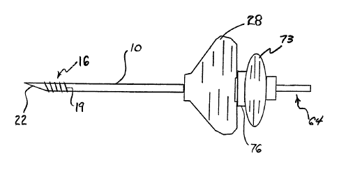
Figure 1 is a side view of a cannula according to
the present invention;
Figure 2 is a side view of a stylet according to
the present invention;
Figure 3 is a side view of a core collector
according to the present invention;
Figure 4 is a cross-sectioned side view of a
portion of the core collector;
CA 02360529 2001-07-27
WO 00/44286 PCT/US00/02302
-4-
Figure 5 is a side view of a core ejector according
to the present invention;
Figure 6 is a cannula liner according to the
present invention;
Figures 7A and 7B show steps of a method according
to the present invention;
Figure 8 is a side view of a cannula assembled with
a stylet according to the present invention;
Figure 9 is a side view of the core collector shown
in Figure 3 assembled with the cannula shown in Figure
1;
Figure 10 is a side view of the core ejector shown
in Figure 5 being used to push a bone sample from the
core collector shown in Figure 3;
Figure 11 shows pituitary forceps used in a method
according to the present invention; and
Figure 12 shows the pituitary forceps shown in
Figure 11 inserted through the cannula shown in Figure
1.
BEST MODE FOR CARRYING OUT THE INVENTION
Figure 1 shows a cannula 10 according to the
present invention. The cannula 10 has a first end 13
having a threaded section 16 with a projecting helical
rib 19. The first end 13 may include a beveled tip 22,
as shown in Figure 1. The cannula 10 also has a second
end 25 having a handle 28. The second end 25 also
CA 02360529 2001-07-27
WO 00/44286 PCT/US00/02302
-5-
includes an apparatus for connecting 31 the cannula 10,
such as a pin lock connector, that permits the cannula
to be connected to another instrument. The cannula
10 has an axial channel 34 extending between the first
5 and second ends 13, 25 of the cannula.
Figure 2 shows a stylet 37 according to the present
invention. The stylet 37 has a first end 40 with a
sharpened point 43. The stylet 37 has a second end 44
that may include a device for connecting 46 the stylet,
10 such as a mating apparatus to the pin lock connector.
The second end 44 may also include a guidance device 49.
Such a guidance device 49 is disclosed more fully in
U.S. Patent No. 5,810,841, and is hereby incorporated by
reference. When the stylet 37 is properly aligned with
a guidance beam 52, a detectable signal 55 is emitted
from the guidance device 49 on the stylet 37. The
detectable signal 55 may be a sound or may be visible
light.
Figure 3 shows a core collector 58 according to the
present invention. The core collector 58 has a first
end 61 and a second end 64. The core collector 58 has
an axial cavity 67 extending between the first and
second ends 61, 64 of the core collector 58. Figure 4
shows a cross-sectioned view of the first end 61 of the
core collector 58. Proximate to the first end 61 of the
core collector 58 are one or more teeth 70 extending
from the core collector 58 into the axial cavity 67.
CA 02360529 2001-07-27
WO 00/44286 PCT/US00/02302
-6-
The second end 64 of the core collector 58 includes a
haft 73, and also includes a device for connecting 76
the core collector, which may be similar to the mating
apparatus to the pin lock connector, described above.
Figure 5 shows a sample ejector 79 having a blunt
end 82 and a handle end 85.
Figure 6 shows a cannula liner 88. The cannula
liner 88 is a tubular instrument having an outside
diameter 91 slightly smaller than the diameter 94 of the
axial channel 34 in the cannula 10. The liner 88 may be
provided with a fitting 97 for attaching a syringe for
injecting filler material, such as bone cement. The
fitting 97 may be a conical fitting with a Luer taper,
such as that described in ISO 594-2, first edition 1991-
05-O1, published by the International Organization for
Standardization.
Figures 7A and 7B show steps of a method according
to the present invention. First, an assembly is formed
(step 200) by providing a stylet and a cannula similar
to those described above, and inserting the stylet into
the axial channel of the cannula so the sharpened point
of the stylet is proximate to the first end of the
cannula. The stylet may have a guidance device similar
to that described above. The sharpened point may extend
from the cannula depending on the particular shape of
the stylet and the cannula, as well as the particular
procedure being performed with the stylet and cannula.
CA 02360529 2001-07-27
WO 00/44286 PCT/US00/02302
The stylet is then connected to the cannula via the
apparatus for connecting the cannula mating with the
device for connecting the stylet. Figure 8 shows the
stylet inserted in the cannula.
Next, the stylet and cannula assembly is inserted
(step 203) into the patient until the first end of the
cannula is proximate to the bone to be sampled. The
stylet cuts soft tissue surrounding the bone to be
sampled, and guides the cannula through the soft tissue
in order to place the first end of the cannula proximate
to the bone at a sample location where the sample is to
be taken. When the stylet includes the guidance device,
the stylet is inserted at an angle and at a location
signified by a guidance beam.
Once the first end of the cannula is proximate to
the sample location, the stylet is disconnected (step
206) from the cannula and the cannula is twisted (step
209) via the handle so the helical rib engages the bone,
and part of the threaded section is screwed into the
bone. Next, the stylet is removed (step 212) from the
cannula. In its place, the core collector is provided
and inserted (step 215) into the channel of the cannula
so the first end of the core collector is proximate to
the bone. Figure 9 shows the core collector inserted in
the cannula. The apparatus for connecting the cannula
is connected to the device for connecting the core
collector. Then the cannula is again twisted (step 218)
CA 02360529 2001-07-27
WO 00/44286 PCT/US00/02302
_g_
via the handle to further screw the threaded section of
the cannula into the bone and to embed the core
collector into the bone.
Next, the core collector is disconnected from the
cannula and pulled out of the bone (step 221) via the
haft. As the core collector is pulled, the teeth of the
core collector engage the bone specimen residing in the
axial cavity of the core collector, and prevent the bone
specimen from being left in the patient. The core
collector, with the bone specimen therein, is removed
from the cannula by further pulling on the haft.
The blunt end of the sample ejector is then
inserted into the axial cavity of the core collector, as
shown in Figure 10. By pushing on the handle end of the
sample ejector, the blunt end is forced from the first
end of the core collector toward the second end of the
core collector in order to eject (step 224) the bone
sample from the core collector.
Next, a cannula liner may be inserted (step 227)
into the cannula, and bone cement may be injected
through the liner to fill the section of bone where the
bone sample was removed. The liner prevents the bone
cement from contacting the cannula and hardening on the
cannula. If the bone cement hardens in the liner, the
liner can be removed and replaced with another liner
without disturbing the cannula. Finally, the cannula is
removed (step 230) from the patient.
CA 02360529 2001-07-27
WO 00/44286 PCT/US00/02302
-9-
In another method according to the present
invention, pituitary forceps 100 shown in Figure 11 are
used in lieu of the core collector 58. Once at least
part of the threaded section 16 is screwed into the bone
as described above in step 209, the cannula 10 is
unscrewed from the bone and the first end 13 is left
proximate to the bone. Pituitary forceps 100 are
inserted through the axial channel 34 of the cannula 10
so the jaws 103 of the pituitary forceps 100 are
proximate to the bone. Next, the forceps 100 are
manipulated by moving the finger rings 106 relative to
each other until the bone sample is grasped by the jaws
103. Figure 12 shows the pituitary forceps 100 inserted
in the cannula 10. The pituitary forceps 100 are then
removed from the cannula 10, with the bone sample in the
jaws 103. The method then continues as described above
by inserting the cannula liner 88 and filing the bone
with bone cement.
The instruments described above can also be used
for other procedures, such as cementoplasty of bones or
a venogram. With regard to the cementoplasty and
venogram procedures, it should be noted the core
collector 58 is not used.
With regard to the syringes mentioned above, it
should be noted that bone cement has a relatively rapid
polymerization time and high viscosity. Therefore, in
performing a method according to the present invention,
CA 02360529 2001-07-27
WO 00/44286 PCT/US00/02302
-10-
it is preferable to inject bone cement using syringes
having a small capacity, for example, a capacity of from
1 to 3 cubic centimeters.
Although the present invention has been described
with respect to one or more particular embodiments, it
will be understood that other embodiments of the present
invention may be made without departing from the spirit
and s cope of the present invention. Hence, the
present invention is deemed limited only by the appended
claims and the reasonable interpretation thereof.
