Note: Descriptions are shown in the official language in which they were submitted.
CA 02360690 2001-07-26
WO 00/44391 PCT/US00/02091
1
PLASMINOGEN KRINGLE 4 REGION FRAGMENTS
AND METHODS OF USE
Related Applications
The present application claims priority to U.S. Provisional
Application Serial No. 60/117,617 filed January 28, 1999.
Field of the Invention
The present invention relates to endothelial inhibitors,
fragments of angiostatin protein, which reversibly inhibit
proliferation of endothelial cells. More particularly, the present
invention relates to kringle 4 region fragments that are useful for
the treatment of angiogenesis-associated diseases such as cancer.
Background of the Invention
As used herein, the term "angiogenesis" means the
generation of new blood vessels into a tissue or organ. Under
normal physiological conditions, humans or animals undergo
angiogenesis only in very specific restricted situations. For
example, angiogenesis is normally observed in wound healing,
fetal and embryonal development and formation of the corpus
luteum, endometrium and placenta. The term "endothelium"
means a thin layer of flat epithelial cells that lines serous cavities,
lymph vessels, and blood vessels.
Both controlled and uncontrolled angiogenesis are thought
to proceed in a similar manner. Endothelial cells and pericytes,
CA 02360690 2001-07-26
WO 00/44391 PCT/US00/02091
2
surrounded by a basement membrane, form capillary blood
vessels. Angiogenesis begins with the erosion of the basement
membrane by enzymes released by endothelial cells and
leukocytes. The endothelial cells, which line the lumen of blood
vessels, then protrude through the basement membrane.
Angiogenic stimulants induce the endothelial cells to migrate
through the eroded basement membrane. The migrating cells
form a "sprout" off the parent blood vessel, where the
endothelial cells undergo mitosis and proliferate. The endothelial
sprouts merge with each other to form capillary loops, creating
the new blood vessel.
Persistent, unregulated angiogenesis occurs in a
multiplicity of disease states, tumor metastasis and abnormal
growth by endothelial cells. The diverse pathological disease
states in which unregulated angiogenesis is present have been
grouped together as angiogenic dependent or angiogenic
associated diseases.
The hypothesis that tumor growth is angiogenesis
dependent was first proposed in 1971. (Folkman J., Tumor
angiogenesis: Therapeutic implications., N. Engl. Jour. Med.
285:1182 1186, 1971 ) In its simplest terms it states: "Once
tumor 'take' has occurred, every increase in tumor cell
population must be preceded by an increase in new capillaries
converging on the tumor." Tumor 'take' is currently
understood to indicate a prevascular phase of tumor growth in
which a population of tumor cells occupying a few cubic
millimeters volume and not exceeding a few million cells, can -
survive on existing host microvessels. Expansion of tumor
volume beyond this phase requires the induction of new capillary
blood vessels.
Amino acid sequence alignment of the kringle domains of
human plasminogen, designated K1, K2, K3 and K4, shows that
all kringle regions display identical gross architecture and
remarkable sequence homology (56-82% identify). Among
these structures, the high-affinity lysine binding kringle, Kl, is
CA 02360690 2001-07-26
WO 00/44391 PCT/US00/02091
3
the most potent inhibitory segment of endothelial cell
proliferation. Of interest, the intermediate-affinity lysine binding
fragment, K4, has previously been shown to lack inhibitory
activity. These data suggest that the lysine binding site of the
kringle structures may not be directly involved in the inhibitory
activity. The amino acid conservation and functional divergence
of these kringle structures provide an ideal system to study the
role mutations caused by DNA replication during evolution.
Similar divergent activities relative to the regulation of
angiogenesis exhibited by a group of structurally related proteins
are also found in the -C-X-C- chemokine and prolactin-growth
hormone families (Maione, T.E., Gray, G.S., Petro, A. J., Hunt,
A.L., and Dormer, S.I. (1990) Science 247, 77-79.; Koch, A.E.,
Polverini, P.J., Kunkel, S.L., Harlow, L.A., DiPietro, L.A., Elner,
V.M., Elner, S.J., and Strieter, R.M. (1992) Science 258, 1798-
1801.; Cao, Y., Chen, C., Weatherbee, J.A., Tsang, M., and
Folkman, J. (1995) J. Exp. Med. 182, 2069-2077.; Strieter,
R.M., Polverini, P.J., Arenberg, D.A., and Kunkel, S.L. (1995)
Shock 4, 155-160.; Jackson, D., Volpert, O.V., Bouck, N., and
Linzer, D.LH. (1994) Science 266, 1581-1584).
Further sequence analysis reveals that K4 contains two
positively charged lysine residues adjacent to cysteines 22 and 78
(Fig. 35). 1H nuclear magnetic resonance (NMR) analysis shows
that these 4 lysines, together with lysine 57, form the core of a
positively charged domain in K4 (Llinas M, unpublished data),
whereas other kringle structures lack such a positively charged
domain. Whether this lysine-enriched domain contributes to the
loss of inhibitory activity of kringle 4 of human plasminogen
remains to be studied. K4 was previously reported to stimulate
proliferation of other cell types and to increase the release of
intracellular calcium (Donate, L.E., Gherardi, E., Srinivasan, N.,
Sowdhamini, R., Aporicio, S., and Blundell, T. L. (1994) Prot.
Sci. 3, 2378-2394). The fact that removal of K4 from
angiostatin potentiates its inhibitory activity on endothelial cells
CA 02360690 2001-07-26
WO 00/44391 PCT/US00/02091
4
suggests that this structure may prevent some of the inhibitory
effect of K1-3.
The mechanism underlying how angiostatin and its related
kringle fragments specifically inhibit endothelial cell growth
remains uncharacterized. It is not yet clear whether the
inhibition is mediated by a receptor that is specifically expressed
in proliferating endothelial cells, or if angiostatin is internalized
by endothelial cells and subsequently inhibits cell proliferation.
Alternatively, angiostatin may interact with an endothelial cell
adhesion receptor such as integrin a~b3, blocking integrin-
mediated angiogenesis (Brooks, P.C., Montgomery, A.M.,
Rosenfeld, M., Reisfeld R.A., Hu, T. HIier, G., and Cheresh, D.A.
(1994) Cell 79, 1157-1164). Of interest, Friedlander et. al.
(Friedlander, M., Brooks, P.C., Shaffer, R.W., Kincaid, C.M.,
Varner, J.A., and Cheresh, D.A. (1995) 270, 1502) reported
recently that in vivo angiogenesis in cornea or chorioallantoic
membrane models (induced by bFGF and by tumor necrosis
factor) was a~b3 integrin dependent. However, angiogenesis
stimulated by VEGF, transforming growth factor a, or phorbol
esters was dependent on a~b5. Antibodies to the individual
integrins specifically blocked one of these pathways, and a cyclic
protein antagonist of both integrins blocked angiogenesis
induced by each cytokine (Friedlander, M., Brooks, P.C.,
Shaffer, R.W., Kincaid, C.M., Varner, J.A., and Cheresh, D.A.
(1995) 270, 1502). Because both bFGF- and VEGF-induced
angiogenesis are inhibited by angiostatin, angiostatin may block a
common pathway involved in integrin-mediated angiogenesis.
An increasing number of endogenous angiogenesis
inhibitors have been identified in the last few decades (Folkman,
J. (1995) N. Engl. J. Med. 333, 1757-1763). Of the nine
characterized endothelial cell suppressors, several inhibitors are
proteolytic fragments. For example, the 16 kDa N-terminal
fragment of human prolactin inhibits endothelial cell proliferation
and blocks angiogenesis in vivo (Clapp, C., Martial, J.A.,
Guzman, R.C., Rentierdelrue, F., and Weiner, R.I. (1993)
CA 02360690 2001-07-26
WO 00/44391 PCT/US00/02091
Endorinology 133, 1292-1299). In a recent paper, D'Angelo et.
al. reported that the antiangiogenic 16 kDa N-terminal fragment
inhibited the activation of mitogen-activated protein kinase
(MAPK) by VEGF and bFGF in capillary endothelial cells
5 (D'Angelo, G., Struman, L, Martial, J., and Weiner, R. (1995)
Proc. Natl. Acad. Sci. 92, 6374-6378). Similar to angiostatin,
the intact parental molecule of prolactin does not inhibit
endothelial cell proliferation nor is it an angiogenesis inhibitor.
Platelet factor 4 (PF-4) inhibits angiogenesis at high
concentrations (Maione, T.E., Gray, G.S., Petro, A. J., Hunt,
A.L., and Dormer, S.I. (1990) Science 247, 77-79; Cao, Y., Chen,
C., Weatherbee, J.A., Tsang, M., and Folkman, J. ( 1995) J. Exp.
Med. 182, 2069-2077). However, the N-terminally truncated
proteolytically cleaved PF-4 fragment exhibits a 30- to 50-fold
increase in its anti-proliferative activity over the intact PF-4
molecule (Gupta, S.K., Hassel, T., and Singh, J.P. (1995) Proc.
Natl. Acad. Sci. 92, 7799-7803). Smaller protein fragments of
fibronectin, murine epidermal growth factor, and
thrombospondin have also been shown to specifically inhibit
endothelial cell growth (Homandberg, G.A., Williams, J.E.,
Grant, D., Schumacher, B., and Eisenstein, R. (1985) Am. J.
Pathol. 120, 327-332; Nelson, J., Allen, W.E., Scott, W.N., Bailie,
J.R., Walker, B., McFerran, N.V., and Wilson, D.J. (1995) Cancer
Res. 55, 3772-3776; Tolsma, S.S., Volpert, O.V., Good, D.J.,
Frazer, W.A., Polverini, P.J., and Bouck, N. (1993) J. Cell Biol.
122, 497-511 ). Proteolytic processing of a large protein may
change the conformational structure of the original molecule or
expose new epitopes that are antiangiogenic. Thus, protease(s)
may play a critical role in the regulation of angiogenesis. To
date, little is known about the regulation of these protease
activities in vivo.
The data also show that the disulfide bond mediated
folding of the kringle structures in angiostatin is preferable to
maintain its inhibitory activity on endothelial cell growth.
Kringle structures analogous to those in plasnunogen are also
CA 02360690 2001-07-26
WO 00/44391 PCT/US00/02091
6
found in a variety of other proteins. For example,
apolipoprotein (a) has as many as 37 repeats of plasminogen
kringle 4 (McLean, J.W., Tomlinson, J.E., Kuang, W.-J., Eaton,
D.L., Chen, E.Y., Fless, G.M., Scanu, A.M., and Lawn, R.M.
(1987) Nature 330, 132-137). The amino terminal portion of
prothrombin also contains two kringles that are homologous to
those of plasminogen (Walz, D.A., Hewett-Emmett, ~ D., and
Seegers, W.H. (1977) Proc. Natl. Acad. Sci. 74, 1969-1973).
Urokinase has been shown to possess a kringle structure that
shares extensive homology with plasminogen (Gunzler, W.A., J.,
S.G., Otting, F., Kim, S.-M. A., Frankus, E., and Flohe, L.
(1982) Hoppe-Seyler's A. Physiol. Chem. 363, 1155-1165). In
addition, surfactant protein B and hepatocyte growth factor
(HGF), also carry kringle structures (Johansson, J., Curstedt, T.,
and Jornvall., H. (1991) Biochem. 30, 6917-6921; Lukker, N.A.,
Presta, L.G., and Godowski, P.J. (1994) Prot. Engin. 7, 895-
903).
Thus, it is clear that angiogenesis plays a major role in the
metastasis of a cancer. If this angiogenic activity could be
repressed or eliminated, then the tumor, although present, would
not grow. In the disease state, prevention of angiogenesis could
avert the damage caused by the invasion of the new
microvascular system. Therapies directed at control of the
angiogenic processes could lead to the abrogation or mitigation
of these diseases.
What is needed therefore is a composition and method
which can inhibit the unwanted growth of blood vessels,
especially into tumors. Also needed is a method for detecting,
measuring, and localizing the composition. The composition
should be able to overcome the activity of endogenous growth
factors in pre-metastatic tumors and prevent the formation of the
capillaries in the tumors thereby inhibiting the growth of the
tumors. The composition, fragments of the composition, and
antibodies specific to the composition, should also be able to
modulate the formation of capillaries in other angiogenic
CA 02360690 2001-07-26
WO 00/44391 PCT/US00/02091
processes, such as wound healing and reproduction. The
composition and method for inhibiting angiogenesis should
preferably be non-toxic and produce few side effects. Also
needed is a method for detecting, measuring, and localizing the
binding sites for the composition as well as sites of biosynthesis
of the composition. The composition and fragments of the
composition should be capable of being conjugated to other
molecules for both radioactive and non-radioactive labeling
purposes
Summary of the Invention
In accordance with the present invention, compositions
and methods are provided that are effective for modulating
angiogenesis, and inhibiting unwanted angiogenesis, especially
angiogenesis related to tumor growth. The present invention
relates to a protein, which has been named "angiostatin",
defined by its ability to overcome the angiogenic activity of
endogenous growth factors such as bFGF, in vitro, and by its
amino acid sequence homology and structural similarity to an
internal portion of plasminogen beginning at approximately
amino acid 98. Angiostatin comprises a protein having a
molecular weight of between approximately 38 kilodaltons and
45 kilodaltons as determined by reducing polyacrylamide gel
electrophoresis and having an amino acid sequence substantially
similar to that of a fragment of murine plasminogen beginning at
amino acid number 98 of an intact murine plasminogen
molecule. Angiostatin protein contains approximately kringle
regions 1 through 4 of a plasminogen molecule.
The present invention relates to fragments of angiostatin
protein in the kringle 4 region. The amino acid sequences of the
kringle 4 region fragments of the present invention vary slightly
depending upon the species. Furthermore, the amino acid
sequences of the kringle 4 region fragments of the present
invention vary slightly at the amino and carboxy terminals.
Therefore, it is to be understood that the number of amino acids
CA 02360690 2001-07-26
WO 00/44391 PCT/US00/02091
8
in the active kringle 4 region fragments may vary and all kringle
4 region amino acid sequences that have endothelial inhibiting
activity are contemplated as being included in the present
invention. The present invention also includes fusion proteins
containing kringle 4 region fragments and other anti-angiogenic
or angiogenic molecules. Examples of other anti-angiogenic
molecules include endostatin protein and fragments of endostatin
protein.
The present invention provides methods and compositions
for treating diseases and processes mediated by undesired and
uncontrolled angiogenesis by increasing the in vivo
concentrations of kringle 4 region fragments in a human or
animal. The in vivo concentrations of kringle 4 region fragments
may be increased by administering to a human or animal a
composition comprising a substantially purified kringle 4 region
fragment in a dosage sufFicient to inhibit angiogenesis.
Additionally, the in vivo concentrations of kringle 4 region
fragments may be increased in a human or animal by the
administration of nucleotides encoding kringle 4 region
fragments or enzymes that release kringle 4 region fragments
from plasminogen or angiostatin. The present invention is
particularly useful for treating, or for repressing the growth of,
tumors. Increasing the in vivo concentrations of kringle 4 region
fragments in a human or animal with prevascularized
metastasized tumors will prevent the growth or expansion of
those tumors.
The present invention also encompasses DNA sequences
encoding kringle 4 region fragments or kringle 4 region fusion
proteins, expression vectors containing DNA sequences
encoding kringle 4 region fragments or kringle 4 region fusion
proteins, and cells containing one or more expression vectors
containing DNA sequences encoding kringle 4 region fragments
or kringle 4 region fusion proteins. The present invention
further encompasses gene therapy methods whereby DNA
sequences encoding kringle 4 region fragments are introduced
CA 02360690 2001-07-26
WO 00!44391 PCT/US00/02091
9
into a patient to modify in vivo angiostatin kringle 4 region
levels.
The present invention also includes diagnostic methods
and kits for detection and measurement of kringle 4 region
fragments in biological fluids and tissues, and for localization of
kringle 4 region fragments in tissues and cells. The diagnostic
method and kit can be in any configuration well known to those
of ordinary skill in the art. The present invention also includes
antibodies specific for the kringle 4 region fragments and
portions thereof, and antibodies that inhibit the binding of
antibodies specific for the kringle 4 region fragments. These
antibodies can be polyclonal antibodies or monoclonal antibodies.
The antibodies specific for the kringle 4 region fragments can be
used in diagnostic kits to detect the presence and quantity of
angiostatin which is diagnostic or prognostic for the occurrence
or recurrence of cancer or other diseases mediated by
angiogenesis. Antibodies specific for kringle 4 region fragments
may also be administered to a human or animal to passively
immunize the human or animal against angiostatin, or kringle 4
region fragments of angiostatin, thereby reducing angiogenic
inhibition.
The present invention also includes diagnostic methods
and kits for detecting the presence and quantity of antibodies
that bind kringle 4 region fragments in body fluids. The
diagnostic method and kit can be in any configuration well
known to those of ordinary skill in the art. The present
invention also includes antibodies that specifically bind to the
angiostatin kringle 4 region receptor and transmit the
appropriate signal to the cell and act as agonists or antagonists.
The present invention also includes kringle 4 region
fragments and analogs that can be labeled isotopically or with
other molecules or proteins for use in the detection and
visualization of angiostatin fragment binding sites with
techniques, including, but not limited to, positron emission
CA 02360690 2001-07-26
WO 00/44391 PCT/US00/02091
tomography, autoradiography, flow cytometry, radioreceptor
binding assays, and immunohistochemistry.
The kringle 4 region fragments and analogs of the present
invention also act as agonists and antagonists at the angiostatin
5 kringle 4 region receptor, thereby enhancing or blocking the
biological activity of angiostatin kringle 4 regions. Such proteins
are used in the isolation of kringle 4 region fragments receptors.
The present invention also includes kringle 4 region
fragment antisera, or angiostatin kringle 4 region receptor
10 agonists and receptor antagonists linked to cytotoxic agents for
therapeutic and research applications. Still further, kringle 4
region fragments, kringle 4 region fragment antisera, kringle 4
region fragment receptor agonists and kringle 4 region fragment
receptor antagonists are combined with pharmaceutically
acceptable excipients, and optionally sustained-release
compounds or compositions, such as biodegradable polymers, to
form therapeutic compositions.
The present invention includes molecular probes for the
ribonucleic acid and deoxyribonucleic acid involved in
transcription and translation of kringle 4 region fragments.
These molecular probes provide means to detect and measure
angiostatin kringle 4 region biosynthesis in tissues and cells.
Accordingly, it is an object of the present invention to
provide a composition comprising a kringle 4 region.
It is another object of the present invention to provide a
method of treating diseases and processes that are mediated by
angiogenesis.
It is another object of the present invention to provide
compositions and methods for increasing the in vivo
concentration of kringle 4 region peptides.
It is an object of the present invention to provide
compounds that modulate or mimic the production or activity of
enzymes that produce kringle 4 region fragments in vivo or in
vitro.
CA 02360690 2001-07-26
WO 00/44391 PCT/US00/02091
11
It is yet another object of the present invention to provide
a diagnostic or prognostic method and kit for detecting the
presence and amount of a kringle 4 region peptide in a body
fluid or tissue.
It is another object of the present invention to provide a
composition for treating or repressing the growth of a cancer.
It is a further object of the present invention to provide
kringle 4 region or anti-kringle 4 region peptide antibodies by
direct injection of angiostatin kringle 4 region DNA into a
human or animal needing such kringle 4 region or anti-kringle 4
region peptide antibodies.
It is an object of present invention to provide a method
for detecting and quantifying the presence of an antibody
specific for a kringle 4 region fragment in a body fluid.
Still another object of the present invention is to provide a
composition consisting of antibodies to kringle 4 region
fragments that are selective for specific regions of the kringle 4
region fragment molecule that do not recognize plasminogen.
It is another object of the present invention to provide a
method for the detection or prognosis of cancer.
It is another object of the present invention to provide a
composition for use in visualizing and quantitating sites of
kringle 4 region fragment binding in vivo and in vitro.
It is yet another object of the present invention to provide
a therapy for cancer that has minimal side effects.
Still another object of the present invention is to provide a
composition comprising kringle 4 region fragments linked to a
cytotoxic agent for treating or repressing the growth of a cancer.
Another object of the present invention is to provide a
method for targeted delivery of kringle 4 region-related
compositions to specific locations.
Yet another object of the invention is to provide
compositions and methods useful for gene therapy for the
modulation of angiogenic processes.
CA 02360690 2001-07-26
WO 00/44391 PCT/US00/02091
12
These and other objects, features and advantages of the
present invention will become apparent after a review of the
following detailed description of the disclosed embodiments and
the appended claims.
Brief Description of the Figures
Fig. 1 shows the production of recombinant murine
angiostatin with a baculovirus expression system.
Fig. 2 shows a gel filtration chromatography of angiostatin
degradation products.
Fig. 3 shows production of fragments from a 52 kDa
recombinant murine angiostatin.
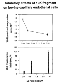
Fig. 4 shows the inhibitory effects of a 10 kDa fragment
on bovine capillary endothelial cells.
Fig. 5 shows the identification of the 10 kDa fragment as
kringle 4 by amino acid microsequencing.
Fig. 6 shows SEQ ID NO:l, the amino acid sequence of
the whole murine plasminogen.
Detailed Description
The present invention includes compositions and methods
for the detection and treatment of diseases and processes that are
mediated by or associated with angiogenesis. The composition is
an angiostatin kringle 4 region, which can be isolated from body
fluids including, but not limited to, serum, urine and ascites, or
synthesized by chemical or biological methods (e.g. cell culture,
recombinant gene expression, protein synthesis, and in vitro
enzymatic catalysis of angiostatin, plasminogen or plasmin to
yield active kringle 4 region peptides). Recombinant techniques
include gene amplification from DNA sources using the
polymerase chain reaction (PCR), and gene amplification from
RNA sources using reverse transcriptase/PCR. These angiostatin
kringle 4 region fragments inhibit the growth of blood vessels
into tissues such as de-vascularized or vascularized tumors.
CA 02360690 2001-07-26
WO 00/44391 PCT/US00/02091
13
The description of angiostatin and other kringle 4 region
fragments can be found, for example, in U.S. Patent Nos.
5,639,725; 5,733,876 and 5,837,682, the entire contents of
which are hereby incorporated by reference.
The present invention also encompasses a composition
comprising, a vector containing a DNA sequence encoding
angiostatin kringle 4 region fragments, wherein the vector is
capable of expressing angiostatin kringle 4 region fragments
when present in a cell, and a method comprising, implanting into
a human or non-human animal a cell containing a vector,
wherein the vector contains a DNA sequence encoding kringle 4
region fragments, and wherein the vector is capable of
expressing kringle 4 region fragments when present in the cell.
The cell may contain one vector or multiple vectors.
Still further, the present invention encompasses kringle 4
region fragments, kringle 4 region antisera, kringle 4 region
receptor agonists or kringle 4 region receptor antagonists that
are combined with pharmaceutically acceptable excipients, and
optionally sustained-release compounds or compositions, such as
biodegradable polymers, to form therapeutic compositions. In
particular, the invention includes a composition comprising an
antibody that specifically binds to a kringle 4 region, wherein the
antibody does not bind to plasminogen.
More particularly, the present invention includes a protein
designated angiostatin kringle 4 region that has a molecular
weight of approximately 10 kilodaltons (kDa) as determined by
reducing polyacrylamide gel electrophoresis that is capable of
overcoming the angiogenic activity of endogenous growth
factors such as bFGF, in vitro. Kringle 4 is typically defined as
encompassing amino acids 377-454 of a human plasminogen
molecule. (The amino acid sequence of the complete murine
plasminogen molecule is shown in Figure 6 and in SEQ
NO:l.) However, the kringle 4 region is surrounded by inter-
kringle domains on either end, portions of which may be
included in functional kringle 4 region fragments of the present
CA 02360690 2001-07-26
WO 00/44391 PCT/US00/02091
14
invention. For example, functional murine kringle 4 region
fragments have been demonstrated herein to have anti-
endothelial cell proliferation activity encompassing amino acids
371-458, 374-458, and 376-458. Therefore, it should be
understood that the term "region" encompasses all such anti-
angiogenic kringle 4 fragments containing varying numbers of
amino acids from the amino and carboxy terminal inter-kringle
domains. It is also to be understood that the present invention is
contemplated to include any derivatives of the angiostatin kringle
4 region fragment that have endothelial inhibitory activity.
These include proteins with angiostatin kringle 4 region activity
that have amino acid substitutions or have sugars or other
molecules attached to amino acid functional groups.
The term "substantially similar," when used in reference to
angiostatin kringle 4 region fragment amino acid sequences,
means an amino acid sequence having anti-angiogenic activity,
which also has a high degree of sequence homology to the
human protein fragment of kringle 4 fragments. A high degree
of homology means at least approximately 60% amino acid
homology, desirably at least approximately 70% amino acid
homology, and more desirably at least approximately 80%
amino acid homology. Homology is often measured using
sequence analysis software, e.g., BLASTIN or BLASTP
(available at http://www.ncbi.nlm.nih.,gov/BLAST). The default
parameters for comparing the two sequences (e.g., "Blast"-ing
two sequences against each other) by BLASTIN (for nucleotide
sequences) are reward for match =l, penalty for mismatch = -2,
open gap = 5, and extension gap = 2. When using BLASTP for
protein sequences, the default parameters are reward for match
= 0, penalty for mismatch = 0, open gap = 11, and extension
gap = 1.
The term "endothelial inhibiting activity" as used herein
means the capability of a molecule to inhibit angiogenesis in
general and, for example, to inhibit the growth of bovine
CA 02360690 2001-07-26
WO 00/44391 PCT/US00/02091
capillary endothelial cells in culture in the presence of fibroblast
growth factor.
The kringle 4 region of angiostatin has been shown to be
capable of inhibiting the growth of endothelial cells in vitro.
5 Angiostatin kringle 4 region does not inhibit the growth of cell
lines derived from other cell types. Specifically, angiostatin
kringle 4 region has no effect on Lewis lung carcinoma cell lines,
mink lung epithelium, 3T3 fibroblasts, bovine aortic smooth
muscle cells, bovine retinal pigment epithelium, MDCk cells
10 (canine renal epithelium), WI38 cells (human fetal lung
fibroblasts) EFN cells (murine fetal fibroblasts) and LM cells
(murine connective tissue). Endogenous angiostatin in a tumor
bearing mouse is effective at inhibiting metastases at a systemic
concentration of approximately 10 mg angiostatin/kg body
15 weight.
Angiostatin has a specific three dimensional conformation
that is defined by the kringle regions of the plasminogen
molecule. (Robbins, K.C., "The plasminogen-plasmin enzyme
system" Hemostasis and Thrombosis, Basic Principles and
Practice, 2nd Edition, ed. by Colman, R.W. et al. J.B. Lippincott
Company, pp. 340-357, 1987) There are five such kringle
regions, which are conformationally related motifs and have
substantial sequence homology, in the NH2 terminal portion of
the plasminogen molecule. Each kringle region of the
plasminogen molecule contains approximately 80 amino acids
and contains 3 disulfide bonds. This cysteine motif is known to
exist in other biologically active proteins. These proteins include,
but are not limited to, prothrombin, hepatocyte growth factor,
scatter factor and macrophage stimulating protein. (Yoshimura,
T, et al., "Cloning, sequencing, and expression of human
macrophage stimulating protein (MSP, MST1) confirms MSP as
a member of the family of kringle proteins and locates the MSP
gene on Chromosome 3" J. Biol. Chem., Vol. 268, No. 21, pp.
15461-15468, 1993). It is contemplated that any isolated kringle
4 region fragment having a three dimensional kringle-like
CA 02360690 2001-07-26
WO 00/44391 PCT/US00/02091
16
conformation or cysteine motif that has anti-angiogenic activity
in vivo, is part of the present invention.
The present invention also includes the detection of the
angiostatin kringle 4 region fragments in body fluids and tissues
for the purpose of diagnosis or prognosis of diseases such as
cancer. The present invention also includes the detection of
angiostatin kringle 4 region fragment binding sites and receptors
in cells and tissues. The present invention also includes methods
of treating or preventing angiogenic diseases and processes
including, but not limited to, arthritis and tumors by stimulating
the production of angiostatin kringle 4 region fragments, and/or
by administering substantially purified angiostatin kringle 4
region fragments, nucleotides encoding angiostatin kringle 4
region fragments, or angiostatin kringle 4 region fragment
agonists or antagonists, and/or angiostatin kringle 4 region
fragment antisera or antisera directed against angiostatin kringle
4 region fragment antisera to a patient. Additional treatment
methods include administration of angiostatin kringle 4 region
fragments, angiostatin kringle 4 region fragment analogs,
angiostatin kringle 4 region fragment antisera, or angiostatin
receptor agonists and antagonists linked to cytotoxic agents. It is
to be understood that the angiostatin kringle 4 region fragments
can be animal or human in origin. Angiostatin kringle 4 region
fragments can be produced synthetically by chemical reaction or
by recombinant techniques in conjunction with expression
systems. Angiostatin kringle 4 region fragments may also be
produced in vitro or in vivo by enzymatically cleaving
angiostatin, plasminogen or plasmin to generate proteins having
anti-angiogenic activity or by using compounds that mimic the
action of endogenous enzymes that cleave angiostatin or
plasminogen into kringle 4 region fragments. Angiostatin
kringle 4 region fragment production may also be modulated by
compounds that affect the activity of plasminogen cleaving
enzymes.
CA 02360690 2001-07-26
WO 00/44391 PCT/US00/02091
17
Passive antibody therapy using antibodies that specifically
bind angiostatin kringle 4 region fragments can be employed to
modulate angiogenic-dependent processes such as reproduction,
development, and wound healing and tissue repair. In addition,
antisera directed to the Fab regions of angiostatin kringle 4
region fragment antibodies can be administered to block the
ability of endogenous angiostatin kringle 4 region fragment
antisera to bind angiostatin kringle 4 region fragments.
The present invention also encompasses gene therapy
whereby the gene encoding an angiostatin kringle 4 region
fragment is regulated in a patient. Various methods of
transfernng or delivering DNA to cells for expression of the
gene product protein, otherwise referred to as gene therapy, are
disclosed in Gene Transfer into Mammalian Somatic Cells in
vivo, N. Yang, Crit. Rev. Biotechn. 12(4): 335-356 (1992), which
is hereby incorporated by reference. Gene therapy encompasses
incorporation of DNA sequences into somatic cells or germ line
cells for use in either ex vivo or in vivo therapy. Gene therapy
functions to replace genes, augment normal or abnormal gene
function, and to combat infectious diseases and other
pathologies.
Strategies for treating these medical problems with gene
therapy include therapeutic strategies such as identifying the
defective gene and then adding a functional gene to either
replace the function of the defective gene or to augment a
slightly functional gene; or prophylactic strategies, such as
adding a gene encoding the protein product that will treat the
condition or that will make the tissue or organ more susceptible
to a treatment regimen. As an example of a prophylactic
strategy, a gene for an angiostatin kringle 4 region fragment
may be placed in a patient and thus prevent occurrence of
angiogenesis; or a gene that makes tumor cells more susceptible
to radiation could be inserted and then radiation of the tumor
would cause increased killing of the tumor cells.
CA 02360690 2001-07-26
WO 00/44391 PCT/US00/02091
18
Many protocols for transfer of angiostatin kringle 4 region
fragment DNA or angiostatin kringle 4 region fragment
regulatory sequences are envisioned in this invention.
Transfection of promoter sequences, other than one normally
found specifically associated with angiostatin, or other sequences
which would increase production of angiostatin kringle 4 region
proteins are also envisioned as methods of gene therapy. An
example of this technology is found in Transkaryotic Therapies,
Inc., of Cambridge, Massachusetts, using homologous
recombination to insert a "genetic switch" that turns on an
erythropoietin gene in cells. See Genetic Engineering News,
April 15, 1994. Such "genetic switches" could be used to
activate an angiostatin kringle 4 region fragment (or the
angiostatin kringle 4 region fragment receptor) in cells not
normally expressing angiostatin kringle 4 region fragment (or
the angiostatin kringle 4 region fragment receptor).
Gene transfer methods for gene therapy fall into three
broad categories: ( 1 ) physical (e.g., eleetroporation, direct gene
transfer and particle bombardment), (2) chemical (lipid-based
Garners, or other non-viral vectors) and (3) biological (virus-
derived vector and receptor uptake). For example, non-viral
vectors may be used which include liposomes coated with DNA.
Such liposome/DNA complexes may be directly injected
intravenously into the patient. It is believed that the
liposome/DNA complexes are concentrated in the liver where
they deliver the DNA to macrophages and Kupffer cells. These
cells are long lived and thus provide long term expression of the
delivered DNA. Additionally, vectors or the "naked" DNA of
the gene may be directly injected into the desired organ, tissue
or tumor for targeted delivery of the therapeutic DNA.
Gene therapy methodologies can also be described by
delivery site. Fundamental ways to deliver genes include ex vivo
gene transfer, in vivo gene transfer, and in vitro gene transfer.
In ex vivo gene transfer, cells are taken from the patient and
grown in cell culture. The DNA is transfected into the cells, the
CA 02360690 2001-07-26
WO 00/44391 PCT/US00/02091
19
transfected cells are expanded in number and then re-implanted
in the patient. In in vitro gene transfer, the transformed cells are
cells growing in culture, such as tissue culture cells, and not
particular cells from a particular patient. These "laboratory cells"
are transfected, the transfeeted cells are selected and expanded
for either implantation into a patient or for other uses.
In vivo gene transfer involves introducing the DNA into
the cells of the patient when the cells are within the patient.
Methods include using virally mediated gene transfer using a
noninfectious virus to deliver the gene in the patient or injecting
naked DNA into a site in the patient and the DNA is taken up
by a percentage of cells in which the gene product protein is
expressed. Additionally, the other methods described herein,
such as use of a "gene gun," may be used for in vitro insertion
of angiostatin kringle 4 region fragment DNA or angiostatin
regulatory sequences.
Chemical methods of gene therapy may involve a lipid
based compound, not necessarily a liposome, used to ferry the
DNA across the cell membrane. Lipofectins or cytofectins, lipid-
based positive ions that bind to negatively charged DNA, make a
complex that can cross the cell membrane and provide the DNA
into the interior of the cell. Biological methods used in gene
therapy techniques may involve receptor-based endocytosis, or
receptor-based phagocytosis, which involve binding a specific
ligand to a cell surface receptor and enveloping and transporting
the ligand across the cell membrane. Specifically, a ligand gene
complex is created and injected into the blood stream and then
target cells that have the receptor will specifically bind the ligand
and transport the ligand-DNA complex into the cell.
Many gene therapy methodologies employ viral vectors to
insert genes into cells. For example, altered retrovirus vectors
have been used in ex vivo methods to introduce genes into
peripheral and tumor-infiltrating lymphocytes, hepatocytes,
epidermal cells, myocytes, and other somatic cells. These altered
CA 02360690 2001-07-26
WO 00/44391 PCT/US00/02091
cells are then introduced into the patient to provide the gene
product from the inserted DNA.
Viral vectors have also been used to insert genes into cells
using in vivo protocols. To accomplish tissue-specific expression
5 of foreign genes, cis-acting regulatory elements or promoters
that are known to be tissue specific can be used. Alternatively,
tissue-specific expression can be achieved using in situ delivery
of DNA or viral vectors to specific anatomical sites in vivo. For
example, gene transfer to blood vessels in vivo was achieved by
10 implanting in vitro transduced endothelial cells in chosen sites on
arterial walls. The virus infected surrounding cells which also
expressed the gene product. A viral vector can be delivered
directly to the in vivo site, by a catheter for example, thus
allowing only certain areas to be infected by the virus, and
15 providing long-term, site specific gene expression. In vivo gene
transfer using retrovirus vectors has also been demonstrated in
mammary tissue and hepatic tissue by injection of the altered
virus into blood vessels leading to the organs.
Viral vectors that have been used for gene therapy
20 protocols include but are not limited to, retroviruses, other RNA
viruses such as poliovirus or Sindbis virus , adenovirus, adeno
associated virus, herpes viruses, SV 40, vaccinia and other DNA
viruses. Replication-defective murine retroviral vectors are the
most widely utilized gene transfer vectors. Murine leukemia
retroviruses are composed of a single strand RNA complexed
with a nuclear core protein and polymerase (pol) enzymes,
encased by a protein core (gag) and surrounded by a
glycoprotein envelope (env) that determines host range. The
genomic structure of retroviruses includes the gag, pol, and env
genes flanked by 5' and 3' long terminal repeats (LTR).
Retroviral vector systems exploit the fact that a minimal vector
containing the 5' and 3' LTRs and the packaging signal are
sufficient to allow vector packaging, infection and integration
into target cells providing that the viral structural proteins are
supplied in traps in the packaging cell line. Fundamental
CA 02360690 2001-07-26
WO 00/44391 PCT/US00/02091
21
advantages of retroviral vectors for gene transfer include
efficient infection and gene expression in most cell types, precise
single copy vector integration into target cell chromosomal
DNA, and ease of manipulation of the retroviral genome.
The adenovirus is composed of linear, double stranded
DNA complexed with core proteins and surrounded with capsid
proteins. Advances in molecular virology have led to the ability
to exploit the biology of these organisms to create vectors
capable of transducing novel genetic sequences into target cells
in vivo. Adenoviral-based vectors will express gene product
proteins at high levels. Adenoviral vectors have high efficiencies
of infectivity, even with low titers of virus. Additionally, the
virus is fully infective as a cell free virion so injection of
expression cell lines is not necessary. Another potential
advantage to adenoviral vectors is the ability to achieve long
term expression of heterologous genes in vivo.
Mechanical methods of DNA delivery include fusogenic
lipid vesicles such as liposomes or other vesicles for membrane
fusion, lipid particles of DNA incorporating cationic lipids such
as lipofectin, polylysine-mediated transfer of DNA, direct
injection of DNA, such as microinjection of DNA into germ or
somatic cells, pneumatically delivered DNA-coated particles,
such as the gold particles used in a "gene gun," and inorganic
chemical approaches such as calcium phosphate transfection.
It has been found that injecting plasmid DNA into muscle
cells yields high percentage of the cells which are transfected and
have sustained expression of marker genes. The DNA of the
plasmid may or may not integrate into the genome of the cells.
Non-integration of the transfected DNA would allow the
transfection and expression of gene product proteins in
terminally differentiated, non-proliferative tissues for a prolonged
period of time without fear of mutational insertions, deletions, or
alterations in the cellular or mitochondrial genome. Long-term,
but not necessarily permanent, transfer of therapeutic genes into
specific cells may provide treatments for genetic diseases or for
CA 02360690 2001-07-26
WO 00/44391 PCT/US00/02091
22
prophylactic use. The DNA could be re-injected periodically to
maintain the gene product level without mutations occurring in
the genomes of the recipient cells. Non-integration of exogenous
DNAs may allow for the presence of several different exogenous
DNA constructs within one cell with all of the constructs
expressing various gene products.
Particle-mediated gene transfer methods were first used in
transforming plant tissue. With a particle bombardment device,
or "gene gun," a motive force is generated to accelerate DNA-
coated high density particles (such as gold or tungsten) to a high
velocity that allows penetration of the target organs, tissues or
cells. Particle bombardment can be used in in vitro systems, or
with ex vivo or in vivo techniques to introduce DNA into cells,
tissues or organs.
Electroporation for gene transfer uses an electrical current
to make cells or tissues susceptible to electroporation-mediated
gene transfer. A brief electric impulse with a given field strength
is used to increase the permeability of a membrane in such a
way that DNA molecules can penetrate into the cells. This
technique can be used in in vitro systems, or with ex vivo or in
vivo techniques to introduce DNA into cells, tissues or organs.
Carrier mediated gene transfer in vivo can be used to
transfect foreign DNA into cells. The carrier-DNA complex can
be conveniently introduced into body fluids or the bloodstream
and then site specifically directed to the target organ or tissue in
the body. Both liposomes and polycations, such as polylysine,
lipofectins or cytofectins, can be used. Liposomes can be
developed which are cell specific or organ specific and thus the
foreign DNA carried by the liposome will be taken up by target
cells. Injection of immunoliposomes that are targeted to a
specific receptor on certain cells can be used as a convenient
method of inserting the DNA into the cells bearing the receptor.
Another carrier system that has been used is the
asialoglycoportein/polylysine conjugate system for carrying DNA
to hepatocytes for in vivo gene transfer.
CA 02360690 2001-07-26
WO 00/44391 PCT/US00/02091
23
The transfected DNA may also be complexed with other
kinds of carriers so that the DNA is carried to the recipient cell
and then resides in the cytoplasm or in the nucleoplasm. DNA
can be coupled to carrier nuclear proteins in specifically
engineered vesicle complexes and carried directly into the
nucleus.
Gene regulation of angiostatin kringle 4 region fragment
may be accomplished by administering compounds that bind to
the angiostatin gene, or control regions associated with the
angiostatin gene, or its corresponding RNA transcript to modify
the rate of transcription or translation. Additionally, cells
transfected with a DNA sequence encoding angiostatin kringle 4
region fragment may be administered to a patient to provide an
in vivo source of angiostatin. For example, cells may be
transfected with a vector containing a nucleic acid sequence
encoding angiostatin. The term "vector" as used herein means a
carrier that can contain or associate with specific nucleic acid
sequences, which functions to transport the specific nucleic acid
sequences into a cell. Examples of vectors include plasmids and
infective microorganisms such as viruses, or non-viral vectors
such as ligand-DNA conjugates, liposomes, lipid-DNA
complexes. It may be desirable that a recombinant DNA
molecule comprising a kringle 4 region DNA sequence is
operatively linked to an expression control sequence to form an
expression vector capable of expressing kringle 4 region
fragments. The transfected cells may be cells derived from the
patient's normal tissue, the patient's diseased tissue, or may be
non-patient cells.
For example, tumor cells removed from a patient can be
transfected with a vector capable of expressing the angiostatin
kringle 4 region fragment of the present invention, and re
introduced into the patient. The transfected tumor cells produce
angiostatin kringle 4 region fragment at levels that inhibit the
growth of the tumor. Patients may be human or non-human
animals. Cells may also be transfected by non-vector, or
CA 02360690 2001-07-26
WO 00/44391 PCT/US00/02091
24
physical or chemical methods known in the art such as
electroporation, ionoporation, or via a "gene gun." Additionally,
angiostatin kringle 4 region fragment DNA may be directly
injected, without the aid of a carrier, into a patient. In particular,
angiostatin kringle 4 region fragment DNA may be injected into
skin, muscle or blood.
The gene therapy protocol for transfecting angiostatin
kringle 4 region fragments into a patient may either be through
integration of the angiostatin DNA into the genome of the cells,
into minichromosomes or as a separate replicating or non-
replicating DNA construct in the cytoplasm or nucleoplasm of
the cell. Angiostatin kringle 4 region fragment expression may
continue for a long-period of time or may be re-injected
periodically to maintain a desired level of the angiostatin kringle
4 region fragment protein in the cell, the tissue or organ or a
determined blood level.
One example of a method of producing angiostatin kringle
4 region fragments using recombinant DNA techniques entails
the steps more fully described in laboratory manuals such as
"Molecular Cloning: A Laboratory Manual" Second Edition by
Sambrook et al., Cold Spring Harbor Press, 1989. The DNA
sequence of human plasminogen has been published (Browne,
M. J., et al., "Expression of recombinant human plasminogen
and aglycoplasminogen in HeLa cells" Fibrinolysis Vol. 5 (4).
257-260, 1991 ) and is incorporated herein by reference
The fragment can also be synthesized by techniques well
known in the art, as exemplified by "Solid Phase Protein
Synthesis: A Practical Approach" E. Atherton and R.C.
Sheppard, IRL Press, Oxford, England. Similarly, multiple
fragments can be synthesized which are subsequently linked
together to form larger fragments. These synthetic protein
fragments can also be made with amino acid substitutions at
specific locations to test for agonistic and antagonistic activity in
vitro and in vivo. Protein fragments that possess high affinity
binding to tissues can be used to isolate the angiostatin kringle 4
CA 02360690 2001-07-26
WO 00/44391 PCT/US00/02091
region fragment receptor on affinity columns. Isolation and
purification of the angiostatin kringle 4 region fragment receptor
is a fundamental step towards elucidating the mechanism of
action of angiostatin kringle 4 regions. Isolation of an angiostatin
5 kringle 4 region fragment receptor and identification of agonists
and antagonists of that receptor will facilitate development of
drugs to modulate the activity of the angiostatin kringle 4 region
fragment receptor. Isolation of the receptor enables the
construction of nucleotide probes to monitor the location and
10 synthesis of the receptor, using in situ and solution hybridization
technology. Further, the gene for the receptor can be isolated,
incorporated into an expression vector and transfected into cells,
such as patient tumor cells to increase the ability of a cell type,
tissue or tumor to bind angiostatin kringle 4 region fragments
15 and inhibit local angiogenesis.
An angiostatin kringle 4 region fragment is effective in
treating diseases or processes that are mediated by, or involve,
angiogenesis. The present invention includes the method of
treating an angiogenesis mediated disease with an effective
20 amount of angiostatin kringle 4 region fragment, or
combinations of kringle 4 region fragments that collectively
possess anti-angiogenic activity, or angiostatin kringle 4 region
agonists and antagonists. The angiogenesis mediated diseases
include, but are not limited to, solid tumors; blood born tumors
25 such as leukemias; tumor metastasis; benign tumors, for example
hemangiomas, acoustic neuromas, neurofibromas, trachomas,
and pyogenic granulomas; rheumatoid arthritis; psoriasis; ocular
angiogenic diseases, for example, diabetic retinopathy,
retinopathy of prematurity, macular degeneration, corneal graft
rejection, neovascular glaucoma, retrolental fibroplasia, rubeosis;
Osier-Webber Syndrome; myocardial angiogenesis; plague
neovascularization; telangiectasia; hemophiliac joints;
angiofibroma; and wound granulation. Angiostatin is useful in
the treatment of disease of excessive or abnormal stimulation of
endothelial cells. These diseases include, but are not limited to,
CA 02360690 2001-07-26
WO 00/44391 PCT/US00/02091
26
intestinal adhesions, Crohn's disease, atherosclerosis,
scleroderma, and hypertrophic scars, i.e., keloids. Angiostatin
kringle 4 region fragment can be used as a birth control agent
by preventing vascularization required for embryo implantation.
Angiostatin kringle 4 region fragment is useful in the treatment
of diseases that have angiogenesis as a pathologic consequence
such as cat scratch disease (Rochele minalia quintosa) and
ulcers (Helicobacter pylori).
Angiostatin kringle 4 region fragments may be used in
combination with other compositions and procedures for the
treatment of diseases. For example, a tumor may be treated
conventionally with surgery, radiation or chemotherapy
combined with angiostatin kringle 4 region fragments and then
angiostatin kringle 4 region fragments may be subsequently
administered to the patient to extend the dormancy of
micrometastases and to stabilize and inhibit the growth of any
residual primary tumor. Additionally, angiostatin kringle 4
region fragments, angiostatin kringle 4 region antisera,
angiostatin kringle 4 region receptor agonists or antagonists, or
combinations thereof, are combined with pharmaceutically
acceptable excipients, and optionally a sustained-release matrix,
such as biodegradable polymers, to form therapeutic
compositions.
The angiogenesis-modulating therapeutic composition of
the present invention may be a solid, liquid or aerosol and may
be administered by any known route of administration.
Examples of solid therapeutic compositions include pills, creams,
and implantable dosage units. The pills may be administered
orally, the therapeutic creams may be administered topically.
The implantable dosage units may be administered locally, for
example at a tumor site, or which may be implanted for systemic
release of the therapeutic angiogenesis-modulating composition,
for example subcutaneously. Examples of liquid composition
include formulations adapted for injection subcutaneously,
intravenously, intraarterially, and formulations for topical and
CA 02360690 2001-07-26
WO 00/44391 PCT/US00/02091
27
intraocular administration. Examples of aersol formulations
include inhaler formulations for administration to the lungs.
The angiostatin kringle 4 region fragments of the present
invention also can be used to generate antibodies that are specific
for the inhibitor and its receptor. The antibodies can be either
polyclonal antibodies or monoclonal antibodies. To enhance the
potential for high specificity in the development of antisera, (or
agonists and antagonists) to angiostatin, protein sequences can be
compared to known sequences using protein sequence databases
such as GenBank, Brookhaven Protein, SWISS-PROT, and PIR
to determine potential sequence homologies. This information
facilitates elimination of sequences that exhibit a high degree of
sequence homology to other molecules. These antibodies that
specifically bind to the angiostatin kringle 4 region fragment or
their receptors, can be used in diagnostic methods and kits that
are well known to those of ordinary skill in the art to detect or
quantify the angiostatin kringle 4 region fragments or receptors
in a body fluid or tissue. Results from these tests can be used to
diagnose or predict the occurrence or recurrence of a cancer or
other angiogenic mediated disease.
Another aspect of the present invention is a method of
blocking the action of excess endogenous angiostatin kringle 4
region fragments. This can be done by passively immunizing a
human or animal with antibodies specific for the undesired
angiostatin kringle 4 region fragment in the system. This
treatment can be important in treating abnormal ovulation,
menstruation and placentation, and vasculogenesis. This
provides a useful tool to examine the effects of angiostatin
kringle 4 region fragment removal on metastatic processes. The
Fab fragment of angiostatin kringle 4 region fragment antibodies
contains the binding site for angiostatin kringle 4 region
fragment. This fragment is isolated from antibodies using
techniques known to those skilled in the art. The Fab fragments
of angiostatin kringle 4 region fragment antisera are then used as
antigens to generate production of anti-Fab fragment serum.
CA 02360690 2001-07-26
WO 00/44391 PCT/US00/02091
28
Infusion of anti-Fab fragment serum prevents angiostatin kringle
4 region fragments from binding to endogenous antibodies. The
net effect of this treatment is to facilitate the ability of
endogenous circulating angiostatin kringle 4 region fragment to
reach target cells, thereby decreasing the spread of metastases.
The proteins and protein fragments with the angiostatin
kringle 4 region fragment activity described above can be
provided as isolated and substantially purified proteins and
protein fragments in pharmaceutically acceptable formulations
using formulation methods known to those of ordinary skill in
the art. These formulations can be administered by standard
routes. In general, the combinations may be administered by the
topical, transdermal, intraperitoneal, intracranial,
intracerebroventricular, intracerebral, intravaginal, intrauterine,
oral, rectal or parenteral (e.g., intravenous, intraspinal,
subcutaneous or intramuscular) route. In addition, the
angiostatin kringle 4 region fragment may be incorporated into
biodegradable polymers allowing for sustained release of the
compound, the polymers being implanted in the vicinity of
where drug delivery is desired, for example, at the site of a
tumor or implanted so that the angiostatin is slowly released
systemically. The biodegradable polymers and their use are
described, for example, in detail in Brem et al., J. NeuroSUrg.
74:441-446 ( 1991 ), which is hereby incorporated by reference in
its entirety. Osmotic minipumps may also be used to provide
controlled delivery of high concentrations of angiostatin kringle
4 region fragment through cannulae to the site of interest, such
as directly into a metastatic growth or into the vascular supply to
that tumor.
The dosage of the angiostatin kringle 4 region fragment of
the present invention will depend on the disease state or
condition being treated and other clinical factors such as weight
and condition of the human or animal and the route of
administration of the compound. For treating humans or
animals, between approximately 0.5 mg/kilogram to 500
CA 02360690 2001-07-26
WO 00/44391 PCT/US00/02091
29
mg/kilogram of the angiostatin kringle 4 region fragment can be
administered. Depending upon the half life of the angiostatin
kringle 4 region fragment in the particular animal or human, it
can be administered between several times per day to once a
week. It is to be understood that the present invention has
application for both human and veterinary use. The methods of
the present invention contemplate single as well as multiple
administrations, given either simultaneously or over an extended
period of time.
The angiostatin formulations may conveniently be
presented in unit dosage form and may be prepared by
conventional pharmaceutical techniques. Such techniques
include the step of bringing into association the active ingredient
and the pharmaceutical carriers) or excipient(s). In general, the
formulations are prepared by uniformly and intimately bringing
into association the active ingredient with liquid carriers or finely
divided solid carriers or both, and then, if necessary, shaping the
product.
Formulations suitable for parenteral administration include
aqueous and non-aqueous sterile injection solutions which may
contain anti-oxidants, buffers, bacteriostats and solutes which
render the formulation isotonic with the blood of the intended
recipient; and aqueous and non-aqueous sterile suspensions
which may include suspending agents and thickening agents.
The formulations may be presented in unit-dose or multi-dose
containers, for example, sealed ampules or vials, and may be
stored in a freeze-dried (lyophilized) condition requiring only the
addition of the sterile liquid Garner, for example, water for
injections, immediately prior to use. Extemporaneous injection
solutions and suspensions may be prepared from sterile powders,
granules and tablets of the kind previously described.
Preferred unit dosage formulations are those containing a
daily dose or unit, daily sub-dose, or an appropriate fraction
thereof, of the administered ingredient. It should be understood
that in addition to the ingredients, particularly mentioned above,
CA 02360690 2001-07-26
WO 00/44391 PCT/US00/02091
the formulations of the present invention may include other
agents conventional in the art having regard to the type of
formulation in question. Optionally, cytotoxic agents may be
incorporated or otherwise combined with angiostatin kringle 4
5 region fragment proteins, or biologically functional protein
fragments thereof, to provide dual therapy to the patient.
Kits for measurement of angiostatin kringle 4 region
fragment, and the receptor, are also contemplated as part of the
present invention. Antisera that possess the highest titer and
10 specificity and can detect angiostatin kringle 4 region fragment
proteins in extracts of plasma, urine, tissues, and in cell culture
media are further examined to establish easy to use kits for
rapid, reliable, sensitive, and specific measurement and
localization of angiostatin kringle 4 region fragments. These
15 assay kits include but are not limited to the following techniques;
competitive and non-competitive assays, radioimmunoassay,
bioluminescence and chemiluminescence assays, fluorometric
assays, sandwich assays, immunoradiometric assays, dot blots,
enzyme linked assays including ELISA, antibody coated strips or
20 dipsticks for rapid monitoring of urine or blood, and
immunocytochemistry. For each kit the range, sensitivity,
precision, reliability, specificity and reproducibility of the assay
are established. Intra-assay and inter-assay variation is
established at 20%, 50% and 80% points on the standard curves
25 of displacement or activity.
In one embodiment of the present invention, a kit is used
for localization of angiostatin kringle 4 region fragments in
tissues and cells. This angiostatin immunohistochemistry kit
provides instructions, angiostatin kringle 4 region fragment
30 antiserum, and possibly blocking serum and secondary antiserum
linked to a fluorescent molecule such as fluorescein
isothiocyanate, or to some other reagent used to visualize the
primary antiserum. Immunohistochemistry techniques are well
known to those skilled in the art.
CA 02360690 2001-07-26
WO 00/44391 PCTNS00/02091
31
This invention is further illustrated by the following
examples, which are not to be construed in any way as imposing
limitations upon the scope thereof. On the contrary, it is to be
clearly understood that resort may be had to various other
embodiments, modifications, and equivalents thereof which, after
reading the description herein, may suggest themselves to those
skilled in the art without departing from the spirit of the present
invention and/or the scope of the appended claims.
Example 1
Characterization of Endothelial Cell Proliferation Inhibiting
Kringle 4 region fragments
Recombinant murine angiostatin protein (Figure 1)
purified from a lysine column as previously described (BBRC,
236:651, 1997) was concentrated to 0.2 ml with Centricon-10
concentrators and applied to a Sephadex G-75 column (46 cm x
1.5 cm) equilibrated with Phosphate Buffered Saline (PBS). The
column was eluted with PBS and 1 ml aliquots were collected.
A single peak of angiostatin protein which appeared as a 52 kDa
band on SDS gel stained with ISS Pro-Blue was obtained
(Figure 3, lane 2).
This 52 kDa angiostatin protein was left at 4°C for
at least seven days. At the end of incubation, the angiostatin
protein sample was analyzed on a Sephadex G-75 column in an
identical manner (Figure 2). Two protein peaks were
discovered. The first peak has a molecular weight of about 37
kDa (Figure 3, lane 4) and the second peak has a molecular
weight of about 10 kDa (Figure 3, lane 5) as analyzed by SDS
gel electrophoresis stained with ISS Pro-Blue. In Figure 3, lane
2 and 3 correspond to angiostatin samples before and after,
respectively, the 4°C incubation. The 52 kDa angiostatin had no
effect on DNA synthesis (Figure 3, lane 2) compared to PBS
(Figure 3, lane 1). However, both the 37 kDa and the 10 kDa
fragments inhibit DNA synthesis of bovine capillary endothelial
CA 02360690 2001-07-26
WO 00/44391 PCT/US00/02091
32
(BCE) cells (Figure 3). The 10 kDa fragment was also
demonstrated to inhibit BCE cell DNA synthesis (Figure 4,
upper panel) and proliferation (Figure 4, lower panel) in a dose-
dependent manner. Amino acid sequence analysis of the 10 kDa
fragment reveals that it consists of a mixture of three different
forms of Kringle 4 of plasminogen (AA377 - AA454)~ and the
variability in the sequences is attributable to the point of cleavage
between Kringle 3 and 4 (Figure 5).
It should be understood that the foregoing relates only to
preferred embodiments of the present invention, and that
numerous modifications or alterations may be made therein
without departing from the spirit and the scope of the invention
as set forth in the appended claims.