Note: Descriptions are shown in the official language in which they were submitted.
1793/PCT CH 000000051
03-02-2001 1.1.2001
1
SURGICAL REAMER AND METHOD OF USING SAME
The invention relates to a device for bone tissue removal, in particular for
expedited
reaming of a medullary canal as defined in the preamble of claim 1 and to a
method
for bone tissue removal, in particular for expedited reaming of a medullary
canal as
defined in the preamble of claim 13.
A wide variety of devices for cutting and removing bone tissue are known in
the art.
Examples of such include those described in U.S. Patent No. 5,269,785 issued
to
Bonutti, U.S. Patent No. 4,830,000 to Shutt, and U.S. Patent No. 5,190,548 to
Davis.
In general, these and similar devices utilize a rotating cutting tip similar
to a drill
displaced at the distal end of drive shaft. Bone cutting devices for use in
reaming the
medullary canal typically use a flexible drive shaft because the medullary
canals of
bones are seldom straight and usually will have some degree of curvature. Most
reamers also have a central bore through both the reamer and the drive shaft.
The
central bore is intended to receive a long, small diameter guide pin or wire
which is
initially inserted into the medullary canal to act as a track for the
advancing reamer.
Reamers are used in orthopedic surgery to prepare the medullary canals of bone
for
a wide variety of surgical procedures. Such procedures include total hip and
knee
replacement, nail insertion to stabilize a long bone fracture, an
intramedullary
osteotomy, and bone harvesting for grafting purposes.
From both a mechanical and a biological point of view, medullary reaming is
particularly beneficial in improving the performance of implants.
Specifically, reaming
expands the medullary canal so that larger diameter implants can be inserted.
These
larger diameter implants are less likely to fail. In fact, certain fractures
require over-
reaming so that larger implants can be used. Without reaming, the surgeon must
use
a "best guess" estimate when selecting the diameter of the implant. The
medical
literature contains numerous case studies reporting the adverse consequences
of an
inaccurate estimate.
Reaming provides a direct measurement of the diameter of the medullary canal,
and
thereby allows for the selection of an implant that precisely fills the canal.
As a
CA 02360867 2001-07-19 AMENDED SHEET
CA 02360867 2001-07-19
WO 00/45714 PCT/CHOO/00051
2
result, the stability of the fracture site is enhanced by achieving endosteal
contact.
When implants do not fill the medullary canal, load sharing between the
implant and
the bone is decreased. This increases the load that is transferred to the
implant and
promotes both implant failure and stress shielding of the bone.
Despite such benefits, negative consequences have also been associated with
medullary reaming. In particular, current procedures for reaming the medullary
cavity
can result in an increase in both temperature and pressure. Like any process
in
which material is being removed, reaming causes generation of heat.
Furthermore, a
hydraulic pressure, which far exceeds that of blood pressure, builds up in the
cavity
during reaming. The reamer acts as a hydraulic piston within the bone cavity,
and if
the contents of the canal, which include a mixture of medullary fat, blood,
blood clots,
and bone debris, enter the blood stream, an embolism can result. Excessive
heat
has been associated with an increased incidence of aseptic necrosis of the
cortex
and elevated pressure has been associated with an increased risk of fat
emboli.
These complications are more likely to occur in patients when extenuating
factors
such as shock, existing lung contusion, multiple traumas, or pre-existing
pulmonary
impairment are present. In these situations, the preferred method of reaming
would
usually not be performed due to the increased risks involved.
Various devices and methods exist for reducing the intramedullary pressure
build-up
during reaming. For example, in prosthetic joint replacement, a distal venting
hole, a
large insertion hole, and a modified technique for cement insertion have all
been
shown to have some success in reducing pressure, and presumably, the chance of
fat embolism. Venting holes in the bone only have little effect because their
diameter
is typically too small and local peak values must be assumed during the
passage of
the reamer. Similarly, reaming the medullary cavity less does not prevent
pressure
increase. In fact, pressure can be high even for reamers of small diameter.
Another technique which has been used in an attempt to reduce temperature and
pressure is to perform the reaming in multiple steps with increasing size of
reamers
with each step. As a result, reaming procedures are done slowly with the
application
of gentle pressure and requiring multiple passes. Usually reaming is performed
in 1
mm diameter increments until the bone cortex is reached and then in 0.5 mm
CA 02360867 2008-02-14
3
increments thereafter. In this regard, the reaming is carried out with less
compression force and the intramedullary pressure can be easily reduced with
most
reaming devices utilizing this slow process. A faster reaming process
utilizing fewer
passes would be desr=rable in order to reduce operating time and medical
costs.
Another disadvantage associated with current devices and methods is the reuse
of
reamers. Because current methods involve the use of multiple reamers of
variable
sizes to create one large opening in the medullary canal, reamers are usually
reused
in subsequent bone reaming procedures. As a result, reamers may become blunt
over time and their continued use can produce greater intramedullary pressures
and
a greater increase in cortical temperature. Consequently, the careful
attention of
surgeons and operating staff to treat the reamers gently and replace them
whenever
necessary is. trying and costly. A single use device is desirable to avoid the
probiems associated with the dulling of reamers which occurs vvith time.
Another disadvantage of current devices is due to the use of reamer designs
with
shallow flutes and large shafts. It has been shown that reamers with small
shafts
and deep flutes are more beneficial in reducing intramedullary pressure and
temperature.
Thus, there exists a need for a device and method for reaming a medullary
canal at
an enhanced rate without increasing the risk of fat emboli and heat necrosis
upon
cutting and removal of bone tissue.
The invention solves this problem by means of a device for bone tissue
removal, in
particular for expedited reaming of a medullary canal and characterized by the
features of a rotatable drive shaft having proximal. and distal ends and
connected at.
a proximal end to a rotational drive element for causing rotation of the drive
shaft;
and a reamer, head coupled to the distal end of the drive shaft, said reamer
head
comprising: a tubular shank having a longitudinal axis and engaging the distal
end of
the drive shaft; and a cutting head integral vvith the shank and having a
plurality of
blades and flutes therebetween for cutting and reaming of bone; wherein each
blade
has inner and outer blade walls, a front cutting portion and a helical side
cutting
portion, vOth the front.cutting portion comprising at least two planar
surfaces and a
front cutting edge defined by an intersection between the inner blade wall and
one of
CA 02360867 2008-02-14
3A
the planar surfaces. The invention further solves this problem by a method for
bone
tissue removal, in particular for expedited reaming of a medullary canal which
is
characterized by the use of the device for. bone tissue removal.
The present invention relates to a device for reaming a medullary canal of a
bone.
The device includes a rotatable drive shaft connected at the proximal end to a
rotational drive element and a reamer head rotatably coupled to the distal end
of the
drive shaft. The reamer head has a tubular shank engaging the distal end of
the
03-02-2001 CH 000000051
4
drive shaft and a cutting head integral with the shank and having a plurality
of blades.
Flutes are located between adjacent'blades. At least some and preferable all
of the
blades have a front cutting portion that includes at least two planar
surfaces. A
helical side cutting portion may be added to any or all of the blades.
Preferably, there
are at least five blades and each blade has at least three planar surfaces.
In one embodiment, each blade has a front cutting edge defined by the
intersection
between the inner blade wall and one of the pianar surfaces. This front
cutting edge
may be oriented at an angle of approximately 300 to 45 with respect to the
longitudinal axis of the tubular shank. In another embodiment, the helical
side cutting
portion further includes a side cutting edge defined by the intersection
between the
inner blade wall and the outer blade wall.
The drive shaft and reamer head each may have a cannulation. These two
cannulations are aligned when the tubular shank is engaged with the drive
shaft to
form a center channel. One use for this channel is for receiving a guide wire
that can
be used to direct the device in the medullary canal.
The preferred embodiment of the device according to the invention includes an
aspiration tube for removing cut material generated by the reamer head. The
aspiration tube has a manifold assembly at a proximal end, a reamer head
retainer at
a distal end, and a lumen configured and dimensioned to receive the drive
shaft.
Preferably, the center channel is in fluid communication with an irrigatiori
source to
provide irrigation to the cutting head. The manifold assembly may include an
irrigation port connected to the irrigation source and an irrigation chamber
in fluid
communication with the irrigation port. The irrigation fluid travels from the
irrigation
chamber through an opening on the drive shaft and into the center channel. In
one
embodiment in which the reamer head is larger than the aspiration tube, the
reamer
head retainer has a substantially spherical outer profile.
The distal end of the lumen of the aspiration tube is in fluid communication
with the
flutes of the reamer head and the proximal end of the lumen is in fluid
communication
with a suction source. Preferably, the manifold assembly includes an
aspiration port
connected to the suction source to assist in the removal of the cut material.
CA 02360867 2001-07-19 AMENDED SHEET
CA 02360867 2001-07-19
WO 00/45714 PCT/CHOO/00051
The invention also relates to a method for removing tissue from a medullary
canal of
a bone. This method includes the steps of reaming an area of the medullary
canal to
remove the material; irrigating the material to be removed while reaming to
reduce
generation of heat and move removed material from the reaming area; and
aspirating
the removed material while reaming to create a negative intramedullary canal
pressure to assist in the removal of the material.
The method may also include the step of inserting an implant in the medullary
canal
after the removal of material. Preferably, the reaming is done with a single
reaming
device, and the device may be guided to the appropriate location in the
medullary
canal using a guide wire which passes through a cannulation in the device. In
another embodiment, the method includes the step of harvesting the removed
tissue
for use as a graft.
Preferred features of the present invention are disclosed in the accompanying
drawings, wherein similar reference characters denote similar elements
throughout
the several views, and wherein:
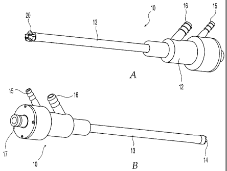
FIG. 1A is a perspective view from the left side of one embodiment of a reamer
device according to the present invention;
FIG. 1 B is a perspective view from the right side of the device of FIG. 1 A;
FIG. 2 is a top view of the reamer device of FIGS. 1A and 1 B;
FIG. 3 is a cross-sectional view of the device taken along line A-A of FIG. 2;
FIG. 4 is a perspective view of one embodiment of a drive shaft assembly
according
to the present invention;
FIG. 5 is a side view of one embodiment of a reamer head according to the
present
Invention;
FIG. 6 is a front view of the reamer head of FIG. 5;
CA 02360867 2001-07-19
WO 00/45714 PCT/CHOO/00051
6
FIG. 7 is a rear view of the reamer head of FIG. 5;
FIG. 8 is a front perspective view of the reamer head of FIG. 5;
FIG. 9 is a rear perspective view of the reamer head of FIG. 5;
FIG. 10 is an enlarged view of the side view of FIG. 5;
FIG. 11 is an enlarged and partially fragmented perspective and cross-
sectional view
of the reamer shown in FIGS. 1A and 1 B;
FIG. 12 shows an exemplary sample of a graph expressing a pressure-time curve
of
a system using the reamer of FIG. 1, the reamer head of FIG. 5, and the drive
shaft
assembly of FIG. 4;
FIG. 13 is a perspective view of a portion of the drive shaft assembly of FIG.
4 with a
guide wire inserted in the cannutation of the drive shaft;
FIG. 14 is a cross-sectional view of the drive shaft assembly taken along line
A-A of
FIG. 13;
FIG. 15 is a top view of another embodiment of a reamer device according to
the
present invention;
FIG. 16 is a front perspective view of another embodiment of a reamer head
according to the present invention; and
FIG. 17 is an enlarged view of the side view of the reamer head of FIG. 16.
For convenience, the same or equivalent elements in the various embodiments of
the
invention illustrated in the drawings have been identified with the same
reference
numerals. Further, in the description that follows, any reference to either
orientation
or direction is intended primarily for the convenience of description and is
not
CA 02360867 2001-07-19
WO 00/45714 PCT/CHOO/00051
7
intended in any way to limit the scope of the present invention thereto.
Referring to FIGS. 1-3, a first embodiment of a reamer 10 according to the
present
invention comprises a reamer head 20 located at a distal end of reamer 10 for
reaming a medullary canal, a flexible aspiration tube 13 for suction and
removal of
the emulsified bone and other material generated by reamer head 20, a reamer
head
retainer 14 for retaining reamer head 20 on aspiration tube 13 while still
allowing
rotation of reamer head 20 with respect to aspiration tube 13, and a manifold
assembly 12 at a proximal end of reamer 10. Thus, as used in this application,
the
term distal designates the end or direction near reamer head 20 and toward the
front
of reamer 10, and the term proximal designates the end or direction near
manifold
assembly 12 and toward the rear of reamer 10. The term longitudinal designates
an
axis central to aspiration tube 13.
Aspiration tube 13 is flexible so that it can bend to accommodate curvature of
the
bone and is preferably made of a translucent material so that the aspirated
material
can be observed. Manifold assembly 12 has an irrigation port 15 and an
aspiration
port 16 for connecting to an irrigation source and aspiration means
respectively. A
drive shaft coupling 17 is located at the proximal end of manifold assembly
12. Drive
shaft coupling 17 can be readily attached and detached to a drive shaft or
some
other means for rotating reamer head 20.
FIG. 4 shows a drive shaft assembly 100 that can be used with reamer 10 to
rotate
reamer head 20 at sufficient speeds to ream the medullary canal. The use of a
drive
shaft assembly 100 with reamer 10 (or any modular system in which the driving
means is contained in an unit that is independent from the reamer) allows
drive shaft
assembly 100 to be reused with many different reamers. Such modularity is
advantageous because different patients and clinical conditions will require
different
sized reamer heads. Furthermore, the reamer head, and not the drive means,
experiences the wear and abrasion of cutting bone. Thus, reamer 10 can be a
single-use, disposable item and drive shaft assembly 100 can be used for an
extended period.
Drive shaft assembly 100 includes a flexible drive shaft 102 having a reamer
head
CA 02360867 2001-07-19
WO 00/45714 PCT/CHOO/00051
8
connector 104 on the distal end for releasably engaging reamer head 20 so that
reamer head 20 rotates when flexible drive shaft 102 rotates, a power source
connector 106 for connection to a source of power to initiate the rotation of
drive
shaft 102, and a manifold coupling 108 located between reamer head and power
source connectors 104, 106 for engaging drive shaft coupling 17. Drive shaft
102 is
sized to fit within the lumen of aspiration tube 13. However, as will be
described in
more detail later, there is sufficient space between the outer wall of drive
shaft 102
and the inner wall of aspiration tube 13 to allow transport of aspirated
material from
reamer head 20 through aspiration tube 13 to aspiration port 16. As was the
case for
aspiration tube 13, drive shaft 102 is flexible to conform to any curvature of
the bone
being reamed. Drive shaft 102 has a cannulation 110 for accommodating a guide
wire 120.
As seen best in FIGS. 11, 13, and 14, there is sufficient space between the
outer wall
of guide wire 120 and the inner wall of cannulation 110 to allow transport of
an
irrigation fluid from irrigation port 15 through cannulation 110 to reamer
head 20.
Drive shaft 102 has an opening 126 that extends from the outer surface of
drive shaft
102 to cannulation 110. Opening 126 is positioned on drive shaft 102 so that
when
drive shaft assembly 100 is coupled to reamer device 10, opening 126 is in
fluid
communication with irrigation port 15 to allow irrigation to flow through
cannulation
110. Opening 126 has curved walls 128, 130. Curved wall 128 bows out to have a
convex profile and curved wall 130 curves inward to have a concave profile.
The
curvature of curved walls 128, 130 helps to draw water into cannulation 110 as
drive
shaft 102 rotates (which with respect to FIG. 14 is in the counter-clockwise
direction).
Any suitable means for releasably joining manifold coupling 108 and drive
shaft
coupling 17 can be used. Preferably, a quick connect mechanism is used for
rapid
coupling and uncoupling. For example, manifold coupling 108 can have a spring
loaded latch mechanism, such as ball bearings, which engage a slot in drive
shaft
coupling 17.
Similarly, any suitable power source and means for securing drive shaft
assembly
100 to the power source can be used. As pneumatic tools are widely used in
orthopaedic surgery, the power source is preferably an air drive such as the
Compact
CA 02360867 2001-07-19
WO 00/45714 PCT/CHOO/00051
9
Air Drive available from Synthes (U.S.A.) of Paoli, Pennsylvania.
Referring back to FIG. 3, housed within manifold assembly 12 is a sealing
element
34 and a sleeve bearing 31. Sealing means 34 and sleeve bearing 31 define an
irrigation chamber 35 and provide a hermetic seal to prevent irrigation fluid
from
escaping irrigation chamber 35 into aspiration port 16 or out the proximal end
of
reamer device 10 during operation. In addition, sleeve bearing 31 prevents the
aspirated emulsified material from entering irrigation chamber 35.
Reamer head 20 is positioned coaxially within reamer head retainer 14 at the
distal
end of aspiration tube 13. FIG. 15 shows a reamer 210 that has a reamer
retainer
14' with a generally spherical outer profile shape. As head retainer 14'
follows
reamer head 20, the shape of head retainer 14' allows head retainer 14' to
glance off
of the medullary canal walls should flexing occur with aspiration tube 13 with
respect
to drive shaft 102. Thus, head retainer 14' can move smoothly while advancing
through the medullary canal, retracting after reaming, and negotiating the
fracture
site.
Reamer head 20 is preferably made of a stainless steel, although any metallic,
polymeric, ceramic, or composite material suitable for cutting bone can be
used. A
reamer cannulation 22 extends from the distal tip to the proximal end of
reamer head
20 (FIGS. 7 and 8). Reamer cannulation 22 is aligned with cannulation 110 of
drive
shaft 102 so that a guide wire can extend from the proximal end of drive shaft
102
through the distal end of reamer head 20.
Although many different reamer heads can be used with reamer 10, 210, one
embodiment is shown in FIGS. 5-10. As shown in these figures, reamer head 20
consists of a cutting head 40 integral with a tubular shank 25. The periphery
of
tubular shank 25 is cylindrical and has a retaining groove 26 indented around
the
periphery which accommodates an extension from the inside of reamer head
retainer
14 and permits reamer head 20 to rotate while maintaining a fixed location
longitudinally at the distal end of the aspiration tube 13. Tubular shank 25
has a
drive shaft receptor 23 at the proximal end which is configured to accommodate
reamer head connector 104 of drive shaft 102 so that reamer head 20 must
rotate
CA 02360867 2001-07-19
WO 00/45714 PCT/CHOO/00051
when drive shaft 102 rotates. Although drive shaft receptor 22 can be of any
shape
conforming to the exterior profile of reamer head connector 104, it is
preferably a
female hex feature.
Cutting head 40 of reamer head 20 has a plurality of blades 41, preferably at
least
five in number, extending radially outwardly from reamer cannulation 22 to
form a
substantially helical pattern. Correlating the number of blades to the
particular blade
geometry and rotation speed is advantageous in order to allow for appropriate
amount of bone material to be removed while providing efficient cutting. When
too
many blades are used with a given blade shape, the flutes become very shallow
and
less bone material can be removed as a result. When an insufficient number of
blades is used, the reamer head is not efficient in cutting bone tissue. In
fact, the
reamer head may bind orjam while cutting bone matter.
Each blade 41 has a multiple surfaced angular distal end with a straight front
cutting
edge 42 joined to a helical side cutting edge 44. Front cutting edge 42 is
defined by
the intersection between an inner blade wall 45 and a planar first lip surface
51. The
angle between inner blade wall 45 and first lip surface 51 is acute. A planar
second
lip surface 52 intersects first lip surface 51 at an obtuse angle to form a
first lip edge
56. A planar third lip surface 53 intersects second lip surface 52 at an
obtuse angle
to form a trailing lip edge 58. Side cutting edge 44 is defined by the
intersection
between inner blade wall 45 and an outer blade surface 46 and is at a constant
radial
distance from the longitudinal axis and extends longitudinally in a helical
fashion.
Outer blade surface 46 whorls radially inward from side cutting edge 44 along
an arc
toward an inner blade wall of an adjacent blade. The space between such
adjacent
blades defines a flute 43 which, during operation, functions to funnel the cut
medullary canal material towards the proximal end of reamer head 20 for
removal
from the bone cavity through aspiration tube 13 under vacuum. Inner blade wall
45
and outer blade surface 46 extend longitudinally on cutting head 40
terminating at the
proximal end in a shoulder surface 48. Shoulder surface 48 abuts tubular shank
25.
FIGS. 16 and 17 show another embodiment of a reamer head 20' according to the
present invention. Reamer head 20' does not have any side cutting edges,
thereby
substantially minimizing the risk of laterally reaming through the cortex of
the bone.
CA 02360867 2001-07-19
WO 00/45714 PCT/CHOO/00051
11
Each blade 41 has a multiple surfaced angular distal end with a straight front
cutting
edge 42. Front cutting edge 42 is defined by the intersection between an inner
blade
wall 45 and a planar first lip surface 51. The angle between inner blade wall
45 and
first lip surface 51 is acute. A planar second lip surface 52 intersects first
lip surface
51 at an obtuse angle to form a first lip edge 56. Outer blade surface 46
whorls
radially inward along an arc toward an inner blade wall of an adjacent blade.
The
space between such adjacent blades defines a flute 43 which, during operation,
functions to funnel the cut medullary canal material towards the proximal end
of the
reamer head 20' for removal from the bone cavity through aspiration tube 13
under
vacuum.
The use of reamer 10, which can be during open surgical, percutaneous, or any
other
minimally invasive procedure, will now be described referring primarily to
FIG. 11. It
should be noted that the use of reamer 210 is analogous to the use of reamer
10, the
primary difference between reamer 10 and reamer 210 being the different
geometries
of head retainer 14 shown in FIG. 2 and head retainer 14' shown in FIG. 15.
After the
bone to be reamed has be accessed, guide wire 120 is inserted into medullary
canal
122 of bone 124. The insertion of guide wire 120 is typically done using
fluoroscopy
to ensure proper placement of guide wire 120. Reamer 10, with an appropriate
cutter
(such as reamer head 20 or 20') attached and coupled with drive shaft 100, is
then
placed over guide wire 120 so that guide wire 120 passes completely through
aspiration tube 13 and provides a track which reamer 10 follows as it reams
canal
122. Preferably, reamer 10 coupled with drive shaft 100 has been connected to
a
driving means prior to insertion into medullary canal 122. Thus, guide wire
120
actually passes through cannulation 110 of drive shaft 102 and cannulation 22
of
reamer head 20.
While reaming medullary canal 122, irrigation and aspiration are applied
simultaneously. The irrigation substantially cools reamer head 20, medullary
canal
122, and bone 124. A preferable irrigation source, which delivers the
irrigation fluid
at a sufficient rate and pressure, is a normal saline bag suspended one meter
above
irrigation port 15. It should also be noted that, in addition to a saline bag,
any
biological compatible solution and delivery system can be used as the
irrigation
source. The irrigation fluid passes from the irrigation source into irrigation
port 15
CA 02360867 2001-07-19
WO 00/45714 PCT/CHOO/00051
12
and enters irrigation chamber 35. The irrigation fluid, traveling along the
path
indicated by arrows I, flows through cannulation 110 in the space between the
inner
wall of cannulation and guide wire 120 and out of reamer head 20.
The aspiration alleviates intramedullary pressure and helps to remove
emulsified
material from reamer head 20. The removal of material not only improves
reaming,
but also provides for the possibility of harvesting the emulsified material
for grafting
purposes. Suction created by an aspiration source travels along the path
indicated
by arrows A. Specifically, the irrigation fluid helps to channel the
emulsified material
generated by reamer head 20 through flutes 43 and into the space between the
outer
wall of drive shaft 102 and the inner wall of aspiration tube 13 to transport
the
emulsified material from reamer head 20 through head retainer 14, aspiration
tube 13
and aspiration port 16 and into a suitable container.
A significant advantage of the system that includes reamer 10, 210, reamer
head 20,
and drive shaft assembly 100 is the ability to ream the medullary canal to the
desired
diameter in one pass, i.e. without the need to use multiple reaming heads of
gradually increasing diameter until the desired reamed size is achieved. In
this
regard, supplying irrigation to reamer head 20 while simultaneously providing
aspiration, and using a reamer head with efficient front cutting geometry (and
optionally a side cutting geometry) produces less pressure and heat than prior
art
reaming devices.
FIG. 12 shows an exemplary sample of a graph expressing a pressure-time curve
of
the system according to the present invention in an animal model. Region I
shows
that no increase in pressure is induced when an access opening to the
medullary
canal is made. The increase in pressure in Region II results from standard
techniques to gain access to the medullary canal. Region III shows that no
increase
in pressure is induced when the guide wire is inserted. As opposed to standard
reaming process, the present invention reduces or eliminates intramedullary
pressure. Specifically, the combined reaming, irrigating and aspirating
functions to
decrease intramedullary pressure below 0,1333 bar (100 mm Hg). In fact, as
shown
in Region IV, a negative intramedullary pressure is achieved with the system
according to the present invention. Because the biologic threshold in the
medullary
CA 02360867 2001-07-19
WO 00/45714 PCT/CHOO/00051
13
canal for fat emboli and pulmonary emboli is known to be greater than or equal
to
0,2666 bar (200 mm Hg), the incidence of fat and pulmonary emboli is reduced.
Additionally, heat necrosis of the cortex is also eliminated due to the
cooling caused
by the flow of fluid during the process.
FIG. 12 shows another important advantage of the system according to the
present
invention. Specifically, the medullary canal reaming (Region IV) requires
approximately 50 seconds. In contrast, conventional reaming in the same animal
model requires approximately 500 seconds. This decrease in reaming time by a
factor of ten means that reaming in clinical situations can be reduced from 30
minutes to 3 minutes. Thus, operating times (and costs) can be significantly
reduced
without any increased risks.
While various descriptions of the present invention are described above, it
should be
understood that the various features can be used singly or in any combination
thereof.
Therefore, this invention is not to be limited to only the specifically
preferred
embodiments depicted herein.
Further, it should be understood that variations and modifications within the
scope of
the invention may occur to those skilled in the art to which the invention
pertains.
Accordingly, all expedient modifications readily attainable by one versed in
the art
from the disclosure set forth herein that are within the scope of the present
invention
are to be included as further embodiments of the present invention. The scope
of the
present invention is accordingly defined as set forth in the appended claims.