Note: Descriptions are shown in the official language in which they were submitted.
CA 02361119 2001-07-27
1
Method, vessel and device for monitoring
metabolic activity of cell cultures
in liquid media
10
The invention relates to a method for monitoring me-
tabolic activity of cells in liquid media, to a ves-
sel particularly appropriate for this, and to a re-
spective device for implementing said method.
In the vessels cells to be cultivated as well as a
liquid medium are contained wherein the latter con-
cerns with a conventional nutritive solution corre-
sponding to the used cells, if necessary. The soluti-
I5 on according to the invention can be employed for the
most different cells and the most different studies,
in particular in the pharmacological field wherein
the metabolic activity of the cells can be monitored
over a longer period. For example, monitoring the ac-
CA 02361119 2001-07-27
2
tion with cytotoxic and biocompatibility tests, and
optimizing the culture conditions for the production
of biological molecules can be carried out.
Commonly, the major nutrient source for cell cultures
is glucose which can be converted into lactate by me-
ans of aerobic glycolysis or oxidatively with the
oxygen consumption and formation of carbon dioxide.
Then, many influences of the physiology of a cell
have an affect on its metabolic activity such that
oxygen consumption is accordingly allowed to vary as
well. Starting from the connection between the meta-
bolic activity of cells with respect to the oxygen
consumption, and e.g. the glucose consumption and L-
glutamine consumption or the generation of lactate it
is allowed to conclude the condition of the monitored
cells, and as a result the influence of the respecti-
ve culture conditions.
Based upon these findings, int. al., in "Noninvasive
Oxygen Measurements and Mass Transfer Considerations
in Tissue Culture Flasks" published in Biotechnology
and Bioengineering, Vol. 51, pp. 466 to 478, it has
been described by Lisa Randers-Eichhorn, how the oxy-
gen consumption of cells cultivated in T-flasks can
be determined by means of an optical measurement.
Therein, it is suggested to arrange sensor membranes
containing fluorescent indicators immediately on the
flask bottom, and in the gas space above the nutriti-
ve solution within such a T-flask. During the measu-
rement of oxygen concentration with such sensor mem-
branes, the well known physical phenomenon of fluo-
CA 02361119 2001-07-27
3
rescent erasure of known fluorescent dyes such as
e.g. complexes of Ruthenium (II) is employed due to
the influence of oxygen wherein the respective fluo-
rescent intensity changes with the continuous excita-
tion according to the oxygen concentration and the
partial pressure of oxygen, respectively. For the de-
termination of oxygen concentration the respective
fluorescent intensity immediately, but also the fluo-
rescence life can be measured, and the oxygen concen-
tration can be determined according to a known cali-
bration.
The set-up of measuring instruments described in this
document, in particular the arrangement of the sensor
membrane on the bottom of the T-flasks, and the ne-
glected determination of some important influence
quantities is not suitable to conclude the metabolic
activity of the culture cells from oxygen consumpti-
on.
The charge of oxygen into the nutritive solution oc-
curs for the most part through the interface of the
nutritive solution toward the gas space, therefore
here toward the gas space in the T-flask, and the
consumption occurs by the cells being present on the
bottom of the T-flask. The maximum enabled oxygen
concentration which can be achieved within the nutri-
tive solution is the saturation concentration of oxy-
gen Csat (of mg/1) which, according to the equation,
Csat = ( ~P - Y*Pw (T) ~ /Po* 0I, (T) *Xo2
CA 02361119 2001-07-27
4
is a function of the total gas pressure p (mbar), the
relative humidity of air y (in values from 0 to 1,
wherein 1 corresponds to a value of 100%, and 0 cor-
responds to a value of 0%), the partial pressure of
water vapour pW(T) (mbar) as a function of the tempe-
rature T, the mole fraction of oxygen Xo2 within the
gas space of the T-flasks, the Bunsen absorption
coefficient a(T) (mg/1) as a function of the tempera-
ture T and the normal pressure po - 1013 mbar . Then,
it is assumed that the gas space and the nutritive
solution have the same temperature, and the gas space
has been filled with atmospheric air which chemical
composition thereof is sufficiently known. These pre-
conditions are commonly present in breeding chambers
in which cells will be cultivated. This saturation
concentration of oxygen appears directly below the
top surface within the nutritive solution. Therefrom,
the oxygen is transported by .means of different ef-
fects such as diffusion and/ or convection toward the
cells being present on the bottom. In such a system
two quantities are significant. On the one hand, this
is the consumption rate k~ at which oxygen is consu-
med by the cells, and on the other hand, the trans-
port rate kT at which oxygen is transported toward
the cells. The two quantities are responsible toge-
ther for that an oxygen gradient results from the top
surface of the nutritive solution toward the bottom
including cells. If the consumption rate is now
slightly smaller than or equal to the transport rate,
thus an oxygen concentration comprising a value of 0
is measured with the oxygen membrane on the bottom
below the cells. In this case, the cells do not suf-
CA 02361119 2001-07-27
fer Prom an oxygen supply since still sufficient oxy-
gen is transported toward the cells, but which does
not arrive toward the sensor membrane below the
cells, and which, accordingly, cannot be measured any
5 longer. If the consumption rate further increases,
e.g. by spreading out the cells, and the consumption
rate becomes greater than the transport rate thus
this cannot be monitored any longer with the oxygen
membrane located on the bottom below the cells.
1o Furthermore, the saturation concentration of oxygen
is a very important quantity in addition to the con-
sumption and transport rates as a function of the to-
tal gas pressure, the humidity, the temperature and
the mole fraction of oxygen and the partial pressure
of oxygen, respectively, within the gas space of the
T-flask. ~A change is causing a change of the oxygen
gradient, and thus a change of oxygen concentration
at any place between the cells and the top surface of
the nutritive solution. Since the parameters of total
pressure, humidity and temperature have not been de-
termined or checked in the mentioned documentation,
it cannot be excluded that measuring results became
falsified due to variations of these parameters.
WO-A-99/~~922 descrihe~s a method for monitoring Che
biological acti.~rity of cells cultivated by liquid me-
dia wherein the cells are cultivated inside the chan-
nels of a.micro type matrix which is placed in a per-
fusion chamber equipped with optical waveguides.
Then, the changes of the oxygen concentration within
the perfusion medium are carried out according to
measurements of fluorescence.
CA 02361119 2001-07-27
5a
US 4 548 907 discloses a fluorescent optical sensor
for the determination of an analyte (e.g. Coz in a
solution) .
US 5 601 979 discloses a method for the examination
of biological activity in liquid media wherein a ves
sel is used which is separated into two parts wherein
in the first part fluorescent marked cells are pre
sent. The cells axe allowed to diffuse through a mem
brane into the adjacent part in which they are opti
cally detected.
Therefore, it is the object of the invention to pre-
determine ways wherein monitoring the oxygen consump-
tion and thus the metabolic activity of culture cells
can be achieved in a cost effective manner and with
an increased accuracy.
According to the invention this object is achieved
with the features of claim 1 for a method, and with
CA 02361119 2001-07-27
6
the features of claim 11 for an appropriate vessel.
Advantageous embodiments and improvements of the in-
vention result from the features mentioned in the
subordinate claims.
The solution according to the invention is now assu-
ming from that said cells will be cultivated in ves-
sels using a liquid medium wherein as a rule here it
concerns with a respective nutritive solution, and
that the metabolic activity thereof takes place
through the measurement of oxygen concentration at a
location within the liquid medium between the cells
consuming oxygen and the part being dominant for the
oxygen charge into the liquid medium, which is here
the top surface of the nutritive solution. Then, the
saturation concentration of oxygen in the liquid me-
dium will be determined according to comparison mea-
surements in a vessel of cell cultures without any
cells and/ or by means of the determination of the
parameters of pressure, humidity, temperature and
with a chemical composition being well known and con-
stant, of the ambient gas space, which are here the
mole fractions of the gas components and the partial
pressures thereof, respectively, in the ambient at-
mosphere. From the comparison between the saturation
concentration of oxygen as a set value and the satu-
ration concentration of oxygen at a location of the
oxygen gradient within the vessel including the cul-
ture cells as an actual value, the oxygen consumption
and thus the metabolic activity of the cells are con-
cluded.
CA 02361119 2001-07-27
7
For monitoring, vessels can readily be used in con-
trast to the prior art which comprise an aperture
such that the top surface of the liquid medium can be
affected by the ambient atmosphere. In certain cases,
however, such an aperture is also allowed to be co-
vered and closed, respectively, with a membrane at
least being permeable to oxygen such that entering of
undesired germs is prevented.
The oxygen concentration will be preferably measured
optically with a sensor membrane suitable thereto
which optical characteristics thereof change as a
function of the respective oxygen concentration.
Thus, in a respective vessel at least one suitable
sensor membrane should be placed which is located
such that it is arranged above the cell cultures cul-
tivated on the vessel bottom, however, below the top
surface of the liquid medium.
If the cells cultivated on the vessel bottom are con-
suming oxygen, the oxygen concentration within the
liquid medium will reduce accordingly, and the oxygen
concentration actually measured with the sensor mem-
brane will be determined by the oxygen consumption
due to the metabolic activity and oxygen quantity
which enters into the liquid medium again occurred
due to the gradient of oxygen concentration.
Since the conformities to natural laws with respect
to the saturation concentration of oxygen in liquid
media are relatively properly known, it is possible
to calculate the respective saturation concentration
CA 02361119 2001-07-27
8
of oxygen within a liquid medium under consideration
of known parameters which are in particular here the
respective temperature, the pressure and the humidi-
ty, such that this calculated value of oxygen concen-
tration can be subjected to a value comparison inclu-
ding the actually measured oxygen concentration to
evaluate the oxygen consumption and metabolic activi-
ty, respectively, of the cell cultures.
However, it is also possible to carry out a reference
measurement wherein a second vessel is used in which
merely a nutrient medium being completely identical
with the used nutritive solutions with respect to the
oxygen diffusion characteristics is used. In such a
reference vessel a respective sensor membrane is ar-
ranged again preferably at the same place whereby the
unaffected oxygen concentration can be measured. The
reference oxygen concentration thus measured is also
allowed to be subjected to a respective value compa-
rison with the oxygen concentration affected by meta-
bolic activity in order to judge the metabolic acti-
vity of the culture cells.
Of course, such a value comparison can also be car-
ried out simultaneously for commonly the calculated
oxygen concentration and measurement signal of oxygen
concentration in the reference vessel including the
oxygen concentration measured under metabolic activi-
ties moderated by culture cells.
Moreover, the meaningfulness with the benchmarking of
metabolic activity of culture cells can be increased
CA 02361119 2001-07-27
9
when sensor membranes are placed additionally within
the vessels in such a manner here again as the oxygen
sensitive membranes which include an oxido-reductase
on an oxygen sensitive membrane, and wherein the
change of oxygen concentration can be measured by a
substrate conversion of the enzyme.
Conveniently, these two different sensor membranes
should be arranged at least approximately in the same
distance of the cell cultures within the liquid medi-
um. For detecting the concentration of the substrate
of oxido-reductase and thus the metabolic activity
the fluorescent signals of the oxygen membrane and
the second oxygen membrane covered with the supple-
mentary membrane are compared. Then, it can be neces-
sary to substract the signal of the first sensor mem-
brane from the signal of the second sensor membrane
so as to achieve the sensor response to the enzymatic
test of the oxygen, and thus to determine the sub-
strate concentration. Then, it can also be advantage-
ous to introduce a factor by means of which the dif-
ferent oxygen transport relations in the two oxygen
membranes are taken into account. Moreover, for an
iteration of the signal of the enzymatic sensor it
can also be necessary to use different calibration
curves depending on the oxygen concentration within
the solution, since oxygen is a co-substrate of the
enzymatic reaction. In any case, by means of a deter-
mination of the enzymatic activity the metabolic ac-
tivity of the cells can further be determined inde-
pendent of oxygen consumption of the culture cells.
CA 02361119 2001-07-27
Since the metabolic activity of the culture cells
changes relatively slowly over longer periods, it is
sufficient to measure the respective concentrations
in longer periods, for example, in intervals of se-
5 veral minutes in order to monitor the metabolic acti-
vity of the respective cell cultures with appropriate
accuracy, which results in the effort required for a
suitable measurement equipment is allowed to be redu-
ced by means of an enabled multiplex operation.
Since the concentration shall be advantageously mea-
sured optically it is necessary to use vessels which
are optically transparent in definite areas such that
the respective change of optical intensity can be
measured with optical waveguides (glass fibers), for
example, and an appropriate optical sensor. Then,
such an optical waveguide has not to be a direct part
of a used vessel, or has not to be connected
therewith immediately, but can be located and aligned
such that it is merely able to detect the area and
parts on one sensor membrane with its aperture. As a
result, the measuring location and measuring vessel
are readily allowed to be separated locally from each
other.
Furthermore, there is a possibility as suggested with
the way of multiplex measurement to commonly monitor
a plurality of vessels in which the same or different
cells are cultivated wherein these each can be taken
into account individually one after another by means
of a respective circuit of at least one multiplexer,
respectively. Hence, a more or less spatial resoluti-
CA 02361119 2001-07-27
11
on type measurement of concentrations can be carried
out. For example, sensor membranes can be used which
change their absorption and reflection characteri-
stics, respectively, as a function of the oxygen con-
s centration. Alternatively, establishing from known
solutions, it is also possible to employ the phenome-
non of fluorescence erasure and to use sensor membra-
nes including known fluorescent dyes which are
capable to be fluorescent with the light of particu-
lar wavelengths when excited wherein the wavelength
of the excitation light and the wavelength of the
fluorescent light are different.
In the last mentioned case the concentration can be
measured once by means of a direct measurement of the
respective fluorescent intensity. However, it is more
favourable to determine the fluorescence life since
in this case aging and subsequently the well known
bleaching behaviour do not affect the measuring accu-
racy.
The oxygen sensitive sensor membranes can be inserted
subsequently as well into the vessels useful for the
solution according to the invention by means of dif-
ferent techniques. Such sensor membranes can be se-
lectively deposited locally by dispensing, spraying,
dipping or glueing as well. Appropriate placing loca-
tions within such vessels are, for example, the inte-
rior wall of the vessel in a predetermined distance
from the vessel bottom, and landing shaped members
projecting beyond the bottom surface of the vessel
are particularly appropriate wherein on the upper end
CA 02361119 2001-07-27
12
face thereof a corresponding sensor membrane can be
formed and applied, respectively. In this case, at
least the landing shaped members are formed from a
material being transparent to the relevant wave-
s lengths such that corresponding monitoring can take
place from below through the vessel bottom. The re-
maining vessel parts then have not to be composed
conclusively of another transparent material.
Within a vessel to be used in accordance with the in-
vention two of such landing shaped members can be lo-
cated spaced apart from each other wherein, on the
one hand, an exclusively oxygen sensitive sensor mem-
brane is deposited, and on the other hand, such a
supplementary membrane can be deposited which is for-
med with a membrane coated with an enzymatic oxidase
sealed against the liquid medium.
For example, if a greater number of samples is to be
simultaneously monitored, it is particularly favou-
rable to form a vessel according to the invention in
an analogous manner with the known MicroWellT''' Plates
wherein in such a MicroWell~ Plate a greater number
of receiving spaces (cavities) for the cells to be
cultivated are located with the liquid medium in a
plurality of rows adjacent to each other. A Micro-
WellT"' Plate formed in this manner can be placed in a
breeding chamber then, for example, for the cell cul-
tivation, wherein the excitation light and the fluo-
rescent light can be directed from an appropriate
light source via optical waveguides upon the sensor
membranes, and the fluorescent light from the sensor
CA 02361119 2001-07-27
13
membranes toward an appropriate optical sensor. Then,
it is possible for both the excitation light and the
fluorescent light to be guided through a single opti-
cal waveguide. Of course, corresponding light guiding
for the two different types of light can be carried
out inside two separate optical waveguides. For this,
the optical waveguides have merely to be fixed and
positioned such that their apertures ensure an opti-
mum fluorescence excitation and an approximately com-
plete detection of the fluorescent light. Then, for
fixing and positioning the optical waveguides separa-
te mechanism plates can be used which will be dimen-
sioned and aligned relative to the vessels to be used
in accordance with the invention, such that the opti-
cal waveguides are located and aligned with respect
to the sensor membranes. This configuration is parti-
cularly appropriate for vessels formed in a Micro-
Well~ Plate shaped manner. Favourably, a cavity
(well) of such a MicroWellTM Plate shaped member can
be used for the reference measurement previously men-
tioned at the beginning, i.e. merely filled with li-
quid medium but without cell cultures.
The aspect for MicroWellTM Plate shaped members for-
med and being useful in accordance with the invention
or even other appropriate vessels can be formed ac-
cording to the common laboratory standards such that
they are able to be used as well in the conventional
form with the different known laboratory instruments.
The spatial resolution type measurement of different
samples cannot only take place with the individual
CA 02361119 2001-07-27
14
optical waveguides associated with the sensor membra-
nes, however, but there is the possibility as well to
use an endoscopy array by means of which the image of
a greater number of sensor membranes detected by such
an array can be directed upon a CCD camera, for ex-
ample, such that an isochronous spatial resolution
type measurement of different oxygen concentrations
is enabled.
l0 In the following the invention will be described in
more detail by way of example.
In the drawings
Figure 1 shows a diagrammatic illustration of an em-
bodiment of a vessel for monitoring metabo-
lic activity of culture cells in liquid me-
dia including sensor membranes and optical
waveguides;
Figure 2 shows a graph diagrammatically illustrating
the oxygen concentration within the liquid
medium from the culture cells containing
bottom of the vessel up to the top surface
of the medium contained in the vessel ac-
cording to three different conditions;
Figure 3 shows diagrammatic illustrations of landing
shaped members including oxygen sensitive
membranes, and alternatively a supplementa-
ry oxido-reductase/ membrane;
CA 02361119 2001-07-27
Figure 4 shows diagrammatically a possibility of an
optical measuring set-up for the generation
and detection of fluorescent signals on a
device according to Figure 1;
5
Figure 5 shows a diagrammatic illustration of
another embodiment of a vessel comprising
an oxygen permeable covering for monitoring
metabolic activity of culture cells within
10 liquid media;
Figure 6 shows a graph illustrating oxygen concen-
tration in the liquid medium from the cul-
ture cells containing bottom of a vessel
15 covered with an oxygen permeable membrane
according to three different conditions up
to the top surface of a liquid medium, and
further up to the environment in which said
vessel is contained;
Figure 7 shows a diagrammatic illustration of a ves-
sel located within a breeding chamber ac-
cording to Figure 1;
Figure 8 shows a multi-purpose vessel in combination
with a mechanism plate for optical wavegui-
des which are connected to a device accor-
ding to Figure 4;
Figure 9 shows a diagrammatic illustration of a mul-
tiplex detection of a plurality of samples;
16
Figure 10 shows an embodiment of a device according
to the invention wherein a Micro4~TellT'" Plate
is monitored by means of an imaging optics,
e.g. an endoscopy array;
Figure 11 shows the optical part of a device accor-
ding to Figure 10 for the generation and
detection of fluorescent signals;
Figure 12 shows an embodiment of an optical system
for the generation and detection of fluo-
rescent signals on a MicroWell~'" plate for
simultaneously monitoring a plurality of
cell cultures.
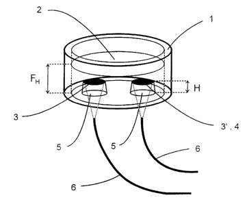
In Figure 1 is shown a diagrammatic illustration of
an embodiment of vessel 1 for monitoring metabolic
activity of cell cultures in liquid media 2.
The vessel 1 illustrated here is predominantly formed
cylinder shaped and comprises a transparent bottom
plate which is open on its upper side, and is also
permeable to oxygen transport into the liquid medium
2, accordingly. The liquid medium 2 is filled with a
particular level FH in which the cells are allowed to
be cultivated. In this embodiment, on the bottom of
the vessel 1 two landing shaped members 5 are formed
from an optically transparent material with the upper
end face thereof is arranged in a particular distance
H from the bottom surface of the vessel 1. Then, H is
generally less than FH.
CA 02361119 2001-07-27
CA 02361119 2001-07-27
17
On the end face of the left landing shaped member 5 a
sensor membrane 3 is formed in order to exclusively
measure oxygen concentration within the liquid medium
2.
On the end face of the right landing shaped member 5
an oxido-reductase membrane 4 is additionally formed
on the oxygen sensitive sensor membrane 3.
Below the vessel 1 optical waveguides 6 including the
light rays thereof (illustrated in dotted lines)
aligned toward the sensor membranes 3 are arranged
for one of the sensor membranes 3 and 3', respective-
ly. By means of the optical waveguides 6 light can be
directed upon the sensor membranes 3 and 3' for fluo
rescence excitation of a fluorescent dye contained in
the sensor membranes 3 and 3'. In contrast, the fluo
rescent light of the sensor membranes 3 and 3' is al
lowed to be trapped by means of the optical wavegui
des 6.
In Figure 2 is illustrated in a graph the oxygen con-
centration in the liquid medium 2 including culture
cells which are located on the bottom, starting from
the bottom of a vessel 1 up to the top surface of the
liquid medium 2 according to three different conditi-
ons within the vessel 1. The bottom plot shows the
course with a great oxygen consumption of the culture
cells on the bottom, and the upper plot according to
low oxygen.
CA 02361119 2001-07-27
18
It will be appreciated from the graph that consider-
ably higher values of oxygen concentration in the li-
quid medium can be measured with a sensor membrane 3
located above the vessel bottom whereas the oxygen
concentration on the bottom of a vessel 1 tends to-
ward 0. This is caused by the oxygen consumption of
the culture cells. Thus, it will be appreciated that
a measurement on the bottom in close proximity to the
cells is not meaningful. In addition, this graph re-
produces the facts that with oxygen consumption of
the culture cells the supply of oxygen into the li-
quid medium 2 can be detected with a sensor membrane
located above the bottom across the surface thereof
up to the cells. Thus, it will be appreciated that an
oxygen measurement is only meaningful in the position
between the culture cells and the places wherein the
oxygen arrives into the liquid medium 2.
In Figure 3 the improvement of landing shaped members
5 is diagrammatically shown wherein the left illu-
stration shows a landing shaped member 5 which is ex-
clusively provided with an oxygen sensitive membrane
3. Here, the landing shaped member 5 is formed in a
truncated shape and is composed of a transparent ma-
terial being at least approximately impermeable to
oxygen wherein it is allowed to achieve that the mea-
suring result will not be falsified by oxygen ente-
ring through the material.
Analogous to this is also formed the landing shaped
member 5 illustrated in figure 3 on the right side
wherein one supplementary oxido-reductase membrane 4
19
is merely provided above the oxygen sensitive membra-
ne 3~.
Of course, the membranes 3, 3' and 4 are not formed,
as illustrated here, above but immediately on the
landing shaped members 5. Then, the oxido-reductase
membrane 4 covers the oxygen sensitive membrane 3'.
In Figure 4 an optical set-up is diagrammatically
shown by means of which light of a light source 20
can be directed through an optical waveguide 6 upon
an oxygen sensitive membrane 3 (not shown here)
within a vessel 1 and a cavity 7, 7', respectively.
The light source 20 preferably radiates approximately
monochromatic light of a fluorescence exciting opti-
cal wavelength through an appropriate lens 27, as the
case may be a filter 21 which essentially allows
light having excitation light wavelength to pass
through upon a dichroic beam splitter 22. Therefrom,
light is coupled through a lens 23 into the optical
waveguide 6. Of course, alternative known input gaps
for optical waveguides 6 can be used as well. It is
also possible to use a multi-spectral light source in
combination with an appropriate filter wherein exclu-
sively the filter provides the monochromatisation.
The fluorescent light then passes opposed to the ex-
citation light through the optical waveguide 6 via
the lens 6, the beam splitter 22, through a respecti-
ve wavelength selected filter 24 which allows to pass
fluorescent light only, as the case may be via
another lens 25, upon an optical detector 26 by means
CA 02361119 2001-07-27
CA 02361119 2001-07-27
of which the intensity of fluorescent light can be
measured as a function of the respective oxygen con-
centration.
5 The vessel 1 illustrated in Figure 5 corresponds in
the most significant points to the vessel already
shown and described, respectively, in Figure 1.
It is merely provided with a covering 28 which is ad-
10 mittedly permeable to oxygen, however, it prevents
contaminations and consequently ensures the sterili-
ty. Moreover, drying out the vessel 1 can be preven-
ted with such a covering 28.
15 In Figure 5 it will be further recognized that the
level FH is below the height GH of the vessel. GH si-
multaneously reproduces the distance of the covering
28 of the vessel 1 from the bottom.
20 The oxygen gradient will be affected through the co-
vering 28.
These facts are reproduced in the graph according to
Figure 6, and it is intimated that oxygen concentra-
tion between the level FH and vessel height GH is
subjected to a particular gradient of oxygen concen-
tration as well which is elicited by the covering 28
during oxygen consumption caused by metabolic activi-
ty.
With the embodiment shown in Figure 7, a vessel 1 ac-
cording to Figure 1 and 5, respectively, has been lo-
CA 02361119 2001-07-27
21
Gated within a typical breeding chamber 9 in which
particularly optimum conditions for the cell cultiva-
tion can be met as is well known. On optimum conditi-
ons it is usually understood a temperature of appro-
ximately 37°C, a relative humidity of air of 100°s at
normal atmospheric pressure, and a partial oxygen
pressure within the gas space corresponding to the
ambient air. The optical waveguide 6 for the two sen-
sor membranes 3 are allowed to be led out of the
breeding chamber 9 such that a local separation bet-
ween the measuring place and measured value detection
can be achieved. Moreover, in this Figure is shown a
gas supply 10 for the breeding chamber 9 through
which atmospheric air having a respective constant
content of oxygen can be supplied. Breeding chambers
of the described type are employed for the cultivati-
on of cells according to the standard.
In Figure 8 there is shown an embodiment for a vessel
1' to be used in accordance with the invention which
is formed as a MicroWell~'' Plate including a plurali-
ty of cavities 7, 7' which is very often used for the
cultivation of cells. Two sensor membranes 3 and 3'
are arranged and configured respectively again in the
cavities 7, 7' wherein each sensor membrane 3' is
further provided with an oxido-reductase membrane 4
in the analogous manner to the vessel 1, as it has
been described and shown in Figure 1.
Such a MicroWellT''' Plate 1' can be located in a bree-
ding chamber 9 again, and the cell cultures are allo-
wed to be accordingly cultivated within the cavities
CA 02361119 2001-07-27
22
7. In the breeding chamber 9 a mechanism plate 8 can
be arranged below the Microir7s11T'" Plate 1' by means
of which the optical waveguides 6 can be fixed and
positioned. Then, the optical waveguides 6 are sup-
s ported to the mechanism plate 8 according to the ar-
rangement of the sensor membranes 3 and 3' in the ca-
vities 7 and 7' of the vessel 1'. The mechanism plate
8 is allowed then to be located within the breeding
chamber 9 in such a distance to the MicroWellTM Plate
1', and here. in particular to the bottom surface the-
reof such that the light rays from the optical wave-
guides 6 are able to detect the complete surface area
of the respective sensor membranes 3 and 3' associa-
ted therewith. Using the mechanism plate 8 permits an
operation being undisturbed for the cell cultivation.
In the breeding chamber the cells will not be subjec-
ted to any further treatment . For such purposes the
MicroWellTM Plate 1' is removed from the breeding
chamber, and thus from the support 8. Outside the
breeding chamber, the treatment of the cell cultures
can be taken place then in the common sense, without
any resrictions such as e.g. a change of the liquid
medium 2 or monitoring the cells with a microscope.
At least one cavity 7' of such a MicroWellTM Plate 1'
can be used for the reference measurement already ex-
plained by filling this cavity 7' without cells only
with liquid medium 2.
In the illustration according to Figure 9 an optical
multiplex operation is diagrammatically shown by me-
ans of which a spatial resolution type measurement in
23
addition to the time resolution type measurement as
well of several samples is enabled which are con-
tained in different vessels 1 and cavities 7 and 7',
respectively. Thus, Figure 9 shows a complex set-up
for implementing the method according to the inventi-
on.
An array of the light source 20, detector 26, lenses
23, 25, 27, filters 21, 24 and beam splitters 22 as
illustrated .in Figure 4 is located within a transmit-
ting and measuring unit 13 outside the breeding cham-
ber 9. The excitation light is guided from this
transmitting and measuring unit 13 via a single opti-
cal waveguide 12 into an optical multiplexer 11
wherein further optical waveguides 6 are connected.
The optical multiplexer 11 sequentially directs the
fluorescent excitation light through the single opti-
cal waveguides 6 for the fluorescent excitation to-
ward the different sensor membranes 3 and 3'.
The fluorescent light of the different sensor membra-
nes 3 and 3' again arrives via the optical waveguides
6 to the optical multiplexer 11, and is sequentially
directed therefrom upon an optical detector which is
included in the unit 13 corresponding to the respec-
tive measurement locations, that is the respective
sensor membranes 3 and 3' according to the respective
sample vessel 7 and 7', respectively, via the opti-
cal waveguide 12. The signals thus detected can be
employed in a correspondingly spatial and time type
resolution manner for the determination of the re-
CA 02361119 2001-07-27
24
spective oxygen concentration and the oxido-reductase
substrate concentration.
From the unit 13 the detected measured values pass to
a benchmarking and control unit 14 which is a perso-
nal computer here by means of which the benchmarking
of the detected measuring signals, and then in parti-
cular the value comparison indicated in the general
part of the description can be carried out by measu-
rement in the reference vessel 7'. With the benchmar-
king and control unit 14 the transmitting and measu-
ring unit 13 and optical multiplexer 11 as well can
be controlled.
Moreover, inside the breeding chamber 9 are arranged
sensors T5 for detecting the temperature, the relati-
ve humidity of air, the gas pressure and chemical
composition as may be the case, including the respec-
tive partial pressure of involved gases of the gase-
ous atmosphere. The measured values detected by the
sensors 15 are directed again via a separate line 16
into the transmitting and control unit 13, and there-
from into the benchmarking and control unit 14 such
that the value comparison indicated in the general
part of the description thus can be carried out. But
there is the possibility either to direct the line 16
immediately from the sensors 15 to the benchmarking
and control unit 14. According to the sensor data the
saturation concentration of oxygen in the reference
vessel 7' can be calculated as shown in the general
part of the description, and thus the value compari-
son is allowed to occur.
CA 02361119 2001-07-27
25
In Figure 10 an embodiment for simultaneously monito-
ring more samples is reproduced which are contained
in a plurality of cavities 7 and 7' of a MicroWellT"'
Plate 1'. Then, below the MicroWellTM Plate 1' a
focussing lens 17 is mounted by means of which at
least that part of the MicroWellTM Plate 1' on which
the membranes 3 and 3' are arranged, on the one hand,
is radiated with the excitation light, and, on the
other hand, the respective fluorescent signal can be
coupled as images into a light guiding system used as
an optical waveguide bundle 6', is allowed to be ima-
ged and passed on.
The excitation and detection by fluorescent signals
on the membranes 3 and 3' within the cavities 7 and
7' is allowed then to occur by way of example as
shown in Figure 11. Therewith, light of a light sour-
ce 20 is coupled again via a lens 27, a filter 21 and
a couple optics 23 at least into one part of the op-
tical waveguide bundle 6', and is directed upon the
membranes 3 and 3' which are located on landing sha-
ped members within the cavities 7 and 7', as already
described a number of times, wherein only light from
the light source 20 having a fluorescence exciting
wavelength is used by means of the filter 21.
The fluorescent light of the different membranes 3
and 3' again arrives, as diagrammatically shown here,
from the other part of the optical waveguide bundle
6' via the two lenses 23, 25 and an accordingly ap-
propriate filter 24 upon an optical detector 26'
CA 02361119 2001-07-27
26
which here is a camera appropriate for a spatial re
solution type measurement. Therewith, the fluorescent
signal of the individual membranes 3 and 3' of the
cavities 7 and 7' can be detected again in a spatial
resolution manner.
In Figure 12 is shown another embodiment of an opti-
cal set-up for simultaneously monitoring different
samples which are located in a plurality of cavities
l0 7 and 7' of a MicroWellTM Plate 1'.
Then, light of a light source 20 is directed again
via a filter 21 for fluorescent excitation upon the
different oxygen sensitive membranes 3 and 3' through
the bottom of the vessel 1' which is formed in a
transparent manner at least in the area of the
landing shaped members 5. For beam forming and beam
guiding again the different lenses 27 and 29 and a
dichroic beam splitter 22 as well are used.
The fluorescent light of the different membranes 3
and 3' passes via the lens 29 through the diochroic
beam splitter 22, and an appropriate filter 24 as may
be the case, upon an optical detector 26' which again
can be a camera 26' here appropriate for a spatial
resolution type measurement. Imaging the fluorescent
light can occur by means of a supplementary lens 25
upon the camera 26'.
CA 02361119 2001-07-27