Note: Descriptions are shown in the official language in which they were submitted.
CA 02361282 2001-11-07
INCISION TEMPLATE AND I~ZE'I'HODS FOR USE
BACKGROUND OF THE I~'VENTION
[O1] Field of the Invention
[02] The present invention relates generally to medical devices and
methods. More particularly, the present invention relates to devices, methods,
and kits for
locating a site on a patient's chest suitable for establishing percutaneous
intercostal access to
the patient's heart for subsequent placement of minimally invasive direct
cardiac massagers,
chest tubes, defibrillation electrodes, and the like.
[03J Sudden cardiac arrest is a leading cause of death in most industrial
societies. In order to resuscitate a victim of cardiac arrest, it is necessary
to provide an
adequate artificial circulation of blood to oxygenate the heart and brain by
re-establishing the
pumping function of the heart during the period between cardiac arrest and
restoration of
normal cardiac activity. Such a cardiac pumping function must be instituted at
the earliest
possible state. While in many cases it is theoretically possible to re-
establish cardiac
function, irreversible damage to vital organs, particularly the brain and the
heart itself, will
usually occur if sufficient blood flow is not re-established within a critical
period of time
from the moment of cardiac arrest. Such a period of time is measured ranging
betlveen four
and six minutes.
(04J A number of techniques have been developed to provide artificial
circulation of blood to oxygenate the heart and brain during the period
bet<veen cardiac arrest
and restoration of normal cardiac activity. Prior to the 1960's, open chest
cardiac massage
(OCM) was a standard treatment for sudden cardiac arrest. Open chest cardiac
massage, as
its name implies, involved opening a patient's chest and manually squeezing
the heart to
pump blood to the body. In the 1960's, closed chest cardiac massage (CCM)
where the heart
2~ is externally compressed through the chest wall became the standard of
treatment. 'Vhen
CCM is combined with airway support, it is known as cardiopulmonary
resuscitation (CPR).
CPR has the advantage that it is much less invasive than OCM and can be
performed by less
skilled individuals. It has the disadvantage, however, that it is not
generally effective: In
particular, the medical literature shows that CCM provides significantly less
cardiac output.
neuroperfusion, and cardiac perfusion than achieved with OCM.
CA 02361282 2001-11-07
[OS] Of particular interest to the present invention is the recent
introduction
of devices for performing minimally invasive direct cardiac massage. Such
devices and
methods are described in co-pending application nos. 09/087,665 filed May 29,
1998, now
U.S. Patent No. 6,200,280; 60/111,934 filed December 1 l, 1998 (now
abandoned);
09/344,440 filed June 25, 1999; 09/356,064 filed July 19, 1999; 09/801,4? 1
filed l~iarch 7,
2001; and 09/898,701 filed July 2, 2001, assigned to the assignee of the
present application.
The full disclosures of each of these prior patents and/or applications are
incorporated herein
by reference. Generally, such methods rely on introducing a plurality of
struts, an expansible
hared bell structure, a laterally oriented expansible structure, or other
expandable member to
engage the heart through a small incision through an intercostal space to a
region over the
pericardium or other heart surface. The heart may then be pumped by directly
engaging the
deployed expansible structure against the pericardium to repeatably compress
the heart,
typically by reciprocating a shaft attached to the member. Additional
minimally invasive
direct cardiac massage devices and methods are also described in 5,582,580;
5,571,074; and
5,484,391 issued to Buckman, Jr. et al. and U.S. Patent Nos. 5,931,850;
5,683,364; and
5,466,221 issued to Zadini et al., licensed to the assignee of the present
application. While
direct cardiac massage approaches offer great promise, certain shortcomings
still exist. For
example, it is sometimes difficult to locate a site on a patient's chest
suitable for establishing
percutaneous intercostal access to the patient's heart, particularly by less
skilled treating
individuals. Misplacement of an access site could lead to serious risks that
may be life
threatening, such as puncturing and/or lacerating an organ, blood vessel (e.g.
internal
mammary artery), or surrounding structure.
[06] For these reasons, it would be desirable to provide devices, methods,
and kits for locating a site on a patient's chest suitable for establishing
percutaneous
intercostal access to the patient's heart. In particular, it would be
desirable to provide
devices, methods, and kits which effectively and rapidly locate an incision
site for intercostal
access by sharp and/or blunt dissection for subsequent placement of minimally
invasive direct
cardiac massagers, chest tubes, defibrillation electrodes, and the like. The
devices, methods,
and kits may be used by persons of minimal experience or training. The
devices, methods,
and kits should be simple and less costly to manufacture and produce. At least
some of these
objectives will be met by the invention described hereinafter.
CA 02361282 2001-11-07
[07) Description of the Background Art
[08] Devices and methods for minimally invasive direct cardiac massage
through intercostal dissection are described co-pending L'.S. Patent
Application No.
09/087,665 filed May 29, 1998, now U.S. Patent No. 6,200,280; U.S. Provisional
Patent
Application No. 60/111,934 filed December 11, 1998 (now abandoned); U.S.
Patent
Application Nos. 09/344,440 filed June 25, 1999; 09/356,064 filed July 19,
1999; 09i801,4? 1
filed March 7, 2001; 09/895,844 filed June 29, 2001; and 09/898,701 filed July
2, 2001,
assigned to the assignee of the present application. U.S. Patent Nos.
5,484,3915, 582,580;
and 5,571,074 to Buckman, Jr. et al. and U.S. Patent Nos. 5,466,221 and
5,683,364 to Zadini
et al., licensed to the assignee of the present application, also describe
devices and methods
for minimally invasive direct cardiac massage through an intercostal space.
Devices and
methods for establishing intercostal access are described in co-pending U.S.
Patent
Application No. 09/768,041 filed January 22, 2001, assigned to the assignee of
the present
application. U.S. Patent No. 3,496,932 describes a sharpened stylet for
introducing a cardiac
massage device to a space between the sternum and the heart. Dissectors
employing
inflatable components are described in U.S. Patent Nos. 5,730,756; 5,730,748;
5,716,325;
5,707,390; 5,702,417; 5,702,416; 5,694,951; 5,690,668; 5,685,826; 5,667,520;
5,667,479;
5,653,726; 5,624,381; 5,618,287; 5,607,443; 5,601,590; 5,601,589; 5,601,581;
5,593,418;
5,573,517; 5,540,711; 5,514,153; and 5,496,345.
[09] The full disclosures of each of the above references are incorporated
herein by reference.
BRIEF SUMMARY OF THE NVENTION
[10] The present invention provides templates, methods, and kits for
locating a site on a patient's chest suitable for establishing percutaneous
intercostal access to
the patient's heart. In particular, the present invention provides templates,
methods, and kits
which effectively and rapidly locate an incision site for intercostal access
by sharp and/or
blunt dissection for subsequent placement of minimally invasive direct cardiac
massagers,
chest tubes, defibrillation electrodes, and the like. Moreover, the present
invention may be
used by persons of minimal skill or training.
[11] In a first aspect of the present invention, an incision template
comprises a structure placeable on a patient's chest. The structure has at
least one marker
which can be aligned with at least one anatomical feature of the patient and a
target zone
CA 02361282 2001-11-07
which lies over a preselected location for intercostal access when the marker
is aligned with
the anatomical feature. The structure may comprise a card structure, wire
structure, or other
framework which is suitable for locating a site on a patient's chest for
establishing intercostal
access to the patient's heart. Preferably, the structure will have a credit
card shape which has
a width in the range from 1 inch to 3 inches, a length in the range from 3
inches to 5 inches.
and a thickness in the range from 0.005 inch to 0.00 inch, and be formed from
plastic, metal,
rubber, wire, or like materials. In some circumstances, a set of templates
having various
shapes, sizes, and/or dimensions may be provided to accommodate different
patient
characteristics.
[12] The template will typically have two markers which together define the
preselected location for intercostal access (i.e. horizontal and vertical
placement). The
template marker may comprise an edge, hole, or line on the structure. For
example, a line
may be placed across a transparent or translucent template that allows a
treating person easily
align the marker with the anatomical feature. The marker is alignable with an
anatomical
feature, such as, a mid-line of a sternum, an intercostal space, a rib (e.g.
forth or fifth rib), or
a nipple. The template target zone (which may also serve as a marker) may
comprise an
opening, groove, notch, or slit in the structure which lies preferably over a
skin surface over a
fourth intercostal space when the marker is aligned with the anatomical
feature.
[13] In a preferred embodiment, the incision template comprises a credit
card structure which has a left edge which can be aligned with a mid-line of a
sternum and a
template opening which lies over a fourth intercostal space. The template
opening will
preferably have a cross-like pattern, wherein a first axis of the structure
opening crosses with
a second axis of the structure opening to define an incision point for
subsequent entry. The
template opening may alternatively have a T-bar pattern, or any other opening
pattern which
serves to define a horizontal incision boundary. In this case, the second axis
may intersect
with the first axis to define the incision boundary. Hence, the second axis
may define either
an incision point or an incision boundary at its intersection with the first
axis. Furthermore,
the structure opening may have more than one intersecting axis (i.e. a third
axis) such that
both the incision point and incision boundary are defined. Such a horizontal
boundary allows
users of the present invention to easily and effectively know how close to the
mid-line
sternum should the preferred location for intercostal access be positioned
without any risks of
unintended damage of blood vessels, such as the internal mammary artery,
organs, or any
other surrounding structures. Typically, the horizontal incision boundary will
be in a range
from about 2.5 cm to about 7.5 cm away from the mid-line sternum.
4
CA 02361282 2001-11-07
[14] In some embodiments, the incision template may additionally comprise
a detachable adhesive skin contacting surface which may be a part of a back or
bottom side of
the structure. The adhesive skin contacting surface will typically be pre-
marked and form a
patch around tissue at the access site to maintain near normal inter-thoracic
pressure after
intercostal access.
[15] In a second aspect of the present invention, an incision template for
locating a site of percutaneous intercostal access comprises a flat-sided body
having an
opening extending between opposite flat sides of the body. The body is
positionable against
a skin surface of an intercostal space so that the opening defines the site
suitable for
subsequent intercostal access.
[16] In a third aspect of the present invention, methods for locating a site
on
a patient's chest suitable for establishing percutaneous intercostal access to
the patient's heart
are provided. One method comprises aligning at least one marker on a template
with at least
one anatomical feature of the patient, the template having a target zone which
lies over the
site when the marker is aligned with the anatomical feature. Aligning
comprises aligning a
first marker with a mid-line of the patient's sternum and aligning a second
marker with a
fourth intercostal space. Preferably, a left edge of the template is aligned
with a sternum mid-
line and a template opening over a fourth intercostal space. In the case where
a set of cards
having various shapes, sizes, and/or dimensions are provided, a user may
choose a particular
template dependent upon patient characteristics prior to alignment.
Furthermore, the
template opening (which may serve as both a marker and a target zone) may
define a
horizontal incision boundary and/or an incision point for subsequent entry.
This incision
point will typically be located between a fourth and fifth rib of the patient,
left of the mid-line
sternum.
[17] The access site on the patient's chest as defined by the target zone on
the template may be appropriately marked by a treating person with a surgical
marker.
Optionally, a pre-marked adhesive skin contacting surface on the back or
bottom side of the
template may be detached on the site so that there is no need to mark the site
with a surgical
marker. Additionally, the adhesive skin contacting surface may form an access
patch around
the site after intercostal access. The incision site and surrounding area may
then be prepped
using antiseptic, a dispenser cup, gauze, and/or procedure drape. Intercostal
access may then
be achieved by sharp dissection, blunt dissection, or preferably by a
combination of sharp and
blunt dissection, wherein a cutting element may be advanced through the site
defined by the
target zone so as to make a small incision or thoracostomy through the skin
overlying an
CA 02361282 2001-11-07
intercostal space and then advancing a blunt member through the intercostal
space above the
heart. The cutting element may comprise a scalpel, surgical knife, lancet,
blade, and the like.
The blunt member may comprise a gloved finger of a treating person, a blunt
shaft or support,
or like structure for clearing access to the heart and verifying the location
of the heart.
Following intercostal access establishment, a direct cardiac massage device
may be
advanced. Exemplary cardiac massage devices are described in co-pending U.S.
Patent
Application No. 09/087,665 filed May 29, 1998, now U.S. Patent No. 6,200,280;
U.S.
Provisional Patent Application No. 60/111,934 filed December 11, 1998 (now
abandoned);
U.S. Patent Application Nos. 09/344,440 filed June 25, 1999; 09/36,064 filed
July 19, 1999;
09/801,421 filed March 7, 2001; and 09/898,701 filed July 2, 2001, assigned to
the assignee
of the present application. Other suitable cardiac massage structures are
described in U.S.
Patent Nos. 5,484,391; 5,582,580; and 5,571,074 issued to Buckman, Jr. et al.
and 5,931,850;
5,683,364; and 5,466,221 issued to Zadini et al., licensed to the assignee of
the present
application.
[18J In a fourth aspect of the present invention, another method for locating
a site on a patient's chest suitable for establishing percutaneous intercostal
access to the
patient's heart is provided. The method comprises aligning a left edge of a
template with a
mid-line of the patient's sternum and aligning an opening in the template with
a fourth
intercostal space so that a target zone of the template lies over the site
following alignment of
the template.
[19] In a fifth aspect of the present invention, an improved method for
establishing intercostal access to a patient's heart is provided. The
improvement comprises
aligning a template with an anatomical feature of the patient and penetrating
an instrument
through tissue between the patient's ribs at a site determined by the
template.
[20] In a sixth aspect of the present invention, kits comprising a template
and instruction for use are provided. The template may comprise any of the
structures
described herein, while instructions for locating a site on a patient's chest
suitable for
establishing percutaneous intercostal access to the patient's heart will
generally recite the
steps for performing one or more of the above described methods. A
conventional package,
which may be in the form of a bag, pouch, box, sealable tray, or the like, may
be used to
contain the template and the instructions for use. The kit may further
comprise a first tray,
holding the incision template and a cardiac massage device, and a second tray,
holding at
least a surgical marking pen, scalpel, gauze, dispenser cup, procedure drape,
clear view
guard, or chest seal. The first and second tray may be hinged together to form
a single unit,
6
CA 02361282 2001-11-07
wherein a tray handle is integrally formed with the first or second tray.
Optionally, but not
necessarily, all tray components may be sterilized. The first and second tray
components
may further be independently sterilizable. The kit may alternatively further
comprise a single
sterile tray holding at least the template, a surgical marking pen, a scalpel,
a cardiac massage
device, gauze, a dispenser cup, a procedure drape, clear view guard, or chest
seal. Still
optionally, the template and a surgical marking pen may be provided outside
the sterile tray
in the package or bag. The instructions will often be printed, optionally
being at least in-part
disposed on packaging. The instructions may alternatively comprise a
videotape, a CD-RO~f
or other machine readable code, a graphical representation, or the like
showing any of the
above described methods.
[21] A further understanding of the nature and advantages of the present
invention will become apparent by reference to the remaining portions of the
specification
and drawings.
BRIEF DESCRIPTION OF THE DRAWINGS
[22] Fig. lA-1D illustrate an exemplary device for locating a site on a
patient's chest suitable for establishing percutaneous intercostal access
constructed in
accordance with the principles of the present invention.
[23] Figs. 2A-2C illustrate an alternative embodiment of the device of the
present invention, showing an incision template with a T-bar opening.
[24] Figs. 3 and 4 illustrate still further embodiments of the device of the
present invention.
[25] Fig. 5 is a cross-sectional view illustrating a heart underneath a
patient's ribs.
[26] Figs. 6A-6E illustrate a method according to the present invention
employing the device of Fig. 1.
(27] Fig. 7 is a perspective view of a cardiac massage device used in
conjunction with the present invention.
[28] Fig. 8 illustrates a distal end of the cardiac massage device of Fig. 7,
3U showing a deployed flared bell structure.
[29] Fig. 9 illustrates a kit according to the present invention comprising an
incision template and instructions for use.
7
CA 02361282 2001-11-07
[30] Fig. 10 illustrates a sterile tray that may be enclosed within the
packaging of the kit of Fig. 9.
[31] Figs. 11A and 11B illustrate an alternative tray unit that may be
enclosed within the packaging of the kit of Fig. 9.
DETAILED DESCRIPTION OF THE IIWENTION
[32] The present invention provides templates, methods, and kits for
locating a site on a patient's chest suitable for establishing percutaneous
intercostal access to
the patient's heart. In particular, the present invention provides templates,
methods, and kits
which effectively and rapidly locate an incision site for intercostal access
by sharp and/or
blunt dissection for subsequent placement of minimally invasive direct cardiac
massagers,
chest tubes, defibrillation electrodes, and the like.
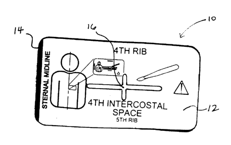
[33] Referring now to Figs. 1A and 1B, an exemplary incision template 10
constructed in accordance with the principles of the present invention for
locating a site on a
patient's chest suitable for establishing percutaneous intercostal access to
the patient's heart
is illustrated. The incision template 10 comprises a structure 12 placeable on
a patient's
chest. The structure 12 has at least one marker 14 which can be aligned with
at least one
anatomical feature of the patient. The structure 12 also has a target zone 16
which lies over a
preselected location for intercostal access when the marker 14 is aligned with
the anatomical
feature. As discussed above, the incision template 10 effectively and rapidly
locates an
incision site and may be used by persons of minimal skill or training. It will
be appreciated
that the following depictions are for illustration purposes only and does not
necessarily reflect
the actual shape, size, or dimension of the incision template 10. This applies
to all depictions
hereinafter.
[34] Preferably, the structure 12 comprises a flat sided body having a credit
card structure which has a width in the range from 1 inch to 3 inches, a
length in the range
from 3 inches to S inches, and a thickness in the range from 0.005 inch to
0.050 inch. In
some instances, a set of cards having various shapes, sizes, and/or dimensions
may be
provided to accommodate different size patients. The structure 12 may be
formed from a
variety of materials, including plastic, metal, rubber, wire, or like
materials.
[35] The structure 12 will typically have two markers which together define
the preselected location for intercostal access (i.e. horizontal and vertical
placement). The
template marker 14 may comprise an edge, hole, or line on the structure. As
shown in Fig.
8
CA 02361282 2001-11-07
1A, a line 14 may be placed across the incision template 10 so as to
facilitate easy alignment
of the marker 14 with the anatomical feature. The marker 14 is alignable with
an anatomical
feature, such as, a mid-line of a sternum and/or an intercostal space between
the forth and
fifth ribs (i.e. fourth intercostal space). The template target zone 16 (which
also serves as a
marker) preferably comprises an opening extending between opposite flat sides
of the body
12, as depicted in Figs. 1 A and 1 B, which lies over a skin surface over a
fourth intercostal
space. As shown in Fig. 1C, the template opening 16 will preferably have a
cross-like
pattern, wherein a first axis 18 of the structure opening crosses with a
second axis 20 of the
structure opening to define an incision point 22 for subsequent entry. FiQ. 1D
illustrates yet
another cross-like pattern for the template opening 16.
[36] Referring now to Figs. 2A and 2B, the template opening 16 may
alternatively comprise a T-bar pattern, or any other opening pattern which
serves to define a
horizontal incision boundary 24. As illustrated in Fig. 2C, the second axis 20
crosses with
the first axis 18 to define an incision boundary 24. Hence, the second axis 20
may define
either an incision point (Fig. 1C) or an incision boundary (Fig. 2C) at its
intersection with the
first axis 18. Furthermore, the structure opening may have more than one
intersecting axis
(i.e. a third axis) such that both the incision point and incision boundary
are defined. The
horizontal boundary 24 allows users of the present invention to easily and
effectively know
how close to the mid-line sternum should the preferred location for
intercostal access be
positioned without any risks of unintended damage to blood vessels, such as
the internal
mammary artery, organs, or any other surrounding structures. Typically, the
horizontal
incision boundary will be in the range from about 2.5 cm to about 7.~ cm away
from the mid-
line sternum. Optionally, the template opening 16 may comprise a groove,
notch, or slit in
the structure 12.
[37] Referring now to Figs. 3 and 4, still further embodiments of the device
of the present invention are illustrated. Fig. 3 illustrates a structure 12
having a marker 14
and a target zone 16. In particular, it will be appreciated that the target
zone may be indicated
in several fashions. In this depiction, the target zone comprises a V shaped
notch or groove
which lies over the site when the marker edge or line 14 is aligned with the
anatomical
feature. Fig. 4 illustrates a wire structure 12 which is suitable for locating
a site on a patient's
chest for establishing intercostal access to the patient's heart. The wire 12
has an edge 14 and
a circular opening I6 which lies over the preferred site after marker 14
alignment.
9
CA 02361282 2001-11-07
[38] Referring now to Fig. 5, a patient's heart H is shown in a cross-section
between ribs R" where n indicates the rib number. The aorta A is also shown
extending from
the top of the heart.
[39] Referring now to Figs. 6A-6E, a first exemplary method for locating a
site S on a patient's chest suitable for establishing percutaneous intercostal
access to the
patient' heart H with the incision template of Figs. 1A and 1B will be
described. As
illustrated in Fig. 6A, at least one marker 14 on a template 10 is aligned
with at least one
anatomical feature of the patient P, the template 10 having a target zone
opening 16 which
lies over the site S when the marker 14 is positioned with the anatomical
feature. Preferably,
a left edge or line 14 of the template 10 is aligned with a sternum mid-line
26 and a template
opening 16 over a fourth intercostal space so that the target zone 16 (which
also serves as a
marker) lies over the site S. In particular, the template opening 16 has a
first axis 18 which
crosses with a second axis 20 of the template opening to define an incision
point 22 for
subsequent entry. The incision point 22 at site S will typically be located
between ribs R,~ and
RS of the patient, left of the mid-line sternum 26.
[40] The access site S on the patient's chest as defined by the target zone 16
on the template 10 may be appropriately marked by a treating person with a
surgical marker.
Optionally, a pre-marked adhesive skin contacting surface on the back or
bottom side of the
template 10 may be detached on the site S so that there is no need to mark the
site with a
surgical marker (not shown). Additionally, the adhesive skin contacting
surface may form an
access patch around the site S after intercostal access. The incision site S
and surrounding
area may then be prepped using antiseptic, a dispenser cup, gauze, and/or
procedure drape.
[41] As shown in Figs. 6B and 6C, intercostal access may then be achieved
by a combination of sharp and blunt dissection. First, a cutting element may
be advanced
through the site S defined by the target zone 16 so as to make a small
incision I or
thoracostomy through the skin, fat, and/or muscle layers overlying an outer
rib surface.
Typically, the incision depth will be in a range from 0.5 cm to about 5 cm,
preferably being
about 3 cm. The cutting element may comprise a scalpel, surgical knife,
lancet, blade, and
the like. A blunt member 28 may then be advanced through the intercostal space
between
ribs R.; and R; above the heart H. The blunt member 28 may comprise a gloved
finger of a
treating person, as shown in Fig. 6C, a blunt shaft or support, or like
structure for clearing
access to the heart H and verifying the location of the heart H.
(42] Following intercostal access establishment, a direct cardiac massage
device 100 may be advanced as illustrated in Figs. 6D and 6E. The cardiac
massage device
CA 02361282 2001-11-07
100, as described in more detail in co-pending U.S. Patent Application Vos.
09/356,064 and
09/898,701, comprises a sleeve 102, a shaft 104 slidably mounted in a central
lumen of the
sleeve 102, and a handle 106 attached to a proximal end of the shaft (Fig. 7).
The sleeve 102
includes a positioning flange 110 near its distal end, typically spaced
proximally of a tip 112
of the device by an optimum distance. A flared bell structure 130, as best
seen in Fig. 8, is
attached to the distal end of shaft 104 and assumes a trumpeted configuration
when fully
deployed. The flared bell structure 130 comprises a plurality of outwardly
curving struts 132
(the illustrated embodiment has a total of eight struts, but this number could
vary). The struts
are preferably formed from a resilient metal, usually formed from a
superelastic alloy, such as
nitinol. To enhance the rigidity and pushability of the structure, re-
enforcing beams 138 may
also be provided. It has been found that the combination of the curved struts
with straight
beam supports provides a useful combination of stiffness over the proximal
portion of the
structure and greater flexibility at the tip portions. The distal tips of the
struts 130 are
preferably connected by a fabric cover 150 having an edge which is folded over
and stitched
to hold the cover in place. The fabric cover may be a light mesh, composed of
polyester or
the like, and will help distribute forces quite evenly over the region of the
pericardium which
is contacted by the flared bell structure.
[43] Turning back to Fig. 6D and 6E, the device 100 is pushed through the
incision until the flange 110 engages the ribs. Usually, the flared bell
structure 130 will have
a contracted profile configuration when introduced through the intercostal
space. Once the
structure is positioned to a region over a pericardium, the flared bell
structure 130 is then
deployed by advancing shaft 104 until a first marker 160 approaches the
proximal end 162 of
the sleeve 102. Once the structure 130 is fully deployed, the handle 106 may
be manually
grasped and the device shaft 104 pumped through the sleeve 102. This will
cause the
deployed flared bell structure 130 to compress the heart, generally shown in
broken line in
Fig. 6E. Once resuscitation has been completed, the device 100 may be
withdrawn by
retracting the shaft 104 relative to the sleeve 102 to draw the structure 130
back into the
sleeve. The structure 130 will be sufficiently retraced as soon as the second
marker 162
becomes visible out of the proximal end of the sleeve. Once the structure 130
is retracted, the
device may be proximally withdrawn through the incision and the incision
closed in a
conventional manner.
[44] Referring now to Fig. 9, an incision template 10 may be packaged
together with instructions for use 30 in a kit 32. A conventional package 34,
which may be in
the form of a bag, pouch, box, or the like, may be used to contain the
template 10 and the
11
CA 02361282 2001-11-07
instructions for use 30. The template 10 may comprise any of the structures
described herein,
while instructions for locating a site on a patient's chest suitable for
establishing
percutaneous intercostal access to the patient's heart will generally recite
the steps for
performing one or more of the above described methods. The instructions 30
will often be
printed on a separate sheet of paper in or on the packaging 34. The
instructions 30 may
alternatively comprise a videotape, a CD-ROM or other machine readable code, a
graphical
representation, or the like showing any of the above described methods.
[45] Referring now to Fig. 10, the kit 32 may further comprise a sterile tray
36 holding at least the template 10, a surgical marking pen 38, a scalpel 40,
a cardiac massage
device 100, gauze 44, a dispenser cup 46, a procedure drape 48, clear view
guard 50, or chest
seal 52. In particular, the gauze 44, dispenser cup 46, and procedure drape 48
may be used to
prep the incision site after it is located with the template 10 and marked
with the pen 38.
Additionally, a clear view guard 50 may be used to maintain a sterilized
environment for both
the patient and an operator of the device. A chest seal 52 may be applied to
the incision site
after a procedure to allow venting of the chest, typically in one directional
fashion where air
is vented out but not back in. Still optionally, the template 10 and the
surgical marking pen
38 may be provided outside the sterile tray 36 in the package 34 or bag (not
shown).
(46] Referring now to Figs. 11 A and 11 B, the kit 32 may alternatively
further comprise a first sterilized tray 56, holding the incision template 10
and a cardiac
massage device 100, and a second non-sterilized tray 54, holding at least a
surgical marking
pen 38, scalpel 40, gauze 44, dispenser cup 46, procedure drape 48, clear view
guard 50, or
chest seal 52. The first 56 and second tray 54 may be hinged 58 together to
form a single unit
60. There may be an interlock seal 62 around a perimeter between the trays 54
and 56 so that
each sealed area 64 of the trays are protected from outside exposure when the
unit 60 is
closed. Additionally, a tray handle 66 may be integrally formed with the
second tray 54,
which may half lock into an undercut of the first tray 56 when the unit 60 is
opened. The
handle 66 allows for easy transportation of the unit 60 and also acts as hinge
lock when the
unit 60 is opened (thereby preventing the hinge 58 from collapsing).
Generally, this hinged
single unit 60 of two separate trays 54 and 56 provides many manufacturing
benefits. For
example, pre-packaged components (e.g. pen 38, scalpel 40, etc.) may be easily
combined
with other custom components (e.g. template 10, device 100, etc.) that may
require separate
sterilization in two separate trays that may be simply locked together to form
a single unit.
This in tum may decrease manufacturing scrap rate, production costs, and allow
flexibility
with respect to which pre-packaged components may be combined with device 100.
12
CA 02361282 2001-11-07
Moreover, the hinged unit 60 is compact so that the kit may be easily stored
in an ambulance
where space constraints are often a concern.
[47] Although certain preferred embodiments and methods have been
disclosed herein, it will be apparent from the foregoing disclosure to those
skilled in the art
that variations and modification of such embodiments and methods may be made
without
departing from the true spirit and scope of the invention. Therefore, the
above description
should not be taken as limiting the scope of the invention which defined by
the appended
claims.
13