Note: Descriptions are shown in the official language in which they were submitted.
CA 02361483 2001-11-08
Because no differences in primary sequence were found between PrPC and PrPSc
(Stahl et
al. (1993) Biochemistry 32, 1991-2002), the two species are believed to differ
only in their
conformation.
The demonstration that vCJD is caused by the same prion strain that causes
bovine
spongiform encephalopathy, has led to concerns about the possibility of a
human epidemic.
Although a limited number of cases of vCJD have been reported to date, it is
likely that
hundreds of thousands of infected cattle entered the human food chain in the
late 1980s and
early 1990s, and the average incubation period of vCJD is unknown.
It is desirable to develop therapeutic approaches to combat prion diseases.
The present invention seeks to overcome problems) associated with the prior
art.
SUMMARY OF THE INVENTION
The present invention is based upon the surprising finding that prion
infection can be treated
or prevented using an agent that cleaves PrPC. The present invention also
relates to a 6H4
monoclonal antibody administered as an encapsulated hybridoma that can be used
for the
treatment or prevention of prion infection. Surprisingly, when 6H4 is
administered to
chronically prion infected cells, the cells remain devoid of PrPSc for 6 weeks
or more.
In a first aspect, the invention provides a method of treating or preventing
prion infection in a
subject comprising administering to said subject a therapeutically effective
amount of an
agent wherein said agent cleaves PrPC.
Preferably, the agent that cleaves PrPC is phosphatidylinositol-specific
phospholipase or a
derivative thereof.
Preferably, a mammalian prion protein causes prion infection in a subject.
More preferably, a
livestock or a human prion protein causes prion infection in a subject.
In a second aspect, the invention provides a pharmaceutical composition
comprising an agent
and a pharmaceutically acceptable carrier, diluent, excipient or adjuvant or
any combination
thereof wherein said agent cleaves PrPC. Preferably, the agent that cleaves
PrPC is
phosphatidylinositol-specific phospholipase or a derivative thereof.
., .. -,o,~:JtH:"
CA 02361483 2001-11-08
In a third aspect, the invention provides a method of treating or preventing
priori infection in a
subject comprising administering to said subject a therapeutically effective
amount of a 6H4
monoclonal antibody wherein said 6H4 monoclonal antibody is administered as an
encapsulated hybridoma.
For some embodiments, preferably, the 6H4 monoclonal antibody is a humanised
antibody.
DETAILED DESCRIPTION OF THE INVENTION
PRION
As used herein the term "priori" refers to a proteinaceous infectious particle
that lacks nucleic
acid.
The term "priori" is a term synonymous with the term "priori protein (PrP)".
Preferably, a mammalian priori protein causes priori infection in a subject.
More preferably, a
livestock or a human priori protein causes priors infection in a subject.
Victor A. McKusick et al on http://www.ncbi.nlm.nih.gov/Omim has presented
background
teachings on prions. The following information concerning prions has been
extracted from
that source:
Mutations in the priori protein gene are associated with Gerstmann-Straussler
disease (GSD),
Creutzfeldt-Jakob disease (CJD), and familial fatal insomnia, and aberrant
isoforms of the
priori protein can act as an infectious agent in these disorders as well as in
kuru and in scrapie
in sheep.
Prusiner ( 1982, 1987) suggested that prions represent a new class of
infectious agent that lacks
nucleic acid. (The term priori, which was devised by Prusiner (1982), comes
from 'protein
infectious agent.') The priori diseases are neurodegenerative conditions
transmissible by
inoculation or inherited as autosomal dominant disorders. Prusiner ( 1994)
reviewed the
pathogenesis of transmissible spongiform encephalopathies and noted that a
protease-resistant
isoform of the priori protein was important in the pathogenesis of these
diseases. Mestel
(1996) reviewed the evidence for and against--and the opinions for and against-
-the existence
of infectious proteins.
3
CA 02361483 2001-11-08 . ...:..u~,':'..~:.'
Tagliavini et al. (1991) purified and characterized proteins extracted from
amyloid plaque
cores isolated from 2 patients of the Indiana kindred. They found that the
major component of
GSD amyloid was an 11-kD degradation product of PrP, whose N-terminus
corresponded to
the glycine residue at position 58 of the amino acid sequence deduced from the
human PrP
cDNA. In addition, amyloid fractions contained larger PrP fragments with
apparently intact N
termini and amyloid P components. Tagliavini et al. ( 1991 ) interpreted these
findings as
indicating that the disease process leads to proteolytic cleavage of PrP,
generating an
amyloidogenic peptide that polymerizes into insoluble fibrils. Since no
mutations of the
structural gene were found in the family, factors other than the primary
structure of PrP may
play a crucial role in the process of amyloid formation.
One interpretation has been that the prion is a sialoglycoprotein whose
synthesis is stimulated
by the infectious agent that is the primary cause of this disorder and
Manuelidis et al. ( 1987)
presented evidence suggesting that the PrP peptide is not the infectious agent
in CJD. Pablos-
Mendez et al. (1993) reviewed the 'tortuous history of prion diseases' and
suggested an
alternative to the idea that prions are infectious, namely, that they are
cytotoxic metabolites.
The authors suggested that studies of the processing of the metabolite PrP and
trials of agents
that enhance the appearance of this protein would be useful ways to test their
hypothesis.
Their model predicted that substances capable of blocking the catabolism of
PrP would lead to
its accumulation. Increasing PrP synthesis in transgenic mice shortens the
latency in
experimental scrapie. The hypothesis of Pablos-Mendez et al. (1993) suggested
an
intracellular derailment of the degradative rather than the synthetic pathway
of PrP.
Forloni et al. (1993) found that the PrP peptide 106-126 has ~ high intrinsic
ability to
polymerize into amyloid-like fibrils in vitro. They also showed that neuronal
death results
from chouronic exposure of primary rat hippocampal cultures to micromolar
concentrations of
a peptide corresponding to this peptide. They suggested that the neurotoxic
effect of the
peptide involves an apoptotic mechanism.
It has been suggested that the infectious, pathogenic agent of the
transmissible spongiform
encephalopathies is a protease-resistant, insoluble form of the PrP protein
that is derived
posmanslationally from the normal, protease-sensitive PrP protein (Beyreuther
and Masters,
1994). Kocisko et al. (1994) reported the conversion of normal PrP protein to
the protease-
resistant PrP protein in a cell-free system composed of purified constituents.
This selective
conversion from the normal to the pathogenic form of PrP required the presence
of preexisting
pathogenic PrP. The authors showed that the conversion did not require
biosynthesis of new
PrP protein, its amino-linked glycosylation, or the presence of its normal
glycosylphosphatidylinositol anchor. This provided direct evidence that the
pathogenic PrP
protein can be formed from specific protein-protein interactions between it
and the normal PrP
protein.
4
CA 02361483 2001-11-08
Rivera et al. (1989) described a 13-year-old male with a severe progressive
neurologic
disorder whose karyotype showed a pseudodicentric chouromosome resulting from
a
telomeric fusion 15p;20p. In lymphocytes the centromeric constriction of the
abnormal
chouromosome was always that of chouromosome 20, whereas in fibroblasts both
centromeres were alternately constricted. The authors suggested that
centromere inactivation
results from a modified conformation of the functional DNA sequences
preventing normal
binding to centromere-specific proteins. They also postulated that the
patient's disorder,
reminiscent of a spongy glioneuronal dystrophy as seen in Creutzfeldt-Jakob
disease, may be
secondary to the presence of a mutation in the prion protein.
Collinge et al. (1990) suggested that 'prion disease', whether familial or
sporadic, may prove
to be a more appropriate diagnostic term. An Indiana kindred with GSD disease
was reported
by Farlow et al. ( 1989) and Ghetti et al. ( 1989). Using PrP gene analysis in
genetic prediction
carries potential problems arising out of uncertainty about penetrance and the
complications of
presymptomatic testing in any inherited late-onset neurodegenerative disorder.
Collinge et al.
(1991) concluded, however, that it had a role to play in improving genetic
counseling for
families with inherited prion diseases, allowing presymptomatic diagnosis or
exclusion of
CJD or GSD in persons at risk.
Gajdusek (1991) provided a chart of the PRNP mutations found to date: 5
different mutations
causing single amino acid changes and 5 insertions of 5, 6, 7, 8, or 9
octapeptide repeats. He
also provided a table of 18 different amino acid substitutions that have been
identified in the
transthyretin gene (TTR; 176300) resulting in amyloidosis and drew a parallel
between the
behavior of the 2 classes of disorders.
Schellenberg et al. (1991) sought the missense mutations at codons 102, 117,
and 200 of the
PRNP gene, as well as the PRNP insertion mutations, which are associated with
CID and
GSSD, in 76 families wish Alzheimer disease, 127 presumably sporadic cases of
Alzheimer
disease, 16 cases of Down syndrome, and 256 normal controls; none was positive
for any of
these mutations. Jendroska et al. ( 1994) used histoblot immunostaining in an
attempt to detect
pathologic prion protein in 90 cases of various movement disorders including
idiopathic
Parkinson disease (PD; 168600), multiple system atrophy, diffuse Lewy body
disease
( 127750), Steele-Richardson-Olszewski syndrome (260540), corticobasal
degeneration, and
Pick disease (172700). No pathologic prion protein was identified in any of
these brain
specimens, although it was readily detected in 4 controls with Creutzfeldt-
Jakob disease. Perry
et al. (1995) used SSCP to screen for mutations at the prion locus in 82
Alzheimer disease
patients from 54 families (including 30 familial cases), as well as in 39 age-
matched controls.
They found a 24-by deletion around codon 68 which removed 1 of the 5 gly-pro
rich
octarepeats in 2 affected sibs and 1 offspring in a late-onset Alzheimer
disease family.
5
CA 02361483 2001-11-08
However, the other affected individuals within the same pedigree did not share
this deletion,
which was also detected in 3 age-matched controls in 6 unaffected members from
a late-onset
Alzheimer disease family. Another octarepeat deletion was detected in 3 other
individuals
from the same Alzheimer disease family, of whom 2 were affected. No other
mutations were
found. Perry et al. (1995) concluded that there was no evidence for
association between prion
protein mutations and Alzheimer disease in their survey.
Hsiao et al. ( 1990) found no mutation in the open reading frame of the PrP
gene in 3 members
of the family analyzed, but Hsiao et al. ( 1992) later demonstrated a phe 198-
to-ser mutation;
see 176640.0011.
Palmer and Collinge (1993) reviewed mutations and polymorphisms in the prion
protein gene.
Chapman et al. ( 1996) demonstrated fatal insomnia and significant thalamic
pathology in a
IS patient heterozygous for the pathogenic lysine mutation at codon 200
(176640.0006) and
homozygous for methionine at codon 129 of the prion protein gene. They
stressed the
similarity of this phenotype to that associated with mutations in codon 178
(176640.0010).
Collinge et al. (1996) investigated a wide range of cases of human prion
disease to identify
patterns of protease-resistant PrP that might indicate different naturally
occurring prion strain
types. They studied protease resistant PrP from 'new variant' CJD to determine
whether it
represents a distinct strain type that can be differentiated by molecular
criteria from other
forms of CJD. Collinge et al. (1996) demonstrated that sporadic CJD and
iatrogenic CJD
(usually due to administration of growth hormone from cadaver brain) is
associated with 3
distinct patterns of protease-resistant PrP on Western blots. Types l and 2
are seen in sporadic
CJD and in some cases of iatrogenic CJD. A third type is seen in acquired
prion diseases with
a peripheral route of exposure to prions. Collinge et al.( 1996) reported that
'new variant' CJD
is associated with a unique and highly consisten appearance of protease-
resistant PrP on
Western blots involving a characteristic pattern of glycosylation of the PrP.
Transmission of
CJD to inbred mice produced a PrP pattern characteristic of the inoculated
CJD. Transmission
of bovine spongiform encephalopathy (BSE) prion produced a glycoform ratio
pattern of PrP
closely similar to that of'new variant' CJD. They found that the PrP from
experimental BSE in
macaques and naturally acquired BSE in domestic cats showed a glycoform
pattern
indistinguishable from that of experimental marine BSE and 'new variant' CJD.
The report of
Collinge et al. (1996) was reviewed by Aguzzi and Weissmann (1996), who
concluded that
Collinge et al. (1996) had reviewed the neuropathologic and clinical features
of the 'new
variant' of CJD that was related to BSE.
Prusiner (1996) provided a comprehensive review of the molecular biology and
genetics of
prion diseases. Collinge (1997) likewise reviewed this topic. He recognized 3
categories of
6
CA 02361483 2001-11-08
human prion diseases: (1) the acquired forms include kuru and iatrogenic CJD;
(2) sporadic
forms include CJD in typical and atypical forms; (3) inherited forms include
familial CJD,
Gerstmann-Straussler-Scheinker disease, fatal familial insomnia, and the
various atypical
dementias. Collinge (1997) tabulated 12 pathogenetic mutations that had been
reported to that
time. Noting that the ability of a protein to encode a disease phenotype
represents a
nonmendelian form of transmission important in biology, Collinge (1997)
commented that it
would be surprising if evolution had not used this method for other proteins
in a range of
species. He referred to the identification of prion-like mechanisms in yeast
(Wickner, 1994;
Ter Avanesyan et al., 1994).
Horwich and Weissman (1997) reviewed the central role of prion protein in the
group of
related transmissible neurodegenerative diseases. The data demonstrated that
prion protein is
required for the disease process, and that the conformational conversion of
the prion protein
from its normal soluble alpha-helical conformation to an insoluble beta-sheet
state is
intimately tied to the generation of disease and infectivity. They noted that
much about the
conversion process remains unclear.
Mallucci et al. ( 1999) described a large English family with autosomal
dominant segregation
of presenile dementia, ataxia, and other neuropsychiatric features. Diagnoses
of demyelinating
disease, Alzheimer disease, Creutzfeldt-Jakob disease, and Gerstmann-
Straussler-Scheinker
syndrome had been made in particular individuals at different times. Mallucci
et al. ( 1999)
also described an Irish family, likely to be part of the same kindred, in
which diagnoses of
multiple sclerosis, dementia, corticobasal degeneration, and 'new variant' CJD
had been
considered in affected individuals. Molecular studies identified the disorder
as prion disease
due to an alall7-to-val mutation in the PRNP gene. They emphasized the
diversity of
phenotypic expression seen in these kindreds and proposed that inherited prion
disease should
be excluded by PRNP analysis in any individual presenting with atypical
presenile dementia
or neuropsychiatric features and ataxia, including suspected cases of 'new
variant' C1D.
Hegde et al. (1999) demonstrated that transmissible and genetic prion diseases
share a
common pathway of neurodegeneration. Hegde et al. (1999) observed that the
effectiveness of
accumulated PrPs°, an abnormally folded isoform, in causing
neurodegenerative disease
depends upon the predilection of host-encoded PrP to be made in a
transmembrane form,
termed CtmPrP. Furthermore, the time course of PrPs~ accumulation in
transmissible prion
disease is followed closely by increased generation of CtmPrP. Thus, the
accumulation of
PrPs° appears to modulate in trans the events involved in generating or
metabolizing CtmPrP.
Hegde et al. (1999) concluded that together these data suggested that the
events of CtmPrP-
mediated neurodegeneration may represent a common step in the pathogenesis of
genetic and
infectious prion diseases.
7
CA 02361483 2001-11-08
PrP', the cellular, nonpathogenic isoform of PrP, is a ubiquitous glycoprotein
expressed
strongly in neurons. Mouillet-Richard et al. (2000) used the murine 1 C 11
neuronal
differentiation model to search for PrP'-dependent signal transduction
thourough antibody-
mediated crosslinking. The 1 C 11 clone is a committed neuroectodermal
progenitor with an
epithelial morphology that lacks neuron-associated functions. Upon induction,
1C11 cells
develop a neural-like morphology, and may differentiate either into
serotonergic or
noradrenergic cells. The choice between the 2 differentiation pathways depends
on the set of
inducers used. Ligation of PrP' with specific antibodies induced a marked
decrease in the
phosphorylation level of the tyrosine kinase FYN (137025) in both serotonergic
and
noradrenergic cells. The coupling of PrP' to FYN was dependent upon caveolin-1
(601047).
Mouillet-Richard et al. (2000) suggested that clathourin (see 118960) might
also contribute to
this coupling. The ability of the 1C11 cell line to trigger PrP'-dependent FYN
activation was
restricted to its fully differentiated serotonergic or noradrenergic
progenies. Moreover, the
signaling activity of PrP' occurred mainly at neurites. Mouillet-Richard et
al. (2000) suggested
that PrP' may be a signal transduction protein.
MAPPING
The human gene for prion-related protein has been mapped to 20p12-pter by a
combination of
somatic cell hybridization and in situ hybridization (Sparkes et al., 1986)
and by spot blotting
of DNA from sorted chouromosomes (Liao et al., 1986). Robakis et al. (1986)
also assigned
the PRNP locus to 20p by in situ hybridization.
By analysis of interstitial 20p deletions, Schnittger et al. (1992)
demonstrated the following
order of loci: pter--PRNP--SCG 1 ( 118920)--BMP2A ( 112261 )--PAX1 ( 167411 )--
cen. Puckett
et al. ( 1991 ) identified 5-prime of the PRNP gene a RFLP that has a high
degree of
heterozygosity; which might serve as a useful marker for the pter-p12 region
of
chouromosome 20.
Riek et al. (1998) used the refined NMR structure of the mouse prion protein
to investigate the
structural basis of inherited human transmissible spongiform encephalopathies.
In the cellular
form of mouse prion protein, no spatial clustering of mutation sites was
observed that would
indicate the existence of disease-specific subdomains. A hydrogen bond between
residues 128
and 178 provided a structural basis for the observed highly specific influence
of a
polymorphism at position 129 in human PRNP on the disease phenotype that
segregates with
the asp178-to-asn (D178N; 176640.0007) mutation. Overall, the NMR structure
implied that
only some of the disease-related amino acid replacements lead to reduced
stability of the
cellular form of PRNP, indicating that subtle structural differences in the
mutant proteins may
affect intermolecular signaling in a variety of different ways.
8
CA 02361483 2001-11-08 ' "°"'""
Windl et al. (1999) searched for mutations and polymorphisms in the coding
region of the
PRNP gene in 578 patients with suspect prion diseases referred to the German
Creutzfeldt-
Jakob disease surveillance unit over a period of 4.5 years. They found 40
cases with a
missense mutation previously reported as pathogenic. Among these, the D 178N
mutation was
the most common. In all of these cases, D178N was coupled with methionine at
codon 129,
resulting in the typical fatal familial insomnia genotype. Two novel missense
mutations and
several silent polymorphisms were found. In their Figure 1, Windl et al.
(1999) diagrammed
the known pathogenic mutations in the coding region of PRNP.
HISTORY
Aguzzi and Brandner ( 1999) reviewed 'the genetics of prions' but raised the
question of
whether this is a contradiction in terms since the prion, which they defined
as an enigmatic
agent that causes transmissible spongiform encephalopathies, is a paradigm of
nongenetic
pathology. The protein-only hypothesis, originally put forward by Griffith
(1967), says that
prion infectivity is identical to scrapie protein, an abnormal form of the
cellular protein, now
referred to as PRNP. Replication occurs by the scrapie prion recruiting
cellular prion and
converting it into further scrapie prion. The newly formed scrapie prion will
join the
conversion cycle and lead to a chain reaction of events that results in an
ever-faster
accumulation of scrapie prion. This hypothesis gained widespread recognition
and acceptance
after Prusiner ( 1982) purified the pathologic protein and Weissmann and his
colleagues
(Oesch et al., 1985; Basler et al., 1986) cloned the gene that encodes the
scrapie protein as
well as its normal cellular counterpart PRNP. Even more momentum was achieved
when
Weissmann's group (Bueler et al., 1993 showed that genetic ablation of Prnp
protects mice
from experimental scrapie on exposure to prions, as predicted by the protein-
only hypothesis.
Aguzzi and Brandner ( 1999) considered the finding of linkage beriveen
familial forms of prion
diseases and mutations in the prion gene to be an important landmark (Hsiao et
al., 1989).
ANIMAL MODEL
The structural gene for prion (Prn-p) has been mapped to mouse chouromosome 2.
A second
marine locus, Prn-i, which is closely linked to Prn-p, determines the length
of the incubation
period for scrapie in mice (Carlson et al., 1986). Yet another gene
controlling scrapie
incubation times, symbolized Pid-1, is located on mouse chouromosome 17. Scott
et al. (1989)
demonstrated that transgenic mice harboring the prion protein gene from the
Syrian hamster,
when inoculated with hamster scrapie prions, exhibited scrapie infectivity,
incubation times,
and prion protein amyloid plaques characteristic of the hamster. Hsiao et al.
(1994) found that
2 lines of transgenic mice expressing high levels of the mutant P101L prion
protein developed
a neurologic illness and central nervous system pathology indistinguishable
from experimental
marine scrapie. Amino acid 102 in human prion protein corresponds to amino
acid 101 in
9
CA 02361483 2001-11-08 ..a;,::::~k'
mouse prion protein; hence, the PIOIL marine mutation was the equivalent of
the pro102-to-
leu mutation (176640.0002) which causes Gerstmann-Straussler disease in the
human. Hsiao
et al. ( 1994) reported serial transmission of neurodegeneration to mice who
expressed the
P I U 1 L transgene at low levels and Syrian hamsters inj ected with brain
extracts from the
transgenic mice expressing high levels of mutant P 101 L prion protein.
Although the high-
expressing transgenic mice accumulated only low levels of infectious prions in
their brains,
the serial transmission of disease to inoculated recipients argued that prion
formation occurred
de novo in the brains of these uninoculated animals and provided additional
evidence that
prions lack a foreign nucleic acid.
Studies on PrP knockout mice have been reported by Bueler et al. ( 1994),
Manson et al.
( 1994), and Sakaguchi et al. ( 1996). Sakaguchi et al. ( 1996) reported that
the PrP knockout
mice produced by them were apparently normal until the age of 70 weeks, at
which point they
consistently began to show signs of cerebellar ataxia. Histologic studies
revealed extensive
loss of Purkinje cells in the majority of cerebellar folia. Atrophy of the
cerebellum and
dilatation of the fourth ventricle were noted. Similar pathologic changes were
not noted in the
PrP knockout mice produced by Bueler et al. ( 1994) and by Manson et al. (
1994). Sakaguchi
et al. (1996) noted that the difference in outcome may be due to strain
differences or to
differences in the extent of the knockout within the PrP gene. Notably, in all
3 lines of PrP
knockout mice described, susceptibility to prion infection was lost.
Based on their studies in PrP null mice, Collinge et al. (1994) concluded that
prion protein is
necessary for normal synaptic function. They postulated that inherited prion
disease may
result from a dominant negative effect with generation of PrPs°, the
posm'anslationally -
modified form of cellular PrP, ultimately leading to progressive loss of
functional PrP (PrP°).
Tobler et al. (1996) reported changes in circadian rhythm and sleep in PrP
null mice and
stressed that these alterations show intriguing similarities with the sleep
alterations in fatal
familial insomnia.
Mice devoid of PrP develop normally but are resistant to scrapie; introduction
of a PrP
transgene restores susceptibility to the disease. To identify the regions of
PrP necessary for
this activity, Shmerling et al. ( 1998) prepared PrP knockout mice expressing
PrPs with amino-
proximal deletions. Surprisingly, PrP with deletion of residues 32-121 or 32-
134, but not with
shorter deletions, caused severe ataxia and neuronal death limited to the
granular layer of the
cerebellum as early as 1 to 3 months after birth. The defect was completely
abolished by
introducing 1 copy of a wildtype PrP gene. Shmerling et al. ( 1998) speculated
that these
truncated PrPs may be nonfunctional and compete with some other molecule with
a PrP-like
function for a common ligand.
CA 02361483 2001-11-08
Telling et al. (1996) reported observations that supported the view that the
fundamental event
in priori diseases is a conformational change in cellular priori protein
whereby it is converted
into the pathologic isoform PrPs'. They found that in fatal familial insomnia
(FFI), the
protease-resistant fragment of PrPs' after deglycosylation has a size of 19
kD, whereas that
from other inherited and sporadic priori diseases is 21 kD. Extracts from the
brains of FFI
patients transmitted disease to transgenic mice expressing a chimeric human-
mouse PrP gene
about 200 days after inoculation and induced formation of the 19-kD PrPs'
fragment, whereas
extracts from the brains of familial and sporadic Creutzfeldt-Jakob disease
patients produced
the 21-kD PrPs' fragment in these mice. The results of Telling et al. (1996)
indicated that the
conformation of PrPs' functions as a template in directing the formation of
nascent PrPs' and
suggested a mechanism to explain strains of prions where diversity is
encrypted in the
conformation of PrPs'.
Lindquist (1997) pointed out that 'some of the most exciting concepts in
science issue from the
I S unexpected collision of seemingly unrelated phenomena.' The case in point
she discussed was
the suggestion by Wickner (1994) that 2 baffling problems in yeast genetics
could be
explained by an hypothesis similar to the priori hypothesis. Two yeast
mutations provided a
convincing case that the inheritance of phenotype can sometimes be based upon
the
inheritance of different protein conformations rather than upon the
inheritance of different
nucleic acids. Thus, yeast may provide important new tools for the study of
priori-like
processes. Furthermore, she suggested that prions need not be pathogenic.
Indeed, she
suggested that self promoted structural changes in macromolecules lie at the
heart of a wide
variety of normal biologic processes, not only epigenetic phenomena, such as
those associated
with altered chouromatin structures, but also some normal, developmentally
regulated events.
Hegde et al. (1998) studied the role of different topologic forms of PrP in
transgenic mice
expressing PrP mutations that alter the relative ratios of the topologic
forms. One form is fully
translocated into the ER lumen and is termed PrP-Sec. Two other forms span the
ER
membrane with orientation of either the carboxy-terminal to the lumen (PrP-
Ctm) or the
amino-terminal to the lumen (PrP-Ntm). F2-generation mice harboring mutations
that resulted
in high levels of PrP-Ctm showed onset of neurodegeneration at 58 +/- 11 days.
Overexpression of PrP was not the cause. Neuropathology showed changes similar
to those
found in scrapie, but without the presence of PrPs'. The level of expression
of PrP-Ctm
correlated with severity of disease.
Supattapone et al. (1999) reported that expression of a redacted PrP of 106
amino acids with 2
large deletions in transgenic (Tg) mice deficient for wildtype PrP (Prnp -/-)
supported priori
propagation. Rocky Mountain laboratory (RML) prions containing full-length
PrPs' produced
disease in Tg(PrP106)Prnp -/- mice after approximately 300 days, while
transmission of
RML106 prions containing PrPs'~°~ created disease in Tg(PrP106)Prnp -/-
mice after
.. ~.,,.:;:..:.:... .. -..~:i::
CA 02361483 2001-11-08
approximately 66 days on repeated passage. This artificial transmission
barrier for the passage
of RML prions was diminished by the coexpression of wildtype mouse PrP' in
Tg(PrP106)Prnp +/- mice that developed scrapie in approximately 165 days,
suggesting that
wildtype mouse PrP acts in trans to accelerate replication of RML 106 prions.
Purified PrPs'm
was protease resistant, formed filaments, and was insoluble in nondenaturing
detergents.
Kuwahara et al. (1999) established hippocampal cell lines from Prnp -/- and
Pmp +/+ mice.
The cultures were established from 14-day-old mouse embryos. All 6 cell lines
studied
belonged to the neuronal precursor cell lineage, although they varied in their
developmental
stages. Kuwahara et al. ( 1999) found that serum removal from the cell culture
caused
apoptosis in the Prnp -/- cells but not in Prnp +/+ cells. Transduction of the
prion protein or
the BCL2 gene suppressed apoptosis in Prnp -/- cells under serum-free
conditions. Prnp -/-
cells extended shorter neurites than Prnp +/+ cells, but expression of PrP
increased their
length. Kuwahara et al. ( 1999) concluded that these findings supported the
idea that the loss of
function of wildtype prion protein may partly underlie the pathogenesis of
prion diseases. The
authors were prompted to try transduction of the BCL2 gene because BCL2 had
previously
been shown to interact with prion protein in a yeast 2-hybrid system. Their
results suggested
some interaction between BCL2 and PrP in mammalian cells as well.
In scrapie-infected mice, prions are found associated with splenic but not
circulating B and T
lymphocytes and in the stroma, which contains follicular dendritic cells.
Formation and
maintenance of mature follicular dendritic cells require the presence of B
cells expressing
membrane-bound lymphotoxin-alpha/beta. Treatment of mice with soluble
lymphotoxin-beta
receptor results in the disappearance of mature follicular dendritic cells
from the spleen.
Montrasio et al. (2000) demonstrated that this treatment abolished splenic
prion accumulation
and retards neuroinvasion after intraperitoneal scrapie inoculation. Montrasio
et al. (2000)
concluded that. their data provided evidence that follicular dendritic cells
are the principal sites
for prion repiication in the spleen.
Chiesa et al. (199$) generated lines of transgenic mice that expressed a
mutant prion protein
containing 14 octapeptide repeats, the human homolog of which is associated
with an
inherited prion dementia. This insertion was the largest identified to that
time in the PRNP
gene and was associated with a prion disease characterized by progressive
dementia and
ataxia, and by the presence of PrP-containing amyloid plaques in the
cerebellum and basal
ganglia (Owen et al., 1992; Duchen et al., 1993; Krasemann et al., 1995). Mice
expressing the
mutant protein developed a neurologic illness with prominent ataxia at 65 or
240 days of age,
depending on whether the transgene array was, respectively, homozygous or
hemizygous.
Starting from birth, mutant PrP was converted into a protease-resistant and
detergent-insoluble
form that resembled the scrapie isoform of PrP, and this form accumulated
dramatically in
12
:._ ~.,_ .:~.:,_::,.,.;u.~:;
CA 02361483 2001-11-08
many brain regions thouroughout the lifetime of the mice. As PrP accumulated,
there was
massive apoptosis of granule cells in the cerebellum.
CLEAVES PrPC
As used herein, the term "cleaves PrPC" refers to the cleavage of PrPC or one
or more entities
associated with PrPC by one or more agents.
An agent may cleave any part of PrPC into one or more smaller fragments. The
agent may
also cleave any part of one or more entities associated with PrPC into one or
more smaller
fragments.
Preferably, the entities associated with PrPC comprise one or more glycerol
moieties - such
as a glycolipid.
The agent may cleave PrPC by the cleavage of one or chemical bonds - such as
chemical
bonds between amino acids or chemical bonds between a phosphorous atom and an
oxygen
atom.
Preferably, the agent cleaves one or more bonds between a phosphorous atom and
an oxygen
atom of one or more glycerol moieties associated with PrPC. More preferably,
the agent
cleaves one or more bonds between a phosphorous atom and an exygen atom at C-1
of a
glycerol moiety of a gycerophospholipid associated with PrPC. More preferably,
the agent
cleaves PrPC at one or more bonds between a phosphorous atom and an oxygen
atom at C-1
of a glycerol moiety of a phosphatidylinositol glycoplipid associated with
PrPC. Most
preferably, the agent cleaves PrPC at one or more bonds between a phosphorous
atom and an
oxygen atom at C-1 of a glycerol moiety of a phosphatidylinositol glycoplipid
associated with
the C-terminus of PrPC.
The formation of PrPSc is believed to occur via a posttranslational process.
During this
process, PrPC undergoes a conformational change whereby the a-helical content
diminishes
and the (3-sheet content increases leading to the formation of PrPSc (Prusiner
( 1998) Proc.
Natl. Acad. Sci 95, 13363-13383). Without wishing to be bound by theory, when
an agent
cleaves PrPC, PrPC can no longer bind to the surface of a cell - such as the
outersurface of a
plasma membrane. Thus, PrPC is prevented from converting in to PrPSc such that
PrPC
cannot be recruited into PrPSc "seeds" which may be located at the cell
surface and/or in the
13
.. : . . ,_.:r",",;,~~,..
CA 02361483 2001-11-08
endocytic/lysosomal compartment of a prion infected cell. Consequently, PrPSe
will
diminish in a cell.
Preferably, PrPSc will diminish in a cell to a level that is lower than before
the agent
described herein is administered. More preferably, PrPSc will diminish in a
cell to a level
that cannot be detected using methods such as cell blotting and Western
blotting. Most
preferably, PrPSc will diminish in a cell to an undetectable level for 2, 3,
4, 5, or 6 or more
weeks.
Thus, by using agents that cleave PrPC, a cell may even be cured of PrPSc and
so the cell is
no longer infected with prions.
The cleavage of PrPC by an agent as described herein may be determined using
various
methods such as those described by Stahl et al. (1990) Biochemistry 29, 5405-
5412. Cells are
incubated with an agent in a buffer - such as phosphate buffered saline - at
room temperature
for about 3 hr. Cell associated and supernatant fractions are separated by
centrifugation at
1000g for 3 min. Proteins are extracted from the cell pellet using TBS with
0.5 % each of
deoxycholate and NP-40. This extract and the supernatant fraction are then
precipitated with
4-10 volumes of ethanol at - 20°C and subjected to SDS-PAGE in 12 %
acrylamide gels and
immunoblotted with a monoclonal antibody that detects PrPC. If the agent has
cleaved PrPC
then one or more bands that cross-react with PrPC may be seen on the
irnmunoblot or the
molecular mass of PrPC may be lower following cleavage with an agent. If the
cell that has
been contacted with an agent also contains PrPSc, PrPC and PrPSc may be
distinguished by
digestion with proteinase K since PrPC is sensitive to proteinase K while
PrPSc loses only its
amino terminus to give rise to a protease-resistant core.
Various methods may be used for the detection of prion proteins - such as
Western blotting
(Collinge et al. 1996, Nature 383, 685-690), immunoassay (described in WO
9837210),
electronic-property probing (described in WO 9831839) and the cell blot
procedure (Bosque
and Prusiner (2000) f Trirol. 74, 4377-4386).
For example, for the cell blotting procedure, cells may be transferred to a
membrane - such as
PVDF membrane - using methods well known in the art and treated with
proteinase K and
denatured. The prion proteins may be immunostained with an antibody - such as
an antibody
that specifically binds bovine, murine or human PrPSc - such as 15B3 (Korth et
al. (1997)
Nature 390, 74-77). Following incubation with a labelled polyclonal antibody -
such as
14
CA 02361483 2001-11-08
horseradish peroxidase-conjugated goat anti-mouse IgGI, prion protein may be
visualised by
enhanced chemiluminescence.
AGENT
As used herein, the term "agent" may be a single entity or it may be a
combination of entities.
The agent may be an organic compound. Typically the organic compound will
comprise two
or more hydrocarbyl groups. Here, the term "hydrocarbyl group" means a group
comprising
at least C and H and may optionally comprise one or more other suitable
substituents.
Examples of such substituents may include halo-, alkoxy-, vitro-, an alkyl
group, a cyclic
group etc. In addition to the possibility of the substituents being a cyclic
group, a
combination of substituents may form a cyclic group. If the hydrocarbyl group
comprises
more than one C then those carbons need not necessarily be linked to each
other. For
example, at least two of the carbons may be linked via a suitable element or
group. Thus, the
hydrocarbyl group may contain hetero atoms. Suitable hetero atoms will be
apparent to those
skilled in the art and include, for instance, sulphur, nitrogen and oxygen.
For some
applications, preferably the agent comprises at least one cyclic group. The
cyclic group may
be a polycyclic group, - such as a non-fused polycyclic group. For some
applications, the
agent comprises at least the one of said cyclic groups linked to another
hydrocarbyl group.
The agent may contain halogen compounds - such as fluoro, chloro, bromo or
iodo groups.
The agent may contain one or more of alkyl, alkoxy, alkenyl, alkylene and
alkenylene groups,
which may be unbranched- or branched-chain.
The agent may be an amino acid molecule, a polypeptide - such as an enzyme -
or a chemical
derivative thereof, or a combination thereof.
Preferably, the agent is an enzyme that cleaves PrPC. More preferably, the
agent is a lipase
that is capable of cleaving a lipid group from PrPC. More preferably, the
agent is a
phospholipase that catalyses the hydrolysis of a glycerophospholipd from PrPC.
More
preferably, the agent is phospholipase C that splits the bond between a
phosphorous atom and
an oxygen atom at C-1 of a glycerol moiety. Most preferably, the agent is
phosphatidylinositol-specific phospholipase C (PIPLC) or a derivative thereof.
...,..r.:w.:t.";;~..~~:."~ ".:..Ye:e:r,W,L,.
CA 02361483 2001-11-08
Without wishing to be bound by theory, PIPLC cleaves the glycosylphosphatidyl
inositol
moiety linking PrP to the outer surface of the plasma membrane, thereby
releasing PrP from
the cell surface.
The agent may be a polynucleotide molecule - which may be a sense or an anti-
sense
molecule.
The agent may be a natural substance, a biological macromolecule, or an
extract made from
biological materials - such as bacteria, fungi, or animal (particularly
mammalian) cells or
tissues, an organic or an inorganic molecule, a synthetic agent, a semi-
synthetic agent, a
structural or functional mimetic, a peptide - such as ~i-sheet breaking
peptides (Soto et al.
(2000) Lancet 355, 192-197), a peptidomimetics, a derivatised agent, a peptide
cleaved from
a whole protein, or a peptides synthesised synthetically (such as, by way of
example, either
using a peptide synthesiser or by recombinant techniques or combinations
thereof, a
recombinant agent, an antibody or fragment thereof, a natural or a non-natural
agent, a
fusion protein or equivalent thereof and mutants, derivatives or combinations
thereof.
The agent may also be an isolated antibody or fragment thereof. The term
"antibody" as used
herein includes but is not limited to, polyclonal, monoclonal, chimeric,
single chain, Fab
fragments and fragments produced by a Fab expression library. Such fragments
include
fragments of whole antibodies which retain their binding activity for a prion
protein - such as
PrPSc, Fv, F(ab') and F(ab')2 fragments - as well as single chain antibodies
(scFv), fusion
proteins and other synthetic proteins which comprise the antigen-binding site
of the antibody.
Furthermore, the antibodies and fragments thereof may be neutralising
antibodies, i.e. those
which inhibit biological activity of the substance polypeptides, are
especially preferred for
diagnostics and therapeutics.
In a preferred aspect of the present invention, the isolated antibody or
fragment thereof is a
6H4 monoclonal antibody.
The 6H4 monoclonal antibody is described in WO 98/37210 and recognises
residues 144-152
of murine PrP and thus binds to its helix 1 (Korth et al., 1997 Nature 390, 74-
77).
The agent may be designed or obtained from a library of compounds, which may
comprise
peptides, as well as other compounds, - such as small organic molecules.
The agent of the present invention may be capable of displaying other
therapeutic properties.
16
.....,.........,. .l::v.i~aJk;:
CA 02361483 2001-11-08
The agent may be used in combination with one or more other pharmaceutically
active agents.
If combinations of active agents are administered - such as PIPLC and 6H4 -
they may be
administered simultaneously, separately or sequentially.
LIVESTOCK
The term "livestock", as used herein refers to any farmed animal. Preferably,
livestock are
one or more of a pig, sheep, cow or bull. More preferably, livestock are a cow
or bull.
TREATMENT
It is to be appreciated that all references herein to treatment refer to the
prevention,
suppression, alleviation or curing of prion infection.
The treatment may be of mammals - such as livestock and/or humans.
DERIVATIVE
The term "derivative" or "derivatised" means an entity that is formed from
another entity to
which it is structurally related. -
This term includes chemical modification. Illustrative of such chemical
modifications would
be replacement of hydrogen by a halo group, an alkyl group, an acyl group or
an amino
group.
STEREO AND GEOMETRIC ISOMERS
The agents may exist as stereoisomers and/or geometric isomers - e.g. they may
possess one
or more asymmetric and/or geometric centres and so may exist in two or more
stereoisomeric
and/or geometric forms. The present invention contemplates the use of the
entire individual
stereoisomers and geometric isomers of those agents, and mixtures thereof,
provided said
forms retain the appropriate functional activity (though not necessarily to
the same degree).
PHARMACEUTICAL SALT
17
CA 02361483 2001-11-08 ~~ v.:r.. ~r
The agent may be administered in the form of a pharmaceutically acceptable
salt.
Pharmaceutically-acceptable salts are well known to those skilled in the art,
and for example
include those mentioned by Berge et al, in J. Pharm. Sci., 66, 1-19 (1977).
Suitable acid
addition salts are formed from acids which form non-toxic salts and include
the
hydrochloride, hydrobromide, hydroiodide, nitrate, sulphate, bisulphate,
phosphate,
hydrogenphosphate, acetate, trifluoroacetate, gluconate, lactate, salicylate,
citrate, tartrate,
ascorbate, succinate, maleate, fumarate, gluconate, formate, benzoate,
methanesulphonate,
ethanesulphonate, benzenesulphonate and p-toluenesulphonate salts.
When one or more acidic moieties are present, suitable pharmaceutically
acceptable base
addition salts can be formed from bases which form non-toxic salts and include
the
aluminium, calcium, lithium, magnesium, potassium, sodium, zinc, and
pharmaceutically-
active amines - such as diethanolamine, salts.
A pharmaceutically acceptable salt of an agent may be readily prepared by
mixing together
solutions of an agent and the desired acid or base, as appropriate. The salt
may precipitate
from solution and be collected by filtration or may be recovered by
evaporation of the
solvent.
An agent may exist in polymorphic form.
An agent may contain one or more asymmetric carbon atoms and therefore exist
in two or
more stereoisomeric forms. Where an agent contains an alkenyl or alkenylene
group, cis (E)
and traps (Z) isomerism may also occur. The present invention includes the
individual
stereoisomers of an agent and, where appropriate, the individual tautomeric
forms thereof,
together with mixtures thereof.
Separation of diastereoisomers or cis- and traps-isomers may be achieved by
conventional
techniques, e.g. by fractional crystallisation, chromatography or H.P.L.C. of
a stereoisomeric
mixture of an agent or a suitable salt or derivative thereof. An individual
enantiomer of an
agent may also be prepared from a corresponding optically pure intermediate or
by resolution,
- such as by H.P.L.C. of the corresponding racemate using a suitable chiral
support - or by
fractional crystallisation of the diastereoisomeric salts formed by reaction
of the
corresponding racemate with a suitable optically active acid or base, as
appropriate.
18
..._ ... __...... . . _.....,.,...,~..
CA 02361483 2001-11-08
The present invention also encompasses all suitable isotopic variations of an
agent or a
pharmaceutically acceptable salt thereof. An isotopic variation of an agent or
a
pharmaceutically acceptable salt thereof is defined as one in which at least
one atom is
replaced by an atom having the same atomic number but an atomic mass different
from the
atomic mass usually found in nature. Examples of isotopes that may be
incorporated into an
agent and pharmaceutically acceptable salts thereof include isotopes of
hydrogen, carbon,
nitrogen, oxygen, phosphorus, sulphur, fluorine and chlorine - such as zH, 3H,
'3C, "C, '5N,
"O,'$O, 3'P,'zP, 355,'8F and'6C1, respectively. Certain isotopic variations of
an agent and
pharmaceutically acceptable salts thereof, for example, those in which a
radioactive isotope -
such as 3H or "C is incorporated are useful in drug and/or substrate tissue
distribution studies.
Tritiated, i.e., 3H, and carbon-14, i.e., '''C, isotopes are particularly
preferred for their ease of
preparation and detectability. Further, substitution with isotopes - such as
deuterium, i.e., zH,
may afford certain therapeutic advantages resulting from greater metabolic
stability, for
example, increased in vivo half life or reduced dosage requirements and hence
may be
preferred in some circumstances. Isotopic variations of an agent of the
present invention and
pharmaceutically acceptable salts thereof of this invention can generally be
prepared by
conventional procedures using appropriate isotopic variations of suitable
reagents.
It will be appreciated by those skilled in the art that an agent may be
derived from a prodrug.
Examples of prodrugs include entities that have certain protected groups) and
which may not
possess pharmacological activity as such, but may, in certain instances, be
administered (such
as orally or parenterally) and thereafter metabolised in the body to form an
agent of the -
present invention which are pharmacologically active.
It will be further appreciated that certain moieties known as "pro-moieties",
for example as
described in "Design of Prodrugs" by H. Bundgaard, Elsevier, 1985 (the
disclosured of which
is hereby incorporated by reference), may be placed on appropriate
functionalities of agents.
Such prodrugs are also included within the scope of the invention.
The present invention also includes the use of zwitterionic forms of an agent
of the present
invention.
SOLVATES
The present invention also includes the use of solvate forms of an agent of
the present
invention.
19
-..:.z~:,.,. , .. ..............t:,.~;a.~,;~
CA 02361483 2001-11-08
PRO-DRUG
As indicated, the present invention may also include the use of pro-drug forms
of an agent.
PHARMACEUTICALLY ACTNE SALT
An agent may be administered as a pharmaceutically acceptable salt. Typically,
a
pharmaceutically acceptable salt may be readily prepared by using a desired
acid or base, as
appropriate. The salt may precipitate from solution and be collected by
filtration or may be
recovered by evaporation of the solvent.
MIMETIC
As used herein, the term "mimetic" relates to any chemical, which includes,
but is not limited
to, a peptide, polypeptide, antibody or other organic chemical, which has the
same qualitative
activity or effect as a reference agent.
PHARMACEUTICAL COMPOSITIONS
Pharmaceutical compositions useful in the present invention may comprise a
therapeutically
effective amount of one or more agents and pharmaceutically acceptable
carrier, diluent or
excipient (including combinations thereof).
Pharmaceutical compositions may be for human or animal usage in human and
veterinary
medicine and will typically comprise any one or more of a pharmaceutically
acceptable
diluent, carrier, or excipient. Acceptable carriers or diluents for
therapeutic use are well
known in the pharmaceutical art, and are described, for example, in
Remington's
Pharmaceutical Sciences, Mack Publishing Co. (A. R. Gennaro edit. 19$5). The
choice of
pharmaceutical carrier, excipient or diluent may be selected with regard to
the intended route
of administration and standard pharmaceutical practice. Pharmaceutical
compositions may
comprise as - or in addition to - the carrier, excipient or diluent any
suitable binder(s),
lubricant(s), suspending agent(s), coating agents) or solubilising agent(s).
Preservatives, stabilisers, dyes and even flavouring agents may be provided in
pharmaceutical
compositions. Examples of preservatives include sodium benzoate, sorbic acid
and esters of
p-hydroxybenzoic acid. Antioxidants and suspending agents may be also used.
... , . . . ..,...,.... .. ..,...;...;:ii.9'itvi
CA 02361483 2001-11-08
There may be different composition/formulation requirements dependent on the
different
delivery systems. By way of example, pharmaceutical compositions useful in the
present
invention may be formulated to be administered using a mini-pump or by a
mucosal route, for
example, as a nasal spray or aerosol for inhalation or ingestable solution, or
parenterally in
which the composition is formulated by an injectable form, for delivery, by,
for example, an
intravenous, intramuscular or subcutaneous route. Alternatively, the
formulation may be
designed to be administered by a number of routes.
Agents may also be used in combination with a cyclodextrin. Cyclodextrins are
known to
form inclusion and non-inclusion complexes with drug molecules. Formation of a
drug-
cyclodextrin complex may modify the solubility, dissolution rate,
bioavailability and/or
stability property of a drug molecule. Drug-cyclodextrin complexes are
generally useful for
most dosage forms and administration routes. As an alternative to direct
complexation with
the drug the cyclodextrin may be used as an auxiliary additive, e.g. as a
carrier, diluent or
solubiliser. Alpha-, beta- and gamma-cyclodextrins are most commonly used and
suitable
examples are described in WO-A-91/11172, WO-A-94/02518 and WO-A-98/55148.
If an agent is a protein, then said protein may be prepared in situ in the
subject being treated.
In this respect, nucleotide sequences encoding said protein may be delivered
by use of non
viral techniques (e.g. by use of liposomes) and/or viral techniques (e.g. by
use of retroviral
vectors) such that the said protein is expressed from said nucleotide
sequence.
ADMINISTRATION
The term "administered" includes delivery by viral or non-viral techniques.
Viral delivery
mechanisms include but are not limited to adenoviral vectors, adeno-associated
viral (AAA
vectos, herpes viral vectors, retroviral vectors, lentiviral vectors, and
baculoviral vectors. Non-
viral delivery mechanisms include lipid mediated transfection, liposomes,
immunoliposomes,
lipofectin, cationic facial amphiphiles (CFAs) and combinations thereof.
The components useful in the present invention may be administered alone but
will generally
be administered as a pharmaceutical composition - e.g. when the components are
in
admixture with a suitable pharmaceutical excipient, diluent or carrier
selected with regard to
the intended route of administration and standard pharmaceutical practice.
For example, the components may be administered (e.g. orally) in the form of
tablets,
capsules, ovules, elixirs, solutions or suspensions, which may contain
flavouring or colouring
21
> :.;_ , .. , h;::: ~.;,:,x."
CA 02361483 2001-11-08
agents, for immediate-, delayed-, modified-, sustained-, pulsed- or controlled-
release
applications.
If the pharmaceutical is a tablet, then the tablet may contain excipients -
such as
microcrystalline cellulose, lactose, sodium citrate, calcium carbonate,
dibasic calcium
phosphate and glycine - disintegrants - such as starch (preferably corn,
potato or tapioca
starch), sodium starch glycollate, croscarmellose sodium and certain complex
silicates - and
granulation binders - such as polyvinylpyrrolidone,
hydroxypropylmethylcellulose (HPMC),
hydroxypropylcellulose (HPC), sucrose, gelatin and acacia. Additionally,
lubricating agents -
such as magnesium stearate, stearic acid, glyceryl behenate and talc may be
included.
Solid compositions of a similar type may also be employed as fillers in
gelatin capsules.
Preferred excipients in this regard include lactose, starch, a cellulose, milk
sugar or high
molecular weight polyethylene glycols. For aqueous suspensions and/or elixirs,
the agent
may be combined with various sweetening or flavouring agents, colouring matter
or dyes,
with emulsifying and/or suspending agents and with diluents - such as water,
ethanol,
propylene glycol and glycerin - and combinations thereof.
The routes for administration (delivery) include, but are not limited to, one
or more of: oral
(e.g. as a tablet, capsule, or as an ingestable solution), topical, mucosal
(e.g. as a nasal spray
or aerosol for inhalation), nasal, parenteral (e.g. by an injectable form),
gastrointestinal,
intraspinal, intraperitoneal, intramuscular, intravenous, intrauterine,
intraocular, intradermal,
intracranial - such as the brain, intratracheal, intravaginal,
intracerebroventricular,
intracerebral, subcutaneous, ophthalmic (including intravitreal or
intracameral), transdermal,
rectal, buccal, vaginal, epidural, sublingual.
It is to be understood that not all of the components of the pharmaceutical
need be
administered by the same route. Likewise, if the composition comprises more
than one active
component, then those components may be administered by different routes.
If a component is administered parenterally, then examples of such
administration include one
or more of: intravenously, infra-arterially, intraperitoneally, intrathecally,
intraventricularly,
intraurethrally, intrasternally, intracranially, intramuscularly or
subcutaneously administering
the component; and/or by using infusion techniques.
For parenteral administration, the component is best used in the form of a
sterile aqueous
solution which may contain other substances, for example, enough salts or
glucose to make
22
CA 02361483 2001-11-08 -'''''°"
the solution isotonic with blood. The aqueous solutions should be suitably
buffered
(preferably to a pH of from 3 to 9), if necessary. The preparation of suitable
parenteral
formulations under sterile conditions is readily accomplished by standard
pharmaceutical
techniques well-known to those skilled in the art.
As indicated, the components) useful in the present invention may be
administered
intranasally or by inhalation and is conveniently delivered in the form of a
dry powder inhaler
or an aerosol spray presentation from a pressurised container, pump, spray or
nebuliser with
the use of a suitable propellant, e.g. dichlorodifluoromethane,
trichlorofluoromethane,
dichlorotetrafluoroethane, a hydrofluoroalkane - such as 1,1,1,2-
tetrafluoroethane (HFA
134A~) or 1,1,1,2,3,3,3-heptafluoropropane (HFA 227EA~) - carbon dioxide or
other
suitable gas. In the case of a pressurised aerosol, the dosage unit may be
determined by
providing a valve to deliver a metered amount. The pressurised container,
pump, spray or
nebuliser may contain a solution or suspension of the active compound, e.g.
using a mixture
of ethanol and the propellant as the solvent, which may additionally contain a
lubricant, e.g.
sorbitan trioleate. Capsules and cartridges (made, for example, from gelatin)
for use in an
inhaler or insufflator may be formulated to contain a powder mix of the agent
and a suitable
powder base - such as lactose or starch.
Alternatively, the components) may be administered in the form of a
suppository or pessary,
or it may be applied topically in the form of a gel, hydrogel, lotion,
solution, cream, ointment
or dusting powder. The components) may also be dermally or transdermally
administered,
for example, by the use of a skin patch. They may also be administered by the
pulmonary or
rectal routes. They may also be administered by the ocular route. For
ophthalmic use, the
compounds may be formulated as micronised suspensions in isotonic, pH
adjusted, sterile
saline, or, preferably, as solutions in isotonic, pH adjusted, sterile saline,
optionally in
combination with a preservative - such as a benzylalkonium chloride.
Alternatively, they may
be formulated in an ointment - such as petrolatum.
For application topically to the skin, the components) may be formulated as a
suitable
ointment containing the active compound suspended or dissolved in, for
example, a mixture
with one or more of the following: mineral oil, liquid petrolatum, white
petrolatum, propylene
glycol, polyoxyethylene polyoxypropylene compound, emulsifying wax and water.
Alternatively, it may be formulated as a suitable lotion or cream, suspended
or dissolved in,
for example, a mixture of one or more of the following: mineral oil, sorbitan
monostearate, a
polyethylene glycol, liquid paraffin, polysorbate 60, cetyl esters wax,
cetearyl alcohol, 2-
octyldodecanol, benzyl alcohol and water.
23
.,. . a .,.,.., ,~W si:,ii,p
CA 02361483 2001-11-08
Daily or frequent administration of an agent may be required if clearance is
rapid and/or
penetration of the blood-brain barrier is slow.
In a preferred aspect, the present invention provides a method of treating or
preventing prion
infection in a subject comprising administering to said subject a 6H4
monoclonal antibody
wherein said 6H4 monoclonal antibody is administered as an encapsulated
hybridoma.
The 6H4 monoclonal antibody may be used as an immune modulator - such as a
vaccine -
that is used for inoculation against prion infection. Preferably, the immune
modulator is used
for passive immunisation, which has its usual meaning in the art and involves
the introduction
of pre-formed antibodies to a particular antigen - such as PrPC and/or PrPSc.
Administration of 6H4 as an encapsulated hybridoma may reduce clearance of the
antibody
and/or improve penetration of the blood-brain barrier. Preferably, the
encapsulated
hybridoma maintains detectable levels of anti-PrP antibodies for several
weeks.
Preferably, encapsulated hybridomas cells are administered by intracerebral,
intraperitoneal or
intrathecal insertion.
The general methodology for making monoclonal antibodies by hybridomas is well
known.
Antibody-producing cell lines may be created by cell fusion, and also by other
techniques -
such as direct transformation of B lymphocytes with oncogenic DNA, or
transfection with
Epstein-Barr virus. Panels of monoclonal antibodies produced against orbit
epitopes may be
screened for various properties; i.e. for isotype and epitope affinity.
Preferably, the hybridoma is a cell line capable of producing the monoclonal
antibody 6H4
deposited under DSM.ACC2295 (WO 98/37210).
Matrices used to encapsulate cells and organisms include matrices based on
alginate gel
technology. For example, US 4401456 and 4400391 disclose processes for
preparing alginate
gel beads containing bioactive materials. The most usual hydroxyl polymers
used for
encapsulating biomaterials are alginate, polyacrylamide, carrageenan, agar, or
agarose.
Alginate and carrageenan may be manufactured in spherical form with
encapsulated material.
by ionotropic gelling, i.e., the alginate is dropped down into a calcium
solution and the
carrageenan into a potassium solution. The resulting beads are stable only in
the presence of
ions (calcium and potassium, respectively). The use of ultrasonic nozzles has
offered a new
24
,.. . .~_ .. :. .... .-~........~~.~.-M ~.~7Ni17.
CA 02361483 2001-11-08
way of making smaller microspheres with very good control over the size of the
droplets
(Ghebre-Sellassie (1989) "Pharmaceutical pelletilization technology," In J.
Swarbrick led.)
Drugs and the pharmaceutical sciences: Vol. 37. Pharmaceutical pelletilization
technology,
New York: Marcel Dekker). Liquid is supplied at low pressure and droplets are
formed at the
tip of the nozzle by ultrasonic frequency.
Cellulose acetate phthalate (CAP) is a polyelectrolyte containing ionizable
carboxyl groups. It
is an enteric coating widely used in the industry for coating tablets. Enteric
coatings protect
the drug from the gastric juices (pH range 1-6) (Yacobi & Walega (1988) Oral
sustained
release formulations: Dosing and evaluation, Pergammon Press). CAP serves this
purpose by
being virtually insoluble below pH 6Ø Aquateric is a commercially available
pseudolatex
containing CAP. Other constituents include Pluronic F-68, Myvacet 9-40,
polysorbate 60 and
<4% free phthalic acid (McGinity [1989], supra). Both CAP and aquateric can be
fabricated
into microspheres by first dissolving them in pH 7.0 distilled deionized water
and dropped in
1 S acidic solution. Others have used coacervation as the method for
microencapsulation
(Merkle & Speiser (1973) JPharmac. Sci. 62:1444-1448).
The use of various matrices to encapsulate cells and organisms for
implantation in the body
has been previously reported (Sun, A. M. ( 1988) "Microencapsulation of
pancreatic islet
cells: A bioartificial endocrine pancreas," In Mosbach, K. led.) Methods in
enzymology. Vol.
137, Academic Press, Inc.). Pancreatic cells have been utilized in vitro and
in vivo for the
production and delivery of insulin. Long term in vivo (in rats) studies of
alginate
microcapsules containing islet cells, implanted in the peritoneal cavity, have
shown great
biocompatibility with no cell adhesion to the capsules and a reversal to
normal of the
previously diagnosed diabetic rats (Sun, A. M., Z. Cai, Z. Shi, F. Ma, G. M.
O'Shea [1987)
Biomaterials, Artificial Cells, and Artificial Organs 15(2):483-496).
In a preferred embodiment, encapsulated hybridomas cells are prepared by
loading cells into
preformed capsules - such as polyethersulfone microporus hollow fibers which
are available
in a wide range of controlled pore sizes. Encapsulated hybridomas cells may
also be prepared
by embedding cells in alginate beads. Alginate solutions containing dispersed
cells may be
gelled by adding into calcium solutions, which results in small beads, 300-500
p.m diameter,
with cells entrapped in the meshes. Such beads retain viable cells (70-80 %)
for many
months, both in vitro and in vivo, and the cell products are discharged into
the medium. This
approach may be used to deliver a variety of proteins to various sites - such
as the brain.
.n... ,.....,.,w e,..,~.,4;:
CA 02361483 2001-11-08
Many variations have been reported for preparing alginate beads - such as the
addition of
polylysine, PLL (a polyelectrolyte), coating with PLL and alginate, use of Bay
rather than
Cap to increase mechanical stability. Immunoglobulins, in particular IgG may
diffuse out of
the bead in vitro and in vivo. Antibodies against alginate have been reported
of which a high
proportion of guluronic acid in the alginate decreased immunogenicity. Also
antibodies
against cells encapsulated in alginate have been observed, due to leaching of
cellular proteins.
However, cells in the beads are not affected, because they are isolated from
cytotoxic T cells
and protected from complement-mediated lysis.
For some embodiments, preferably, the immune modulator comprises or is based
on a
humanised 6H4 monoclonal antibody.
Humanised antibodies may be obtained using various methods well known in the
art (for
example as described in US-A-239400). Monoclonal antibodies may be obtained by
immunising immunologically humanised mice with for example, a recombinant
substantially
purified preparation of PrP. Immunologically humanised mice are commercially
available
through Abgenix or Medarex for example.
The agent may be administered in combination with an adjuvant to provide a
generalised
stimulation of the immune system.
-The encapsulated hybridoma may be administred before or after prion infection
has been
determined in a subject.
DOSE LEVELS
Typically, a physician will determine the actual dosage, which will be most
suitable for an
individual subject. The specific dose level and frequency of dosage for any
particular patient
may be varied and will depend upon a variety of factors including the activity
of the specific
compound employed, the metabolic stability and length of action of that
compound, the age,
body weight, general health, diet, mode and time of administration, rate of
excretion, drug
combination, the severity of the particular condition, and the individual
undergoing therapy.
PIPLC administered before and/or during exposure of a susceptible cell to
prions in
quantities of 0.25, 0.5, 1 and 2 units/ml prevents appearance of PrPSc.
26
CA 02361483 2001-11-08
6H4 administered before and/or during exposure of a susceptible cell to prions
in quantities
of 2.5, 5, 10 and 20 pg/ml prevents appearance of PrPSc.
PIPLC administered in quantities of 0.25 or 0.5 units/ml causes rapid loss of
PrPSc when
administered to susceptible cells chronically infected with prions.
6H4 administered in quantities of 3 pg/ml causes rapid loss of PrPSc when
administered to
susceptible cells chronically infected with prions.
2.5, 5, 10 and 20 pg/ml of 6H4 administered to cells chronically infected with
prions remain
devoid of PrPSc for 2, 4 or 6 weeks after removal of the agent.
Thus, in a preferred aspect, 0.25 units/ml or greater of PIPLC and/or 2.5
pg/ml or greater of
6H4 are administered for the treatment or prevention of prion infection in a
subject.
FORMULATION
The components) may be formulated into a pharmaceutical composition, - such as
by mixing
with one or more of a suitable carrier, diluent or excipient - by using
techniques that are
known in the art.
FIGURES
T'he present invention will now be described by way of example, in which
reference is made to
the following Figures:
Figure 1 represents the susceptibility to scrapie infection and PrP level of
various sublines
of N2a cells. N2a populations as propagated routinely in the lab and single
clones
transformed with a PrP expression or a control vector are seeded into 24-well
plates (2 x 10⁴
cells/well) and grown to confluence. (a) Cultures are exposed for 3 days to
purified mouse
PrPSc (RML strain, 20 ng/ml), cultured for 29 days (8 passages) and assayed
for PrPSc
formation by the cell blot assay. (b) Prion-susceptible N2a/Bos2 and resistant
N2a/2M11
cells are exposed for 3 days to the dilutions indicated of infected 10% brain
homogenate,
cultured for 14 days (3 passages) and assayed for PrPSc formation. Cells
exposed to a 10~°
dilution are still slightly positive (c) Western blot analysis of N2a sublines
is performed
using monoclonal anti-PrP antibody 6H4. Cells transfected with the expression
plasmid for
mouse PrP', MHM2 PrP or MH2M PrP are indicated by mo, M2 or 2M, respectively.
BOS
27
.. ~ .. .. . ...,,u.",n,
CA 02361483 2001-11-08
designates cells cotransfected with pSVneo and pEF-BOS-EX. N2a, the original
uncloned
cells, as well as the highly susceptible N2a/Bos2 cell line show similar, low
expression of
PrPC as compared to the non-susceptible mo5 or 2M11 lines.
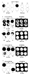
Figure 2 represents anti PrP antibody 6H4 and PIPLC preventing infection of
N2a/Bos2 cell
with scrapie prions and abrogate PrPSc accumulation in chronically infected
cells. (a)
N2aBos2 cells are incubated for 2 h with antibody 6H4 or PIPLC at the
concentrations
indicated and exposed to 0.1 % scrapie-infected brain homogenate (final
concentration) for 3
days. After culturing for 14 days (3 passages) in the absence of PIPLC or in
the continued
presence of 6H4, PrPSc expression is determined. (b,c) Chronically scrapie-
infected
N2aBos2 cells are cultured for (b) 3 days at the levels of antibody 6H4 or of
PIPLC
indicated or (c) 14 days (3 passages) at the concentrations of antibody 6H4
indicated, and
PrPSc accumulation is monitored. (d) Chronically scrapie-infected N2a/Bos2
cells are
exposed to 6H4 at the concentrations indicated for 2 weeks and further
cultured in the
absence of the antibody for 6 weeks. Cultures are split 1:5 every 3-4 days.
There is no
reappearance of PrPSc. "Cell staining" refers to staining of the membranes
with ethidium
bromide to monitor efficiency of transfer of the cell layer. IN, chronically
scrapie-infected
N2a/Bos2 cells.
Figure 3 represents chronically infected N2a/Bos2 cells "cured" of PrPSc by
antibody 6H4
treatment continuing to produce PrP and their susceptibility to reinfection.
(a) Chronically
infected- N2a/Bos2 cells, treated with antibody 6H4 for 2 weeks at the
concentrations
indicated and propagated in its absence for 66 days were not exposed (top 2
rows) or
exposed to 0.1 % RML-infected mouse brain homogenate for 3 days (bottom 2
rows). After
culturing for 14 days, PrPSc is monitored by the cell blot assay. (b) Relative
susceptibility
to prions of N2alBos2 cells {BOS2) and N2a/Bos2 cells "cured" by exposure to
antibody
6H4 at 20 pg/ml is determined by exposing cultures to various dilutions of RML-
infected
mouse brain homogenate for 3 days and determining PrPSc as above. (c) Levels
of PrPC
and PrPSc in various sublines are determined by Western blotting. Chronically
infected
N2aBos2 cells, treated for 2 weeks with antibody 6H4 at the concentrations
indicated are
passaged for 84 days after antibody withdrawal. Cells are lysed and samples
corresponding
to 2.25 x 10⁵ cells are incubated in the presence (+PK) or absence (-PK) of
proteinase K (5
p.g/ml) for 90 minutes at 37°C. Western blotting is performed as
described in Methods. L1N,
uninfected N2a/Bos2 cells and I-BOS2, chronically prion-infected BOS2 cells.
Molecular
weight markers are indicated at the left of each panel.
Figure 4 represents a model to explain abolition of PrPSc by anti PrP antibody
(or PIPLC).
28
CA 02361483 2001-11-08
PrPC is attached to the membrane by a glycosylphosphatidyl inositol anchor and
cycles
between the cell surface and an endocytic comparhnent (43). In scrapie-
infected cells, PrPC
is recruited into PrPSe "seeds" (44), which may be located at the cell surface
and/or in the
endocytic/lysosomal compartment. PrPSc is degraded with a half life of about
15h (37); if
PrPC is prevented from converting to PrPSc by either a blocking antibody or by
being
stripped from the cell surface by PIPLC, PrPSc will diminish and ultimately
disappear.
EXAMPLES
Materials and Methods
Cell culture.
Mouse neuroblastoma N2a cells are cultured in OPTI-MEM supplemented with 10 %
fetal
calf serum (FCS) and penicillin G-streptomycin (complete medium) at 37
°C in 5 % CO2.
Cells are routinely split 1:5 every 3 to 4 days and grown in 10-cm dishes
(Corning Costar,
New York, NY, USA). PIPLC was from Sigma (St.Louis, MO, USA). Antibodies used
are
of the monoclonal IgGl subtype: anti PrP (6H4, Prionics AG), anti APP (2X11,
Boehringer Mannheim, Germany) and anti (3-actin (AC-15, Sigma, St.Louis MO,
USA).
Prion infection of N2a cells.
Brains from scrapie-sick mice infected with Rocky Mountain Laboratory (RML)
mouse-
adapted prions are homogenized by passing 8 times each through 21G and 25G
needles and
adjusted to 10 % (w/v) with I x Dulbecco's phosphate-buffer saline (D-PBS;
Gibco BRL,
Glasgow, UK). After centrifuging 5 min at 1,000 rpm (Eppendorf Centrifuge
5415c,
Hamburg, Germany) and room temperature, supernatants are recovered and stored
at -80°C.
Mouse PrPSc is partially purified as described by Bolton et al. (1987) Arch.
Biochem.
Biophys. 258, 579-590. Cells (2-5 x 10° in 1 ml medium) are seeded into
24-wells plates
(Corning Costar) and cultured for 1-2 days before exposure to either 20 ng/ml
of purified
PrPSc (RML strain) or RML-infected mouse brain homogenate, diluted as
indicated with
complete medium. The inoculum is removed after 3 days and the cells are split
1:5 every 3-
4 days. After 14 days the cells are assayed for PrPSc by the cell blot
procedure (Bosque, P.
J. & Prusiner, S. B. (2000) J. Virol. 74, 4377-4386).
Selection of prion-susceptible and prion-resistant N2a sublines.
29
CA 02361483 2001-11-08
N2a cells are cotransfected with pSVneo (Southern, P. J. & Berg, P. ( 1982) J.
Mol. Appl.
Genet. 1, 327-341) that confers neomycin resistance and a plasmid (pEF-Bos-EX;
Murai et
al. (1998) Proc. Natl. Acad. Sci. USA 95, 3461-3466) containing a "half
genomic" PrP
transcription unit (Fischer et al. (1996) EMBO J. 15, 1255-1264) with the open
reading
frame of wild type PrP, MHM2 PrP (Scott et al. (1993) Cell 73, 979-988; Scott
et al. (1992)
Protein Sci. l, 986-997) or MH2M PrP (Scott et al. (1993) Cell 73, 979-988;
Scott et al.
( 1992) Protein Sci. 1, 986-997) under the control of the EF 1 a promoter. The
aim is to
generate N2a cell lines that overexpress mouse or hamster-mouse hybrid PrP
molecules and
thereby facilitate infection with mouse and/or hamster prions (Scott et al. (
1992) Protein
Sci. 1, 986-997). As a negative control, cotransformation is performed with
pSVneo and
pEF-BOS-EX (devoid of an open reading frame). After selection in 6418 at 0.7
mg/ml,
clones of each group are tested for susceptibility to infection by RML scrapie
prions, as
described above. Clone N2a/Bos2 is selected as the most susceptible subline
and aliquots
thereof and of N2alBos2 cells chronically infected with RML prions are kept at
-80°C.
Cell blot assay for PrPSc.
The assay is performed as described (Bosque, P. J. & Prusiner, S. B. (2000) J.
Virol. 74,
4377-4386). In short, cells are transferred to a PVDF membrane, treated with
proteinase K,
denatured, immunostained with antibody 6H4 followed by horseradish peroxidase-
conjugated goat anti-mouse IgGI, and visualized by enhanced chemiluminescence
(ECL
kit; Pierce, Rockford ILL, USA). After exposure, the membrane is stained for
15 min with
0.5 ~g/ml ethidium bromide and photographed in UV light, to document the
transfer of the
cell layer.
Western blot analysis.
Samples (15 pg total protein) are run through 16 % SDS-polyacrylamide gels (8
x 8 cm) and
transferred to a PVDF membrane by electroblotting at 35 V for 1 h. PrP is
detected by
incubation with 200 ng/ml monoclonal anti-PrP antibody 6H4 (Korth et al.
(1997) Nature
390, 74-77) (Prionics AG, Zurich, Switzerland) followed by horseradish
peroxidase-
conjugated goat anti-mouse IgGl (Zymed, South San Francisco, CA, USA; 10'000-
fold
dilution in TBST containing 1 % dry milk) at room temperature for 1 h. The
immune complex
is visualized by enhanced chemiluminescence (ECL kit, Pierce, Rockford, ILL,
USA).
,~~:.:.,::;:_,~.
CA 02361483 2001-11-08
EXAMPLE 1
Isolation of cell lines susceptible and resistant to RML scrapie prions.
N2a cells are transformed with PrP expression (or control) plasmids with the
intent of
raising their PrP level and thereby rendering them more susceptible to
infection with prions
from various sources. Transformed clones are assayed for their susceptibility
to RML prion
infection by an immuno-blotting assay (Bosque, P. J. & Prusiner, S. B. (2000)
.I. Virol. 74,
4377-4386) (Figure la). Unexpectedly, the most susceptible line, N2a/Bos2, was
derived
from cells that had been transformed with the control plasmid. In order to
assess the
proportion of cells susceptible to infection, N2a/Bos2 cells were subcloned
and assayed by
the blotting procedure; 49% of the cells were susceptible to infection (data
not shown).
N2aBos2 cells and a prion-resistant line, N2a/2M11, (transformed with a PrP
expression
plasmid) are challenged with mouse-scrapie-infected brain extract at various
dilutions and
passaged for 2 weeks. N2a/Bos2 cultures become PrPSc-positive after exposure
to scrapie-
infected 10 % brain homogenate diluted to 10~', while the resistant line
remains negative at
all dilutions tested (Figure 16).
The susceptible cell line N2aBos2 expresses PrP at about the same low level as
the original
N2a cells, while the resistant line expresses PrP at a level 10 or more times
higher, as shown
by Western blot analysis (Figure lc). PrP is expressed at the cell surface of
all cell lines, as
evidenced by biotinylating intact cells, immunoprecipitating the extracted
proteins with anti
PrP antibody and subjecting them to Western blot assay with horseradish
peroxidase-labelled
streptavidin (data not shown).
Thus, cell lines susceptible and resistant to RML, scrapie prions are
prepared.
EXAMPLE 2
PIPLC prevents infection of the susceptible N2a cell line by scrapie prions.
PIPLC is added to a medium containing susceptible N2a cells at various
concentrations 2 h
before and during exposure of NZa/Bos2 cells to 0.1 % scrapie-infected brain
homogenate.
After 3 days, the cells are washed and further cultured for 2 weeks, splitting
1:5 every 3-4
days. As assayed by the blotting procedure after 14 days, PIPLC at 0.25
units/ml su~ced to
prevent appearance of PrPSc (Figure 2a).
31
CA 02361483 2001-11-08
Thus, it is demonstrated that PIPLC prevents the infection of susceptible N2a
cells by scrapie
prions when the cells are contacted with PIPLC either before or during
exposure to prions.
EXAMPLE 3
Anti PrP monoclonal antibody 6H4 prevents infection of the susceptible N2a
cell line by
scrape pnons.
Antibody 6H4 is added to the medium at various concentrations 2 h before and
during
exposure of N2aBos2 cells to 0.1 % scrapie-infected brain homogenate. After 3
days, the
cells are washed and further cultured for 2 weeks, splitting 1:5 every 3-4
days. During this
period antibody 6H4 treatment is continued. As assayed by the blotting
procedure after 14
days, antibody 6H4 at 2.5 pg/ml prevents appearance of PrPSc (Figure 2a). Two
other IgGl
monoclonal antibodies, anti APP and anti (3-actin, at concentrations up to 5
~.g/ml are used as
controls (data not shown).
Accordingly, it is demonstrated that 6H4 prevents the infection of susceptible
N2a cells by
scrapie prions when the cells are contacted with 6H4 either before or during
exposure to
prions.
EXAMPLE 4 -
Rapid loss of PrPSc elicited by PIPLC.
Chronically prion-infected N2aBos2 cells are exposed to various concentrations
of PIPLC
and cultured on coverslips without passaging. After 3 days, PrPSc is barely
detectable
using PIPLC at 0.25 units/ml (Figure 2b).
Therefore, it is shown that PIPLC can cure chronically prion-infected N2aBos2
cells of
infection by scrapie prions.
EXAMPLE 5
Rapid loss of PrPSc elicited by anti PrP monoclonal antibody 6H4.
32
_. .. _ _..
CA 02361483 2001-11-08
Chronically prion-infected N2aBos2 cells are exposed to various concentrations
of
antibody 6:H4 and cultured on coverslips without passaging. After 3 days,
PrPSc is barely
detectable with 6H4 at 3 ~g/ml (Figure 2b). After culturing for 2 weeks,
splitting 1:5 every
3-4 days, as little as 1 ~g/ml 6H4 leads to disappearance of PrPSc (Figure
2c).
To determine whether the depletion of PrPSc is transient or permanent,
chronically infected
N2aBos2 cells are cultured for 2 weeks with various concentrations of antibody
6H4 and
subsequently in its absence. Samples assayed 2, 4 (data not shown) or 6 weeks
(Figure 2d)
after removal of the antibody are devoid of PrPSc, in contrast to the
chronically infected,
untreated cells. "Cured" cells are still susceptible to scrapie prion
infection (Figure 3a,6) and
express PrPC at the original level (Figure 3c).
Therefore, it is shown that 6H4 can cure chronically prion-infected N2aBos2
cells of
infection by scrapie prions.
EXAMPLE 6
Humanised anti-PrP antibodies
"Human" monoclonal antibodies are obtained by immunising "immunologically
humanised"
mice (available from Abgenix or Medarex) with PrPSc and producing the selected
antibody in
w hybridomas.
30
EXAMPLE 7
Encapsulation of hybridoma cells
Hybridoma cells are loaded into preformed capsules (polyethersulfone
microporus hollow
fibers).
EXAMPLE 8
Administration of encapsulated hybridoma cells intracerebrally or
intraperitoneally
25 alginate beads containing or not containing hybridoma cells are injected in
a final volume
of 25 p1, intracereberally into 4 mice each.
33
CA 02361483 2001-11-08
100 and 500 beads, respectively, are injected that contain anti-PrP producing
hybridoma cells
are injected intraperitoneally into 4 mice each.
Antibody levels are determined after 1 and 4 weeks.
EXAMPLE 9
Intracerebral inoculation; encapsulated hybridoma cells administered
intracerebrally or
intraperitoneally
5 or 25 beads (suspended in 20 ~1 PBS; a bead of X00 um diameter has a volume
of about
0.06 p1) Containing anti-PrP hybridoma cells or mock hybridoma cells are
injected
stereotaxically into the brain of tga20 mice (a) simultaneously with RML
inoculation (b) 35
days after inoculation. Inoculation is intracerebrally with t % RML-infected
brain
homogenate in a volume of 10 p1.
The control used is mice inoculated intracerebrally with RML,, without or
(simultaneously)
with intracerebrally or intraperitoneally administered "mock" beads.
The monoclonal antibody in serum during experiment and the survival time of
the mice is
monitored.
Histopathology, brain and spleen infectivity, Western blots of brain and
spleen and the
presence of the monoclonal antibody in brain, spleen and serum after 60 days
and at the
terminal stage or after 200 days is determined.
EXAMPLE 10
Intraperitoneal administration of encapsulated hybridoma cells
100 or 500 beads (in 100 u1 PBS) are injected intraperitoneally into tga20
mice (a)
simultaneously with intraperitoneal scrapie inoculation (b) 35 days after
inoculation.
Inoculation is with 100 p.1 1 % RML-infected brain homogenate,
intraperitoneally.
Mice inoculated with RML intraperitoneally without or (simultaneously) with
intraperitoneally administered "mock" beads are used as the control.
34
CA 02361483 2001-11-08
During the experiment, the monoclonal antibody in the serum is determined.
Additionally,
the survival time, histopathology, brain and spleen infectivity, monoclonal
antibody in brain,
spleen and serum at time of death are also determined. Two mice of each set to
be sacrificed
60 days after infection are examined.
. . -. :,~. , . ...
. ~.~.~~",
CA 02361483 2001-11-08
All publications mentioned in the specification are herein incorporated by
reference. Various
modifications and variations of the described methods and systems of the
invention will be
apparent to those skilled in the art without departing from the scope and
spirit of the invention.
Although the invention has been described in connection with specific
preferred embodiments,
it should be understood that the invention as claimed should not be unduly
limited to such
specific embodiments. Indeed, various modifications of the described modes of
carrying out
the invention which are obvious to those skilled in molecular biology or
related fields are
intended to be within the scope of the following claims.
15
36
. . . , .. a:~;~~~.
CA 02361483 2001-11-08
PRION REFERNCES
Aguzzi, A.; Brandner, S. Lancet 354: 22-25, 1999.
Aguzzi, A.; Weissmann, C. A suspicious signature. Nature 383: 666-667, 1996.
Basler, K. et al., Cell 46: 417-428, 1986.
Beyreuther, K.; Masters, C. L. Nature 370: 419-420, 1994.
Bueler, H. et al., Cell 73: 1339-1347, 1993.
Bueler, H. et al., Molec. Med. 1: 19-30, 1994.
Carlson, G. A. et al., Cell 46: 503-511, 1986.
Chapman, J. et al., Neurology 46: 758-761, 1996.
Chiesa et al. Neuron 21: 1339-1351, 1998.
Collinge, J. Hum. Mol. Genet. 6: 1699-1705, 1997.
Collinge, J. et al., Lancet 336: 7-9, 1990.
Collinge, J et al. Lancet 337: 1441-1442, 1991.
Collinge, J. et al., Nature 383: 685-690, 1996.
Collinge, J. et al., Nature 370: 295-297, 1994.
Duchen, L. W et al. Brain 116: 555-567, 1993.
Farlow, M. R. Neurology 39: 1446-1452, 1989.
Forloni, G.; et al., Nature 362: 543-546, 1993.
Gajdusek, D. C. Europ. J. Epidemiol. 7: 567-577, 1991.
Ghetti, B et al., Neurology 39: 1453-1461, 1989.
Griffith, J. S: : Self replication and scrapie. Nature 215: 1043-1044, 1967.
Hegde, R. S. et al., Science 279:827-834, 1998.
Hegde, R. S. et al.,. Nature 402: 732-736, 1999.
Horwich & Weissman, Cell 89: 499-510, 1997.
Hsiao, K. et al., Nature 338: 342-345, 1989.
Hsiao, K et al., Neurobiol. Aging 1 l: 302, 1990.
Hsiao, K et al.,. Nature Genet. 1: 68-71, 1992.
Hsiao, K. K et al., Proc. Nat. Acad. Sci. 91: 9126-9130, 1994.
Jendroska, K et al., J. Neurol. Neurosurg. Psychiat. 57: 1249-1251, 1994.
Kocisko, D. A et al., Nature 370: 471-474, 1994.
Krasemann, S et al., Molec. Brain Res. 34: 173-176, 1995.
Kuwahara, C. et al., Nature 400: 225-226, 1999.
Liao, Y.-C. J. et al., Science 233: 364-367, 1986.
Lindquist, S. Cell 89: 495-498, 1997.
Mallucci, G. R et al., Brain 122: 1823-1837, 1999.
37
~~.::.,<:
CA 02361483 2001-11-08
Manson, J. C et al., Neurodegeneration 3: 331-340, 1994.
Manuelidis, L et al., EMBO J. 6: 341-347, 1987.
Mestel, R. Science 273: 184-189, 1996.
Montrasio F et al. Science 2000;288: 1257-9.
Oesch, B et al.,. Cell 40: 735-746, 1985.
Owen, F et al., Molec. Brain Res. 13: 155-157, 1992.
Pablos-Mendez, et al., Lancet 341: 159-161, 1993.
Palmer, M. S.; Collinge, J. Hum. Mutat. 2: 168-173, 1993.
Perry, R. T, et al., Am. J. Med. Genet. 60: 12-18, 1995.
Prusiner, S. B. Annu. Rev. Med. 38: 381-398, 1987.
Prusiner, S. B. Ann. Rev. Microbiol. 48: 655-686, 1994.
Prusiner, S. B. Science 216: 136-144, 1982.
Prusiner, S. B. Cold Spring Harbor Symp. Quant. Biol. 61: 473-493, 1996.
Puckett, C et al.,. Am. J. Hum. Genet. 49: 320-329,1991.
Riek, R et al., Proc.Nat. Acad. Sci. 95: 11667-11672, 1998.
Rivera, H et al.,. J. Med. Genet. 26: 626-630, 1989.
Robakis, N.. K et al., Biochem. Biophys. Res. Commun. 140: 758-765, 1986.
Sakaguchi, S et al., Nature 380: 528-531, 1996.
Schellenberg, G. D. et al., Am. J. Hum. Genet. 49: 511-517, 1991.
Schnittger, S et al., Genomics 14: 740-744, 1992.
Scott, M et al., Cell 59: 847-857, 1989.
Shmerling, D et al., Cell 93: 203-214, 1998.
Sparkes, R. S et al., Proc. Nat. Acad. Sci. 83: 7358-7362, 1986.
Supattapone, S et al., Cell 96: 869-878, 1999.
Tagliavini, F et al., EMBO J. 10: 513-519, 1991.
Telling, G. C et al., Science 274: 2079-1962, 1996.
Ter-Avanesyan, M. D. et al., Genetics 137: 671-676, 1994.
Tobler, I et al., Nature 380: 639-642, 1996.
Wickner, R. B. Science 264: 566-569, 1994.
Windl, O et al., Hum. Genet. 105: 244-252, 1999.
38
CA 02361483 2001-11-08 " " w""''
AGENT
FIELD OF INVENTION
The present invention relates to a method. In particular, the present
invention relates to a
method of treating or preventing prion infection in a subject.
BACKGROUND TO THE INVENTION
A prion protein (PrP) is a transmissable particle devoid of nucleic acid. The
PrP gene
encodes prion proteins. The most notable prion diseases are Bovine Spongiform
Encephalopathy (BSE), Scrapie of Sheep and Creutzfeldt-Jakob Disease (CJD) of
humans.
The most common manifestation of CJD is sporadic CJD (sCJD) which occurs
spontaneously
in individuals. Iatrogenic CJD (iCJD) is a disease that results from
accidental infection.
Familial CJD (fCJD) is a form of CJD that occurs rarely in families and is
caused by
mutations of the human PrP gene. Gerstmann-Strassler-Scheinker Disease (GSS)
is an
inherited form of human prion disease and the disease occurs from an autosomal
dominant
disorder. New variant' CJD (vCJD) of humans is a distinct strain type of CJD
that is
associated with a pattern of PrP glycoforms that are different from those
found for other types
of CJD. It has been suggested that BSE may have passed from cattle resulting
in vCJD in
humans.
The "protein only" hypothesis proposes that the causative agent of
transmissible spongiform
encephalopathies, the prion, is identical with a conformational isoform of
PrPC (Prusiner, S.
B. (1989) Annu. Rev. Microbiol. 43, 345-374). PrPC is a normal host protein
(Oesch et al.
(1985) Cell 40, 735-746; Chesebro et al. (1985) Nature 315, 331-333; Basler et
al. (1986)
Cell 46, 417-428.) that occurs in most organs, but most abundantly in the
brain. It carries
up to two N-linked glycans, is anchored at the outer surface of the plasma
membrane by a
glycosylphosphatidyl inositol tail and is associated with caveolae, at least
in cultured cells
(Vey et al. (1996) Proc. Natl. Acad. Sci. USA 93, 14945-14949; Harmey et al.
(1995)
Biochem. Biophys. Res. Commun. 210, 753-759). The abnormal conformer, when
introduced into the organism, causes conversion of PrPC into a likeness of
itself called
PrPSc (Prusiner, S. B. (1989) Annu. Rev. Microbiol. 43, 345-374).
During the course of prion disease, the largely protease-resistant and
aggregated PrPSc
accumulates mainly in the brain, and may be the main or only constituent of
the prion
(Oesch et al. (I985) Cell 40, 735-746; McKinley et al. (1991) J. Virol. 65,
1340-1351).