Note: Descriptions are shown in the official language in which they were submitted.
CA 02361900 2001-11-13
C'ANNULATION DEVICE AND APPARATUS
SPECIFICATION
FIEhD OF THE INV~1TION
My present invention relates to a cannulation device
and method and, more particularly, to a device which permits the
introduction of a cannula into tissue of a patient, especially
cardiac tissue and most specifically for ventricular cannulation
as may be required for the installation of ventricular-assist
devices which augment cardiac blood circulation.
BACKGROUND OF THE INVENTION
Cardiac-assist devices can support circulation in cases
of severe heart failure. Cardiac-assist devices, also referred
to as ventricular-assist devices (VAD) draw blood from the left
ventricle and eject it into the aorta. The blood is withdrawn
through a tube or cannula introduced into the left ventricle, is
displaced by a pump and is ejected through a tube which is
inserted into the aorta.
The insertion of the tube into, for example, the
ventricle through the cardiac tissue is referred to as
ventricular cannulation and it is such introduction of the
cannula that is the concern of this invention.
It should be noted that the implantation of a
ventricular-assist device can be quite costly because the
- 1 -
CA 02361900 2001-11-13
surgical procedure also requires intensive care required over a
period of, say, twenty days. The high cost, long recovery time
and related factors reduce the utility of the procedure for many
patients. The assist device itself can be quite expensive in
addition.
In addition to the high cost, conventional techniques
involve major intervention and a traumatic procedure at lesat in
part because of the need to connect the patient to a heart/lung
machine. The latter technique is widely used but prolongs the
duration of the surgery and increases the recovery time and the
complexity of the equipment required for surgery. This major
intervention increases the mortality and morbidity.
Conventional techniques for ventricular cannulation
have involved piercing the cavity wall with a sharp tube over
which the cannula can be fitted. Alternatively, a piece of
tissue may be cut out of the ventricle with a coring knife. Both
techniques require supporting the heart muscle which is pierced
by the tool against the axial force applied against to the
ventricle wall and cannot easily be accomplished while the heart
is beating and full of blood.
The bleeding from the site can be extensive and can
prevent the surgeon from seeing the action at the cardiac muscle
wall. To assist having to handle the bleeding heart, the
cardiac/pulmonary bypass approach has been used.
- 2 -
CA 02361900 2001-11-13
OBJECTS OF T8E INVENTION
It is, therefore, the principal object of the present
invention to provide an improved device for beating-heart
cannulation, especially ventricular cannulation, that is simple
to operate, inexpensive and eliminates the need to connect the
patient to a heart/lung bypass circulation and thereby obviates
the drawbacks described.
Another object is to provide an improved cannulation
device which simplifies the surgical procedures involved,
especially in cannulation for ventricular-assist devices, reduces
complications in such surgical procedures and minimizes
morbidity, mortality and cost.
It is also an object of this invention to provide an
improved cannulation method whereby disadvantages of earlier
cannulation systems are obviated.
SDI~lARY OF THE INV~1TION
I have found that the disadvantages of earlier systems
with respect to the introduction of a cannula into tissue and
especially the ventricular cannulation can be eliminated by
eliminating the axial forces which are applied by the cannulation
device implementation to the tissue while guiding the cannula
into place, and, therefore, by spreading an incision in the
tissue purely radially for this purpose. The term "cannulation
device" when broadly used in this description and the appended
claims can include the surgical tools, therapeutic devices
- 3 -
CA 02361900 2001-11-13
(balloons), diagnostic sensors, optical devices and any equipment
for automating and/or monitoring the procedures described.
According to the invention, the procedure involves
inserting into an incision in the tissue a narrow array of
members which are then pressed outwardly without applying an
axial component of force to the tissue, thereby spreading the
incision and forming a circular opening while guiding within that
array of members a cannula into the incision. Upon retraction of
the members, therefore, the cannula is seized by the edges of the
opening or incision as they naturally elastically contract around
the cannula. The device can be anchored to the tissue by
subsequent suturing to prevent it from sliding in or out.
More particularly, the cannulation device for the
purposes described can comprise
a handle;
a plurality of pins in a circular array projecting from
an end of the handle;
an actuator on the handle operatively connected with
the pins for spreading the array from a narrow configuration in
which the pins are insertable through an incision in tissue in
which cannulation is to be effected into a wide position in which
the tissue is radially and elastically spread at the incision;
and
a cannula in the handle insertable through the array in
the wide position for anchoring in the tissue by contraction of
the tissue around the incision.
- 4 -
CA 02361900 2001-11-13
According to a feature of the invention the pins are
formed as generally linear shanks on respective wire springs,
each of the wire springs having a pivot portion parallel to the
respective shank but offset laterally therefrom and a connecting
portion between the respective shank and the respective pivot
portion, the actuator including a sleeve provided with formations
engaging the wire springs and rotating the connecting portions
about axes of the pivot portions to radially displace the shanks
between the positions.
The pivot portions are fixed on the handle whereby each
of the wire springs is twisted about the respective axis by the
respective formation, thereby torsionally stressing the
respective wire spring, the shanks returning toward the narrow
position by spring force resulting from the torsional stressing
of the wire springs upon release of the actuator.
The actuator can have a lever projecting laterally form
the handle and enabling rotation of the sleeve by a hand of a
user holding that handle. The handle itself can be hollow and
provided with a cut-out through which the cannula can be pressed
through the array of pins into the opening by a finger of the
user.
A cover can be fitted over the sleeve and the pivot
portions of the wire spring at the end of the handle and can have
an opening through which the shanks can project. The canaula, in
turn, can have a tapered end adapted to lie in a body organ and
- 5 -
CA 02361900 2001-11-13
an opposite end which can be connected to, for example, the
intake side of the ventricular assist device.
It has been found to be advantageous to provide the
formations as projections on an end of the sleeve. The array
should include at least eight pins or shanks and the sleeve can
project from an end of the handle and can have a cylindrical
extension received in the handle. The sleeve and the handle can
be connected by a rib-and-groove connection for axially fixing
the sleeve to the handle.
The cannulation method can comprise the steps of:
(a) forming an incision in tissue into which a cannula
is to be introduced;
(b) inserting into the incision a narrow array of pins
(c) spreading the pins into a wide array to thereby
radially expand the incision elastically and form an opening
without axially stressing the tissue;
(d) inserting a cannula into the opening within the
wide array of pins thereby plugging the opeaings and
(e) withdrawing the pins from the tissue and leaving
the cannula in the opening to eliminate bleeding or spilling of
visceral fluids whereby the cannula is particularly retained is
the tissue by elastic contraction of the tissue around the
cannula.
The tissue is usually a ventricle wall and the caanula
is inserted into a ventricle of the patient. The cannula is
pushed into the opening simultaneously with the widening thereof
- 6 -
CA 02361900 2001-11-13
by the pin. The cannula can thus be inserted into a beating
heart without removal of the tissue from the ventricle wall.
With the system of the invention, once the incision is
made, the only forces applied to the cavity wall are radial
forces which spread the orifice to the diameter required by the
cannula and no forces are exerted in the axial or inward
direction. During the axial movement of the cannula, it is
guided in a track formed by the shanks of the pins. The
synchronous radial opening of the orifice and inward movement of
the cannula seals the opening in the wall. The synchronous
action of spreading the orifice and inserting the cannula can
easily be accomplished by the single hand of a surgeon, but may
be synchronized by a computer-controlled system operating
respective actuators for spreading the opening and inserting the
cannula. The pins preferably are of circular or semicircular
cross section but can have other shapes as well. The shapes of
the springs can also be varied and the shanks can be rigid or
flexible.
BRI$F D$SCRIPTION OF THB DRAWING
The above and other objects, features, and advantages
will become more readily apparent from the following description,
reference being made to the accompanying drawing in which:
CA 02361900 2001-11-13
FIG. 1 is a diagram showing an initial step in the
cannulation of a ventricle in accordance with the invention;
FIG. 2 is a diagram similar to FIG. 1 showing a second
step in the process;
FIG. 3 is a diagram similar to FIGS. 1 and 2
illustrating a third step in the process;
FIG. 4 is a cross sectional view through a manual
device for cannulation of the ventricle;
FIG. 5 is a detail section of a portion of the device;
FIG. 6 is an end view thereof;
FIG. 7 is a cross sectional view taken along the line
VII-VII of FIG. 5;
FIG. 8 is a diagram illustrating manual synchroniza-
tion;
FIG. 9 is a detail perspective view showing the
apparatus in the closed position;
FIG. 10 is a view similar to FIG. 9 showing the open
position;
FIG. 11 is a perspective view of the device in the
closed position;
FIG. 12 is a perspective view of the device showing the
open position; and
FIG. 13 is a view similar to FIG. 12 but with the front
casing removed.
_ g _
CA 02361900 2001-11-13
SP$CIFIC D$SCRIPTION
According to the invention, an incision 10 is made in a
beating heart 11 through a ventricle wall 12 thereof by a scalpel
13 and without removal of the tissue of this wall.
Then an array of pins 14 is inserted into the incision
and spread outwardly to radially spread the edges of the incision
as represented by the arrows 15 simultaneously with the advance
of a tapered end 16 of a cannula 17 along the track formed by the
pins 14 into the incision. Once the cannula is lodged in the
incision, the pins are withdrawn, e.g. on a handle 18 in which
the pins may be held.
The device thus creates radial forces only on the
tissue and the tissue stretches and opens the circular track
without imposing any forces in the inward direction. No inward
compressive forces are thus exerted on the ventricle and no
distortion of the lumen can occur. Since the tissue is
elastically pressed away from the incision, radial elasticity of
the tissue holds the cannula firmly and the elastic engagement of
the pins prevents the ventricle from slipping away from the
device. The ventricle wall can then be sutured around the
cannulation for reinforcement. Cannulation is thus effected with
a minimum invasive approach and can be effected through a small
opening in the chest wall (Minimally invasive procedure).
Hleeding during cannulation is minimized because the
ventricle wall is plugged by the advancing canaula as it follows
the radial opening. The setting of the cannula from the time of
_ g _
CA 02361900 2001-11-13
insertion of the pins into the incision can take less than a
second and the skill required is minimal.
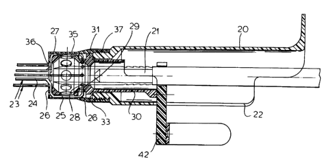
As can be seen from FIGS. 4-7, the device itself can
comprise a handle 20 which is hollow to enable the cannula 21 to
be inserted. A slot 22 in a lateral wall of the handle enables a
finger to press the cannula into place through the track formed
by the pins 23.
Each of the pins 23 is a bent wire spring having a
shank 24 parallel to a pivot portion 25 of the spring which is
connected to the shank by a transverse portion 26 and is offset
outwardly therefrom. The spring pins 23 are held within the
handle 20 at 26 and thus can be bent about the portion 25, i.e.
the spring twisted, for deflection of the shanks 24 outwardly.
To actuate the device, a sleeve 27 (see also FIG. 5) is provided
and with the holes 28 into which bent legs 29 of the spring can
be inserted. A cylindrical portion 30 is connected to the sleeve
by a portion 31 having an outwardly-extending rib 32 engageable
in a groove,33 of the handle 20.
At the front end of the sleeve 27 are projections 34
which serve to twist the individual springs as will be described
in connection with FIGS. 9-13. A casing 35 surrounds the sleeve
and is formed with an opening 36 through which pins emerge, the
casing 36 also protecting the springs and being fitted at 37 onto
an end of the housing 20.
- 10 -
CA 02361900 2001-11-13
The cylindrical portion 30 is provided with a lever 41
in FIG. 8 and 42 in FIG. 4 or 43 in FIGS. 11-13, which serves to
rotate the sleeve.
When the array of pins, which may be eight in number or
more is in its closed position, it is inserted in the incision.
The closed array has been shown at 44 in FIG. 9 and the tapered
tip 45 of the cannula is there seen directly behind the array.
When the sleeve 27 is rotated, the projections 34 twist
the pins about the axes of the offset portions 25 which are fixed
at 26 so that the array is spread (FIG. 10) simultaneously with
the advance of the cannula 21 with its tapered tip 45.
From FIGS. 11 and 12, it is clear that a casing from
cover 35 surrounds the sleeve 27 and the portions of the springs
mounted thereon while the pins 44 pass through the opening 36 in
that casing. The movement of the cannula in the lateral wall 22
is visible by comparing FIGS. 11 and 12. The simultaneous
spreading of the pins and insertion of the cannula has been
illustrated diagraamnatically in FIG. 8 and, once the cannula is
set, of course, the pins can be pulled out.
FIG. 13 shows the device with the casing removed.
Advantageously, the diameter of the array of pins in
the closed position is about 2 ran and the perimeter smaller than
the circumference of the scalpel blade which forms the incision
in the ventricle wall tissue. The incision is made through the
thickness of the cavity wall but causes no bleeding because
- 11 -
CA 02361900 2001-11-13
residual stresses in the ventricle wall tend to seal the puncture
site.
The rotation of the handle, by flexion of the second
finger (see FIG. 8) which rotates the sleeve about its axis
through about 15°, spreads the array which should be composed of
at least eight pins to a maximum radial opening which can be,
say, 25 man. Simultaneously with the widening of the track
defined by the array of pins, the cannula is pushed forward by
the thumb of the operator and the synchronization is effected by
coordinating the action of the two fingers.
The cannulation method and device of the invention have
a number of advantages over prior cardiac cannulation methods
including elimination of the need for circulatory bypass or the
use of a cardiopulmonary apparatus and the lack of need to empty
the blood from the heart.
There is no need to arrest the heart and the method and
apparatus work with a beating heart. The system operates with a
relatively small chest opening and hence a minimum invasive
approach since there is no need to insert both hands into the
thorax to grab the heart.
The system is capable of rapid insertion of the
cannula, normally taking less than 15 seconds, not including the
thoracotomy and suturing.
The insertion of the cannula without axial force is
along a well-defined track radially opened through the tissue and
the cannula is anchored by the radial forces of contraction of
- 12 -
CA 02361900 2001-11-13
the tissue to form a tight anchor. There is no need to seize the
tissue during cannulation.
Axial forces that may push the tissue inward and
distort the geometry of the cavity are precluded and thus there
are no changes in the cardiac hemodynamics during and after
cannulation.
No ventricle mass is removed and since the tissue
retains its integrity, it readily seals by contraction so that
there is better recovery and tissue repair once the cannula is
pulled out.
- 13 -