Note: Descriptions are shown in the official language in which they were submitted.
CA 02362206 2001-11-14
PROCESS FOR ACQUIRING
SCANNED IMAGE DATA RELATING
TO AN EXTERNAL BODY PORTION
AND/OR A PRODUCT APPLIED THERETO
BACKGROUND OF THE INVENTION
Field of the Invention
The present invention relates to a process for acquiring scanned image
data relating to an external body portion and/or a product applied thereto,
wherein the process involves the use of a transfer member. Such a process
could include sending of the scanned information from a first location to a
second location remote from the first location. Another aspect of the present
invention relates a process for providing a treatment recommendation for the
external body portion and/or monitoring of the external body portion. An
additional aspect relates to evaluating a product.
Description of Related Art
Accurate diagnosis of cosmetic and dermatological related conditions
often requires consultations with professionals having the requisite level of
proper skill and training. In the past, such consultations required the
individual seeking advice to travel to a site and conduct a personal meeting
with a professional who would visualize the individual's skin condition, for
example, sometimes with special instruments, and prescribe a corrective
treatment plan involving one or more cosmetic and/or dermatological
products.
Recently, advances in technology have led to a number of attempts at
obviating the need for some of these personal, face-to-face meetings
requiring travel. In particular, some consultants provide remote cosmetic or
dermatological consultations where the individual in need of the consultation
can be located at a geographic location different from that of the skilled
cosmetician or dermatologist. These attempts have been primarily limited
because there has been no easy way of sending all of the necessary
information to the remote professional.
GB 2 288 511 discloses a method and apparatus for use in diagnosing
medical conditions, such as skin conditions, where there are visual
CA 02362206 2001-11-14
2
symptoms. This reference discloses operating either a video camera, a
camcorder, or a digital still camera to generate an electrical signal that is
digitized, compressed, and transmitted to an expert consultant, such as a
dermatologist. This technique, however, suffers from a number of drawbacks
and limitations. In particular, it is time consuming to operate the video
camera, run the software necessary to digitize and compress the image
captured by the camera, and then send the compressed image to the
consultant. An even more significant limitation relates to the fact that there
is
no easy way to standardize the image. For example, incorrect lighting,
inferior equipment, non-uniform cameras and software programs, incorrect
camera operation, or other variables may produce a video image that does
not clearly show all of the details of the original area that was recorded. In
some cases, the video image submitted to the remote professional might be
either completely unusable or result in an incorrect evaluation being made.
Another more simplistic approach involves a consumer filling out a
preestablished questionnaire and then sending the completed questionnaire
(via a delivery service or the Internet) to a cosmetic product distributor
that
suggests one or more cosmetic products after reviewing the information
provided on the competed questionnaire. Such a process can provide
general information useful in advising about some types of basic cosmetic
products, but such information is limited by the level of detail in the
description provided by the individual and is, therefore, inadequate for many
treatments, especially those that are highly specialized and advanced. In
addition, both the completion of the questionnaire by the consumer and the
evaluation of completed questionnaire by the cosmetic distributor can be time
consuming.
In addition to the limitations associated with current means of remote
diagnosis, there are also drawbacks associated with some diagnostic
methods used during face-to-face consultations with a professional. For
example, to examine certain types of skin conditions, some dermatologists
use very specialized photographic equipment to obtain a photograph of a skin
region being examined. In using one such professional photographic system,
CA 02362206 2001-11-14
3
called DERMAPHOT, a uniquely designed camera lens is placed in contact
with the skin and light is emitted through the lens before taking a photograph
of the skin. This technique, however, suffers from a number of drawbacks
and limitations. In particular, it is time consuming to properly set up the
system, correctly operate the camera, and request a service to develop the
film. An even more significant limitation relates to the fact that the
specialized
photographic equipment is very expensive. Further, the resolution of
photographic images obtained with such systems is not always acceptable.
In light of the foregoing, there is a need in the art for improving
processes used for collecting data relating to a person's external body
portion.
SUMMARY OF THE INVENTION
Accordingly, processes consistent with the present invention preferably
may obviate one or more of the limitations of the related art. Such processes
have particular advantages in the field of cosmetics and/or dermatology, but
may also be used in other areas.
One aspect of the invention includes a process for acquiring scanned
image data relating to an external body portion and/or a product applied to
the external body portion. The process includes placing a transfer member in
contact with an external portion of an individual to obtain a transfer image
on
the transfer member. The transfer image is scanned with an optical image
scanner to obtain scanned image data. This scanned image data is for an
image representative of at least one characteristic of the external body
portion
and/or at least one product applied to the external body portion.
The external portion could be on many different areas of the body of
the individual. For example, the external portion could include an area of the
skin of the individual, at least one strand of hair of the individual, at
least one
fingernail of the individual, at least one toe nail of the individual, and at
least
one tooth of the individual. When the external portion includes the skin of
the
individual, the external portion may be located on the hand, foot, arm, leg,
torso, and/or face (i.e., lips) of the individual. When the external portion
CA 02362206 2001-11-14
4
includes at least one strand of hair, the strand may be from the scalp, the
eyelashes or the eyebrows.
In one possible practice of the process, the transfer image on the
transfer member indicates a condition of the external portion. There are
many different types of transfer members that could be used. The transfer
member could even be part of the image scanner itself. For example, the
transfer member could be a window of the scanner that defines the scanner's
scanning region.
In one example of the process, the transfer member includes adhesive
material provided on a backing, the adhesive material of the transfer member
being placed in contact with skin (or another external body portion) and the
transfer member being removed from the skin to transfer cells from the skin of
the individual to the transfer member. In this example, the amount of cells
transferred to the transfer member could be analyzed, based on the scanned
data, to diagnose the condition of the dryness of the skin. The adhesive
material of the transfer member may be placed in contact with adhesive
material of a second transfer member and the transfer members may then be
separated to transfer a portion of the skin cells to the second transfer
member.
In one other example of the process, the transfer member is placed in
contact with an external body portion having a product, such as a cosmetic
product, applied thereto, and the image of the scanned image data is
representative of at least one characteristic of the product. For example, the
external portion could include the lips and the product could be a lip care
product or a lip makeup product, such as lipstick.
In an exemplary process where the image of the scanned image data
is representative of a product applied to the external body portion, a
transfer
member in the form of a sheet of material could be placed in contact with lips
of the individual and a lip product could be transferred from the lips to the
sheet of material. This enables analysis of the non-retention and/or non-
transferability characteristics of a makeup product, such as lipstick on the
lips.
CA 02362206 2001-11-14
In another example, the transfer member could be placed in contact
with skin, such as facial skin, having foundation makeup applied thereto.
Such a process could be used to analyze non-retention and/or non-
transferability characteristics of the foundation makeup. For example, when
5 the transfer member is a piece of fabric or an entire article of fabric
clothing,
such as a blouse, the method could be used to evaluate whether a product
causes soiling of clothing and/or whether the product remains on the skin
during a period of time.
In a further example, where the transfer member includes a moldable
material, the moldable material is placed in contact with the skin of the
individual to produce, on the moldable material, the surface profile of the
skin.
In yet another example, where the transfer member is a hair comb or a
hair brush, the comb or brush is passed through hair, and the image on the
transfer member includes hair strands and/or skin cells.
Still another example involves the use of a transfer member configured
to change color in response to a condition of the external portion. For
example, the transfer member could be formed of litmus paper.
The above-mentioned mode of scanning using the transfer member
may be combined with other scanning modes. For example, the transfer
member scanning mode could be combined with a direct scanning mode
where an external body portion is directly scanned with the image scanner.
These modes could occur either simultaneously or one after the other.
In the direct scanning mode, the external portion of the individual is
placed in the vicinity of a scanning region of the scanner, and the external
portion is scanned with the image scanner to obtain the scanned image data.
The external portion of the individual is preferably placed into contact with
the
scanning region (i.e., the glass window pane) of the scanner. In one
embodiment, the scanner is a flat bed scanner and the external portion of the
individual is moved into contact with the scanning region. In another
embodiment, the scanner is a hand-held scanner and the scanner is moved
into contact with the external portion of the individual. Optionally, a liquid
is
placed between the external body portion and the scanning region, the liquid
CA 02362206 2001-11-14
6
altering the index of refraction to improve visualization of the
characteristic of
the external body portion and/or the product applied thereto. The direct
scanning mode may also involve placing a dye and/or a pigment on the
external portion to improve viewing of the characteristic.
In one preferred embodiment, a first computer associated with the
image scanner is located at a first location, and the process further
comprises
transferring the scanned image data from the first computer to a second
computer located at a second location remote from the first location. The
transferring may include transmitting the scanned image data via the Internet,
or shipping a data storage medium, such as a CD ROM or computer disk, to
the second location.
Other information may also be transferred to the second location. For
example, questionnaire answers relating to the condition of the external
portion and/or the product applied to the external portion may be transferred
to the second location. Billing and/or payment information could also be sent
to the second location.
One aspect of the invention relates to a process of analyzing one or
more characteristics of an external body portion andlor a product applied
thereto. This aspect preferably involves displaying an image corresponding
to the scanned image data. The displayed image is preferably viewed to
analyze the characteristics. Based on this analysis, a diagnosis of the
condition of the external portion and/or an evaluation of the product may be
determined.
The characteristics of the external portion that are analyzed are
preferably characteristics of non-dermatoglyphic body portions. As used
herein, the term "non-dermatoglyphic" relates an external area of the body
substantially free of dermatoglyphs, wherein dermatoglyphs are features that
do not change as a person ages. For example, dermatoglyphs are located on
the inferior surface of the hand in the form of fingerprints and palm lines.
Some examples of characteristics of "non-dermatoglyphic" body portions
include wrinkles, crows eyes, blood vessel networks visible through the skin,
skin pores, cosmetic materials applied to an external body portion, viewable
CA 02362206 2001-11-14
7
features of hair strands including roots, viewable features of skin including
pigmentations and groups of skin cells, viewable features of fingernails and
toe nails, and exteriorly viewable features of teeth.
There are many different characteristics of products that could be
analyzed with the process according to the invention. For example, the
process could be practiced to analyze product characteristics, such as non-
transferability, especially for lipstick and foundation makeup; product
coverage (i.e., homogeneity), especially for nail enamel or hair conditioner;
brilliancy, especially for nail enamel; coloring, especially for various types
of
makeup products; greasiness, especially for skin lotions; various interactions
between the skin and the product, especially for products designed to make
wrinkles less visible and products designed to change transparency of the
skin; and thickness or amount of the product on the external portion,
especially for hair products such as conditioners.
In another preferred practice of the invention, the process includes
sending the scanned image data to a plurality of locations so that the
characteristics) may be analyzed numerous times.
Another aspect of the process includes monitoring status of the
external portion during treatment of the external portion. For example, the
monitoring process may include repeating at least the acquiring of the
scanned image data. A recommendation for an additional treatment could be
provided based on the monitored status. In addition, the individual may be
provided with information regarding the effectiveness of the treatment.
A further aspect of the present invention relates to a process for
recommending treatment for an external body portion. The characteristics of
the external portion are analyzed and one or more treatment
recommendations for the external body portion are determined. The
treatment recommendation is provided so that the external portion of the
individual may be treated according to the recommendation. A second,
remotely located computer may at least partially determine the treatment
recommendation.
CA 02362206 2001-11-14
The recommendation could be a recommendation regarding use of a
cosmetic product and/or a dermatological product, such as a makeup
product, a care product, a hair product, a skin product, and a sun exposure
product. For example, it could be a recommendation regarding application of
the product to the external portion. Optionally, product ordering information
is
provided along with the recommendation.
The treatment recommendation may be provided to the individual
and/or a treatment provider. This information may be sent via the Internet or
any other form of communication means.
Another aspect relates to evaluating a product applied to the external
body portion. Such a process involves the analysis of one or more
characteristics of a product, such as a cosmetic product.
The process may also involve one or more databases. For example,
the process may include collecting information relating to the scanned
external portion to form a database for use in diagnoses, treatment
recommendation determinations, product evaluations, and product
formulations. In another example, the analysis of the characteristic may
include comparing an image formed from the scanned image data to at least
one image formed from image data stored in an image database.
In another aspect, the scanned image data includes data regarding
color of the transfer image. This enables the analysis at the second location
to include an evaluation of the color of the characteristic(s).
In yet another aspect, the scanner emits light on the transfer member
during scanning. Preferably, the scanner is configured in the form of a
scanner for scanning documents.
In a further aspect, a calibration member is scanned along with the
image of the transfer member. The calibration member preferably has a
predetermined size and/or a predetermined color.
In an even further aspect, the scanned image data includes data
relating to multiple scanned images or a single scanned image.
In yet another aspect, the transfer member is treated to enhance the
image on the transfer member.
CA 02362206 2001-11-14
9
In still another aspect, the transfer member and/or the external body
portion may be treated so that when the transfer member is removed from the
external body portion an increased amount of material is transferred to the
transfer member.
In yet another aspect, the external body portion and/or the transfer
member could be analyzed with various types of analysis equipment. In
addition, a trained person, such as a clinician, could conduct an analysis of
the external portion.
In an even further aspect, the process could include providing a grade
indicative of the condition of the external portion and/or the performance of
the product, and information relating to this grade could be stored in a
database.
It is to be understood that both the foregoing general description and
the following detailed description are exemplary, and are intended to provide
further explanation of the invention as claimed.
BRIEF DESCRIPTION OF THE DRAWINGS
The accompanying drawings are included to provide a further
understanding of the invention and are incorporated in and constitute a part
of
this specification. The drawings illustrate various aspects of embodiments of
the process according to the invention and, together with the description,
serve to explain the principles of the invention. In the drawings,
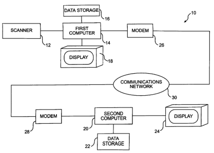
Fig. 1 is a schematic view of an example of system capable of being
used to practice the process of the present invention;
Fig. 2 is a plan view of an adhesive transfer member configured to be
used to obtain scanned image data;
Fig. 3 is a schematic view showing the adhesive transfer member of
Fig. 2 being placed in contact with facial skin of an individual;
Fig. 4 is a plan view of the adhesive transfer member of Fig. 3 showing
skin cells transferred to the transfer member after removal of the transfer
member from the skin;
Fig. 5 is a perspective view of scanned image data being obtained by
scanning the transfer member of Fig. 4 with a scanner shown in Fig. 1;
CA 02362206 2001-11-14
Fig. 6 is a view of a scanned image of skin cells transferred to a
transfer member, wherein data for the scanned image was obtained in the
manner shown in Fig. 5;
Fig. 7 is a view of a scanned image of a tissue paper transfer member
5 including a lipstick imprint of lips, wherein data for the scanned image was
obtained in a manner similar to that shown in Fig. 5;
Fig. 8 shows a schematic view of scanned image data being obtained
by directly scanning an external portion of the body with the image scanner
show in Fig. 1;
10 Fig. 9 is a view of a scanned image showing dry skin from a leg,
wherein data for the image was obtained according to the direct scanning
mode of Fig. 8;
Fig. 10a is a view of a scanned image showing pigment spots on skin,
wherein data for the image was obtained according to the direct scanning
mode of Fig. 8;
Fig. 10b is a view of a scanned image similar to that of Fig. 10a,
wherein contact oil has been placed on the spot prior to scanning;
Fig. 11 is a view of a scanned image of skin including pigmentation
and micro cuts, wherein data for the scanned image was obtained in a
manner similar to that shown in Fig. 8;
Fig. 12 is a view similar to that of Fig. 11 showing skin from another
external portion of the body, wherein data for the scanned image was
obtained in a manner similar to that shown in Fig. 8;
Fig. 13 is a view of a scanned image showing dry skin, wherein data
for the scanned image was obtained in a manner similar to that shown in Fig.
8;
Fig. 14 is a view of a scanned image of skin of the cheek showing
small micro vessels and facial hair, wherein data for the scanned image was
obtained in a manner similar to that shown in Fig. 8;
Fig. 15 is a view of a scanned image of root portions of two strands of
hair, wherein data for the image was obtained according to the direct
scanning mode of Fig. 8;
CA 02362206 2001-11-14
11
Fig. 16 is view of a scanned image of an entire strand of hair, wherein
data for the image was obtained according to the direct scanning mode of
Fig. 8;
Fig. 17 is a view of a scanned image of a nail clipping of a fingernail,
wherein data for the image was obtained according to the direct scanning
mode of Fig. 8;
Fig. 19 is a view of a scanned image of a nail showing bed capillaries,
wherein a liquid is used to modify the index of refraction and wherein data
for
the image was obtained according to the direct scanning mode of Fig. 8;
Fig. 18 is a view of a scanned image of a finger tip showing a
fingernail, wherein data for the image was obtained according to the direct
scanning mode of Fig. 8; and
Fig. 20 is a view of a scanned image of top, front teeth, wherein data
for the scanned image was obtained in a manner similar to that shown in Fig.
8.
DETAILED DESCRIPTION OF EMBODIMENTS
Reference will now be made in detail to embodiments of the invention,
examples of which are illustrated in the accompanying drawings. Wherever
possible, the same reference symbols are used in the drawings and the
description to refer to the same or like parts.
Fig. 1 shows an example of a system 10 that could be used to practice
a process according to the present invention. The system 10 includes an
optical image scanner 12, a first computer 14 associated (via any type of
communication link, including a phone line) with the scanner 12, a data
storage 16 for the first computer 14, and a visual display screen 18 for the
first computer 14. The system 10 also includes a second computer 20 linked
to a data storage 22 and a visual display screen 24. Preferably, the optical
image scanner 12 and first computer 14 are provided at a first location remote
from a second location where the second computer 20 is located. Respective
modems 26 and 28 are provided to link communication between the
computers 14 and 20 via a communication network 30, such as the Internet.
CA 02362206 2001-11-14
12
The first and second computers 14 and 20 could be configured in
many different ways. In one implementation, the computers 14 and 20 are
conventional personal computers typically found in home or office
environments. Many other types of devices, including those that are hand-
held, may also be used as long as they are capable of processing scanned
image data generated by an image scanner.
One of the initial stages of the process according to the present
invention involves obtaining scanned image data with the optical image
scanner 12. Preferably, the optical image scanner 12 is a conventional,
optical, image scanner typically used to scan documents and/or photographs
in a home or office environment. Many different types of commercially
available image scanners could be used in the practice of the present
invention. For example, the scanner could be a flat bed scanner, a hand-held
scanner, a slide scanner, or even a combined scanner and facsimile device.
Preferred scanners have a resolution high enough to produce a 2-
dimensional scanned image showing viewable details that are normally taken
into account during analysis of the condition of an external portion of an
individual. For example, the image scanner 12 could have a resolution of up
to about 4800 dots per inch (dpi).
Scanners for use in the process of the present invention preferably
emit light on an object being scanned. The object being scanned may absorb
part of this light, reflect part of it, and/or permit passage of part of it
through
the object. The scanner preferably detects the reflected portion of light. The
emitting of light during scanning enables the scanned image to be relatively
standardized and relatively unaffected by ambient light conditions because
preferably all, or a substantial portion, of the light detected by the scanner
originates from the scanner.
Preferably, the scanner 12 includes one or more light-emitting
scanning elements that are moved relative to the object being scanned.
Alternatively, the scanner 12 could be configured such that the object being
scanned is moved relative to the light-emitting scanning elements. Rather
than providing an instantaneous scan of an entire object being scanned, the
CA 02362206 2001-11-14
13
scanner 12 is preferably configured to sequentially scan different portions of
an object in either a block-by-block, line-by-line, or point-by-point manner,
for
example.
The preferred scanner may have a relatively short depth of field for its
scanning (i.e., the scanner and the object being scanned are preferably
located at a close, predetermined distance to one another during scanning).
In one preferred embodiment, the object being scanned is placed in contact
with a support during scanning. For example, the support could be part of the
scanner, such as a window defining a scanning region, or the support could
be separate from the scanner.
The preferred scanner is also preferably a color scanner configured to
produce scanned image data including color data. A color scanner is
preferred because it enables a skin diagnosis, for example, that takes into
account color: One possible scanner, used to produce the scanned images
shown in the drawings, is an EPSON Perfection, model 1200 Photo scanner
having a maximum resolution of 1200 dpi. Another type of possible scanner
is a QUBYX Lynx A3 scanner having a resolution of between 2400 and 4800
dpi.
In accordance with the present invention, scanned image data is
acquired by placing a transfer member in contact with an external portion of
an individual to provide an image on the transfer member, and then scanning
the image of the transfer member with the scanner to obtain the scanned
image data. There are many different types of transfer members that could
be used. For example, the transfer member could include either adhesive
material provided on a backing, a sheet of absorbent material, a piece of
fabric, an article of fabric clothing (i.e., a blouse), a piece of moldable
material, a hair brush or comb, or even a portion of the scanner 12, such as a
window defining a scanning region 32, as shown in Fig. 5.
Fig. 2 shows an example of a transfer member 34 including adhesive
material provided on a backing. In one preferred embodiment of the
invention, the transfer member 34 is a commercially available product called
SEBUTAPE. Many other types of alternative configurations are also possible.
CA 02362206 2001-11-14
14
For example, this type of transfer member could simply be a piece of
relatively transparent plastic tape, such as SCOTCH tape manufactured by
3M. As shown in Fig. 3, the adhesive material of the transfer member 34 is
placed in contact with skin (i.e., of the face) and, as shown in Fig. 4, when
the
transfer member is removed from the skin, skin cells and possibly also sebum
are transferred from the skin of the individual to the transfer member. As
shown in Fig. 5, the transfer member 34 is then scanned with the scanner 12
(for example, by placing it in contact with a glass window pane defining the
scanning region 32) to obtain a scanned image showing the transferred skin
cells and/or sebum. An example of this type of scanned image is shown in
Fig. 6, wherein open areas between aggregates of skin cells show cohesion
between the skin cells, separation of skin cells, and valleys in the skin.
With
such an arrangement, the amount of cells transferred to the transfer member
could be analyzed to diagnose the condition of the dryness of the skin. In
addition, this could be used to diagnose desquamation.
When the removal of the transfer member 34 from the skin results in a
significant amount of skin cells and/or sebum being transferred to the
transfer
member 34, the amount may be reduced by placing the adhesive side of the
transfer member 34 in contact with the adhesive of another transfer member
and then separating the two transfer members to transfer amounts of the skin
cells and/or sebum to both transfer members. Such a procedure could be
used in order to diagnose the size of individual cells where an overabundance
of cells on the transfer member make the analysis difficult.
In an example of the process, a transfer member may placed in
contact with an external body portion having a product, such as a cosmetic
product, applied thereto, so that a transfer image relating to one or more
characteristics of the product is created on the transfer member. For
example, the external portion could include the lips and the product could be
a lip care product or a lip makeup product, such as lipstick. One possible
type of transfer member is a sheet of absorbent material and this sheet could
be in the form of a paper sheet, such as a facial tissue, toilet tissue, or
paper
towel. The sheet of material could be placed in contact with lips of an
CA 02362206 2001-11-14
individual to transfer a lip product, such as lipstick, from the lips to the
sheet
of material. Fig. 7 shows an example of a scanned image of tissue paper
including an imprint of lips formed, for example, from lipstick. This type of
scanned image could be used to diagnose the non-retention and/or non-
5 transferability characteristics of lipstick over time. In other words, the
process
could be used to determine the ability of the lipstick to remain on the lips
as a
function of time and/or as a function of the number of events when the lips
come in contact with other things, such as by kissing: Additionally, such a
process could be used to determine coverage of the product on the external
10 body portion.
In another example, a transfer member in the form of a piece of fabric
or an article of fabric clothing (i.e., a blouse) could be placed in contact
with
skin, such as facial skin, having foundation makeup applied thereto. The
amount of any foundation makeup transferred to the transfer member could
15 then be scanned with the scanner 12. Such a process could be used to
analyze non-retention and/or non-transferability characteristics of the
foundation makeup. In particular, the method could be used to evaluate
whether a product causes soiling of clothing and/or whether the product
remains on the skin during a period of time.
In a further example, the transfer member includes a moldable
material, such as modeling clay or a malleable paste. The moldable material
could be pressed against the surface of the skin to produce the surface
profile
of the skin on the moldable material. The moldable material could then be
scanned to produce a scanned image. Such a scanned image could be used
for the analysis of micro-reliefs in the skin.
In still another example, the transfer member could be the window of
the scanning region 32. In such an arrangement, a visible image would be
created on the window after contact of an external body portion with the
window, and removal of the body portion prior to scanning. For instance, a lip
imprint like that of Fig. 7 could be placed on the window, for example with
lipstick. This could be used in the analysis of the non-transferability of a
lip
product.
CA 02362206 2001-11-14
16
The process of the present invention could be practiced to determine
both the coverage and non-transferability of a product applied to the external
body portion. For example, after applying a cosmetic product to a skin
portion, the skin portion could be placed in contact with the scanning region
32 during scanning to obtain image data for an image representing coverage
(i.e., homogeneity) of the product on the skin portion. After removing the
skin
portion from the scanning region 32, any of the product transferred from the
skin portion to the scanning region 32 (which is also the transfer member in
this example) could then be scanned to obtain scanned image data for an
image relating to the non-transferability of the product.
A hair brush or a hair comb could also provide a transfer member.
With this type of an arrangement, the brush or comb would be passed
through the hair to collect hair strands and/or skin cells and then the brush
or
comb would be scanned in a manner like that of Fig. 5. This could be used to
diagnose the extent of hair loss or dandruff, for example.
In an alternative process according to the invention, the transfer
member may be configured to change color when the transfer member is
placed in contact with the external body portion and the color change may
provide an indication of the condition of the external body portion. For
example, the transfer member could be configured in the form of litmus paper
capable of measuring PH of the skin by changing color.
Optionally, the transfer member and/or the external body portion could
be treated before the transfer member is placed on the external body portion.
Such treatment might enhance gathering of material on the transfer member
and/or viewing of features on the transfer member.
In addition to using the transfer member to acquire scanned image
data, scanned image data may also be acquired in other scanning modes.
Fig. 8 shows an example of the direct scanning mode. In the direct mode, the
external portion of an individual (i.e., the arm shown in Fig. 8) is placed in
the
vicinity of a scanning region 32 of the scanner, and the external portion is
scanned with the image scanner 12 to obtain scanned image data. In the
example shown in Fig. 8, the scanner 12 includes a scanning region 32
CA 02362206 2001-11-14
17
configured in the form of a glass window pane that makes contact with an
object being scanned, the external portion of the individual is preferably
placed into contact with this scanning region 32 during the scanning.
Preferably, the scanner shown in Figs. 8 is a flat bed scanner, and the
external portion of the individual is moved into contact with the glass window
pane of the scanner 12. If, on the other hand, the scanner is a hand-held
scanner (not shown), the scanner can be moved to place its scanning region
into contact with the external portion of the individual.
The direct scanning mode and the scanning mode using the transfer
member could be combined in a number of different ways to obtain scanned
image data relating to an exterior portion of a body. For example, both a
transfer member and an external body portion could be placed in contact with
the scanning region 32 and then scanned substantially simultaneously.
Alternatively, scanning in the different modes may occur one after the other
so that scanned image data from both of these modes may be used.
In one example of a process combining multiple modes and analysis of
both external portion characteristic and product characteristics, skin of the
cheek could be placed in contact with the scanning region 32 during scanning
to obtain image data relating to an image representative of normal
transparency of the skin. Then, a hydrating cream that improves skin
transparency could be applied to the cheek and cheek could be again
scanned while in direct contact with the scanning region 32 to obtain scanned
image data relating to the improved visibility provided by the cream. After
the
cheek is finally removed from the scanning region 32, any cream transferred
to the scanning region 32 (i.e., the scanner window provides the transfer
member) could then be scanned to obtain scanned image data relating to
non-transferability of the cream.
The direct scanning mode could be used for the diagnosis of a skin
condition. For example, when diagnosing a skin condition, such as dry skin,
the skin of an individual's face, arm, leg, hand, foot, or torso could be
brought
in the vicinity of (i.e., placed near or against) the scanning region 32 of
the
scanner 12 during scanning. Fig. 9 shows an example of a scanned image
CA 02362206 2001-11-14
18
showing dry skin from a leg, wherein the image was scanned while the
scanning region 32 was in contact with the skin. The direct mode scanning
might also be used in the diagnosis of many other skin conditions, such as
psoriasis, vitiligo, or melanoma, for example
Scanning in the direct mode could also be used to diagnose certain
pigmented areas on the skin and/or blood vessels, such as micro vessels,
visible through the skin. Fig. 10a shows an example of a direct mode
scanned image showing a skin region containing pigment spots P and a
visible micro vessel MV. Fig. 10b is an example of a scanned image showing
the skin region of Fig. 8a wherein a liquid (i.e., contact oil) has been
placed
on the spot prior to scanning in order to alter the index of refraction and
thereby improve viewing of the skin characteristics, such as the pigment spots
P and the micro vessel MV. This aspect of the process may be used to
diagnose the condition of blood vessels visible through the skin and to detect
acrosyndromes or couperosis, for example.
To further enhance viewing, a dye and/or pigment (i.e., a fluorescent
pigment) could be placed on the skin prior to the scanning.
Fig. 11 is an example of another direct mode scanned image showing
a skin region including pigment spots P and micro-cuts MC caused, for
example, by shaving. Fig. 12 is another example of a direct mode scanned
image showing a skin region similar to that of Fig. 11 and also including
wrinkles W.
Fig. 13 is an example of a direct mode scanned image showing a skin
region having cracks indicating a significant number of dry and/or dead skin
cells. Fig. 14 shows another example of a direct mode scanned image of a
skin region from an area such as the cheek, wherein the skin region includes
micro vessels MV an a number of facial hair strands H, some of which have
been shaved shorter than others.
In addition to being used in analysis of skin, the direct mode could also
be used to scan the image of a strand of hair for use in the diagnosis of
certain hair conditions, such as determining the thickness or length of a
strand of hair or the status of a hair root. For example, the strand could be
CA 02362206 2001-11-14
19
either a strand of hair from the scalp of the individual, an eyelash of the
individual, or an eyebrow hair of the individual. Fig. 15 shows an example of
a scanned image of the root portions of two separate strands of hair. Fig. 16
shows an example of a scanned image of an entire strand of hair. Each of
the images of Figs. 15 and 16 was scanned while the hair strand was placed
against the scanning region 32.
The hair strands shown in Figs. 15 and 16 could be obtained in a
variety of different ways. For example, the hair strands could be pulled from
the skin of the individual, removed during brushing or combining, or collected
from clothing or a drain of a shower or bath.
The direct scanning mode could also be used to scan an image of a
fingernail or a toenail for use in diagnosis relating to pathology, ungual
state,
onychomycosis, or split nails, for example. Fig. 17 shows an example of a
scanned image of a nail clipping of the fingernail, wherein the image was
obtained by scanning when the nail clipping was in contact with the scanning
region 32. A scanned image like that of Fig. 17 may be used in the diagnosis
of nail delamination. Fig. 18 shows an example of a scanned image showing
a fingernail and cuticle, wherein the image was obtained by scanning when
the finger tip was in contact with the scanning region of the scanner. Fig. 19
shows a scanned image similar to Fig. 18, wherein a liquid (i.e., oil) was
placed on the finger prior to scanning to improve visualization of capillary
loops CL near the cuticle of the finger. Such an image could be used for the
diagnosis of acrosyndromes, such as Raynaud's syndrome.
The direct mode scanning could also be used to scan other exterior
portions of the body. For example, Fig. 20 shows an example of a scanned
image of top front teeth. Such an arrangement could be used to diagnose a
number of different conditions of the teeth.
Optionally, a calibration member may be scanned along with the
transfer member and/or external body portion. For example, the calibration
member could have a predetermined size and or color that would enable
calibration of an image formed from the scanned image data (for example, via
CA 02362206 2001-11-14
image processing software such as Photoshop) to provide a more exact
indication of the size and/or color of characteristics.
When the scanned image data has been obtained, the process
according to the present invention could further include analyzing one or more
5 characteristics of the external body portion and/or the product applied
thereto,
and determining a diagnosis of one or more conditions of the external portion
and/or one or more features of the product. There are many different ways in
which this analysis and determination may take place. For example, the
analysis could include a person viewing an image (displayed on the first or
10 second display screen 18, 24 for example) formed from the scanned image
data obtained with the image scanner 12, and making a determination of a
diagnosis based on this viewing. Alternatively, a computer program running
on the first or second computer 14, 20 could perform at least a portion of
the.
analysis and diagnosis. The person and/or computer performing the analysis
15 and/or diagnosis could provide a grade indicating the condition of the
external
portion and/or product performance, and this grade could be stored in one of
the data storages16, 22.
The analysis according to the present invention could combine both an
analysis of one or more images formed from the scanned image data and any
20 other type of analysis for external body portions and/or cosmetic products.
For example, the other analysis could be an analysis using conventional
analysis equipment used for analyzing external body portions and/or cosmetic
products applied to external body portions. In particular, the process could
include usage of equipment typically used by specialists during examinations.
For example, the process of the present invention could include the use of
corneometer, a dermal torque meter, an image analyzer, a sebumeter, a PH
meter, or a device for measuring hydration of the skin. The other analysis
could also be an analysis performed by a trained person, such as a clinician,
directly viewing the external portion, for example, at a location where
products
are sold. The additional analysis could be used to confirm the results of the
analysis via the scanned image data. During the course of this analysis, a
grade representative of the condition of the external portion and/or
CA 02362206 2001-11-14
21
performance of the product could be provided. This grade could then be
stored, for example, in one of the data storages 16, 22.
In one preferred practice of the present invention, the scanned image
data is transferred from the first computer 14 to the second computer 20 via
the communications network 30. Alternatively, the data could be stored on a
data storage medium, such as a computer disk, CD, or other information
storage means, and this data storage medium could be shipped to the
location of the second computer 20. In addition, the scanned image data
could be stored in the first and/or second data storages 16, 22.
Optionally, the scanned image data could be transferred to the second
computer 20 along with written information, such as answers to a brief
questionnaire regarding the condition of the exterior body portion and/or any
product applied to the external portion. These questionnaire answers may
then be considered in conducting the analysis and diagnosis. In addition,
billing information and/or payment information may also be sent along with
the scanned image data.
Preferably, the second computer 20 is located at a diagnosis area
where an image can be created from the scanned image data transferred
from the first computer 14. Optionally, this image could be displayed at the
second location on the second display screen 24. The image preferably
contains representations of one or more of the characteristics of the external
body portion and/or the product applied to the external portion. These
characteristics are analyzed at the second location to provide a diagnosis of
one or more conditions of the external portion and/or one or more features of
any product applied to the external portion. At least part of the analysis
could
involve a person viewing a displayed image at the second location. In
addition, some or at least substantially all of the analysis could be
performed
automatically by the second computer 20. For example, the image could be
analyzed at least partially by means of an image analysis software operating
on the second computer 20.
Optionally, the first computer 14 and/or the second computer 20 could
modify the scanned image data to improve the viewing of certain
CA 02362206 2001-11-14
22
characteristics of the external body portion and/or the product. For example,
image modification software, such as Photoshop, could be used to enhance
viewing of the characteristics shown in the images. Such software could be
used to digitally magnify portions of images being displayed to facilitate
analysis and diagnosis.
Preferably, the results of the diagnosis are provided to the individual
and/or a treatment provider for the individual. For example, the diagnosis
could be sent via the communications network 30.
When one or more conditions of an individual's external portion have
been diagnosed, a recommendation for treatment of the conditions) may be
determined. Preferably, this recommendation is provided to the individual
and/or a treatment provider so that the external portion of the individual may
be treated according to the recommendation.
The recommendation could be determined at least in part by a manual
process or an automated process. For example, the recommendation could
be determined by selecting, from one of the data storages 16 and 22,
treatments based on the diagnosed condition. The recommendation could be
provided to the individual and/or treatment provider by sending it via the
communications network 30. In addition, information relating to the diagnosis
could also be provided along with the recommendation.
In one aspect of the present invention, the recommendation is a
recommendation regarding use of at least one of a cosmetic product and a
dermatological product. A wide variety of products could be recommended
using the technique. For example, the recommended products may be
chosen from makeup products, care products (both therapeutic and non-
therapeutic), hair products, skin products, and sun exposure products (i.e.,
sun screen or after-sun products). The recommendation could be a
prescription for a particular product.
The treatment recommendation may include a recommendation
regarding application of a product to the external portion. Optionally,
product
ordering information may be provided along with the recommendation.
CA 02362206 2001-11-14
23
In certain circumstances, the treatment recommendation might not
involve usage of a particular product. For example, the treatment
recommendation could be advice regarding hygiene or cleaning for a body
portion.
The scanned image information transferred from the first computer 14
to the second computer 20 could also be used for monitoring the status of the
condition of the external portion during treatment. For example, skin
pigmentations could be monitored over time to determine effectiveness of a
treatment; or sizes of skin cells could be monitored over time to determine
skin cell renewal rate. Optionally, an additional recommendation for a
treatment could be provided based on the monitored status. Such a
recommendation could be a recommendation regarding application of at least
one cosmetic product and dermatological product to the external portion, and
product ordering information can be provided along with the recommendation.
In addition, the process could involve providing the individual with
information
regarding the effectiveness of the recommended treatment. The monitoring
could include repeating the obtaining of the scanned image data and the
analysis. Each monitoring could include providing a grade representative of
the condition of the external portion and/or product performance.
One more additional aspect involves collecting information relating to
the scanned external portion to form a database for use in at least one of
further diagnoses, further recommendation determinations, further product
evaluations, and/or product formulations. For example, a neural network
could be established that would add information to its database and establish
some form of artificial intelligence system. Such a database could be used
when conducting further analysis of characteristics of external body portions
and/or products. For example, an image formed from the scanned
information could be compared to an image formed from a database stored in
one of the data storages 16 and 22. The database could also be used to
evaluate different product formulations to select an appropriate formulation.
Optionally, the database could include information relating to one or more
CA 02362206 2001-11-14
24
grades representative of the condition of the external portion and/or product
performance.
Another possible practice of the present invention involves sending the
scanned image data to a plurality of different locations, for example via the
communications network 30, to permit substantially simultaneous analysis at
a plurality of different areas. For example, such a practice of the present
invention could permit a team of experts in different areas to diagnose
external body conditions and/or evaluate products, such as cosmetic
products, somewhat simultaneously.
The process of the present invention could be practiced to diagnose
many different types of conditions. For example, the process could be
practiced to diagnose skin conditions, such as elasticity, dryness,
cellulitis,
sweating, aging, wrinkles, melanoma, exfoliation, desquamation,
homogeneity of color, micro-circulation, shininess, softness, smoothness,
matitty, hydration, sebum production, cleanliness, irritation, redness,
vasomotion, vasodilation, vasoconstriction, pigmentation (including freckles),
PH, whitening, dying or coloring, insect bites, growths, lesions, wounds, post
surgical incisions, wound healing, etc., for example.
With regard to hair, the process may be practiced to diagnose dying,
curling, scales, keratin plugs, length, dryness, oiliness, dandruff, lice or
other
parasites, thickness, density, root conditions, split ends, hair loss,
staging,
etc., for example.
For fingernails or toenails, the process could be practiced to diagnose
lines, spots, thickness, skin at the base of the nail, delamination,
curvature,
brilliancy, length, psoriasis, etc., for example. In addition, diagnoses
relating
to the teeth may include color, enamel coverage, surface smoothness,
whiteness, etc., for example.
When the process involves a treatment recommendation for the
external portion, there are a variety of different treatment recommendations
that could be provided. For example, treatment recommendations for skin
conditions could include use of nourishing cream, anti-wrinkle cream,
moisturizer, or keratinous cream; applying a solution of salicylic acid; or
CA 02362206 2001-11-14
removal of dead skin cells via exfoliation, etc., for example. Possible hair
treatment recommendations may include use of special shampoos or other
products for treating hair loss, split ends, dandruff; or types of hair
trimming,
etc., for example. For nails, possible treatment recommendations include,
5 pushing of the cuticles, applying cuticle cream, softening of the cuticles,
polishing nails, use of nail varnish, application of nail care creams (i.e.,
for
treating psoriasis), etc. for example. Regarding the teeth, possible treatment
recommendations relate to brushing, flossing, and use of whiteners, tart
removers, or nicotine removers, etc., for example.
10 The process according to the present invention could preferably have a
number of different advantages. For example, the process preferably could
obtain an image with a very high resolution as long as the scanner has that
capability. Commercially available document scanners have resolutions up to
4800 dpi, for example. Such high resolutions are greater than those of
15 conventional photographs, and not obtainable with a simple direct viewing
of
an external portion through a magnifying glass.
The invention could preferably be practiced with equipment typically
available to most Internet users.
When a color scanner is used, the color image allows for very accurate
20 diagnosis of color related conditions.
The use of a scanner is advantageous because lighting can be
automatically standardized with this type of digitizer. The external portion
or
transfer member can be scanned by directly placing it in contact with the
scanning region, for example, and directly acquiring, point by point,
25 colorimetric coordinates of the image. This is not the case with pictures
(either film-based or digital) produced in a home or professional setting.
One other relatively significant advantage relates to the fact that the
images are directly transferable to a cosmetician or dermatologist
electronically, preferably without any manipulation.
Another advantage relates to the ability to create the image without
regard to the level of external lighting.
CA 02362206 2001-11-14
26
A further advantage relates to the ability to monitor the change in
pathology or effectiveness of a treatment without having to travel.
Of course, many aspects of the invention could be practiced without
necessarily accomplishing one or more of these advantages.
It will be apparent to those skilled in the art that various modifications
and variations can be made to the structure and methodology of the present
invention without departing from the scope or spirit of the invention. In view
of
the foregoing, it is intended that the present invention cover modifications
and
variations of this invention, provided they fall within the scope of the
following
claims and their equivalents.