Note: Descriptions are shown in the official language in which they were submitted.
CA 02364294 2009-09-03
1
Testing endosymbiont cellular organelles and compounds identifiable
therewith.
The invention relates to diagnosis of disease and/or determination of
functioning of cellular organisms, being of multi-cellular or unicellular
nature,
being visible by the naked eye or being a micro-organism.
A diagnostician of disease studying (mal) functioning of cellular
organisms, classically can employ a broad range of inroads into said organism
to obtain relevant information as to the various aspects of said
malfunctioning.
These inroads vary widely, examples range from detecting relative ratios of
kidney stones by studying urinary samples obtained from various patients,
from probing for the presence or absence of intestinal ulcers via endoscopy,
from scanning for detectable tumors by nuclear magnetic resonance (NMR),
from detecting diabetes by testing for insulin levels and/or glucose
concentration in blood plasma, to determining cancer proneness by
determining transcriptional levels of oncogenes, and so on.
Currently, detection of disease or malfunctioning (or vice versa, of
health and proper functioning) of higher organisms, such as animals and
plants relies very much on testing samples obtained from these organisms and
studying these samples in a laboratory. Often when a fruitful method capable
of determining, identifying or detecting (aspects of) a disease of
malfunctioning
of an organism has been found it is in general also useful in testing or
screening for compounds or methods useful for treatment of (aspects of) said
disease or malfunctioning or useful in testing or screening for compounds or
methods involved in causing (aspects of) said disease or malfunctioning; by
using the same or a similar method as used in diagnosis it is generally
possible
to generate an assessment of the usefulness of such candidate compounds or
methods in treating and/or causing the disease or malfunctioning in question.
Clearly, life science laboratories are always in the need of yet other inroads
into organisms to obtain yet more information relating to disease or
CA 02364294 2009-09-03
2a
malfunctioning and to compounds and methods related to cause and/or treatment
of disease
or malfunctioning.
The invention provides a method for determining (mal)functioning of a cellular
organism comprising determining the relative ratio of an endosymbiont cellular
organelle nucleic acid and/or gene product thereof in relation to another
nucleic acid or
gene product present in a sample obtained from said organism. In terms of the
invention, by
a relative ratio is meant the amount of said first endosymbiont cellular
organelle nucleic
acid and/or gene product thereof in relation to the amount of said second
nucleic acid
and/or gene product thereof Said relative ratio can for instance be determined
by (among
other things) dividing the amount of said first nucleic acid or gene product
thereof by the
amount of said second nucleic acid or gene product thereof, or vice versa. The
amount of
one or both compounds can also be divided by, or subtracted from, a reference
value.
By determining functioning of a cellular organism is meant herein determining
whether
said cellular organism is in its natural healthy state, or whether said
organism is
somehow affected, for instance by a disease and/or a (toxic) compound. Said
disease
and/or (toxic) compound may affect said organism to such extent that clinical
symptoms
are present. Alternatively, said disease or (toxic) compound may have an
influence upon
said organism while clinical symptoms are not (yet) manifested.
According to an embodiment of the invention, there is provided a method for
determining whether an HIV infected animal is affected by an HIV related
disease,
comprising determining the amount of a mitochondrial nucleic acid and/or gene
product thereof in a blood sample obtained from said animal, determining the
amount
of a nuclear nucleic acid and/or gene product thereof in said blood sample,
determining
the relative ratio of said mitochondrial nucleic acid and/or gene product
thereof in
relation to the amount of said nuclear nucleic acid and/or gene product
thereof, and
comparing said relative ratio to a reference value, wherein alteration of said
relative
ratio as compared to a natural ratio of said mitochondrial nucleic acid and/or
gene
product thereof in relation to the amount of said nuclear nucleic acid and/or
gene
product thereof is indicative for said animal being affected by said HIV-
related disease.
Further, according to an embodiment of the invention, there is provided a
method for determining whether an HIV infected animal is affected by an HIV-
related
disease, comprising determining the amount of a first mitochondrial nucleic
acid and/or
gene product thereof in a blood sample obtained from said animal, determining
the
amount of a second mitochondrial nucleic acid and/or gene product thereof in
said
blood sample, determining the relative ratio of said first mitochondrial
nucleic acid
CA 02364294 2009-09-03
2b
and/or gene product thereof in relation to the amount of said second
mitochondrial
nucleic acid and/or gene product thereof detectable in said sample, and
comparing said
relative ratio to a reference value, wherein alteration of said relative ratio
as compared
to a natural ratio of said first mitochondrial nucleic acid and/or gene
product thereof in
relation to said second mitochondrial nucleic acid and/or gene product thereof
is
indicative for said animal being affected by said HIV-related disease.
Additionally, an embodiment of the invention provides a method for
determining the staging of an HIV related disease, comprising determining the
amount
of a mitochondrial nucleic acid and/or gene product thereof in a blood sample
obtained
from an animal suffering from or at risk of suffering from said HIV related
disease,
determining the amount of a nuclear nucleic acid and/or gene product thereof
or the
amount of a second mitochondrial nucleic acid and/or gene product thereof in
said
blood sample, determining the relative ratio of said mitochondrial nucleic
acid and/or
gene product thereof in relation to the amount of said nuclear nucleic acid
and/or gene
product thereof, or in relation to the amount of said second mitochondrial
nucleic acid
and/or gene product thereof, and comparing said relative ratio to a reference
value,
wherein alteration of said relative ratio as compared to a natural ratio of
said first
mitochondrial nucleic acid and/or gene product thereof in relation to said
second
mitochondrial nucleic acid and/or gene product thereof is indicative for said
staging.
An alternative embodiment of the invention provides a method for determining
therapeutic activity and/or possible side-effects of a candidate compound for
treatment
of an HIV infected animal which has been provided with said candidate
compound,
comprising determining the amount of a mitochondrial nucleic acid and/or gene
product thereof in a blood sample of said animal, determining the amount of a
nuclear
nucleic acid and/or gene product thereof or the amount of a second
mitochondrial
nucleic acid and/or gene product thereof in said blood sample, determining the
relative
ratio of said mitochondrial nucleic acid and/or gene product thereof in
relation to the
amount of said nuclear nucleic acid and/or gene product thereof, or in
relation to the
amount of said second mitochondrial nucleic acid and/or gene product thereof,
and
comparing said relative ratio to a reference value, wherein alteration of said
relative
ratio after said animal is provided with said compound as compared to before
said
animal is provided with said compound is indicative for said therapeutic
activity and/or
possible side effects.
An embodiment of the invention additionally provides a method for determining
therapeutic activity and/or possible side-effects of a pharmaceutical
compound,
CA 02364294 2009-09-03
2c
comprising determining the amount of a mitochondrial nucleic acid and/or gene
product thereof in a blood sample obtained from an animal which has been
provided
with said pharmaceutical compound, determining the amount of a nuclear nucleic
acid
and/or gene product thereof or the amount of a second mitochondrial nucleic
acid
and/or gene product thereof in said blood sample, determining the relative
ratio of said
mitochondrial nucleic acid and/or gene product thereof in relation to the
amount of said
nuclear nucleic acid and/or gene product thereof, or in relation to the amount
of said
second mitochondrial nucleic acid and/or gene product thereof, and comparing
said
relative ratio to a reference value, wherein alteration of said relative ratio
after said
animal is provided with said compound as compared to before said animal is
provided
with said compound is indicative for said therapeutic activity and/or possible
side-
effects.
Additionally, an embodiment of the invention provides a method for
determining toxic activity of a candidate compound in an animal provided with
said
compound, comprising determining the amount of a mitochondrial nucleic acid
and/or
gene product thereof in a blood sample obtained from said animal, determining
the
amount of a nuclear nucleic acid and/or gene product thereof or the amount of
a second
mitochondrial nucleic acid and/or gene product thereof in said blood sample,
determining the relative ratio of said mitochondrial nucleic acid and/or gene
product
thereof in relation to the amount of said nuclear nucleic acid and/or gene
product
thereof, or in relation to the amount of said second mitochondrial nucleic
acid and/or
gene product thereof, and comparing said relative ratio to a reference value,
wherein
alteration of said relative ratio after said animal is provided with said
compound as
compared to before said animal is provided with said compound is indicative
for said
toxic activity.
Endosymbiont cellular organelles are those organelles of a eukaryotic cell
that
are thought to have been derived of prokaryotic bacteria very early on in the
evolution
of eukaryotic cells; these bacteria (as it is thought) have engaged in a
symbiosis with
early eukaryotic cells, and at present, eukaryotic cells comprising these
endosymbiont
organelles in general cannot live without them; none of the present eukaryotic
cells
would function properly without mitochondria, and most plant cells would at
least
considered to be malfunctioning when no proplastids, or organelles derived
thereof, such as
CA 02364294 2001-12-04
3
chloroplasts, etioplasts, amyloplasts, elaioplasts or chromoplasts were
present.
These organelles in general appear to be at least partially self-replicating
bodies which, although under some nuclear controls, still possess considerable
autonomy.
In particular, the invention provides a method whereby said relative
ratio of an endosymbiont cellular organelle nucleic acid and/or gene product
thereof is determined in relation to the amount of essentially nuclear nucleic
acid detectable in said sample (be it DNA or RNA), or in relation to gene
products (derivable by transcription and/or translation, such as mRNA or
(poly)peptides) of said nuclear nucleic acid, (nuclear nucleic acid herein
comprises chromosomal DNA and the RNA transcribed therefrom) for example
present in nuclear or cytoplasmatic fractions or parts of said sample. DNA or
corresponding mRNA encoding components of small nuclear ribonucleoprotein
(SNRNP), or other essentially common nucleic acid derived from chromosomal
DNA, is particularly useful to test, because of its ubiquitous presence. In
this
way, the invention provides a method for studying for example endosymbiont
cellular organelle related disease, like mitochondrial and/or proplastid
related
disease. By endosymbiont cellular organelle related disease is meant herein a
condition wherein the amount and/or at least one property of nucleic acid of
said endosymbiont cellular organelle, and/or gene product thereof, is altered
as
compared to the natural situation. For instance, expression of said nucleic
acid
may be reduced. Endosymbiont cellular organelle related disease, e.g. encoded
by defects in said organelle's DNA, manifests in many different syndromes and
is often variable in its expression (and thus in general hard to detect by
testing
for clinical parameters alone) due to heteroplasmy, whereby mutant and wild
type nucleic acid can be found in one cell, whereby its distribution can vary.
Endosymbiont cellular organelle related disease is often aggravated with
increasing age of the affected individual. Endosymbiont cellular organelle
related disease can also often be observed after treatment against other
disease with various drugs, and then contributes to various side-effects of
CA 02364294 2001-12-04
4
those drugs that one would like to avoid during treatment. Those side effects
can now be better studied by using a method as provided herein.
Furthermore, the invention provides a method whereby said relative
ratio of a first endosymbiont cellular organelle nucleic acid and/or gene
product thereof is determined in relation to the amount of a second (distinct)
endosymbiont cellular organelle nucleic acid detectable in said sample (be it
DNA or RNA), or in relation to gene products (derivable by transcription
and/or translation, such as mRNA or (poly)peptides) of said endosymbiont
cellular organelle nucleic acid. In one aspect of the invention the method
involves determining a ratio between organelle DNA, such as mtDNA, and the
corresponding transcriptionally derivable organelle RNA, in the example the
related mtRNA, or translated gene product. This way, the level of
transcription and/or translation can be determined. An alteration of the level
of transcription and/or translation, as compared to the natural level of
transcription and/or translation, is indicative for an altered functioning of
said
organelle. Said altered functioning may be malfunctioning of said organelle,
because of a disease and/or because of side-effects of a certain treatment.
Said
malfunctioning may for instance comprise a decreased level of transcription.
Alternatively, said altered functioning may be an improved functioning of said
organelle, for instance during treatment and/or curing of an endosymbiont
cellular organelle related disease.
Said malfunctioning may as well comprise an increased level of transcription.
A disease, or a treatment of a disease, may involve decrement of the amount of
endosymbiont organelle DNA. However, said decrement can at least in part be
compensated by an increase in transcription of said DNA, at least in the first
stage of said disease. This way, the amount of RNA derived from said
endosymbiont organelle DNA may not be decreased at all, or relatively less
decreased as compared to the amount of said endosymbiont organelle DNA.
Symptomatic side-effects of said disease or treatment may then not be (fully)
80 sensed yet. However, upon further decrement of the amount of said
CA 02364294 2001-12-04
endosymbiont organelle DNA, the amount of RNA derived from said DNA will
eventually also drop significantly. Side-effects can then occur.
Conventionally,
upon manifestation of side-effects, a disease is treated or a treatment is
reduced or stopped. However, in this conventional way, a patient already
5 suffers from said side-effect(s). With a method of the invention, however,
side-
effect(s) involving clinical symptoms can be predicted- For instance, an
altered
level of transcription and/or translation of an endosymbiont cellular
organelle
nucleic acid is indicative for altered functioning of a cellular organism, for
instance malfunctioning of said organism involving (future) side-effects. An
alteration of the relative ratio of endosymbiont cellular organelle DNA and/or
gene product thereof in relation to the amount of nuclear nucleic acid or gene
product thereof is also indicative for altered functioning of a cellular
organism.
In yet another aspect of the invention, the ratio between two distinct
organelle DNA's or related gene products is determined. In one aspect, a
method of the invention is provided wherein said first endosymbiont cellular
organelle nucleic acid and said second endosymbiont cellular organelle nucleic
acid are obtained from the same kind of organelle. Said organelle for instance
comprises a mitochondrion.
A method of the invention is particularly suitable for staging of a
disease. An organism can already be affected by a disease, while no or little
clinical symptoms are essentially present yet. However, although no clinical
symptoms are essentially present, the relative ratio of a first endosymbiont
cellular organelle nucleic acid and/or gene product thereof in relation to the
amount of a second nucleic acid and/or gene product thereof can already be
altered. As shown in the examples, said alteration of said relative ratio can
be
determined before clinical symptoms and/or conventional tests, like
determination of the lactate pyruvate ratio, indicate an altered functioning
of
an organism. Thus, said relative ratio is very suitable for determining the
stage of a certain disease. The invention therefore provides in one aspect a
CA 02364294 2001-12-04
6
method for determining the staging of a disease, comprising determining the
relative ratio of an endosymbiont cellular organelle nucleic acid and/or gene
product thereof in a sample obtained from an organism suffering from or at
risk of suffering from said disease.
A method of the invention for staging of a disease can be used for
diagnosis. For instance, people can be routinely tested by a method of the
invention with certain time intervals. Alternatively, people can be tested at
the moment that they have some clinical symptoms. An alteration in said
relative ratio is indicative for a certain degree of disease. The kind of said
disease need not be diagnosed by a method of the invention.
Other possible uses of the invention lay in candidate drug testing, for
beneficial activity and/or side effects of possible medicaments or
pharmaceutical compositions such as candidate anti-parasitic compounds,
antibiotic compounds, cytostatic compounds, and so on. For example, the
invention provides a method for determining therapeutic activity and/or
possible side-effects of a candidate compound, for example in determining its
usefulness for treatment of malfunctioning of a cellular organism, comprising
determining the relative ratio of an endosymbiont cellular organelle nucleic
acid and/or gene product thereof in a sample obtained from said organism,
preferably said organism or an essentially related organism, such as belonging
to the same species or genus, having been provided with said compound. If the
relative ratio of an endosymbiont cellular organelle nucleic acid, and/or gene
product thereof, of a certain organism is altered after said candidate
compound
is administered to said organism, this indicates therapeutic activity and/or
side-effects involved with said compound when administered 'to said organism.
Additionally, this also indicates therapeutic activity and/or side-effects
involved with said compound in an essentially related organism. Therefore, for
determining therapeutic activity and/or ~titie=efiecis~f -candidate compound
for treatment of malfunctioning of a cellular organism, it is not necessary to
CA 02364294 2001-12-04
7
use exactly the same organism in a method of the invention. An essentially
related organism can also be used.
In another aspect, the invention provides a method for determining
therapeutic activity and/or possible side-effects of a medicament comprising
determining the relative ratio of an endosymbiont cellular organelle nucleic
acid and/or gene product thereof in a sample obtained from an organism,
preferably said organism having been provided with said medicament.
In terms of the invention, therapeutic activity means the capability of at
least
in part treating a disease. In one embodiment of the.inventtion,=said. .....==
therapeutic activity comprises a therapeutic activity against an HIV-related
disease and/or a tumor-related disease. Said medicament may for instance
comprise a cytostaticum, optionally combined with other antiretroviral
therapy. According to the ATHENA-study in the Netherlands, forty percent of
the patients undergoing an antiretroviral therapy need to change
antiretroviral therapy because of adverse side-effects. Therefore, a method of
the invention is very much desired during such therapies, because said method
can detect side-effects before (severe) clinical symptoms are essentially
present. Said therapy can then already be stopped and/or changed before said
clinical effects are essentially present. In that case said clinical symptoms
may
not, or to a lesser extent, become present. This will prevent a lot of
suffering.
Thus, in a preferred aspect a method of the invention is provided wherein said
side-effects are not essentially manifested at the moment that said method is
performed. In terms of the invention, by'not essentially manifested' is meant
that said side-effect is not (yet), or only partly, manifested by clinical
symptoms.
In one aspect a method of the invention is provided wherein said
compound or medicament comprises a cytostaticum. Commonly used
cytostatica for instance comprise alkylating compounds, antimitotoxic
80 cytostatica, antitumor antibiotica, and topo-isomerase inhibitors. Non-
limiting
CA 02364294 2001-12-04
8
examples thereof comprise chloorambucil, cyclofosfamide, estramustine,
ifosamide, melfalanthiotepabusulfan, treosulfancarmustne,
lomustinecisplatine, carboplatine, oxaliplatinedacarbazine, procarbazine,
temozolomide vinblastine, vincristine, vindesinedocetaxel,
paclitaxeldaunorubicine, doxorubicine, epirubicine, idarubicine,
mitoxa.nthronbleomycine, dactinomycine, mitomycineirinotecan,
topotecanetoposide, teniposide amsacrine, asparaginase, cladribine,
hydroxycarbamide, pentostatine methotrexaat and/or raltitrexed.
During antiretroviral treatment, and/or treatment of tumour-related disease, a
nucleoside and/or nucleotide analogue is often used. These analogues involve a
high risk of side-effects, because they interfere with replication and/or
transcription processes in an organism. The amount of endosymbiont cellular
organelle nucleic acid is then often altered as well. Therefore, a method of
the
invention is very suitable when an organism is treated with a medicament
involving nucleoside and/or nucleotide analogues.
In one aspect the invention provides a method of the invention wherein
said compound or medicament comprises a nucleoside and/or nucleotide
analogue. Non-limiting examples of such analogues are fludarabine,
mercaptopurine, thioguanine, cytarabine, flunrneuraril, and/nr geme,Ertabine.
In yet another aspect a method of the invention is provided wherein said
compound or medicament comprises AZT, ddl, ddC, d4T, STC and/or tenofofir.
In a method of the invention, said organism or an essentially related organism
has preferably been provided with said compound or organism.
Treatment of certain diseases, like for instance an HIV-related disease,
has to be performed during a long period. A method of the invention is
particularly suitable during treatment of a disease during a long period of
time. During said long period, many side-effects can evolve, and a patient can
now be monitored regularly even though no clinical symptoms are present
(yet). Therefore, in one aspect a method of the invention is provided wherein
CA 02364294 2001-12-04
9
said medicament is used during at least 3 months, preferably during at least 6
months, more preferably during at least 12 months. In one aspect, said
medicament is used for treatment of a chronic disease. By a chronic disease is
meant herein a disease which cannot be completely cured. Once an individual
has acquired said disease, said disease is always present in said individual,
albeit the clinical symptoms may vary widely. Said symptoms may sometimes
even be unnoticed by said individual. A chronic disease for instance comprises
an HIV-related disease.
By a side effect of a compound is meant herein another effect than the
purpose of said compound. Said side-effect may be an unwanted effect. For
instance, a therapeutic compound may counteract a disease and
simultaneously reduce the metabolism of an organism- Said reductinn of paid
metabolism is then referred to as a (negative) side-effect. Alternatively, a
side-
effect of a compound may be a beneficial effect, like for instance immunity
against yet another disease.
Also use for (selective) toxin testing, of e.g. herbicides, insecticides, anti-
parasitic compounds, antibiotic compounds is provided herein. The invention
provides a method for determining toxic activity of a candidate compound, for
example in determining its usefulness for causing malfunctioning of a cellular
organism, e.g. by having a cytostatie or even cytotoxic effect, comprising
determining the relative ratio of an endosymbiont cellular organelle nucleic
acid and/or or gene product thereof in a sample obtained from an organism,
preferably said organism or related organism having been provided with said
compound.
In a preferred embodiment, selectivity is also tested, using or applying
the method as provided herein (preferably in parallel experiments) on or to a
first organism and on or to an essentially unrelated second organism, if
desired belonging to a different family or order, but preferably belonging to
at
least a different class or phylum, most preferably belonging to a different
CA 02364294 2001-12-04
kingdom of organisms. Selectivity aspects are for example tested by testing
the
compounds in (if desired only in cells of) a first target organism (such as a
bacterium or parasite) as well as of testing the host or cells thereof, being
an
essentially unrelated second organism, for example a mammal or plant, or by
5 testing of a crop plant or cells thereof as well as testing an essentially
unrelated weed plant or cells thereof with said compound, to determine for
example selective toxic or selective therapeutic effects. It is also provided
to
test normal cells derived from an individual in parallel or comparison with
aberrant cells, such as tumour cells derived from the same individual, to
detect
10 or screen for a tumour-specific or at least selective cytostatic or
cytotoxic
compound for use in therapy of said individual or others with similar or
related disease.
With a method of the invention, a relative ratio is for instance
determined by measuring the amount of said nucleic acid(s) and/or gene
product(s) present in said sample, usually after at least one processing step,
like for instance amplification of target nucleic acid. After said amounts
have
been measured, said relative ratio can be determined by dividing one amount
by another.
Minute amounts of target nucleic acid can be detected and quantified by
using enzymatic amplification. Examples of enzymatic amplification
techniques are a polymerase chain reaction (PCR)1, nucleic acid sequence-
based amplification (NASBA)2, SDA, PMA, and others. Specific amplification
of a target nucleic acid sequence can be achieved by adding two primer
sequences to a reaction. An amplified region can be detected at the end of an
amplification reaction by probes that are specific for said amplified region.
Alternatively, an amplified region can be detected during generation of said
amplified nucleic acid in said amplification reaction3. In the latter protocol
a
signal of a label attached to a probe can become detectable after said probe
has
hybridised to a complementary nucleic acid. Examples of such probes that
CA 02364294 2008-06-12
11
enable real-time homogenous detection in amplification reactions are TaqMan3
and Molecular Beacon probes4;5.
Quantification of a target nucleic acid sequence is commonly
accomplished by adding a competitor molecule, which is amplified using the
same primers and which contains sequences that allow discrimination between
competitor and target nucleic acid sequence2;6. The ratio between amplified
competitor and target nucleic acid sequence can be used to quantify said
target
nucleic acid sequence. Detection of competitor or target nucleic acid sequence
can for instance be achieved at the end of the amplification reaction by
probes
that are specific for said amplified region of competitor or target nucleic
acid
sequence or during generation of said amplified nucleic acid in the
amplification reaction. In the latter protocol a signal of a label attached to
a
probe can become detectable after said probe has hybridised to a
complementary target nucleic acid and when said target has exceeded a
threshold level; the time or cycle number to positivity. In other methods for
quantification, the time to positivity can be used for quantification without
addition of a competitor7.
A method of the invention is very suitable for, among others,
determining (mal)functioning of a cellular organism, candidate drug testing
and selective toxin testing. Many reactions have been carried out using a
method of the invention, which has proven to be a useful tool (see examples).
An even more precise result can be obtained using a method of the invention
when double spreading in the result is avoided. Generally, double spreading in
the result of a method of the invention is obtained due to varieties in
conditions in different reaction mixtures. For instance, to be able to detect
and
quantify specific nucleic acids present in a sample, an amplification step is
often necessary. However, the temperature of the reaction mixture of nucleic
acid 1 may be slightly higher than the temperature of the reaction mixture of
nucleic acid 2. This may result in a higher yield of nucleic acid 1 and,
hence, in
*Trade-mark
CA 02364294 2001-12-04
12
a higher ratio of the amount of nucleic acid 1 versus nucleic acid 2 than
would
have been obtained if the temperature of reaction mixture 1 had been exactly
the same as the temperature of reaction mixture 2. Because of said
temperature difference in said reaction mixtures, the determined ratio is not
exactly the same as the real ratio of the two nucleic acids present in the
initial
sample. Likewise, minute variations in other conditions like for instance the
amount of enzyme added can lead to variations in the determined amounts of
nucleic acids 1 and 2. Thus, the measured amounts of nucleic acids 1 and 2
may vary independently from each other. Independent variations in said
determined amounts may result in an even larger variation in the calculated
ratio of said measured amounts. This is called the double spreading in the
result. Thus, by double spreading is meant herein at least one variation in an
obtained result, due to a variety of at least one reaction condition in at
least
two reaction mixtures. For instance, also the total amount of volume may
differ slightly between two reaction mixtures.
In some particular cases, double spreading in a result may exceed the
variations of the relative ratio of an endosymbiont cellular organelle nucleic
acid and/or gene product thereof in an organism which is due to a certain
disease or treatment. For instance, inhibitors of viral polymerase are often
used for treatment of HIV. Inhibitors of viral polymerase may also affect
mitochondrial polymerase gamma. Thus, the amount of mitochondrial
polymerase gamma may be reduced during said treatment of HIV, which may
result in a decreased amount of mitochondria per cell. A decrement of :for
instance 50% of the mitochondria may result in side-effects. The ratio of
mitochondrial DNA versus nuclear DNA may be diminished by a factor 2.
However, a decrement of mitochondrial DNA by a factor 2 can in some cases
lie within the double spreading of the measurement of said ratio because of
the
mentioned variations in conditions. Therefore this biologically important
difference in amount of mitochondria may not reliably be detected because of
CA 02364294 2001-12-04
13
double spreading in the result. Thus, double spreading can in some cases
reduce the reliability of detection of biologically important differences in a
ratio of nucleic acids and/or their gene products. Therefore, one embodiment
of
the present invention provides a method for determining functioning of a
cellular organism, without double spreading in the result, comprising
determining the relative ratio of a first endosymbiont cellular organelle
nucleic
acid and/or gene product thereof in a sample obtained from said organism in
relation to the amount of a second nucleic acid and/or gene product thereof.
Said double spreading can in a preferred embodiment of the present invention
be prevented by determination of said ratio in the same assay. This means
that a processing step and/or a measurement of the amounts of at least 2
nucleic acids and/or gene products thereof is performed in the same assay. In
terms of the invention, an assay typically utilises one reaction mixture.
Preferably, all components of an assay of the invention are mixed randomly in
said assay. Said reaction mixture may be present in one reaction tube.
However, a person skilled in the art can think of more methods to
prevent double spreading in the result. He/she can for instance use a reaction
vessel which is divided in different parts by a (semi)permeable membrane. As
long as at least one reaction condition varies dependently in said different
parts, double spreading is avoided and the obtained result will be more
accurate.
In one embodiment of the current invention at least two target
sequences are amplified in one assay. Said two target sequences may be said
endosymbiont cellular organelle nucleic acid and said second nucleic acid.
Thus in one embodiment of the current invention a method of the invention is
provided, comprising amplification of said endosymbiont cellular organelle
nucleic acid and said second nucleic acid in the same assay. When at least two
target sequences are amplified in one assay, varieties in reaction conditions
in
said assay can influence the obtained amount of each sequence present in said
CA 02364294 2001-12-04
14
assay dependently. For instance, the obtained amount of each sequence
present in said assay will be influenced by the same temperature, the same
overall volume, and so on. Detection of said two target sequences can be
achieved by using two specific probes during the generation of amplified
nucleic acids during an amplification reaction. Said two probes may each have
a different label allowing discrimination between said two probes and thereby
between said two different target sequences. Quantification can be achieved by
relating the time to positivity as well as the slope of the relative
fluorescence
increase of both real time amplification reactions. Preferably, a reference
curve
is created before. The quantification of said nucleic acid can then be
performed
by comparing the obtained value(s) with said reference curve. Thus there is no
need for an internal standard like for instance a competitor molecule. A
method of relative quantification of two targets in one assay has an improved
accuracy compared to quantification in two separate assays, and requires less
handling time and reagents. We found that duplexing of two amplification
reactions in the same tube gives an immediate indication of the ratio of the
two targets. The conditions of both amplification reactions are the same,
ruling
out variations of those conditions without the necessity for internal or
external
calibrators. Hence, double spreading in the result is now avoided. Thus, in
one
aspect the invention provides a method, wherein a relative ratio is determined
directly by dividing one amount of nucleic acid by another. Preferably, said
relative ratio is determined by comparison with a reference curve. In terms of
the invention, determined directly means that an immediate indication of the
ratio of the two targets is possible, for instance by comparing the intensity
of
said two different fluorescent labels of said two specific probes. In this
embodiment, dividing one amount of nucleic acid by another is performed by
dividing the intensity of the corresponding fluorescent label by another. No
internal standards are used in a method of the invention wherein said relative
ratio is determined directly.
CA 02364294 2001-12-04
In one aspect, a method of the invention is provided wherein said
cellular organelle nucleic acid, said gene product thereof, said second
nucleic
acid and/or said gene product thereof is obtained from a peripheral blood
mononuclear cell (PBMC) and/or a fibroblast. Especially the use of PBMCs is
5 preferred because then a blood sample from said organism can be used. A
blood sample is easy to obtain and relative large amounts are often available,
Therefore, in a preferred embodiment a method of the invention is provided
wherein said sample comprises a blood sample.
10 A method of the invention is especially useful to quantify a target
nucleic acid and/or gene product thereof with a variable content in relation
to a
target nucleic acid and/or gene product thereof with a constant content. An
example is the quantification of the variable cellular content of
mitochondrial
DNA to the constant cellular content of the DNA of a nuclear gene (two per
15 diploid cell). Another example comprises the quantification of variably
expressed RNA like mitochondrial RNA to constitutively expressed RNA that
is essential for cell survival like the SNRP U1A encoding RNA involved in
splicing or other essentially common nucleic acids derived from nuclear DNA
with an ubiquitous presence. We found that it is possible to determine a
relative ratio of a factor 2 it 3.
In one aspect, the invention provides a method of the invention wherein
said first nucleic acid comprises RNA and wherein said second nucleic acid
comprises DNA. A method of the invention is for instance particularly suitable
for the quantification of the cellular content of mitochondrial RNA to the
cellular content of the DNA of a nuclear gene like U1A. This is shown in
example 22.
Furthermore, the invention provides a diagnostic kit comprising at least
one means for performing a method according to the invention, said kit
comprising at least one primer or probe set selective for the amplification
and
CA 02364294 2001-12-04
16
detection of a nucleic acid related to or derived from endosymbiont cellular
organelles and, when so desired, necessary amplification reagents, such as can
be found examplified in the detailed description herein or which are otherwise
known in the art. In particular, the invention provides a diagnostic kit
wherein said kit comprises more than one primer or probe set for the
amplification of nucleic acid sequences related to cellular organelles,
preferably supplemented with a primer or probe set for the amplification of
nucleic acid related to the chromosomes, such as a SNRP specific primer or
probe. In particular the invention provides a kit comprising at least one
primer
or probe from table 1 for the amplification of nucleic acid sequences related
to
cellular organelles. It is of course preferred that said amplification
reagents,
when provided with the kit, comprise an enzyme with reverse transcriptase
activity, such as required for PCR or NASBA amplification. Of course, a kit
comprising a means for the detection of a gene product other than nucleic
acid,
for use in a method according to the invention is herewith also provided.
The invention furthermore provides the use of a compound obtainable or
detectable by a method according to the invention in the preparation of a
medicament, a herbicide, insecticide, anti-parasiticum, cytostatic, etc, and a
medicament, herbicide, insecticide, anti-parasiticum etc. obtainable or
derivable or identifiable by a method according to the invention.
The invention is further explained in the detailed description herein,
wherein most examples are directed by way of example at testing of
mitochondriae, being central to the provision and use of energy in a cell,
however, it will easily be understood that the same principles apply to tests
using other endosymbiont organelles, such as chioroplasts, being central to
the
provision of carbohydrates to a plant cell.
CA 02364294 2001-12-04
17
Examples
Used ingredients and general methodology
In table 1 the primers and probes used in the examples are summarised.
Standard NASBA nucleic acid amplification reactions were performed in a
2041 reaction volume and contained- 40mM Tris-pH 8.5, 70mM KC1, 12mM
MgCl2, 5mM dithiotreitol, 1mM dNTP's (each), 2mM rNTP's (each), 0.,2 1M
primer (each), 0.0514M molecular beacon, 375mM sorbitol, 0.105 g/ 1 bovine
serum albumin, 6.4 units AMV RT, 32 units T7 RNA polymerase, 0.08 units
RNAse H and input nucleic acid. The complete mixture, except the enzymes,
sorbitol and/or bovine serum albumin was, prior to adding the enzyme
mixture, heated to 65 C for 2 minutes in order to denature any secondary
structure in the RNA and to allow the primers to anneal. After cooling the
mixture to 41 C the enzymes were added. The amplification took place at 41 C
for 90 min in a fluorimeter (CytoFluor 2000) and the fluorescent signal was
measured every minute (using the filter set 530/25 nm and 485/30 am). For
amplification of DNA target sequences the 65 C denaturation step was
replaced with a 95 C denaturation step for 2 to 5 minutes.
To achieve quantification, a dilution series of target sequence for a
particular
primer set was amplified and the time points at which the reactions became
positive (the time to positivity, TTY') were plotted against the input amounts
of
nucleic acid. This way a calibration curve was created that could be used to
read TTP values of reactions with unknown amounts of input and deduce the
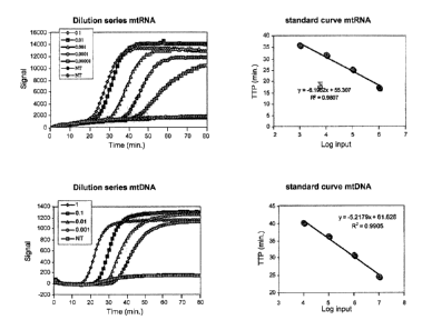
input amount. Examples of typical standard curves for quantification of RNA
and DNA are shown in figure 1.
For some of the target sequences no dilution series were available with
reliable
absolute amount of copies determined. Those series were given an arbitrary
unit as measurement instead of DNA or RNA copies, e.g. cell-equivalent or ET-
CA 02364294 2001-12-04
18
unit. As a result it sometimes seems that there is less RNA than DNA, which
is quite the opposite of what is expected.
Cells (fibroblasts and PBMC's) were cultured under standard conditions in
standard media known to persons skilled in the art with addition of drugs or
putative toxic or stimulating compounds as defined in the examples. Nucleic
acids were isolated from the cells with the method described by Boom et al
(Boom R, Sol CJ, Salimans MM, Jansen CL, Wertheim-van Dillen PM, van der
Noordaa J, 1990. Rapid and simple method for purification of nucleic acids. J
Clin Microbiol; 28(3):495-503) or with dedicated isolation kits purchased from
Qiagen (Qiagen GmbH, Max Volmer Strasse 4, 40724 Hilden, Germany) and
used according to the manufacturer's protocols. A small aliquot of the
isolated
nucleic acid was analysed on an agarose gel and the remainder stored at -80 C
until further analysis. Usually the nucleic acid was diluted 10 times with
water and of the diluted nucleic acid usually 5 gl was used as input in the
NASBA amplification reactions.
Example 1
In this example it is explained what kind of ratio's can be measured with a
method according to the invention and the meaning they can have in
diagnostic sense:
The invention for example provides determining the relative ratio of organelle
DNA to chromosomal DNA. This ratio, when compared with normal values or
determined at at least two points in time, shows the decline or increase of
organelles per cell. Also is provided determining the ratio of organelle RNA
to
chromosome encoded RNA. This ratio when compared with normal values or
determined at at least two points in time, shows the organelle transcription
activity decline or increase per cell, normalised for the active state (i.e.
transcription state) of the cell.
CA 02364294 2001-12-04
19
Determining the ratio of organelle RNA to chromosomal DNA is also provided.
This ratio when compared with normal values or determined at at least two
points in time, shows the organelle transcription activity decline or increase
per cell.
Determining the ratio of organelle DNA to organelle RNA is also provided.
This ratio, when compared with normal values or determined at at least two
points in time, shows the decline or increase of transcription in the
organelle,
indicating regulation at the transcriptional level to achieve a certain mRNA
(and therefore protein) level.
Determining the ratio of organelle DNA to chromosome encoded RNA is also
provided. This ratio, when compared with normal values or determined at at
least two points in time, shows the decline or increase of transcription in
the
cell, in relation to chromosomal RNA transcription levels, indicating the
activity state of the organelle, which is especially useful when chromosomal
RNA is determined that encodes an organelle protein or other component
thereof.
Example 2
Fibroblast cells were cultured in vitro in the presence of the anti viral
drugs
DDC, AZT and D4T at two concentrations each, 3 pM and 30 PM, respectively,
for 4 weeks. As controls cell cultures with ethidium bromide and without drugs
were also performed. Fthidium bromide is known to deplete mitochondrial
DNA completely from cells and is a positive control in terms of achieving an
effect on the mitochondria content of cells. At one week intervals part of the
cells was harvested and analyzed for amount of mitochondrial DNA (primers
MtD p1 and MtD p2 and probe MtD mb) and chromosomal DNA (primers
SnrpD p1 and SnrpD p2 and probe SnrpD mb) in the described NASBA
protocol. The cultures with AZT, D4T and without additive showed no
measurable change in mitochondrial DNA to chromosomal DNA ratio in the
CA 02364294 2001-12-04
culture period of 4 weeks. The culture with Ethidium bromide showed a
decline in mitochondria) DNA content as expected. The results for DDC are
shown in figure 2.
The data in figure 2 clearly show a decline in the amount of mitochondrial
5 DNA per cell with more than 2 logs and therewith the mitochondrial toxicity
of
the antiviral drug DDC.
Example 3
Fibroblast cells were cultured in vitro in the presence of the anti viral
drugs
10 DDC, AZT and D4T at two concentrations each, 8 pM and 30 pM, respectively,
for 4 weeks. As controls cell cultures with ethidiusn bromide and without
drugs
were also performed. Ethidium bromide is known to deplete mitochondrial
DNA completely from cells and is a positive control in terms of achieving an
effect on the mitochondria content of cells. At one week intervals part of the
15 cells was harvested and analyzed for amount of mitochondrial RNA (primers
MtR p 1 and MtR p2 and probe MtR mob) and chromosome encoded RNA
(primers SnrpR p1. and SnrpR p2 and probe SnrpR mb) in the described
NASBA protocol. The cultures with AZT, D4T and without additive showed no
measurable change in mitochondrial RNA to chromosome encoded RNA ratio
20 in the culture period of 4 weeks. The culture with Ethidium bromide showed
a
decline in mitochondrial RNA content as expected. The results for DDC are
shown in figure S. The data in figure 3 clearly show a decline in the amount
of
mitochondrial RNA per cell with at least 2 logs and therewith the
mitochondrial toxicity of the antiviral drug DDC. The time point at 3 weeks
has a very low value and presumably this is somewhat of an outlier
measurement.
CA 02364294 2001-12-04
21
Example 4
Fibroblast cells were cultured in vitro in the presence of the anti viral
drugs
DDC, AZT and D4T at two concentrations each, 3 p.M and 30 uM., respectively,
for 4 weeks. As controls cell cultures with ethidium bromide and without drugs
were also performed. Ethidium bromide is known to deplete mitochondrial
DNA completely from cells and is a positive control in terms of achieving an
effect on the mitochondria content of cells. At one week intervals part of the
cells was harvested and analyzed for amount of mitochondrial RNA (primers
MtR p1 and MtR p2 and probe MtR mb) and chromosomal DNA (primers
SnrpD p1 and SnrpD p2 and probe Snrpl) mb) in the described NASBA
protocol.
The cultures with AZT, D4T and without additive showed no measurable
change in mitochondrial RNA to chromosomal DNA ratio in the culture period
of 4 weeks. The culture with Ethidium bromide showed a decline in
mitochondrial RNA content as expected. The results for DDC are shown in
figure 4.
The data in figure 4 clearly show a decline in the amount of mitochondria)
RNA per cell with almost 3 logs and therewith the mitochondrial toxicity of
the
antiviral drug DDC. The time point at 3 weeks has a very low value and
presumably this is somewhat of a outlier measurement.
Example 5
Fibroblast cells were cultured in vitro in the presence of the anti viral
drugs
DDC, AZT and D4T at two concentrations each, 3 pM and 30 pM, respectively,
for 4 weeks. As controls cell cultures with ethidium bromide and without drugs
were also performed. Ethidium bromide is known to deplete mitochondrial
DNA completely from cells and is a positive control in terms of achieving an
effect on the mitochondria content of cells. At one week intervals part of the
cells was harvested and analyzed for amount of mitochondrial RNA (primers
CA 02364294 2001-12-04
22
MtR p 1 and MtR p2 and probe MtR rab) and mitochondrial DNA (primers MtD
p 1 and MtD p2 and probe MtD mb) in the described NASBA protocol.
The cultures with AZT, D4T and without additive showed no
measurable change in mitochondrial RNA to mitochondrial DNA ratio in the
culture period of 4 weeks. The culture with Ethidium bromide showed a
decline in mitochondrial RNA and DNA content as expected. The results for
DDC are shown in figure 5.
The data in figure 5 clearly show that the ratio of mitochondrial DNA to RNA
in not significantly changing over the period of 4 weeks. The time point at 3
weeks in figure 5 has a low value for mitochondrial RNA that shows up, this
measurement is presumably somewhat of an outlier measurement.
Example 6
Fibroblast cells were cultured in vitro in the presence of the anti viral
drugs
DDC, AZT and D4T at two concentrations each, 3 pM and 30 kuNI, respectively,
for 4 weeks. As controls cell cultures with ethidium bromide and without drugs
were also performed. Ethidium bromide is known to deplete mitochondrial
DNA completely from cells and is a positive control in terms of achieving an
effect on the mitochondria content of cells. At one-week intervals part of the
cells was harvested and analyzed for amount of chromosome encoded RNA
(primers SnrpR p1 and SnrpR p2 and probe SnrpR mb) and chromosomal DNA
(primers SnrpD p1 and SnrpD p2 and probe SnrpD nab) in the described
NASBA protocol.
The cultures with AZT, D4T, ethidium bromide and without additive showed
no measurable change in ratio in the culture period of 4 weeks. The results
for
DDC are shown in figure 6.
The data in figure 6 clearly show that the ratio of chromosomal DNA to RNA
in not significantly changing over the period of 4 weeks.
CA 02364294 2001-12-04
23
Example 7
Fibroblast cells were cultured in vitro in the presence of the anti viral drug
DDC at a concentration of 30 p.M for 4 weeks. After that period the cell
culture
continued but now in the absence of DDC. During this period of culture
without DDC part of the cells was harvested and analyzed for amount of
mitochondrial DNA (primers MtD pl and Mtf) p2 and probe 1VItD mb) and
chromosomal DNA (primers SnrpD p 1 and SnrpD p2 and probe SnrpD mb) in
the described NA-SBA protocol at two-week intervals for a period of 12 weeks.
The results of the analysis are shown in figure 7.
The results in figure 7 clearly show that the amount of mitochondria per cell
increases with more than 2 logs after DDC is removed from the culture. This
result shows that the toxic effect of DDC can be reversed if there are still
some
mitochondria left in the cells to repopulate the new growing cells.
Example 8
Fibroblast cells were cultured in vitro in the presence of the anti viral drug
DDC at a concentration of 30 pM for 4 weeks. After that period the cell
culture
continued but now in the absence of DDC. During this period of culture
without DDC part of the cells was harvested and analyzed for amount of
mitochondrial RNA (primers MtR p1 and MtR p2 and probe MtR mb) and
chromosome encoded RNA (primers SnrpR p1 and SnrpR p2 and probe SnrpR
mb) in the described NASBA protocol at two-week intervals for a period of 12
weeks. The results of the analysis are shown in figure S.
The results in figure 8 clearly show that the amount of mitochondrial RNA per
cell increases with more than 2 logs after DDC is removed from the culture.
This results shows that the toxic effect of DDC can be reversed and that the
function of the mitochondria comes back as shown by synthesis of RNA and
subsequently proteins.
CA 02364294 2001-12-04
24
Example 9
Fresh peripheral blood mononuclear cells (PBMC's) from a healthy blood donor
were cultured in vitro in the presence of the anti viral drugs DDC, AZT and
D4T at two concentrations each, 6 pM and 60 pM, respectively, for 5 days. As
controls cell cultures with DMSO and without drugs were also performed.
DMSO is part of the solvent in which the drugs are solublelized. After 5 days
the cells were harvested and analyzed for amount of mitochondrial DNA
(primers MtD p1 and MtD p2 and probe MtD mb) and chromosomal DNA
(primers SnrpD p1 and SnrpD p2 and probe SnrpD mb) in the described
NASBA protocol.
The cultures with AZT, D4T, DMSO and without additive showed no
measurable change in ratio in the culture period 5 days. The results for DDC
are shown in figure 9.
The results in figure 9 clearly show the decline in PBMC's of mitochondrial
DNA per cell of more than 1 log during the 5 day culture period.
Example 10
Fresh peripheral blood mononuclear cells (PBMC's) from a healthy blood donor
were cultured in vitro in the presence of the anti viral drugs DDC, AZT and
D4T at two concentrations each, 6 ltM and 60 pM, respectively, for 5 days. As
controls cell cultures with DMSO and without drugs were also performed.
DMSO is part of the solvent in which the drugs are solublelized. After 5 days
the cells were harvested and analyzed for amount of mitochondrial RNA
(primers MtR p1 and MtR p2 and probe MtR mb) and chromosome encoded
RNA (primers SnrpR pI and SnrpR p2 and probe SnrpR mb) in the described
NASBA protocol.
The cultures with AZT, D4T, DMSO and without additive showed no
measurable change in ratio in the culture period 5 days. The results for DDC
are shown in figure 10. Interestingly, the results in figure 10 do not clearly
CA 02364294 2001-12-04
show a decline in PBMC's of mitochondria) RNA per cell during the 5-day
culture period at the highest concentration of DDC used. This is in contrast
to
the mitochondrial DNA as shown in example 9. Probably the decline in
mitochondrial DNA is compensated by an increase in transcription,
5 maintaining the level of mitochondrial RNA. This mechanism delays the
decline of mitochondrial RNA.
Consequently, one can say that the mitochondrial RNA is a reflection of the
current status of the functionality of the mitochondria and that mitochondrial
DNA is predictive of what will happen in the (near) future with the
10 mitochondrial function and therefore has a more prognostic character.
Example 11
Using the primers and probes Rubisco-DNA pl, Rubisco-DNA p2, Rubisco-
DNA MB, Rubisco-RNA p 1, Rubisco-RNA p2 and Rubisco-RNA-MB (table 1)
15 the chloroplast DNA and RNA of Oryza satauum (rice) can be quantified and
the ratio to the chromosomal DNA and RNA can be determined by using
primers and probes OryzaDNA. p 1, OryzaDNA p2, OryzaDNA mb, OryzaRNA
p 1, OryzaRNA p2, OryzaRNA mb (table 1). During the application of herbicide
(or other) compounds the conditions of the plants can be assessed by
20 measurement of the chloroplast nucleic acid content of the cells using
amplification methods like PCR and NASBA that are known to persons skilled
in the art. At the same time, using primer sets suitable for weeds, the
deterioration of the unwanted plants can be monitored. It is clear that these
molecular tools are very suited in the research for new herbicides that
25 specifically attack one group of plants and not others.
Example 12
In this example the NASBA nucleic acid amplification reactions for DNA
target sequences were performed in a 20 l reaction volume and contained:
CA 02364294 2001-12-04
26
40mM Tris-pH 8.5, 70mM KCI, 12mM MgC12, 5mM dithiotreitol, 1mM dNTP's
(each), 2mM rNTP's (each), 0.2 M primer (each), 0.05 M molecular beacon, 1.5
units restriction enzyme Msp 1, 375mM sorbitol, 0.106 kglpl bovine serum
albumin, 6.4 units AMV RT, 32 units T7 RNA polymerase, 0.08 units R.NAse H
and input nucleic acid. The complete mixture, except the enzymes, sorbitol and
bovine serum albumin was, prior to adding the enzyme mixture, incubated at
37 C for 25 minutes and subsequently heated to 95 C for two minutes in order
to denature the DNA and to allow the primers to anneal. After cooling the
mixture to 41 C the enzyme mixture was added. The amplification took place
at 41 C for 90 min in a fluorimeter (CytoFluor 2000) and the fluorescent
signal
was measured every minute (using the filter set 530/25 nm and 485/30 nm).
To achieve quantification, a dilution series of target sequence for a
particular
primer set was amplified and the time points at which the reactions became
positive (the time to positivity, TTP) were plotted against the input amounts
of
nucleic acid. This way a calibration curve was created that could be used to
read TTP values of reactions with unknown amounts of input and deduce the
input amount. Fresh peripheral blood mononuclear cells (PBMC's) from a
healthy blood donor were cultured in vitro for 5 days. After 5 days the cells
were harvested and analyzed for amount of chromosomal DNA (primers SnrpD
p1 and SnrpD2 p2 and probe SnrpD mb) with the described NASBA protocol in
the chapter "Used ingredients and general methodology" and compared with
the NASBA protocol as described in this example. As can be clearly seen in
figure 11 the DNA NASBA reactions with pre-treatment of restriction enzyme
perform much better than without. The rationale for this observation is the
direct extension from the Msp 1 created 3' over the T7 promoter part of the p
l
primer.
CA 02364294 2001-12-04
27
Example 13
Using the primers and probes tRNA-L-D p1, tRNA-L-D p2, tRNA-L-D MB,
petB RNA pl, petB RNA p2 and petB RNA MB (table 1) the chloroplast DNA
and RNA of Oryza sativum (rice) can be quantified and the ratio to the
chromosomal DNA and RNA can be determined by using primers and probes
OryzaDNA p1, OryzaDNA p2, OryzaDNA mb, OryzaRNA p1, OryzaRNA p2,
OryzaRNA mb (table 1). During the application of herbicide (or other)
compounds the conditions of the plants can be assessed by measurement of the
chloroplast nucleic acid content of the cells using amplification methods like
PCR and NASBA that are known to persons skilled in the art. At the same
time, using primer sets suitable for weeds, the deterioration of the unwanted
plants can be monitored. It is clear that these molecular tools are very
suited
in the research for new herbicides that specifically attack one group of
plants
and not others.
Example 14
Thousand molecules of plasmid containing Snrp DNA were mixed with 4 x 10⁵,
2 x 10⁵, 105, 5 x 10⁴, 2.5 x 10⁴, or 104 molecules of plasmid containing
mitochondrial DNA, and the mixture was used as input for the reactions. A
reaction mix was prepared similar to that of example 12, except that primers
and beacons differed in order to amplify Snrp-nuclear and mitochondrial DNA
in one tube. The reaction mix (duplex-mix) contained two sets of primers and
beacon: SnrpD p1 and SnrpD p2, and MtD p1_2 and MtD p2_2 (each 0.2 4M)
with beacons SnrpD rub (ROX-labeled) and MtD mb_2 (FAM-labeled) (each
0.05 M). Restriction enzyme digestion, amplification, and detection were
performed as in example 12. Filter sets of the fluorimeter (CytoFluor 2000)
were adapted to simultaneously measure the FAM and the ROX-label (485/20
and 580/25 for FAM; 590/20 and 645/40 for ROX). In a duplex reaction with
two competing amplifications the ratio of the slope of the curves of
fluorescence
CA 02364294 2001-12-04
28
in time is proportional to the ratio of the amount of molecules of each
amplified species (see figure 12).
Example 15
PBMC were cultured in the absence and presence of 5 M ddC. After 5 days
PBMC samples were drawn. Nucleic acids were isolated from 105 PBMC
according to the method described by Boom at al. and dissolved in 50 Al DNAse
and RNAse free water. A 1::10 and 1:100 dilution was made, and 5 l of the
dilution (equivalent to 1,000 or 100 PBMC, respectively) was put in the
reaction mix to amplify the specific targets. In parallel, 103 molecules of
plasmid containing Snrp DNA was mixed with 4 x 10⁵, 2 x 10⁵, 105, or 5 x 10
molecules of plasmid containing mitochondrial DNA, and the mixture was
used as input for the reactions. A reaction mix was prepared similar to that
of
example 12, except that primers and beacons differed in order to amplify Snrp-
nuclear and mitochondrial DNA in one tube. The reaction mix (duplex-mix)
contained two sets of primers and beacon: SnrpD p 1 and SnrpD p2, and MtD
pl_2 and MtD p2_2 (each 0.2 M) with beacons SnrpD mb (ROX-labeled) and
MtD mb_2 (FAM-labeled) (each 0.05 M). Restriction enzyme digestion,
amplification, and detection were performed as in example 12. Filter sets of
the fluorimeter (CytoFluor 2000) were adapted to simultaneously measure the
FAM and the ROX-label (485/20 and 530/25 for FAM; 590/20 and 645/40 for
ROX). In a duplex reaction with two competing amplifications the ratio of the
slope of the curves of fluorescence in time is proportional to the ratio of
the
amount of molecules of each amplified species. The data of the plasmid Snrp/
mitochondrial DNA mixtures were used to create a standard curve on which
the unknown ratio of mitochondrial to Snrp nuclear DNA of the PBMC
samples in the dilutions 1:10 and 1:100 in the absence and presence of 5 M
ddC could be assessed (see figure 13).
CA 02364294 2001-12-04
29
Example 16
From an HIV-1 infected patient that died as a result of severe lactic acidosis
4
blood samples were analysed for the mitochondrial content of the peripheral
blood mononuclear cells (PBMC). Sample 1 was taken 1 year prior to the
moment of death, sample 2 was taken 3 months before the moment of death,
sample 3 was taken 1.5 months before the moment of death and sample 4 was
taken just before death. The blood was used to prepare peripheral blood
mononuclear cells (PBMC) by Ficoll-Isopaque purification. PBMC were viably
frozen in medium plus 5% DMSO and stored in liquid nitrogen until use.
Nucleic acids were extracted from 105 PBMC using the Boom method. Nucleic
acids equivalent of 1,000 PBMC were used as input for the NASBA that
measures mitochondrial DNA (primers MtD pl and MtD p2 and probe MtD
mb) and the NASBA that measures chromosomal DNA (primers SnrpD p1 and
SnrpD p2 and probe SnrpD mb). See table 1 for primer and probe sequences.
The result of this assay is expressed as the mitochondrial DNA copies per
chromosomal DNA copy (see figure 14).
Example 17
Different ratios of mitochondrial and chromosomal DNA targets in plasmids
were analyzed in this example: 2x10$ Ula DNA / 8x10$ Mt DNA, 2x10³ Ula
DNA 12x104 Mt DNA, 2x10³ U la DNA 14x104 Mt DNA, 2x10³ Ula DNA / 105
Mt DNA, 2x10³ Ula DNA / 2x10⁵ Mt DNA, 2x10³ Ula DNA / 4x10⁵ Mt DNA,
and 2x10³ Ula DNA / 8x10⁵ Mt DNA molecules were included. A reaction mix
was prepared similar to that of example 12, except that primers and beacons
differed in order,to amplify chromosomal and mitochondrial DNA in one tube.
The reaction mix (duplex-mix) contained two sets of primers and beacons:
SnrpD P1 and SnrpD2 P2 (first primer set, each 0.2 .tM), and MtD P1_2 and
MtD P2_2 (second primer set, each 0.3 M) with beacons SnrpD mb_2 (FAM-
CA 02364294 2001-12-04
labeled) and MtD mb_S (ROX-labeled) (each 0.04 1M). See table 1 for primer
and probe sequences. Restriction enzyme digestion, amplification, and
detection were performed as in example 12. Filter sets of the fluorimeter
(CytoFluor 2000 or EasyQ analyzer) were adapted to simultaneously measure
5 the FAM and the ROX-label (485/20 and 530/25 for FAM; 690/20 and 645/40
for ROX). In a duplex reaction with two competing amplifications the ratio of
the slope of the curves of fluorescence in time is proportional to the ratio
of the
amount of molecules of each amplified species. The results are shown in figure
16. The relation between the ratio of the slopes of FAM and ROX signal is
10 linear to the ratio of mitochondria) DNA and chromosomal DNA in the input.
This result can be used to generate a calibration curve and the number of
mitochondria) DNA copies per cell can be calculated from this standard
calibration curve.
Example 18
Fibroblasts were cultured in the presence of the anti-retroviral drug ddC (30
M) for 4 weeks. After that period, the cell culture continued, in the
presence,
but also in the absence of ddC for another 6 weeks. During this period of
culture, part of the cells were harvested and analyzed for the ratio of
lactate-
pyruvate using standard methods known by person skilled in the art. The
results of the lactate-pyuvate ratio measurements are shown in figure 17.
The data in figure 17 clearly show that in the presence of ddC the lactate-
pyruvate ration increases, but significant increase can only be observed after
4
weeks of culture. During continued culture in the presence of ddC the lactate-
pyruvate ratio remains high, however, in continued culture after week 4 in the
absence of ddC the lactate-pyruvate ratio drops to normal levels.
CA 02364294 2001-12-04
31
Furthermore, the same samples were used to determine the ratio of
mitochondrial DNA and chromosomal DNA as described in example 17. The
results are shown in figure 18.
CA 02364294 2001-12-04
32
The data in figure 18 clearly show that in the presence of ddC the fibroblasts
lose their mitochondrial DNA (decline of the black line in top panels). A
significant decrease in the mitochondrial DNA content can already be observed
after 2 weeks and hardly any mitochondrial DNA can be observed after 3
weeks of culture in the presence of ddC. These data are in contrast to the
traditional lactate-pyruvate measurements were a significant change could
only be observed after 4 weeks. These results clearly show the predictive
value
of measurement of mitochondrial DNA content for effects on functionality in
time.
In the continued culture in the presence of ddC the amount of mitochondrial
DNA remains very low (bottom left two panels). Continued culture in the
absence of ddC shows a clear rebound in the amount of mitochondrial DNA in
the fibroblasts (bottom right two panels).
Example 19
PBMC's were cultured in the presence of the anti-retroviral drug ddC (5 M)
and with a corresponding concentration of the solvent (DMSO) of the drug as a
control, for 11 days. During.this period of rnlturw; every two days Hart of
the
cells were harvested and analyzed for the ratio of Mitochondrial DNA and UJla
DNA as described in example 17. The results are shown in figure 19.
The data of this P?Ep imant rlaarly chow that the mitnchondrial DNA content
of PBMC in culture in the presence of ddC rapidly declines. At day two the
mitochondrial DNA content of PBMC cultured in the presence of ddC has
decreased to 20%, compared to control cultures. The number or mitochondrial
DNA copies in PBMC further declines to undetectable levels at day 11 of the
culture in the presence of ddC.
CA 02364294 2008-06-12
33
Example 20
Forty-eight HIV-1 infected patients were randomized for antiviral therapy
with either AZT, AZT+ddl, or AZT+ddC. Blood was drawn at week 0, 4, 24,
and 48 weeks after the start of therapy. The blood was used to prepare
peripheral blood mononuclear cells (PBMC) by Ficoll-Isopaque purification.
PBMC were viably frozen in medium plus 5% DMSO and stored in liquid
nitrogen until use.
Nucleic acids were extracted from 105 PBMC using the Boom method. Nucleic
acids equivalent of 1,000 PBMC were used as input for the one-tube real-time
duplex-NASBA that measures both mitochondrial and chromosomal DNA as
described in example 17. The result of this assay is expressed as the
mitochondrial DNA content per cell (i.e., PBMC) of the patient sample. The
results are summarized in table 2.
The mtDNA content of the PBMC of the patients at start of therapy was
compared to the mtDNA content at week 4, 24, and 48 and analyzed for
statically significant changes (see table 3 and figures 20 + 21). The data
clearly
show that patients undergoing therapy containing AZT+ddI or ddC experience
a significant decline in the mitochondrial DNA content of their PBMC.
Example 21
Different ratios of mitochondrial RNA target and chromosomal DNA target in
a plasmid were analyzed in this example: 2x10³ Ufa DNA/5x104 Mt RNA,
2x10³ Ula DNA/ 2.5x10⁵ Mt RNA, 2x10³ Ufa DNA/5x105 Mt RNA, 2x10³ Ufa
DNA/2.5x106 Mt RNA, 2x10³ Ula DNA/5x106 Mt RNA, 2x10³ Ufa DNA/107 Mt
RNA, 2x10³ Ula DNA/2.5x107 Mt RNA molecules were included. A reaction
mix was prepared similar to that of example 12, except that primers and
beacons differed in order to amplify chromosomal DNA and mitochondrial
RNA in one tube. The reaction mix (duplex-mix) contained two sets of primers
*Trade-mark
CA 02364294 2001-12-04
34
and beacons: SnrpD P1 and SnrpD2 P2 (first primer set, each 0.1 M) and MtR
P1_2 and MtR P2-2 (first primer set, each 0.4 M) with beacons SnrpD mb
(ROX-labeled) and MtR mb (FAM-labeled) (each 0.04 M). See table 1 for
primer and probe sequences. Restriction enzyme digestion, amplification, and
detection were performed as in example 12. Filter sets of the fluorimeter
(CytoF'luor 2000 or EasyQ) were adapted to simultaneously measure the FAM
and the ROX-label (485/20 and 580/25 for FAM; 590/20 and 645/40 for ROX).
In a duplex reaction with 'two competing amplifications the ratio of the slope
of
the curves of fluorescence in time is proportional to the ratio of the amount
of
molecules of each amplified species. The results are shown in figure 22. The
relation between the ratio of the slopes of FAM and ROX signal is linear to
the
ratio of mitochondrial RNA and chromosomal DNA in the input. This result
can be used to generate a calibration curve and the number of mitochondrial
RNA copies per cell can be calculated from this standard calibration curve.
Example 22
Fibroblasts were cultured in the presence of the anti-retroviral drug ddC (80
.&M) for 8 weeks. After that period, the cell culture continued, in the
presence,
but also in the absence of ddC for another 8 weeks. During this period of
culture, part of the cells were harvested at different timepoints and analyzed
for the ratio of Mitochondrial RNA and chromosomal DNA as described in
example 21. The results are shown in figure 23.
The data in figure 23 clearly show that in the presence of ddC the fibroblasts
lose their mitochondrial RNA In the continued culture in the presence of ddC
the amount of mitochondrial RNA remains very low. Continued culture in the
absence of ddC shows a clear rebound in the amount of mitochondrial RNA in
the fibroblasts (week 10, 12, 14 and 16 timepoints).
CA 02364294 2001-12-04
Example 23
Two HIV-1 infected patients (patient 1 and 2) treated with antiviral therapy
(AZT + ddl) were analyzed for the mitochondrial RNA content in their PBMC.
Blood was drawn at week 0, 4, 24, and 48 weeks after the start of therapy. The
5 blood was used to prepare peripheral blood mononuclear cells (PBMC) by
Ficoll-Isopaque purification. PBMC were viably frozen in medium plus 5%
DMSO and stored in liquid nitrogen until use.
Nucleic acids were extracted from 103 PBMC using the Boom method.. Nucleic
acids equivalent of 1,000 PBMC were used as input for the one-tube real-time
10 duplex-NASBA that measures both mitochondrial RNA and chromosomal DNA
as described in example 21. The result of this assay is expressed as the
mitochondrial RNA content per cell (i.e., PBMC) of the patient sample. The
results are summarized in table 4.
15 The mitochondrial RNA content of the PBMC of the patients 1 and 2 does not
seem to vary significantly in the time of this study and with the therapies
(drugs and dosis) applied. The current study will be expanded to encompass
more individuals and different therapies to get an even better assessment of
the changes in mitochondrial RNA caused by therapies encompassing
20 nucleoside analogues.
CA 02364294 2012-02-29
36
Table 1. Sequences of primers and probes used in the examples.
Name Sequence'
MtD pl 5' AATTCTAATACGACTCACTATAGGGAGAAGAGCCGTTGAGTTGTGGTA 3'
MtD p2 5' TCTCCATCTATTGATGAGGGTCTTA 3'
MtD mb 5' GCATGCCCCTCCTAGCCTTACTACTAATGCATGC3'
MtD p1_2 5'AAT TCT AAT ACG ACT CAC TAT AGG GAA GAA CCG GGC TCT GCC ATC TTA A
3'
MtD p2_2 5' GTA ATC CAG GTC GGT TTC TA 3'
MtD mb 2 5' GGA CCC CCC ACA CCC ACC CAA GAA CAG GGT CC 3'
SnrpD pl 5' AATTCTAATACGACTCACTATAGGGAGAGGCCCGGCATGTGGTGCATAA 3'
SnrpD p2 5' TTCCTTACATCTCTCACCCGCTA 3'
SnrpD sub 5' GCATGCTGTAACCACGCACTCTCCTCGCATGC3'
SnrpD2 p2 5' TGCGCCTCTTTCTGGGTGTT 3'
MtR pl 5' AATTCTAATACGACTCACTATAGGGAGGAGAAGATGGTTAGGTCTAC 3'
MtR p2 5' CGATATGGCGTTCCCCCGCATAAA 3'
MtR mb 5' GCTCCG AAGCTTCTGACTCTTACCTCCC CGGAGC 3'
MtR pl_2 5' AAT TCT AAT ACG ACT CAC TATAGG GAG AGG AGA CAC CTG CTA GGT GT 3'
MtR pl_3 5'AAT TCT AAT ACG ACT CAC TAT AGG GAG AAG GGT AGA CTG TTC AAC CTG TT
3'
MtR p2_2 5' GGT GCC CCC GAT ATG GCG TTC C 3'
MtR p2_3 5' GTA ATA ATC TTC TTC ATA GTA A 3'
SnrpR pl 5' AATTCTAATACGACTCACTATAGGG AGAGGCCCGGCATGTGGTGCATAA 3'
SnrpR p2 5' CAGTATGCCAAGACCGACTCAGA 3'
SnrpR mb 5' CGTACGAGAAGAGGAAGCCCAAGAGCCACGTACG 3'
SnrnpR pl_2 5'AAT TCT AAT ACG ACT CAC TATAGG G A GAA GAA GAT GAC AAA GGC CTG
GCC 3'
SnrnpR pl-3 5' AAT TCT AAT ACG ACT CAC TATAGG G A GAA AAA GGC CTG GCC CCT CAT
CTT 3'
SnrnpR p2_2 5' TCC ATG GCA GTT CCC GAG A 3'
SnrnpR p2-3 5' CAC TAT TTA TAT CAA CAA CC 3'
SnrnpR p2_4 5' TCA ATG AGA AGA TCA AGA A 3'
SnrnpR mb_2 5C GA TCG AGT CCC TGT ACG CCA TCT TC CGA TCG 3'
Rubisco-DNA p1 5' AATTCTAATACGACTCACTATAGGGGGATAATTTCATTACCTTCACGAG 3'
Rubisco-DNA p2 5' GGAGTCCTGAACTAGCCGCAG 3'
Rubisco-DNA MB 5' GCATGCGGTAGATAAACTAGATAGCTAGGCATGC 3'
Rubisco-RNA pl 5' AATTCTAATACGACTCACTATAGGGGAGTTGTTGTTATTGTAAGTC 3'
Rubisco-RNA p2 5' CAAGTCTTATGAATTCCTATAG 3'
Rubisco-RNA-MB 5' GCTAGCACACAGGGTGTACCCATTATGCTAGC 3'
OryzaDNA pl 5' AA7TCTAATACGACTCACTATAGGGGGATCTTAATTACATGCCGTTCA 3'
CA 02364294 2012-02-29
37
OryzaDNA p2 5' AAAGGTGCCGGTTCTCACTA 3'
OryzaDNA mb 5' GCTAGCCTCTGCAAGCTTCATCAGTAATAGGCTAGC 3'
OryzaRNA pl 5' AATTCTAATACGACTCACTATAGGGGCTAATGCCCTTT TCTTTTCITCCTC 3'
OryzaRNA p2 5' CATATTGGCT TTCGAAGATT 3'
OryzaRNA mb 5' GCTAGCCTTCAGCCATTATTCAAGAT GGTGGCTAGC 3'
tRNA-L-D pl 5' AATTCTAATACGACTCACTATAGGGGGGTfCTAGTTCGAGAACCGCTTG 3'
tRNA-L-D p2 5' GCGAAATCGGTAGACGCTACG 3'
tRNA-L-D MB 5' GCTAGCCAACTTCCAAATTCAGAGAAGCTAGC 3'
petB RNA pl 5' AATTCTAATACGACTCACTATAGGGAAACCGGTAGCAACTTGTACTAG 3'
petB RNA p2 5' GGTTTCGGTATCTCTGGAATATGAG 3'
petB RNA MB 5' GCTAGCGAGGAACGTCTTGAGATTCAGCTAGC 3'
SnrnpD mb-2 5' CGCATGC TGTAACCACGCACTCTCCTC GCATGCG 3'
MtD mb 3 5' CGTACG TGATATCATCTCAACTTAGTAT CGTACG 3'
1. The T7 promoter part of primer pl sequences is shown in italics, the stem
sequences of the molecular beacon probes are
shown in bold. The molecular beacon sequences were labelled at the 3' end with
DABCYL (the quencher) and at the 5' end
with 6-FAM (the fluorescent label).
CA 02364294 2001-12-04
38
Table 2. Mitochondrial DNA content in P8_MC of patients undergoing
different therapy regimens during 48 week follow up.
Ofdeek õ Median n?k c t range
0 196 111-252
4 157 103-191
AZT 24 182 123 -224
48 155 110 -224
o 174 150 -243
4 126 89 - 235
AZT / ddl 24 93 42 -200
48 112 66 - 170
0 132 83 -200
4 _ 48 36-76
AZT / ddC 24 68 29- 107
48 - ---- 74 - - f---- 51-83
CA 02364294 2001-12-04
39
Table 3. Analysis of significant changes in mitochondrial DNA content of
PBMC of patients undergoing different regimens of therapy
Ant ka! drugs Week % decrease p,value
4 11% 0.22
AZT 24 1% 0.80
48 5% 0, 55
4 130/0 0.04
AZT+ ddI 24 24% 0.09
48 16% 0.02
4 22% 0.002
AZT+4dC 24 2211/o 0.06
48 25% 0.04
Table 4. Mitochondrial RNA content in PBMC of patients undergoing different
therapy regimens during 48 week follow up-
0 632 880
4 1482 605
24 516 1106
46 448 not valid
CA 02364294 2001-12-04
Table 5. Mitochondrial toxicities of nucleoside and nucleotide analogue HIV-1
RT-inhibitors. From: A. Carr, DA Cooper. Lancet 2000; 356; 1423-1430
Affected Clinical Laboratory Rats (%) Drug(s)
organ features features
Muscle Fatigue, Creative 17 AZT
myalgia, kinaset
proximal
weakness,
wasting--
Heart Dilated Rare AZT
cardiomyopathy
Nerve Distal pain, 10-30 ddC = d4T >
numbness, ddl > 3TC
paraesthesia.,
reduced,
reflexes/ power
Liver Hepatomegaly, Lactic <1 All except,
nausea, ascites, acidosis 3TC, ABC
oederrna, Serum
dyspnoea, lactate
encphalophathy Liver
enzymesT
Anion gap 1
BicarbonateT
Pancreas Abdominal pain Amylase <1-6 . ddI>3TClddC
Fat Peripheral 50 d4T> others
atrophy
Li od stro h
5
CA 02364294 2001-12-04
41
Brief description of the drawings
Figure 1. Examples of standard curves for DNA and RNA target sequences.
Figure 2. Ratio of mitochondrial DNA and chromosomal DNA in fibroblast
cells cultured in the presence of DDC.
Figure 3. Ratio of mitochondrial RNA and chromosome encode RNA in
fibroblast cells cultured. in the presence of DDC.
Figure 4. Ratio of mitochondrial RNA and chromosomal DNA in fibroblast
cells cultured in the presence of DDC.
Figure .5.Ratio .of.mitochondrial .RNA. and. raitochondrial DNA in .fibroblast
cells cultured in the presence of DDC.
Figure 6. Ratio of chromosome encoded RNA and chromosomal DNA in
fibroblast cells cultured in the presence of DDC.
Figure 7. Ratio of mitochondrial DNA and chromosomal DNA in fibroblast
cells cultured in the absence of DDC after being cultured with DDC for 4
weeks.
Figure 8. Ratio of mitochondrial RNA and chromosome encoded RNA in
fibroblast cells cultured in the absence of DDC after being cultured with DDC
for 4 weeks.
Figure 9. Ratio of mitochondrial DNA and chromosomal DNA in PBMC's
cultured in the presence of DDC for 5 days.
CA 02364294 2001-12-04
42
Figure 10. Ratio of mitochondrial RNA and chromosome encoded RNA in
PBMC's cultured in the presence of DDC for 5 days.
Figure 11. Comparison of SNRNP DNA NASBA reactions with and without
pre-treatment with restriction enzyme Msp 1.
Figure 12. Fluorescence in time of the reactions of 1000 molecules plasmid
containing Snrp DNA mixed with 4 x 10⁵ (A), 2 x 10⁵ (B), 105 (C), 5 x 10⁴ (D),
2.5 x 10⁴ (E) or 104 (F) molecules of plasmid containing mitochondrial DNA.
The curve (G) of the ratio of the amount of molecules of amplified
mitochondrial DNA to Snrp nuclear DNA plotted against ratio of the slope of
the corresponding fluorescence in time
Figure 13. Fluorescence in time of the reactions of 1000 molecules plasmid
containing Snrp DNA mixed with 4 x 10⁵ (A), 2 x 10⁵ (B), 105 (C), or 5 x 10⁴
(D)
molecules of plasmid containing mitochondrial DNA. The standard curve (E) of
the ratio of the amount of molecules of amplified plasmid mitochondrial DNA
to plasmid Snrp nuclear DNA plotted against ratio of the slope of the
corresponding fluorescence in time as derived from the figures A-D; closed
circles indicate data points. The 1:10 (F, H) and 1:100 (G,1) dilutions of
PBMC
in the absence (F, 0) and presence of 5 M ddC (H, I). In figure E, the
squares
represent the PBMC samples cultured in the absence of ddC and the diamonds
represent PBMC samples cultured in the presence of 5 M ddC.
Figure 14. Mitochondrial DNA copies per chromosomal DNA copy in 4 blood
PBMC samples of a HIV-i infected patient that died of lactic acidosis. For
further explanation of time points see text.
CA 02364294 2001-12-04
43
Figure 15 A. CD4 positive cell numbers and HN-1 RNA load of an HIV-1
infected individual. Bars labeled with ddC and AZT below the X-axis indicate
the time period of treatment with these drugs. The 4 arrows below the X-axis
indicate the time points at which samples of PBMC were analyzed for
mitochondrial DNA content and lactate-pyruvate ratio. Approximately one
month after time point 4 the patient died of lactate acidosis.
Figure 15 B. The left panel shows the lactate-pyruvate ratio's of the PBMC
samples number 1 to 4. No increase in lactate-pyruvate ratio can be measured
in these PBMC. The right panel shows the mitochondrial DNA content of
PBMC in samples 1 to 4. In this experiment a clear decrease in mitochondrial
DNA content can be observed.
Figure 16. Fluorescence in time of ROX (chromosomal DNA, grey lines) and
FAM (mitochondrial DNA, black lines) fluorescent signal using different ratios
of mitochondrial DNA to chromosomal DNA as input. In the lower panel the
linear relation between the ratio of signal and the ratio of DNA's is shown.
Figure 17. Lactate-pyruvate ratio as measured in fibroblasts cultured in the
presence of ddC for the first 4 weeks, after which the culture was continued
both in th epresence and absence of ddC.
Figure 18. Fluorescence in time of ROX (chromosomal DNA, grey lines) and
FAM (mitochondrial DNA, black lines) fluorescent signal of fibroblasts
cultured in the presence of ddC. Panels from top left to top right: culture in
the
presence of ddC for respectivel;y 1, 2, 3 and 4 weeks. Bottom left two panels:
culture continued in the presence of ddC to respectively week 7 and week 10.
Bottom right two panels: culture continued in the absence of ddC to
respectively week 7 and week 10
CA 02364294 2001-12-04
44
Figure 19. The bars represent the percent of mitochondria in-PBMC during
culture in the absence (dotted bars) and presence (striped bars) of ddC. The
amount of mitochondrial DNA in the controls (DMSO) is set at 100% at each
given time point.
Figure 20. Decrease of mitochondrial DNA content in S patient groups treated
with AZT, AZT + ddI and AZT + ddC, respectively. P-values above the bars
indicate significant changes in mitochondrial DNA content compared to time
point zero, the start of therapy.
Figure 21. The mitochondrial DNA content of 3 individual patients during
treatment with AZT, AZT + ddl and AZT + ddC, respectively.
Figure 22. Fluorescence in time of ROX (chromosomal DNA, grey lines) and
FAM (mitochondrial RNA, black lines) fluorescent signal using different ratios
of mitochondrial RNA to chromosomal DNA as input. In the lower panel the
linear relation between the ratio of signal and the ratio of RNA and DNA's is
shown.
Figure 23. Bars represent the amount of mitochondrial RNA in fibroblatst
cultured in the presence of ddC for the first 8 weeks, after which the culture
was continued both with and without ddC until week 16.
Figure 24. ATHENA-study of patients changing anti retroviral treatment
because of adverse side-effects.
Figure 25. Schematic representation of DNA-NASBA amplification.
Figure 26. Genetic map of the mitochondrial DNA with two regions indicated
where part of the amplification primers as shown in table 1 are located. Other
CA 02364294 2001-12-04
amplification primers shown in table 1 are located in other regions of the
mitochondrial genome and are not indicated in this figure.
CA 02364294 2001-12-04
46
References:
1. Saiki RK, Gelfand DH, Stoffel S, Scharf SJ, Higuchi R, Horn GT, Mullis
KB, Erlich HA: Primer-directed enzymatic amplification of DNA with a
thermostable DNA polymerase. Science 239: 487-491, 1988
2. Van Gemen B, van Beuningen R, Nabbe A, Van Strijp D, Jurriaans S,
Lens P, Kievits'I: A one-tube quantitative HIV-1 RNA NASBA nucleic
acid amplification assay using electrochemiluminescent (ECL) labelled
probes. J.Virol.Methods 49: 157-167, 1994
3. Heid CA, Stevens J, Livak KJ, Williams PM: Real time quantitative PCR.
Genome Res. 6: 986-994, 1996
4. Tyagi S, Kramor FR: Molecular beacons: probes that fluoresce upon:.
hybridization. Nat.Biotechnol. 14: 303-308, 1996
5. Leone G, van Schijndel H, Van Gemen B, Kramer FR, Schoen CD:
Molecular beacon probes combined with amplification by NASBA enable
homogeneous, real-time detection of RNA. Nucleic Acids Res. 26: 2150-
2155,1998
6. Piatak M, Luk KC, Williams B, Lifson JD: Quantitative competitive
polymerase chain reaction for accurate quantitation of HIV DNA and
RNA species. Biotechaniques 14:70-081, 1995
7. De Baar, MP, van Dooren, MW, de Rooij, E, Bakker, M, Van Gemen, B,
Goudsmit, J, and de Ronde, A. Single rapid real-time monitored
isothermal RNA amplification assay for quantification of HIV-1 isolates
from group M, N, and 0. J. Clin. Microbiol. 39(4): 1378-1384, 2001
8. Boom R, Sol CJ, Salimans MM, Jansen CL, Wertheim-van Dillen PM, van
der NJ: Rapid and simple method for purification of nucleic acids.
CA 02364294 2001-12-04
47
J.Clin.Microbiol. 28: 495-503, 1990
CA 02364294 2002-03-04
48
SEQUENCE LISTING
(1) GENERAL INFORMATION:
(i) APPLICANT:
(A) NAME: Primagen B.V.
(B) STREET: Twin-2, Building R-South
(C) CITY: Meibergdreef 59
(D) STATE/PROVINCE: 1105 BA Amsterdam
(E) COUNTRY: Netherlands
(F) POSTAL CODE/ZIP:
(G) TELEPHONE:
(I) TELEFAX:
(ii) TITLE OF INVENTION: Testing Endosymbiont Cellular
Organelles and Compounds Identifiable Therewith
(iii) NUMBER OF SEQUENCES: 46
(iv) CORRESPONDENCE ADDRESS:
(A) ADDRESSEE: Borden Ladner Gervais LLP
(B) STREET: 1100-100 Queen Street
(C) CITY: Ottawa
(D) PROVINCE: Ontario
(E) COUNTRY: CANADA
(F) POSTAL CODE: K1P 1J9
(v) COMPUTER READABLE FORM:
(A) MEDIUM TYPE: Floppy disk
(B) COMPUTER: IBM PC compatible
(C) OPERATING SYSTEM: PC-DOS/MS-DOS
(D) SOFTWARE: Patentln Ver. 2.1
(vi) CURRENT APPLICATION DATA:
(A) APPLICATION NUMBER: 2,364,294
(B) FILING DATE: 04-DEC-2001
(vii) PRIOR APPLICATION DATA:
(A) APPLICATION NUMBER: EP 00204322.2
(B) FILING DATE: 04-DEC-2000
(vii) PRIOR APPLICATION DATA:
(A) APPLICATION NUMBER: EP 01202168.9
(B) FILING DATE: 06-jun-2001
(viii) ATTORNEY/AGENT INFORMATION:
(A) NAME: Fritz, Joachim
CA 02364294 2002-03-04
49
(B) REGISTRATION NUMBER: 4173
(C) REFERENCE/DOCKET NUMBER: PAT 50696-1
(ix) TELECOMMUNICATION INFORMATION:
(A) TELEPHONE: (613) 237-5160
(B) TELEFAX: (613) 787-3558
(2) INFORMATION FOR SEQ ID NO:1:
(i) SEQUENCE CHARACTERISTICS:
(A) LENGTH: 48
(B) TYPE: nucleic acid
(C) STRANDEDNESS: single
(D) TOPOLOGY: linear
(ii) MOLECULE TYPE: DNA
(ix) FEATURE:
(A) NAME/KEY: Artificial Sequence: primer MtD pl
(B) LOCATION:
(xi) SEQUENCE DESCRIPTION: SEQ ID NO:l:
aattctaata cgactcacta tagggagaag agccgttgag ttgtggta 48
(2) INFORMATION FOR SEQ ID NO:2:
(i) SEQUENCE CHARACTERISTICS:
(A) LENGTH: 25
(B) TYPE: DNA
(C) STRANDEDNESS: single
(D) TOPOLOGY: linear
(ii) MOLECULE TYPE: nucleic acid
(ix) FEATURE:
(A) NAME/KEY:
(B) LOCATION:
(xi) SEQUENCE DESCRIPTION: SEQ ID NO:2:
tctccatcta ttgatgaggg tctta 25
(2) INFORMATION FOR SEQ ID NO:3:
(i) SEQUENCE CHARACTERISTICS:
(A) LENGTH: 34
(B) TYPE: Nucleic acid
(C) STRANDEDNESS: Single
(D) TOPOLOGY: Linear
CA 02364294 2002-03-04
(ii) MOLECULE TYPE: DNA
(ix) FEATURE:
(A) NAME/KEY: Artificial Sequence: probe MtD mb
(B) LOCATION:
(xi) SEQUENCE DESCRIPTION: SEQ ID NO:3:
gcatgcccct cctagcctta ctactaatgc atgc 34
(2) INFORMATION FOR SEQ ID NO:4:
(i) SEQUENCE CHARACTERISTICS:
(A) LENGTH: 49
(B) TYPE: Nucleic acid
(C) STRANDEDNESS: Single
(D) TOPOLOGY: Linear
(ii) MOLECULE TYPE: DNA
(ix) FEATURE:
(A) NAME/KEY: Artificial Sequence: probe MtD p1_2
(B) LOCATION:
(xi) SEQUENCE DESCRIPTION: SEQ ID NO:4:
aattctaata cgactcacta tagggaagaa ccgggctctg ccatcttaa 49
(2) INFORMATION FOR SEQ ID NO: 5:
(i) SEQUENCE CHARACTERISTICS:
(A) LENGTH: 20
(B) TYPE: Nucleic acid
(C) STRANDEDNESS: Single
(D) TOPOLOGY: Linear
(ii) MOLECULE TYPE: DNA
(ix) FEATURE:
(A) NAME/KEY: Artificial Sequence: probe MtD p2_2
(B) LOCATION:
(xi) SEQUENCE DESCRIPTION: SEQ ID NO: 5:
gtaatccagg tcggtttcta 20
(2) INFORMATION FOR SEQ ID NO: 6:
(i) SEQUENCE CHARACTERISTICS:
(A) LENGTH: 32
(B) TYPE: Nucleic acid
(C) STRANDEDNESS: Single
(D) TOPOLOGY: Linear
CA 02364294 2002-03-04
51
(ii) MOLECULE TYPE: DNA
(ix) FEATURE:
(A) NAME/KEY: Artificial Sequence: probe MtD mb 2
(B) LOCATION:
(xi) SEQUENCE DESCRIPTION: SEQ ID NO: 6:
ggacccccca cacccaccca agaacagggt cc 32
(2) INFORMATION FOR SEQ ID NO: 7:
(i) SEQUENCE CHARACTERISTICS:
(A) LENGTH: 49
(B) TYPE: Nucleic acid
(C) STRANDEDNESS: Single
(D) TOPOLOGY: Linear
(ii) MOLECULE TYPE: DNA
(ix) FEATURE:
(A) NAME/KEY: Artificial Sequence: primer SnrpD pl
(B) LOCATION:
(xi) SEQUENCE DESCRIPTION: SEQ ID NO: 7:
aattctaata cgactcacta tagggagagg cccggcatgt ggtgcataa 49
(2) INFORMATION FOR SEQ ID NO: 8:
(i) SEQUENCE CHARACTERISTICS:
(A) LENGTH: 23
(B) TYPE: Nucleic acid
(C) STRANDEDNESS: Single
(D) TOPOLOGY: Linear
(ii) MOLECULE TYPE: DNA
(ix) FEATURE:
(A) NAME/KEY: Artificial Sequence: primer SnrpD p2
(B) LOCATION:
(xi) SEQUENCE DESCRIPTION: SEQ ID NO: 8:
ttccttacat ctctcacccg cta 23
(2) INFORMATION FOR SEQ ID NO: 9:
(i) SEQUENCE CHARACTERISTICS:
(A) LENGTH: 32
(B) TYPE: Nucleic acid
CA 02364294 2002-03-04
52
(C) STRANDEDNESS: Single
(D) TOPOLOGY: Linear
(ii) MOLECULE TYPE: DNA
(ix) FEATURE:
(A) NAME/KEY: Artificial Sequence: probe SnrpD mb
(B) LOCATION:
(xi) SEQUENCE DESCRIPTION: SEQ ID NO: 9:
gcatgctgta accacgcact ctcctcgcat gc 32
(2) INFORMATION FOR SEQ ID NO: 10:
(i) SEQUENCE CHARACTERISTICS:
(A) LENGTH: 20
(B) TYPE: Nucleic acid
(C) STRANDEDNESS: Single
(D) TOPOLOGY: Linear
(ii) MOLECULE TYPE: DNA
(ix) FEATURE:
(A) NAME/KEY: Artificial Sequence: primer SnrpD2 p2
(B) LOCATION:
(xi) SEQUENCE DESCRIPTION: SEQ ID NO: 10:
tgcgcctctt tctgggtgtt 20
(2) INFORMATION FOR SEQ ID NO: 11:
(i) SEQUENCE CHARACTERISTICS:
(A) LENGTH: 47
(B) TYPE: Nucleic acid
(C) STRANDEDNESS: Single
(D) TOPOLOGY: Linear
(ii) MOLECULE TYPE: DNA
(ix) FEATURE:
(A) NAME/KEY: Artificial Sequence: primer MtR pl
(B) LOCATION:
(xi) SEQUENCE DESCRIPTION: SEQ ID NO: 11:
aattctaata cgactcacta tagggaggag aagatggtta ggtctac 47
(2) INFORMATION FOR SEQ ID NO: 12:
(i) SEQUENCE CHARACTERISTICS:
(A) LENGTH: 24
CA 02364294 2002-03-04
53
(B) TYPE: Nucleic acid
(C) STRANDEDNESS: Single
(D) TOPOLOGY: Linear
(ii) MOLECULE TYPE: DNA
(ix) FEATURE:
(A) NAME/KEY: Artificial Sequence: primer MtR p2
(B) LOCATION:
(xi) SEQUENCE DESCRIPTION: SEQ ID NO: 12:
cgatatggcg ttcccccgca taaa 24
(2) INFORMATION FOR SEQ ID NO: 13:
(i) SEQUENCE CHARACTERISTICS:
(A) LENGTH: 34
(B) TYPE: Nucleic acid
(C) STRANDEDNESS: Single
(D) TOPOLOGY: Linear
(ii) MOLECULE TYPE: DNA
(ix) FEATURE:
(A) NAME/KEY: Artificial Sequence: probe MtR mb
(B) LOCATION:
(xi) SEQUENCE DESCRIPTION: SEQ ID NO: 13:
gctccgaagc ttctgactct tacctccccg gagc 34
(2) INFORMATION FOR SEQ ID NO: 14:
(i) SEQUENCE CHARACTERISTICS:
(A) LENGTH: 47
(B) TYPE: Nucleic acid
(C) STRANDEDNESS: Single
(D) TOPOLOGY: Linear
(ii) MOLECULE TYPE: DNA
(ix) FEATURE:
(A) NAME/KEY: Artificial Sequence: probe MtR p1_2
(B) LOCATION:
(xi) SEQUENCE DESCRIPTION: SEQ ID NO: 14:
aattctaata cgactcacta tagggagagg agacacctgc taggtgt 47
(2) INFORMATION FOR SEQ ID NO: 15:
(i) SEQUENCE CHARACTERISTICS:
CA 02364294 2002-03-04
54
(A) LENGTH: 50
(B) TYPE: Nucleic acid
(C) STRANDEDNESS: Single
(D) TOPOLOGY: Linear
(ii) MOLECULE TYPE: DNA
(ix) FEATURE:
(A) NAME/KEY: Artificial Sequence: probe MtR p1_3
(B) LOCATION:
(xi) SEQUENCE DESCRIPTION: SEQ ID NO: 15:
aattctaata cgactcacta tagggagaag ggtagactgt tcaacctgtt 50
(2) INFORMATION FOR SEQ ID NO: 16:
(i) SEQUENCE CHARACTERISTICS:
(A) LENGTH: 22
(B) TYPE: Nucleic acid
(C) STRANDEDNESS: Single
(D) TOPOLOGY: Linear
(ii) MOLECULE TYPE: DNA
(ix) FEATURE:
(A) NAME/KEY: Artificial Sequence: probe MtR p2_2
(B) LOCATION:
(xi) SEQUENCE DESCRIPTION: SEQ ID NO: 16:
ggtgcccccg atatggcgtt cc 22
(2) INFORMATION FOR SEQ ID NO: 17:
(i) SEQUENCE CHARACTERISTICS:
(A) LENGTH: 22
(B) TYPE: Nucleic acid
(C) STRANDEDNESS: Single
(D) TOPOLOGY: Linear
(ii) MOLECULE TYPE: DNA
(ix) FEATURE:
(A) NAME/KEY: Artificial Sequence: probe MtR p2_3
(B) LOCATION:
(xi) SEQUENCE DESCRIPTION: SEQ ID NO: 17:
gtaataatct tcttcatagt as 22
(2) INFORMATION FOR SEQ ID NO: 18:
CA 02364294 2002-03-04
(i) SEQUENCE CHARACTERISTICS:
(A) LENGTH: 49
(B) TYPE: Nucleic acid
(C) STRANDEDNESS: Single
(D) TOPOLOGY: Linear
(ii) MOLECULE TYPE: DNA
(ix) FEATURE:
(A) NAME/KEY: Artificial Sequence: primer SnrpR pl
(B) LOCATION:
(xi) SEQUENCE DESCRIPTION: SEQ ID NO: 18:
aattctaata cgactcacta tagggagagg cccggcatgt ggtgcataa 49
(2) INFORMATION FOR SEQ ID NO: 19:
(i) SEQUENCE CHARACTERISTICS:
(A) LENGTH: 23
(B) TYPE: Nucleic acid
(C) STRANDEDNESS: Single
(D) TOPOLOGY: Linear
(ii) MOLECULE TYPE: DNA
(ix) FEATURE :
(A) NAME/KEY: Artificial Sequence: primer SnrpR p2
(B) LOCATION:
(xi) SEQUENCE DESCRIPTION: SEQ ID NO: 19:
cagtatgcca agaccgactc aga 23
(2) INFORMATION FOR SEQ ID NO: 20:
(i) SEQUENCE CHARACTERISTICS:
(A) LENGTH: 34
(B) TYPE: Nucleic acid
(C) STRANDEDNESS: Single
(D) TOPOLOGY: Linear
(ii) MOLECULE TYPE: DNA
(ix) FEATURE:
(A) NAME/KEY: Artificial Sequence: probe SnrpR mb
(B) LOCATION:
(xi) SEQUENCE DESCRIPTION: SEQ ID NO: 20:
cgtacgagaa gaggaagccc aagagccacg tacg 34
CA 02364294 2002-03-04
56
(2) INFORMATION FOR SEQ ID NO: 21:
(i) SEQUENCE CHARACTERISTICS:
(A) LENGTH: 50
(B) TYPE: Nucleic acid
(C) STRANDEDNESS: Single
(D) TOPOLOGY: Linear
(ii) MOLECULE TYPE: DNA
(ix) FEATURE:
(A) NAME/KEY: Artificial Sequence: probe SnrnpR pl_2
(B) LOCATION:
(xi) SEQUENCE DESCRIPTION: SEQ ID NO: 21:
aattctaata cgactcacta tagggagaag aagatgacaa aggcctggcc 50
(2) INFORMATION FOR SEQ ID NO: 22:
(i) SEQUENCE CHARACTERISTICS:
(A) LENGTH: 50
(B) TYPE: Nucleic acid
(C) STRANDEDNESS: Single
(D) TOPOLOGY: Linear
(ii) MOLECULE TYPE: DNA
(ix) FEATURE:
(A) NAME/KEY: Artificial Sequence: probe SnrnpR pl_3
(B) LOCATION:
(xi) SEQUENCE DESCRIPTION: SEQ ID NO: 22:
aattctaata cgactcacta tagggagaaa aaggcctggc ccctcatctt 50
(2) INFORMATION FOR SEQ ID NO: 23:
(i) SEQUENCE CHARACTERISTICS:
(A) LENGTH: 19
(B) TYPE: Nucleic acid
(C) STRANDEDNESS: Single
(D) TOPOLOGY: Linear
(ii) MOLECULE TYPE: DNA
(ix) FEATURE:
(A) NAME/KEY: Artificial Sequence: probe SnrnpR p2_2
(B) LOCATION:
(xi) SEQUENCE DESCRIPTION: SEQ ID NO: 23:
tccatggcag ttcccgaga 19
CA 02364294 2002-03-04
57
(2) INFORMATION FOR SEQ ID NO: 24:
(i) SEQUENCE CHARACTERISTICS:
(A) LENGTH: 20
(B) TYPE: Nucleic acid
(C) STRANDEDNESS: Single
(D) TOPOLOGY: Linear
(ii) MOLECULE TYPE: DNA
(ix) FEATURE:
(A) NAME/KEY: Artificial Sequence: probe SnrnpR p2_3
(B) LOCATION:
(xi) SEQUENCE DESCRIPTION: SEQ ID NO: 24:
cactatttat atcaacaacc 20
(2) INFORMATION FOR SEQ ID NO: 25:
(i) SEQUENCE CHARACTERISTICS:
(A) LENGTH: 19
(B) TYPE: Nucleic acid
(C) STRANDEDNESS: Single
(D) TOPOLOGY: Linear
(ii) MOLECULE TYPE: DNA
(ix) FEATURE:
(A) NAME/KEY: Artificial Sequence: probe SnrnpR p2_4
(B) LOCATION:
(xi) SEQUENCE DESCRIPTION: SEQ ID NO: 25:
tcaatgagaa gatcaagaa 19
(2) INFORMATION FOR SEQ ID NO: 26:
(i) SEQUENCE CHARACTERISTICS:
(A) LENGTH: 32
(B) TYPE: Nucleic acid
(C) STRANDEDNESS: Single
(D) TOPOLOGY: Linear
(ii) MOLECULE TYPE: DNA
(ix) FEATURE:
(A) NAME/KEY: Artificial Sequence: probe SnrnpR mb_2
(B) LOCATION:
(xi) SEQUENCE DESCRIPTION: SEQ ID NO: 26:
CA 02364294 2002-03-04
58
cgatcgagtc cctgtacgcc atcttccgat cg 32
(2) INFORMATION FOR SEQ ID NO: 27:
(i) SEQUENCE CHARACTERISTICS:
(A) LENGTH: 49
(B) TYPE: Nucleic acid
(C) STRANDEDNESS: Single
(D) TOPOLOGY: Linear
(ii) MOLECULE TYPE: DNA
(ix) FEATURE:
(A) NAME/KEY: Artificial Sequence: primer Rubisco-DNA pl
(B) LOCATION:
(xi) SEQUENCE DESCRIPTION: SEQ ID NO:27:
aattctaata cgactcacta tagggggata atttcattac cttcacgag 49
(2) INFORMATION FOR SEQ ID NO: 28:
(i) SEQUENCE CHARACTERISTICS:
(A) LENGTH: 21
(B) TYPE: Nucleic acid
(C) STRANDEDNESS: Single
(D) TOPOLOGY: Linear
(ii) MOLECULE TYPE: DNA
(ix) FEATURE:
(A) NAME/KEY: Artificial Sequence: primer Rubisco-DNA p2
(B) LOCATION:
(xi) SEQUENCE DESCRIPTION: SEQ ID NO: 28:
ggagtcctga actagccgca g 21
(2) INFORMATION FOR SEQ ID NO: 29:
(i) SEQUENCE CHARACTERISTICS:
(A) LENGTH: 34
(B) TYPE: Nucleic acid
(C) STRANDEDNESS: Single
(D) TOPOLOGY: Linear
(ii) MOLECULE TYPE: DNA
(ix) FEATURE:
(A) NAME/KEY: Artificial Sequence: primer Rubisco-DNA MB
(B) LOCATION:
CA 02364294 2002-03-04
59
(xi) SEQUENCE DESCRIPTION: SEQ ID NO: 29:
gcatgcggta gataaactag atagctaggc atgc 34
(2) INFORMATION FOR SEQ ID NO: 30:
(i) SEQUENCE CHARACTERISTICS:
(A) LENGTH: 46
(B) TYPE: Nucleic acid
(C) STRANDEDNESS: Single
(D) TOPOLOGY: Linear
(ii) MOLECULE TYPE: DNA
(ix) FEATURE:
(A) NAME/KEY: Artificial Sequence: primer Rubisco-RNA pi
(B) LOCATION:
(xi) SEQUENCE DESCRIPTION: SEQ ID NO: 30:
aattctaata cgactcacta taggggagtt gttgttattg taagtc 46
(2) INFORMATION FOR SEQ ID NO: 31:
(i) SEQUENCE CHARACTERISTICS:
(A) LENGTH: 22
(B) TYPE: Nucleic acid
(C) STRANDEDNESS: Single
(D) TOPOLOGY: Linear
(ii) MOLECULE TYPE: DNA
(ix) FEATURE:
(A) NAME/KEY: Artificial Sequence: primer Rubisco-RNA p2
(B) LOCATION:
(xi) SEQUENCE DESCRIPTION: SEQ ID NO: 31:
caagtcttat gaattcctat ag 22
(2) INFORMATION FOR SEQ ID NO: 32:
(i) SEQUENCE CHARACTERISTICS:
(A) LENGTH: 32
(B) TYPE: Nucleic acid
(C) STRANDEDNESS: Single
(D) TOPOLOGY: Linear
(ii) MOLECULE TYPE: DNA
(ix) FEATURE:
(A) NAME/KEY: Artificial Sequence: probe Rubisco-RNA MB
(B) LOCATION:
CA 02364294 2002-03-04
(xi) SEQUENCE DESCRIPTION: SEQ ID NO: 32:
gctagcacac agggtgtacc cattatgcta gc 32
(2) INFORMATION FOR SEQ ID NO: 33:
(i) SEQUENCE CHARACTERISTICS:
(A) LENGTH: 48
(B) TYPE: Nucleic acid
(C) STRANDEDNESS: Single
(D) TOPOLOGY: Linear
(ii) MOLECULE TYPE: DNA
(ix) FEATURE:
(A) NAME/KEY: Artificial Sequence: primer oryzaDNA pl
(B) LOCATION:
(xi) SEQUENCE DESCRIPTION: SEQ ID NO: 33:
aattctaata cgactcacta tagggggatc ttaattacat gccgttca 48
(2) INFORMATION FOR SEQ ID NO: 34:
(i) SEQUENCE CHARACTERISTICS:
(A) LENGTH: 20
(B) TYPE: Nucleic acid
(C) STRANDEDNESS: Single
(D) TOPOLOGY: Linear
(ii) MOLECULE TYPE: DNA
(ix) FEATURE:
(A) NAME/KEY: Artificial Sequence: primer oryzaDNA p2
(B) LOCATION:
(xi) SEQUENCE DESCRIPTION: SEQ ID NO: 34:
aaaggtgccg gttctcacta 20
(2) INFORMATION FOR SEQ ID NO: 35:
(i) SEQUENCE CHARACTERISTICS:
(A) LENGTH: 36
(B) TYPE: Nucleic acid
(C) STRANDEDNESS: Single
(D) TOPOLOGY: Linear
(ii) MOLECULE TYPE: DNA
(ix) FEATURE :
(A) NAME/KEY: Artificial Sequence: probe oryzaDNA mb
(B) LOCATION:
CA 02364294 2002-03-04
61
(xi) SEQUENCE DESCRIPTION: SEQ ID NO: 35:
gctagcctct gcaagcttca tcagtaatag gctagc 36
(2) INFORMATION FOR SEQ ID NO: 36:
(i) SEQUENCE CHARACTERISTICS:
(A) LENGTH: 51
(B) TYPE: Nucleic acid
(C) STRANDEDNESS: Single
(D) TOPOLOGY: Linear
(ii) MOLECULE TYPE: DNA
(ix) FEATURE:
(A) NAME/KEY: Artificial Sequence: primer oryzaRNA pl
(B) LOCATION:
(xi) SEQUENCE DESCRIPTION: SEQ ID NO: 36:
aattctaata cgactcacta taggggctaa tgcccttttc ttttcttcct c 51
(2) INFORMATION FOR SEQ ID NO: 37:
(i) SEQUENCE CHARACTERISTICS:
(A) LENGTH: 20
(B) TYPE: Nucleic acid
(C) STRANDEDNESS: Single
(D) TOPOLOGY: Linear
(ii) MOLECULE TYPE: DNA
(ix) FEATURE:
(A) NAME/KEY: Artificial Sequence: primer oryzaRNA p2
(B) LOCATION:
(xi) SEQUENCE DESCRIPTION: SEQ ID NO: 37:
catattggct ttcgaagatt 20
(2) INFORMATION FOR SEQ ID NO: 38:
(i) SEQUENCE CHARACTERISTICS:
(A) LENGTH: 36
(B) TYPE: Nucleic acid
(C) STRANDEDNESS: Single
(D) TOPOLOGY: Linear
(ii) MOLECULE TYPE: DNA
(ix) FEATURE:
(A) NAME/KEY: Artificial Sequence: probe oryzaRNA mb
CA 02364294 2002-03-04
62
(B) LOCATION:
(xi) SEQUENCE DESCRIPTION: SEQ ID NO: 38:
gctagccttc agccattatt caagatggtg gctagc 36
(2) INFORMATION FOR SEQ ID NO: 39:
(i) SEQUENCE CHARACTERISTICS:
(A) LENGTH: 49
(B) TYPE: Nucleic acid
(C) STRANDEDNESS: Single
(D) TOPOLOGY: Linear
(ii) MOLECULE TYPE: DNA
(ix) FEATURE:
(A) NAME/KEY: Artificial Sequence: primer tRNA-L-D pl
(B) LOCATION:
(xi) SEQUENCE DESCRIPTION: SEQ ID NO: 39:
aattctaata cgactcacta taggggggtt ctagttcgag aaccgcttg 49
(2) INFORMATION FOR SEQ ID NO: 40:
(i) SEQUENCE CHARACTERISTICS:
(A) LENGTH: 21
(B) TYPE: Nucleic acid
(C) STRANDEDNESS: Single
(D) TOPOLOGY: Linear
(ii) MOLECULE TYPE: DNA
(ix) FEATURE:
(A) NAME/KEY: Artificial Sequence: primer tRNA-L-D p2
(B) LOCATION:
(xi) SEQUENCE DESCRIPTION: SEQ ID NO: 40:
gcgaaatcgg tagacgctac g 21
(2) INFORMATION FOR SEQ ID NO: 41:
(i) SEQUENCE CHARACTERISTICS:
(A) LENGTH: 32
(B) TYPE: Nucleic acid
(C) STRANDEDNESS: Single
(D) TOPOLOGY: Linear
(ii) MOLECULE TYPE: DNA
CA 02364294 2002-03-04
63
(ix) FEATURE:
(A) NAME/KEY: Artificial Sequence: probe tRNA-L-D mb
(B) LOCATION:
(xi) SEQUENCE DESCRIPTION: SEQ ID NO: 41:
gctagccaac ttccaaattc agagaagcta gc 32
(2) INFORMATION FOR SEQ ID NO: 42:
(i) SEQUENCE CHARACTERISTICS:
(A) LENGTH: 48
(B) TYPE: Nucleic acid
(C) STRANDEDNESS: Single
(D) TOPOLOGY: Linear
(ii) MOLECULE TYPE: DNA
(ix) FEATURE:
(A) NAME/KEY: Artificial Sequence: primer petB RNA pl
(B) LOCATION:
(xi) SEQUENCE DESCRIPTION: SEQ ID NO: 42:
aattctaata cgactcacta tagggaaacc ggtagcaact tgtactag 48
(2) INFORMATION FOR SEQ ID NO: 43:
(i) SEQUENCE CHARACTERISTICS:
(A) LENGTH: 25
(B) TYPE: Nucleic acid
(C) STRANDEDNESS: Single
(D) TOPOLOGY: Linear
(ii) MOLECULE TYPE: DNA
(ix) FEATURE:
(A) NAME/KEY: Artificial Sequence: primer petB RNA p2
(B) LOCATION:
(xi) SEQUENCE DESCRIPTION: SEQ ID NO: 43:
ggtttcggta tctctggaat atgag 25
(2) INFORMATION FOR SEQ ID NO: 44:
(i) SEQUENCE CHARACTERISTICS:
(A) LENGTH: 32
(B) TYPE: Nucleic acid
(C) STRANDEDNESS: Single
(D) TOPOLOGY: Linear
(ii) MOLECULE TYPE: DNA
CA 02364294 2002-03-04
64
(ix) FEATURE:
(A) NAME/KEY: Artificial Sequence: probe petB RNA MB
(B) LOCATION:
(xi) SEQUENCE DESCRIPTION: SEQ ID NO: 44:
gctagcgagg aacgtcttga gattcagcta gc 32
(2) INFORMATION FOR SEQ ID NO: 45:
(i) SEQUENCE CHARACTERISTICS:
(A) LENGTH: 34
(B) TYPE: Nucleic acid
(C) STRANDEDNESS: Single
(D) TOPOLOGY: Linear
(ii) MOLECULE TYPE: DNA
(ix) FEATURE:
(A) NAME/KEY: Artificial Sequence: probe SnrnpD mb_2
(B) LOCATION:
(xi) SEQUENCE DESCRIPTION: SEQ ID NO: 45:
cgcatgctgt aaccacgcac tctcctcgca tgcg 34
(2) INFORMATION FOR SEQ ID NO: 46:
(i) SEQUENCE CHARACTERISTICS:
(A) LENGTH: 34
(B) TYPE: Nucleic acid
(C) STRANDEDNESS: Single
(D) TOPOLOGY: Linear
(ii) MOLECULE TYPE: DNA
(ix) FEATURE:
(A) NAME/KEY: Artificial Sequence: probe MtD mb_3
(B) LOCATION:
(xi) SEQUENCE DESCRIPTION: SEQ ID NO: 46:
cgtacgtgat atcatctcaa cttagtatcg tacg 34