Note: Descriptions are shown in the official language in which they were submitted.
CA 02364670 2001-09-17
WO 00/55194 PCT/US00/07196
TUBERCULOSIS ANTIGENS AND METHODS OF USE THEREFOR
TECHNICAL FIELD
The present invention relates generally to the detection and treatment of
tuberculosis. The invention is more specifically related to polypeptides
comprising at
least a portion of a Mycobacterium tuberculosis antigen, or a portion or other
variant
thereof, and to the use of such polypeptides for the serodiagnosis and
immunotherapy of
M. tuberculosis infection.
BACKGROUND OF THE INVENTION
Tuberculosis is a chronic, infectious disease that is generally caused by
infection with Mycobacterium tuberculosis. It is a major disease in developing
countries, as well as an increasing problem in developed areas of the world,
with about
eight million new cases and three million deaths each year. Although the
infection may
be asymptomatic for a considerable period of time, the disease is most
commonly
manifested as an acute inflammation of the lungs, resulting in fever and a
nonproductive
cough. If left untreated, M. tuberculosis infection generally results in
serious
complications and death.
Inhibiting the spread of tuberculosis requires accurate, early diagnosis of
the disease. The most common method of diagnosis is a skin test, which
involves
intradermal exposure to tuberculin PPD (protein-purified derivative). Antigen-
specific
T cell responses result in measurable indubation at the injection site within
48-72 hours
after injection, which indicates exposure to mycobacterial antigens. Although
the
tuberculin test is used throughout the world, it suffers from problems with
sensitivity
and specificity. For example, individuals vaccinated with Bacillus Calmette-
Guerin
(BCG) cannot be distinguished from infected individuals. In addition,
tuberculosis is a
frequent occurrence in AIDS patients, but the sensitivity of the tuberculin
skin test is
substantially reduced during HIV infection.
CA 02364670 2001-09-17
WO 00/55194 PCT/US00/07196
2
Accordingly, there is a need in the art for improved diagnostic methods
for detecting tuberculosis infection, particularly in HIV-infected
individuals. The
present invention fulfills these needs and further provides other related
advantages.
SUMMARY OF THE INVENTION
Briefly stated, this invention provides compositions and methods for the
detection and therapy of tuberculosis. In certain aspects, isolated
polypeptides are
disclosed that comprise an immunogenic portion of one or both of the M.
tuberculosis
antigens referred to herein as Mtb-81 or Mtb-67.2. Alternatively, such
polypeptides
may comprise a variant of either antigen that differs in one or more
substitutions,
deletions, additions and/or insertions such that the ability of the variant to
react with
antigen-specific antisera is not substantially diminished. Within certain
embodiments,
the polypeptide comprises an amino acid sequence recited in Figures lA-1F (SEQ
ID
N0:2) or Figure 5 (SEQ ID NO:S). Fusion proteins comprising such polypeptides
in
combination with a known M. tuberculosis antigen are also provided.
Polynucleotides that encode all or a portion of an Mtb-81 or Mtb-67.2
polypeptide are also provided, as are antisense polynucleotides that comprise
at least 15
consecutive nucleotides complementary to a sequence recited in Figures lA-1F
(SEQ
ID NO:1) or Figure 4 (SEQ ID N0:4). Recombinant expressions vectors comprising
such polynucleotides, and host cells transformed or transfected with such
polynucleotides, are also provided.
Within further aspects, the present invention provides antibodies, and
antigen-binding fragments thereof, that specifically bind to Mtb-81 or Mtb-
67.2. Such
antibodies may be polyclonal or monoclonal.
Within certain aspects, the present invention provides methods for
determining the presence or absence of M. tuberculosis infection in a
biological sample.
Certain such methods comprise the steps of: (a) contacting a biological sample
with a
polypeptide as recited above or an antigen-presenting cell that expresses such
a
polypeptide; (b) detecting in the sample an amount of immunocomplexes formed
between the polypeptide and antibodies in the biological sample; and (c)
comparing the
CA 02364670 2001-09-17
WO 00/55194 PCT/US00/07196
3
amount of polypeptide with a cut-off value. Biological samples include, but
are not
limited to, whole blood, serum, sputum, plasma, saliva, cerebrospinal fluid
and urine.
Other methods comprise the steps of: (a) contacting a biological sample
that comprises T cells with an isolated polypeptide as described above; (b)
detecting in
S the sample an amount of T cells that specifically react with the
polypeptide; and (c)
comparing the amount of T cells detected to a cut-off value.
Still further methods comprise the steps of: (a) detecting in a biological
sample an amount of mRNA encoding a polypeptide as described above; and (b)
comparing the amount of polynucleotide detected to a cut-off value. Within
certain
embodiments, the amount of mRNA is detected via polymerase chain reaction
using, for
example, at least one oligonucleotide primer that hybridizes to a
polynucleotide that
encodes a polypeptide as recited above, or a complement of such a
polynucleotide.
Within other embodiments, the amount of mRNA is detected using a hybridization
technique, employing an oligonucleotide probe that hybridizes to a
polynucleotide that
encodes a polypeptide as recited above, or a complement of such a
polynucleotide.
Other such methods comprise the steps of: (a) contacting a biological
sample with an antibody or antigen-binding fragment as described above and (b)
detecting in the sample an amount of immunocomplexes formed between antibody
or
antigen-binding fragment thereof and proteins in the biological sample. Such
immunocornplexes may be detected, for example, using an ELISA or competitive
assay.
Within related aspects, the present invention provides methods for
determining the presence or absence of M. tuberculosis infection in a patient.
Such
methods may generally be performed using any of the methods provided above for
determining the presence or absence of M. tuberculosis infection in a
biological sample,
with the biological sample obtained from a patient.
Within related aspects, methods are provided for monitoring therapy for
M. tuberculosis infection in a patient. Certain methods comprise the steps of:
(a)
contacting a biological sample obtained from a M. tuberculosis-infected
patient at a first
time point with an isolated polypeptide or antigen-presenting cell as
described above;
(b) detecting an amount of immunocomplexes formed between the polypeptide and
CA 02364670 2001-09-17
WO 00/55194 PCT/US00/07196
4
antibodies in the biological sample that specifically bind to the polypeptide;
(c)
repeating steps (a) and (b) using a biological sample obtained at a second
time point,
wherein the second time point follows at least a portion of therapy for M.
tuberculosis
infection; and (d) comparing the amount of immunocomplexes detected in step
(a) with
the amount detected in step (c).
Within other aspects, method for monitoring M. tuberculosis therapy in a
patient may comprise the steps of: (a) detecting, in a biological sample
obtained from a
M. tuberculosis-infected patient at a first time point, an amount of a mRNA
encoding a
polypeptide as described above; (b) detecting an amount of such mRNA in a
biological
sample obtained from the patient at a second time point, wherein the second
time point
follows at least a portion of a therapy for M. tuberculosis infection; and (c)
comparing
the amount of mRNA detected in step (a) to the amount detected in step (b).
Other such methods comprise the steps of: (a) contacting a biological
sample obtained from a M. tuberculosis-infected patient at a first time point
with an
antibody or antigen-binding fragment as described above; (b) detecting in the
sample an
amount of immunocomplexes formed between the antibody or antigen-binding
fragment and proteins in the biological sample; (c) repeating steps (a) and
(b) using a
biological sample obtained at a second time point, wherein the second time
point
follows at least a portion of therapy for M. tuberculosis infection; and (d)
comparing the
amount of immunocomplexes detected in step (a) with the amount detected in
step (c).
Within any of the methods recited above, the patient may be infected
with HIV.
Within further aspects, diagnostic kits are provided. Such kits generally
comprise a polypeptide, polynucleotide or antibody as described above. In
addition,
such kits may comprise a detection reagent or solid support material for use
within the
assays provided herein.
The present invention further provides, within other aspects,
pharmaceutical compositions comprising: (a) a Mtb-81 or Mtb-67.2 polypeptide
as
described above; a polynucleotide encoding such a polypeptide; an antigen-
presenting
cell that expresses such a polypeptide; or an antibody or antigen-binding
fragment
CA 02364670 2001-09-17
WO 00/55194 PCT/US00/07196
thereof that specifically binds to Mtb-81 (SEQ ID N0:2) or Mtb-67.2 (SEQ ID
NO:S);
and (b) a physiologically acceptable carrier.
Within further aspects, the present invention provides vaccines
comprising:(a) a Mtb-81 or Mtb-67.2 polypeptide as described above; a
polynucleotide
5 encoding such a polypeptide; or an antigen-presenting cell that expresses
such a
polypeptide; and (b) a non-specific immune response enhancer.
Methods are further provided, within other aspects, for inhibiting the
development of tuberculosis in a .patient, comprising administering to a
patient an
effective amount of (a) a polypeptide as described above, (b) a polynucleotide
encoding
such a polypeptide, (c) an antigen presenting cell that expresses a
polypeptide or (d) an
antibody or antigen-binding fragment thereof that specifically binds to Mtb-81
(SEQ ID
N0:2) or Mtb-67.2 (SEQ ID NO:S), and thereby inhibiting the development of
tuberculosis in the patient.
The present invention further provides methods for stimulating and/or
expanding T cells specific for Mtb-81 or Mtb-67.2, comprising contacting T
cells with
one or more of: (i) a polypeptide as described above; (ii) a polynucleotide
encoding
such a polypeptide; andlor (iii) an antigen presenting cell that expresses
such a
polypeptide; under conditions and for a time sufficient to permit the
stimulation and/or
expansion of T cells. Isolated T cell populations prepared by such methods are
also
provided, as are methods for inhibiting the development of tuberculosis in a
patient,
comprising administering to a patient an effective amount of such a T cell
population.
Within related aspects, the present invention provides methods for
inhibiting the development of tuberculosis in a patient, comprising the steps
of: (a)
incubating CD4+ and/or CD8+ T cells isolated from a patient with one or more
of: (i) a
polypeptide as described above; (ii) a polynucleotide encoding such a
polypeptide; or
(iii) an antigen-presenting cell that expresses such a polypeptide; such that
T cells
proliferate; and (b) administering to the patient an effective amount of the
proliferated
T cells, and thereby inhibiting the development of tuberculosis in the
patient.
Within further aspects, methods are provided for inhibiting the
development of tuberculosis in a patient, comprising the steps of: (a)
incubating CD4+
CA 02364670 2001-09-17
WO 00/55194 PCT/US00/07196
6
and/or CD8+ T cells isolated from ,a patient with one or more of: (i) a
polypeptide as
described above; (ii) a polynucleotide encoding such a polypeptide; or (iii)
an antigen-
presenting cell that expresses such a polypeptide; such that T cells
proliferate; (b)
cloning proliferated T cells; and (c) administering to the patient an
effective amount of
the proliferated T cells, and thereby inhibiting the development of
tuberculosis in the
patient.
These and other aspects of the present invention will become apparent
upon reference to the following detailed description and attached drawings.
All
references disclosed herein are hereby incorporated by reference in their
entirety as if
each was incorporated individually.
BRIEF DESCRIPTION OF THE DRAWINGS
Figures lA-1F depict a M. tuberculosis genomic sequence that includes a
nucleotide sequence encoding Mtb-81. The predicted amino acid sequence of Mtb-
81 is
shown below the nucleotide sequence and is indicated by the solid black line.
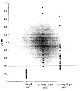
Figure 2 is a graph illustrating the seroreactivity of Mtb-81 in patients
infected with HIV. Mtb-81 was used to detect reactive antibodies in sera from
patients
who were normal (uninfected with M. tuberculosis); HIV-positive and M.
tuberculosis-
positive; or HIV-negative and M. tuberculosis-positive, as indicated. ODQSO
was
indicative of antibody binding. Values above the cut-off value (indicated by
the line)
were considered positive for M. tuberculosis infection.
Figure 3 is a graph illustrating the seroreactivity of Mtb-67.2 in
tuberculosis patients co-infected with HIV. Mtb-67.2 was used to detect
reactive
antibodies in sera from patients who were normal (uninfected with M.
tuberculosis);
HIV-positive and M. tuberculosis-positive; or HIV-negative and M. tuberculosis-
positive, as indicated. OD4so was indicative of antibody binding. Values above
the cut-
off value (indicated by the line) were considered positive for M. tuberculosis
infection.
Figure 4 shows an M. tuberculosis DNA sequence encoding Mtb-67.2.
Figure 5 shows an amino acid sequence of M. tuberculosis Mtb-67.2.
CA 02364670 2001-09-17
WO 00/55194 PCT/US00/07196
7
DETAILED DESCRIPTION OF THE INVENTION
As noted above, the present invention is generally directed to compounds
and methods for the diagnosis and therapy of M. tuberculosis infection. This
invention
is based, in part, on the discovery of two M. tuberculosis antigens (Mtb-81
and Mtb-
67.2). Compounds provided herein include Mtb-81 polypeptides, which comprise
at
least an immunogenic portion of Mtb-81 or a variant thereof, and Mtb-67.2
polypeptides, which comprise at least an immunogenic portion of Mtb-67.2 or a
variant
thereof. Mtb-81 is an 8lkD M. tuberculosis antigen having the sequence recited
in SEQ
ID N0:2 and Figure 2. Mtb-67.2 has the sequence recited in SEQ ID NO:S and
Figure
5. Nucleic acid sequences encoding at least a portion of such polypeptides (or
complements of such nucleic acid sequences) are also provided. Compounds
provided
herein also include binding agents such as antibodies (i. e., immune system
proteins, or
antigen-binding fragments thereof). Mtb-81 and Mt-67.2 polypeptides,
polynucleotides
and antibodies may be used within a variety of serodiagnostic methods for
tuberculosis
1 S detection, and provide enhanced sensitivity in patients infected with HIV.
Such
compounds may also be used for immunotherapy of tuberculosis.
MTB-81 AND MTB-67.2 POLYNUCLEOTIDES
Any polynucleotide that encodes an Mtb-81 or Mtb-67.2 polypeptide, as
described herein, is encompassed by the present invention. Preferred
polynucleotides
comprise at least 10 consecutive nucleotides, preferably at least 15
consecutive
nucleotides, and more preferably at least 30 consecutive nucleotides, that
encode a
portion of Mtb-81 or Mtb-67.2. Within certain embodiments, a polynucleotide
may
encode an immunogenic portion of Mtb-81 or Mtb-67.2. Polynucleotides
comprising at
least 15 consecutive nucleotides complementary to any such sequences are also
encompassed by the present invention. Polynucleotides may be single-stranded
(coding
or antisense) or double-stranded, and may be DNA (genomic, cDNA or synthetic)
or
RNA molecules. Additional coding or non-coding sequences may, but need not, be
present within a polynucleotide of the present invention, and a polynucleotide
may, but
need not, be linked to other molecules and/or support materials.
CA 02364670 2001-09-17
WO 00/55194 PCT/US00/07196
8
Polynucleotides may comprise a native sequence (i.e., an endogenous M.
tuberculosis sequence that encodes Mtb-81, Mtb-67.2 or a portion thereof) or
may
comprise a variant of such a sequence. Certain polynucleotide variants may
contain one
or more substitutions, additions, deletions and/or insertions such that the
immunogenicity of the encoded polypeptide is not diminished, relative to
native Mtb-81
or Mtb-67.2. The effect on the immunogenicity of the encoded polypeptide may
generally be assessed as described herein. Variants preferably exhibit at
least about
70% identity, more preferably at least about 80% identity and most preferably
at least
about 90% identity to a native polynucleotide sequence that encodes Mtb-81,
Mtb-67.2
or a portion thereof. The percent identity may be readily determined by
comparing
sequences using computer algorithms well known to those of ordinary skill in
the art,
such as Megalign, using default parameters. Certain variants are substantially
homologous to a native gene, or a portion or complement thereof. Such
polynucleotide
variants are capable of hybridizing under moderately stringent conditions to a
naturally
occurring DNA sequence. Suitable moderately stringent conditions include
prewashing
in a solution of 5 X SSC, 0.5% SDS, 1.0 mM EDTA (pH 8.0); hybridizing at
50°C-65°
C, 5 X SSC, overnight; followed by washing twice at 65°C for 20 minutes
with each of
2X, O.SX and 0.2X SSC containing 0.1% SDS.
It will be appreciated by those of ordinary skill in the art that, as a result
of the degeneracy of the genetic code, there are many nucleotide sequences
that encode
an Mtb-81 or Mtb-67.2 polypeptide as described herein. Some of these
polynucleotides
bear minimal homology to the nucleotide sequence of any native gene.
Nonetheless,
polynucleotides that vary due to differences in codon usage are specifically
contemplated by the present invention.
Polynucleotides may be prepared using any of a variety of techniques.
For example, a polynucleotide may be amplified via polymerase chain reaction
(PCR)
from cDNA or genomic DNA prepared from M. tuberculosis. For this approach,
sequence-specific primers may be designed based on the sequences provided
herein,
and may be purchased or synthesized.
CA 02364670 2001-09-17
WO 00/55194 PCT/US00/07196
9
An amplified portion may be used to isolate a full length gene from a
suitable library (e.g., an M. tuberculosis genomic or cDNA library) using well
known
techniques. Within such techniques, a library is screened using one or more
polynucleotide probes or primers suitable for amplification. Preferably, a
library is
size-selected to include larger molecules. Random primed libraries may also be
preferred for identifying 5' and upstream regions of genes.
For hybridization techniques, a partial sequence may be labeled (e.g., by
nick-translation or end-labeling with 32P) using well known techniques. A
bacterial or
bacteriophage library is then screened by hybridizing filters containing
denatured
bacterial colonies (or lawns containing phage plaques) with the labeled probe
(see
Sambrook et al., Molecular Cloning: A Laboratory Manual, Cold Spring Harbor
Laboratories, Cold Spring Harbor, NY, 1989). Hybridizing colonies or plaques
are
selected and expanded, and the DNA is isolated for further analysis. cDNA
clones may
be analyzed to determine the amount of additional sequence by, for example,
PCR using
a primer from the partial sequence and a primer from the vector. Restriction
maps and
partial sequences may be generated to identify one or more overlapping clones.
The
complete sequence may then be determined using standard techniques, which may
involve generating a series of deletion clones. The resulting overlapping
sequences are
then assembled into a single contiguous sequence. A full length cDNA molecule
can be
generated by ligating suitable fragments, using well known techniques.
Alternatively, there are numerous amplification techniques for obtaining
a full length coding sequence from a partial cDNA sequence. Within such
techniques,
amplification is generally performed via PCR. Any of a variety of commercially
available kits may be used to perform the amplification step. Primers may be
designed
using, for example, software well known in the art. Primers are preferably 22-
38
nucleotides in length, have a GC content of at least 50% and anneal to the
target
sequence at temperatures of about 56°C to 72°C. The amplified
region may be
sequenced as described above, and overlapping sequences assembled into a
contiguous
sequence.
CA 02364670 2001-09-17
WO 00/55194 PCT/US00/07196
One such amplification technique is inverse PCR (see Triglia et al., Nucl.
Acids Res. 16:8186, 1988), which uses restriction enzymes to generate a
fragment in the
known region of the gene. The fragment is then circularized by intramolecular
ligation
and used as a template for PCR with divergent primers derived from the known
region.
5 Within an alternative approach, sequences adjacent to a partial sequence may
be
retrieved by amplification with a primer to a linker sequence and a primer
specific to a
known region. The amplified sequences are typically subjected to a second
round of
amplification with the same linker primer and a second primer specific to the
known
region. A variation on this procedure, which employs two primers that initiate
10 extension in opposite directions from the known sequence, is described in
WO
96/38591. Another such technique is known as "rapid amplification of cDNA
ends" or
RACE. This technique involves the use of an internal primer and an external
primer,
which hybridizes to a polyA region or vector sequence, to identify sequences
that are 5'
and 3' of a known sequence. Additional techniques include capture PCR
(Lagerstrom et
al., PCR Methods Applic. 1:111-19, 1991) and walking PCR (Parker et al., Nucl.
Acids.
Res. 19:3055-60, 1991). Other methods employing amplification may also be
employed to obtain a full length cDNA sequence.
A genomic M. tuberculosis DNA sequence that includes the coding
region Mtb-81 is presented in Figure 1 (SEQ ID N0:3). In this figure, encoded
amino
acid residues are also indicated (SEQ ID N0:2), with the coding region for Mtb-
81
(SEQ ID NO:l) indicated by the solid black bar. A DNA sequence (SEQ ID N0:4)
encoding Mtb-67.2 is presented in Figure 4, and the encoded amino acid
residues are
shown in Figure S (SEQ ID NO:S). These coding regions, as well as portions
thereof
and sequences complementary to all or a portion thereof, are specifically
encompassed
by the present invention.
Polynucleotide variants may generally be prepared by any method
known in the art, including chemical synthesis by, for example, solid phase
phosphoramidite chemical synthesis. Modifications in a polynucleotide sequence
may
also be introduced using standard mutagenesis techniques, such as
oligonucleotide-
directed site-specific mutagenesis (see Adelman et al., DNA 2:183, 1983).
Certain
CA 02364670 2001-09-17
WO 00/55194 PCT/US00/07196
11
portions may be used to prepare an encoded polypeptide, as described herein. A
portion
of a coding sequence or a complementary sequence may also be designed as a
probe or
primer to detect gene expression. Probes may be labeled by a variety of
reporter
groups, such as radionuclides and enzymes, and are preferably at least 15
nucleotides in
length, more preferably at least 30 nucleotides in length and still more
preferably at
least 50 nucleotides in length. Primers, as noted above, are preferably 22-38
nucleotides in length.
Any polynucleotide may be further modified to increase stability in vivo.
Possible modifications include, but are not limited to, the addition of
flanking
sequences at the 5' and/or 3' ends; the use of phosphorothioate or 2' O-methyl
rather
than phosphodiesterase linkages in the backbone; and/or the inclusion of
nontraditional
bases such as inosine, queosine and wybutosine, as well as acetyl- methyl-,
thio- and
other modified forms of adenine, cytidine, guanine, thymine and uridine.
Nucleotide sequences as described herein may be joined to a variety of
other nucleotide sequences using established recombinant DNA techniques. For
example, a polynucleotide may be cloned into any of a variety of cloning
vectors,
including plasmids, phagemids, lambda phage derivatives and cosmids. Vectors
of
particular interest include expression vectors, replication vectors, probe
generation
vectors and sequencing vectors. In general, a vector will contain an origin of
replication
functional in at least one organism, convenient restriction endonuclease sites
and one or
more selectable markers. Other elements will depend upon the desired use, and
will be
apparent to those of ordinary skill in the art.
Within certain embodiments, polynucleotides may be formulated so as to
permit entry into a cell of a mammal, and expression therein. Such
formulations are
particularly useful for therapeutic purposes, as described below. Those of
ordinary skill
in the art will appreciate that there are many ways to achieve expression of a
polynucleotide in a target cell, and any suitable method may be employed. For
example, a polynucleotide may be incorporated into a viral vector such as, but
not
limited to, adenovirus, adeno-associated virus, retrovirus, or vaccinia or
other pox virus
(e.g., avian pox virus). Techniques for incorporating DNA into such vectors
are well
CA 02364670 2001-09-17
WO 00/55194 PCT/US00/07196
12
known to those of ordinary skill in the art. A retroviral vector may
additionally transfer
or incorporate a gene for a selectable marker (to aid in the identification or
selection of
transduced cells) and/or a targeting moiety, such as a gene that encodes a
ligand for a
receptor on a specific target cell, to render the vector target specific.
Targeting may
also be accomplished using an antibody, by methods known to those of ordinary
skill in
the art.
Other formulations for therapeutic purposes include colloidal dispersion
systems, such as macromolecule complexes, nanocapsules, microspheres, beads,
and
lipid-based systems including oil-in-water emulsions, micelles, mixed
micelles, and
liposomes. A preferred colloidal system for use as a delivery vehicle in vitro
and in
vivo is a liposome (i. e., an artificial membrane vesicle). The preparation
and use of
such systems is well known in the art.
MTB-81 AND MTB-67.2 POLYPEPTIDES
Within the context of the present invention, Mtb-81 polypeptides
comprise at least an immunogenic portion of Mtb-81 (Figures lA-1F; SEQ ID
N0:2) or
a variant thereof, as described herein. Mtb-67.2 polypeptides comprise at
least an
immunogenic portion of Mtb-67.2 (Figure 5; SEQ ID NO:S) or a variant thereof.
Polypeptides as described herein may be of any length. Additional sequences
derived
from the native protein and/or heterologous sequences may be present, and such
sequences may (but need not) possess further immunogenic or antigenic
properties.
An "immunogenic portion," as used herein is a portion of an antigen that
is recognized (i.e., specifically bound) by a B-cell and/or T-cell surface
antigen
receptor. Such immunogenic portions generally comprise at least 5 amino acid
residues, preferably at least 9, more preferably at least 15, and still more
preferably at
least 50 amino acid residues of Mtb-81, Mtb-67.2 or a variant of either
antigen.
Immunogenic portions may generally be identified using well known techniques,
such
as those summarized in Paul, Fundamental Immunology, 3rd ed., 243-247 (Raven
Press,
1993) and references cited therein. Such techniques include screening
polypeptides for
the ability to react with antigen-specific antibodies, antisera and/or T-cell
lines or
CA 02364670 2001-09-17
WO 00/55194 PCT/US00/07196
13
clones. As used herein, antisera and antibodies are "antigen-specific" if they
specifically bind to an antigen (i.e., they react with the antigen in an ELISA
or other
immunoassay, and do not react detectably with unrelated proteins). Such
antisera and
antibodies may be prepared as described herein, and using well known
techniques. An
S immunogenic portion of Mtb-81 or Mtb-67.2 is a portion that reacts with such
antisera
and/or T-cells at a level that is not substantially less than the reactivity
of the full length
polypeptide (e.g., in an ELISA and/or T-cell reactivity assay). Immunogenic
portions
may react within such assays at a level that is similar to or greater than the
reactivity of
the full length polypeptide. Such screens may generally be performed using
methods
well known to those of ordinary skill in the art, such as those described in
Harlow and
Lane, Antibodies: A Laboratory Manual, Cold Spring Harbor Laboratory, 1988.
For
example, a polypeptide may be immobilized on a solid support and contacted
with
patient sera to allow binding of antibodies within the sera to the immobilized
polypeptide. Unbound sera may then be removed and bound antibodies detected
using
a detection reagent, such as'ZSI-labeled Protein A.
As noted above, a polypeptide may be a variant of Mtb-81 or Mtb-67.2.
A polypeptide "variant," as used herein, is a polypeptide that differs from
native Mtb-81
or Mtb-67.2 in one or more substitutions, deletions, additions and/or
insertions, such
that the immunogenicity of the polypeptide is not substantially diminished. In
other
words, the ability of a variant to react with antigen-specific antisera or T
cells may be
enhanced or unchanged, relative to the native antigen, or may be diminished by
less
than 50%, and preferably less than 20%, relative to the native antigen. Such
variants
may generally be identified by modifying one of the above polypeptide
sequences and
evaluating the reactivity of the modified polypeptide with antigen-specific
antibodies or
antisera as described herein. Polypeptide variants preferably exhibit at least
70%, more
preferably at least 90% and most preferably at least 95% identity to Mtb-81 or
Mtb-
67.2.
Preferably, a variant contains conservative substitutions. A
"conservative substitution" is one in which an amino acid is substituted for
another
amino acid that has similar properties, such that one skilled in the art of
peptide
CA 02364670 2001-09-17
WO 00/55194 PCT/US00/07196
14
chemistry would expect the secondary structure and hydropathic nature of the
polypeptide to be substantially unchanged. Amino acid substitutions may
generally be
made on the basis of similarity in polarity, charge, solubility,
hydrophobicity,
hydrophilicity and/or the amphipathic nature of the residues. For example,
negatively
charged amino acids include aspartic acid and glutamic acid; positively
charged amino
acids include lysine and arginine; and amino acids with uncharged polar head
groups
having similar hydrophilicity values include leucine, isoleucine and valine;
glycine and
alanine; asparagine and glutamine; and serine, threonine, phenylalanine and
tyrosine.
Other groups of amino acids that may represent conservative changes include: (
1 ) ala,
pro, gly, glu, asp, gln, asn, ser, thr; (2) cys, ser, tyr, thr; (3) val, ile,
leu, met, ala, phe;
(4) lys, arg, his; and (5) phe, tyr, trp, his. A variant may also, or
alternatively, contain
nonconservative changes. Variants may also (or alternatively) be modified by,
for
example, the deletion or addition of amino acids that have minimal influence
on the
immunogenicity, secondary structure and hydropathic nature of the polypeptide.
As noted above, polypeptides may comprise a signal (or leader)
sequence at the N-terminal end of the protein which co-translationally or post-
translationally directs transfer of the protein. The polypeptide may also be
conjugated
to a linker or other sequence for ease of synthesis, purification or
identification of the
polypeptide (e.g., poly-His), or to enhance binding of the polypeptide to a
solid support.
For example, a polypeptide may be conjugated to an immunoglobulin Fc region.
Polypeptides may be prepared using any of a variety of well known
techniques. Recombinant polypeptides encoded by DNA sequences as described
above
may be readily prepared from the DNA sequences using any of a variety of
expression
vectors known to those of ordinary skill in the art. Expression may be
achieved in any
appropriate host cell that has been transformed or transfected with an
expression vector
containing a DNA molecule that encodes a recombinant polypeptide. Suitable
host
cells include prokaryotes, yeast and higher eukaryotic cells. Preferably, the
host cells
employed are E. coli, yeast or a mammalian cell line such as COS or CHO.
Supernatants from suitable host/vector systems which secrete recombinant
protein or
polypeptide into culture media may be first concentrated using a commercially
available
CA 02364670 2001-09-17
WO 00/55194 PCT/US00/0'7196
filter. Following concentration, the concentrate may be applied to a suitable
purification matrix such as an affinity matrix or an ion exchange resin.
Finally, one or
more reverse phase HPLC steps can be employed to further purify a recombinant
polypeptide.
5 Portions and other variants having fewer than about 100 amino acids,
and generally fewer than about 50 amino acids, may also be generated by
synthetic
means, using techniques well known to those of ordinary skill in the art. For
example,
such polypeptides may be synthesized using any of the commercially available
solid-
phase techniques, such as the Merrifield solid-phase synthesis method, where
amino
10 acids are sequentially added to a growing amino acid chain. See Merrifield,
J. Am.
Chem. Soc. 85:2149-2146, 1963. Equipment for automated synthesis of
polypeptides is
commercially available from suppliers such as Applied BioSystems, Inc. (Foster
City,
CA), and may be operated according to the manufacturer's instructions.
Within certain specific embodiments, a polypeptide may be a fusion
15 protein that comprises a polypeptide as described herein. For example, such
fusion
proteins may further comprise one or more known M. tuberculosis antigens, or
variants) of such antigens. Representative known M. tuberculosis antigens
include the
38 kD antigen described in Andersen and Hansen, Infect. Immun. 57:2481-2488,
1989
(GenBank Accession No. M30046) and ESAT-6 (Sorensen et al., Infect. Immun.
63:1710-1717, 1995). Fusion proteins may generally be prepared using standard
techniques. For example, a fusion protein may be prepared recombinantly.
Briefly,
DNA sequences encoding the polypeptide components may be assembled separately,
and ligated into an appropriate expression vector. The 3' end of the DNA
sequence
encoding one polypeptide component is ligated, with or without a peptide
linker, to the
5' end of a DNA sequence encoding the second polypeptide component so that the
reading frames of the sequences are in phase. This permits translation into a
single
fusion protein that retains the biological activity of both component
polypeptides.
A peptide linker sequence may be employed to separate the first and the
second polypeptide components by a distance sufficient to ensure that each
polypeptide
folds into its secondary and tertiary structures. Such a peptide linker
sequence is
CA 02364670 2001-09-17
WO 00/55194 PCT/US00/07196
16
incorporated into the fusion protein using standard techniques well known in
the art.
Suitable peptide linker sequences may be chosen based on the following
factors:
( 1 ) their ability to adopt a flexible extended conformation; (2) their
inability to adopt a
secondary structure that could interact with functional epitopes on the first
and second
polypeptides; and (3) the lack of hydrophobic or charged residues that might
react with
the polypeptide functional epitopes. Preferred peptide linker sequences
contain Gly,
Asn and Ser residues. Other near neutral amino acids, such as Thr and Ala may
also be
used in the linker sequence. Amino acid sequences which may be usefully
employed as
linkers include those disclosed in Maratea et al., Gene 40:39-46, 1985; Murphy
et al.,
Proc. Natl. Acad. Sci. USA 83:8258-8262, 1986; U.S. Patent No. 4,935,233 and
U.S.
Patent No. 4,751,180. The linker sequence may generally be from 1 to about 50
amino
acids in length. Linker sequences are not required when the first and second
polypeptides have non-essential N-terminal amino acid regions that can be used
to
separate the functional domains and prevent steric interference.
The ligated DNA sequences are operably linked to suitable
transcriptional or translational regulatory elements. The regulatory elements
responsible for expression of DNA are located only 5' to the DNA sequence
encoding
the first polypeptides. Similarly, stop codons required to end translation and
transcription termination signals are only present 3' to the DNA sequence
encoding the
second polypeptide.
Fusion proteins are also provided that comprise a polypeptide of the
present invention together with an unrelated immunogenic protein. Preferably
the
immunogenic protein is capable of eliciting a recall response. Examples of
such
proteins include tetanus, tuberculosis and hepatitis proteins (see, for
example, Stoute
et al. New Engl. J. Med., 336:86-91, 1997).
In general, polypeptides (including fusion proteins) and polynucleotides
as described herein are isolated. An "isolated" polypeptide or polynucleotide
is one that
is removed from its original environment. For example, a naturally-occurring
protein is
isolated if it is separated from some or all of the coexisting materials in
the natural
system. Preferably, such polypeptides are at least about 90% pure, more
preferably at
CA 02364670 2001-09-17
WO 00/55194 PCT/US00/07196
17
least about 95% pure and most preferably at least about 99% pure. A
polynucleotide is
considered to be isolated if, for example, it is cloned into a vector that is
not a part of
the natural environment.
BINDING AGENTS
The present invention further provides agents, such as antibodies and
antigen-binding fragments thereof, that specifically bind to Mtb-81 or Mtb-
67.2. As
used herein, an antibody, or antigen-binding fragment thereof, is said to
"specifically
bind" to Mtb-81 or Mtb-67.2 if it reacts at a detectable level (within, for
example, an
ELISA) with Mtb-81 or Mtb-67.2, and does not react detectably with unrelated
proteins
under similar conditions. As used herein, "binding" refers to a noncovalent
association
between two separate molecules (each of which may be in solution or present on
the
surface of a cell or solid support) such that a "complex" is formed. The
ability to bind
may be evaluated by, for example, determining a binding constant for the
formation of
the complex. The binding constant is the value obtained when the concentration
of the
complex is divided by the product of the component concentrations. In general,
two
compounds are said to "bind," in the context of the present invention, when
the binding
constant for complex formation exceeds about 103 L/mol. The binding constant
maybe
determined using methods well known in the art.
Binding agents are further capable of differentiating between patients
with and without M. tuberculosis infection, using the representative assays
provided
herein. In other words, antibodies or other binding agents that bind to Mtb-81
or Mtb-
67.2 will generate a signal indicating the presence of M. tuberculosis
infection in at
least about 20% of patients with such infection, and will generate a negative
signal
indicating the absence of such infection in at least about 90% of uninfected
individuals.
In general, a signal is considered positive if it is greater than the mean
signal obtained
from an uninfected sample plus three standard deviations. To determine whether
a
binding agent satisfies this requirement, biological samples (e.g., blood,
sera, plasma,
saliva, cerebrospinal fluid or urine) from patients with and without M.
tuberculosis
infection (as determined using a standard diagnostic test) may be assayed as
described
CA 02364670 2001-09-17
WO 00/55194 PCT/US00/07196
18
herein for the presence of polypeptides that bind to the binding agent. It
will be
apparent that a statistically significant number of samples with and without
the infection
should be assayed. Each binding agent should satisfy the above criteria;
however, those
of ordinary skill in the art will recognize that binding agents may be used in
combination to improve sensitivity.
Any agent that satisfies the above requirements may be a binding agent.
For example, a binding agent may be a ribosome with or without a peptide
component,
an RNA molecule or a polypeptide. In a preferred embodiment, a binding agent
is an
antibody or an antigen-binding fragment thereof. Such antibodies may be
polyclonal or
monoclonal. In addition, the antibodies may be single chain, chimeric, CDR-
grafted or
humanized.
Binding agents may be further linked to a reporter group, to facilitate
diagnostic assays. Suitable reporter groups will be apparent to those of
ordinary skill in
the art, and include enzymes (such as horseradish peroxidase), substrates,
cofactors,
inhibitors, dyes, colloids (e.g., colloidal gold), radionuclides, luminescent
groups,
fluorescent groups and biotin. The conjugation of antibody to reporter group
may be
achieved using standard methods known to those of ordinary skill in the art.
Antibodies may be prepared by any of a variety of techniques known to
those of ordinary skill in the art. See, e.g., Harlow and Lane, AntiTSOdies: A
Laboratory
Manual, Cold Spring Harbor Laboratory, 1988. In general, antibodies can be
produced
by cell culture techniques, including the generation of monoclonal antibodies
as
described herein, or via transfection of antibody genes into suitable
bacterial or
mammalian cell hosts, in order to allow for the production of recombinant
antibodies.
To generate antibodies, a polypeptide immunogen may be the full length Mtb-81
or
Mtb-67.2, or may be an immunogenic portion of either antigen. If an
immunogenic
portion is employed, the resulting antibody should indicate the presence of M.
tuberculosis infection in substantially all (i.e., at least 80%, and
preferably at least 90%)
of the patients for which M. tuberculosis infection would be indicated using
an antibody
raised against the full length antigen. The antibody should also indicate the
absence of
M. tuberculosis infection in substantially all of those samples that would be
negative
CA 02364670 2001-09-17
WO 00/55194 PCT/US00/07196
19
when tested with an antibody raised against the full length antigen. The
representative
assays provided herein, such as the two-antibody sandwich assay, may generally
be
employed for evaluating the ability of an antibody to detect tuberculosis.
In one technique, an immunogen comprising the polypeptide is initially
injected into any of a wide variety of mammals (e.g., mice, rats, rabbits,
sheep or goats).
In this step, the polypeptides of this invention may serve as the immunogen
without
modification. Alternatively, particularly for relatively short polypeptides, a
superior
immune response may be elicited if the polypeptide is joined to a carrier
protein, such
as bovine serum albumin or keyhole limpet hemocyanin. The immunogen is
injected
into the animal host, preferably according to a predetermined schedule
incorporating
one or more booster immunizations, and the animals are bled periodically.
Polyclonal
antibodies specific for the polypeptide may then be purified from such
antisera by, for
example, affinity chromatography using the polypeptide coupled to a suitable
solid
support.
Monoclonal antibodies specific for the antigenic polypeptide of interest
may be prepared, for example, using the technique of Kohler and Milstein, Eur.
J.
Immunol. 6:511-519, 1976, and improvements thereto. Briefly, these methods
involve
the preparation of immortal cell lines capable of producing antibodies having
the
desired specificity (i.e., reactivity with the polypeptide of interest). Such
cell lines may
be produced, for example, from spleen cells obtained from an animal immunized
as
described above. The spleen cells are then immortalized by, for example,
fusion with a
myeloma cell fusion partner, preferably one that is syngeneic with the
immunized
animal. A variety of fusion techniques may be employed. For example, the
spleen cells
and myeloma cells may be combined with a nonionic detergent for a few minutes
and
then plated at low density on a selective medium that supports the growth of
hybrid
cells, but not myeloma cells. A preferred selection technique uses HAT
(hypoxanthine,
aminopterin, thymidine) selection. After a sufficient time, usually about 1 to
2 weeks,
colonies of hybrids are observed. Single colonies are selected and their
culture
supernatants tested for binding activity against the polypeptide. Hybridomas
having
high reactivity and specificity are preferred.
CA 02364670 2001-09-17
WO 00/55194 PCT/US00/07196
Monoclonal antibodies may be isolated from the supernatants of growing
hybridoma colonies. In addition, various techniques may be employed to enhance
the
yield, such as injection of the hybridoma cell line into the peritoneal cavity
of a suitable
vertebrate host, such as a mouse. Monoclonal antibodies may then be harvested
from
5 the ascites fluid or the blood. Contaminants may be removed from the
antibodies by
conventional techniques, such as chromatography, gel filtration,
precipitation, and
extraction. The polypeptides of this invention may be used in the purification
process
in, for example, an affinity chromatography step.
Within certain embodiments, the use of antigen-binding fragments of
10 antibodies may be preferred. Such fragments include Fab fragments, which
may be
prepared using standard techniques. Briefly, immunoglobulins may be purified
from
rabbit serum by affinity chromatography on Protein A bead columns (Harlow and
Lane,
Antibodies: A Laboratory Manual, Cold Spring Harbor Laboratory, 1988) and
digested
by papain to yield Fab and Fc fragments. The Fab and Fc fragments may be
separated
15 by affinity chromatography on protein A bead columns.
Monoclonal antibodies of the present invention may be coupled to one or
more therapeutic agents for use in the therapeutic methods provided herein.
Suitable
agents in this regard include radionuclides, differentiation inducers, drugs,
toxins, and
derivatives thereof. Preferred radionuclides include 9°Y, 'Z~I, 'ZSI,
'3'I, '$6Re, 'BgRe, 2"At,
20 and 2'ZBi. Preferred drugs include methotrexate, and pyrimidine and purine
analogs.
Preferred differentiation inducers include phorbol esters and butyric acid.
Preferred
toxins include ricin, abrin, diptheria toxin, cholera toxin, gelonin,
Pseudomonas
exotoxin, Shigella toxin, and pokeweed antiviral protein.
A therapeutic agent may be coupled (e.g., covalently bonded) to a
suitable monoclonal antibody either directly or indirectly (e.g., via a linker
group). A
direct reaction between an agent and an antibody is possible when each
possesses a
substituent capable of reacting with the other. For example, a nucleophilic
group, such
as an amino or sulflrydryl group, on one may be capable of reacting with a
carbonyl-
containing group, such as an anhydride or an acid halide, or with an alkyl
group
containing a good leaving group (e.g., a halide) on the other.
CA 02364670 2001-09-17
WO 00/55194 PCT/US00/07196
21
Alternatively, it may be desirable to couple a therapeutic agent and an
antibody via a linker group. A linker group can function as a spacer to
distance an
antibody from an agent in order to avoid interference with binding
capabilities. A
linker group can also serve to increase the chemical reactivity of a
substituent on an
agent or an antibody, and thus increase the coupling efficiency. An increase
in
chemical reactivity may also facilitate the use of agents, or functional
groups on agents,
which otherwise would not be possible.
It will be evident to those skilled in the art that a variety of bifunctional
or polyfunctional reagents, both homo- and hetero-functional (such as those
described
in the catalog of the Pierce Chemical Co., Rockford, IL), may be employed as
the linker
group. Coupling may be effected, for example, through amino groups, carboxyl
groups,
sulfllydryl groups or oxidized carbohydrate residues. There are numerous
references
describing such methodology, e.g., U.S. Patent No. 4,671,958, to Rodwell et
al.
Where a therapeutic agent is more potent when free from the antibody
portion of the immunoconjugates of the present invention, it may be desirable
to use a
linker group which is cleavable during or upon internalization into a cell. A
number of
different cleavable linker groups have been described. The mechanisms for the
intracellular release of an agent from these linker groups include cleavage by
reduction
of a disulfide bond (e.g., U.S. Patent No. 4,489,710, to Spitler), by
irradiation of a
photolabile bond (e.g., U.S. Patent No. 4,625,014, to Senter et al.), by
hydrolysis of
derivatized amino acid side chains (e.g., U.S. Patent No. 4,638,045, to Kohn
et al.), by
serum complement-mediated hydrolysis (e.g., U.S. Patent No. 4,671,958, to
Rodwell
et al.), and acid-catalyzed hydrolysis (e.g., U.S. Patent No. 4,569,789, to
Blattler et al.).
It may be desirable to couple more than one agent to an antibody. In one
embodiment, multiple molecules of an agent are coupled to one antibody
molecule. In
another embodiment, more than one type of agent may be coupled to one
antibody.
Regardless of the particular embodiment, immunoconjugates with more than one
agent
may be prepared in a variety of ways. For example, more than one agent may be
coupled directly to an antibody molecule, or linkers which provide multiple
sites for
attachment can be used. Alternatively, a carrier can be used.
CA 02364670 2001-09-17
WO 00/55194 PCT/US00/07196
22
A carrier may bear the agents in a variety of ways, including covalent
bonding either directly or via a linker group. Suitable carriers include
proteins such as
albumins (e.g., U.S. Patent No. 4,507,234, to Kato et al.), peptides and
polysaccharides
such as aminodextran (e.g., U.S. Patent No. 4,699,784, to Shih et al.). A
carrier may
also bear an agent by noncovalent bonding or by encapsulation, such as within
a
liposome vesicle (e.g., U.S. Patent Nos. 4,429,008 and 4,873,088): Garners
specific for
radionuclide agents include radiohalogenated small molecules and chelating
compounds. For example, U.S. Patent No. 4,735,792 discloses representative
radiohalogenated small molecules and their synthesis. A radionuclide chelate
may be
formed from chelating compounds that include those containing nitrogen and
sulfur
atoms as the donor atoms for binding the metal, or metal oxide, radionuclide.
For
example, U.S. Patent No. 4,673,562, to Davison et al. discloses representative
chelating
compounds and their synthesis.
A variety of routes of administration for the antibodies and
immunoconjugates may be used. Typically, administration will be intravenous,
intramuscular or subcutaneous. It will be evident that the precise dose of the
antibody/immunoconjugate will vary depending upon the antibody used and the
rate of
clearance of the antibody.
2O METHODS FOR DETECTING TUBERCULOSIS
In general, M. tuberculosis infection may be detected in a patient based
on the presence of one or more of the following in a biological sample
obtained from a
patient: (a) antibodies that specifically bind to Mtb-81 or Mtb-67.2; (b) T-
cells that
specifically react with Mtb-81 or Mtb-67.2; (c) Mtb-81 or Mtb-67.2 antigen or
(d)
mRNA encoding Mtb-81 or Mtb-67.2 antigen. In other words, Mtb-81 and/or Mtb-
67.2
may be used as a marker to indicate the presence or absence of M. tuberculosis
infection
in a patient. Mtb-81 or Mtb-67.2 polypeptides, as well as polynucleotides
encoding
such polypeptides and antigen-presenting cells that express such polypeptides,
may be
used to detect the presence of specific antibodies or T-cells. The binding
agents
provided herein generally permit detection of the level of Mtb-81 or Mtb-67.2
antigen
CA 02364670 2001-09-17
WO 00/55194 PCT/US00/07196
23
in the biological sample. Polynucleotide primers and probes may be used to
detect the
level of mRNA encoding Mtb-81 or Mtb-67.2.
Diagnostic methods provided herein have advantages over existing
methods in sensitivity. In particular, methods provided herein may be used to
detect M.
tuberculosis infection in AIDS patients. M. tuberculosis and HIV co-infection
is
common in such patients, but the tuberculosis has been difficult to detect
using previous
diagnostic methods. Further, Mtb-81 appears to be an early stage marker for M.
tuberculosis infection, permitting early detection of the disease.
A biological sample may be any sample obtained from one or more
human or non-human animals that would be expected to contain the target
substance in
infected individuals. For example, to detect M. tuberculosis infection based
on the
presence of Mtb-81- or Mtb-67.2-specific antibodies, any antibody-containing
sample
may be used. Such samples include whole blood, sputum, serum, plasma, saliva,
cerebrospinal fluid and urine. Preferred biological samples include blood,
serum and
plasma obtained from a patient or blood supply.
Within the methods provided herein, Mtb-81 and/or Mtb-67.2 may, but
need not, be used in combination with one or more known M. tuberculosis
antigens. In
such embodiments, the antigens used are preferably complementary (i.e., one
antigen
will tend to detect infection in samples where the infection would not be
detected by the
other antigen). Complementary antigens may generally be identified by using
each
polypeptide individually to evaluate serum samples obtained from a series of
patients
known to be infected with M. tuberculosis. After determining which samples
test
positive (as described below) with each polypeptide, combinations of two or
more
polypeptides may be formulated that are capable of detecting infection in
most, or all, of
the samples tested. Such polypeptides are complementary.
There are a variety of assay formats known to those of ordinary skill in
the art for using one or more polypeptides to detect antibodies in a sample.
See, e.g.,
Harlow and Lane, Antibodies: A Laboratory Manual, Cold Spring Harbor
Laboratory,
1988, which is incorporated herein by reference. In a preferred embodiment,
the assay
involves the use of polypeptide immobilized on a solid support to bind to and
remove
CA 02364670 2001-09-17
WO 00/55194 PCT/US00/07196
24
the antibody (in the form of an immunocomplex with polypeptide) from the
sample.
The immunocomplex may then be detected using a detection reagent that contains
a
reporter group. Suitable detection reagents include antibodies that bind to
the
immunocomplex and free polypeptide labeled with a reporter group (e.g., in a
semi-
s competitive assay). Alternatively, a competitive assay may be utilized, in
which an
antibody that binds to the polypeptide is labeled with a reporter group and
allowed to
bind to the immobilized antigen after incubation of the antigen with the
sample. The
extent to which components of the sample inhibit the binding of the labeled
antibody to
the polypeptide is indicative of the reactivity of the sample with the
immobilized
polypeptide.
The solid support may be any solid material known to those of ordinary
skill in the art to which the antigen may be attached. For example, the solid
support
may be a test well in a microtiter plate or a nitrocellulose or other suitable
membrane.
Alternatively, the support may be a bead or disc, such as glass, fiberglass,
latex or a
plastic material such as polystyrene or polyvinylchloride. The support may
also be a
magnetic particle or a fiber optic sensor, such as those disclosed, for
example, in U.S.
Patent No. 5,359,681.
The polypeptides may be bound to the solid support using a variety of
techniques known to those of ordinary skill in the art, which are amply
described in the
~20 patent and scientific literature. In the context of the present invention,
the term "bound"
refers to both noncovalent association, such as adsorption, and covalent
attachment
(which may be a direct linkage between the antigen and functional groups on
the
support or may be a linkage by way of a cross-linking agent). Binding by
adsorption to
a well in a microtiter plate or to a membrane is preferred. In such cases,
adsorption may
be achieved by contacting the polypeptide, in a suitable buffer, with the
solid support
for a suitable amount of time. The contact time varies with temperature, but
is typically
between about 1 hour and 1 day. In general, contacting a well of a plastic
microtiter
plate (such as polystyrene or polyvinylchloride) with an amount of polypeptide
ranging
from about 10 ng to about 1 ~,g, and preferably about 100 ng, is sufficient to
bind an
adequate amount of antigen.
CA 02364670 2001-09-17
WO 00/55194 PCT/US00/07196
Covalent attachment of polypeptide to a solid support may generally be
achieved by first reacting the support with a bifunctional reagent that will
react with
both the support and a functional group, such as a hydroxyl or amino group, on
the
polypeptide. For example, the polypeptide may be bound to supports having an
5 appropriate polymer coating using benzoquinone or by condensation of an
aldehyde
group on the support with an amine and an active hydrogen on the polypeptide
(see,
e.g., Pierce Immunotechnology Catalog and Handbook, 1991, at A12-A13).
In certain embodiments, the assay is an enzyme linked immunosorbent
assay (ELISA). This assay may be performed by first contacting a polypeptide
antigen
10 that has been immobilized on a solid support, commonly the well of a
microtiter plate,
with the sample, such that antibodies to the polypeptide within the sample are
allowed
to bind to the immobilized polypeptide. Unbound sample is then removed from
the
immobilized polypeptide and a detection reagent capable of binding to the
immobilized
immunocomplex is added. The amount of detection reagent that remains bound to
the
15 solid support is then determined using a method appropriate for the
specific detection
reagent.
More specifically, once the polypeptide is immobilized on the support as
described above, the remaining protein binding sites on the support are
typically
blocked. Any suitable blocking agent known to those of ordinary skill in the
art, such
20 as bovine serum albumin or Tween 20TM (Sigma Chemical Co., St. Louis, MO)
may be
employed. The immobilized polypeptide is then incubated with the sample, and
antibody is allowed to bind to the antigen. The sample may be diluted with a
suitable
diluent, such as phosphate-buffered saline (PBS) prior to incubation. In
general, an
appropriate contact time (i.e., incubation time) is that period of time that
is sufficient to
25 detect the presence of antibody within a M. tuberculosis-infected sample.
Preferably,
the contact time is sufficient to achieve a level of binding that is at least
95% of that
achieved at equilibrium between bound and unbound antibody. Those of ordinary
skill
in the art will recognize that the time necessary to achieve equilibrium may
be readily
determined by assaying the level of binding that occurs over a period of time.
At room
temperature, an incubation time of about 30 minutes is generally sufficient.
CA 02364670 2001-09-17
WO 00/55194 PCT/US00/07196
26
Unbound sample may then be removed by washing the solid support
with an appropriate buffer, such as PBS containing 0:1% Tween 20TM. Detection
reagent may then be added to the solid support. An appropriate detection
reagent is any
compound that binds to the immobilized antibody-polypeptide complex and that
can be
detected by any of a variety of means known to those in the art. Preferably,
the
detection reagent contains a binding agent (such as, for example, Protein A,
Protein G,
immunoglobulin, lectin or free antigen) conjugated to a reporter group.
Preferred
reporter groups include enzymes (such as horseradish peroxidase), substrates,
cofactors,
inhibitors, colloids, dyes, radionuclides, luminescent groups, fluorescent
groups and
biotin. The conjugation of binding agent to reporter group may be achieved
using
standard methods known to those of ordinary skill in the art. Common binding
agents
may also be purchased conjugated to a variety of reporter groups from many
commercial sources (e.g., Zymed Laboratories, San Francisco, CA, and Pierce,
Rockford, IL).
The detection reagent is then incubated with the immobilized antibody-
polypeptide complex for an amount of time sufficient to detect the bound
antibody. An
appropriate amount of time may generally be determined from the manufacturer's
instructions or by assaying the level of binding that occurs over a period of
time.
Unbound detection reagent is then removed and bound detection reagent is
detected
using the reporter group. The method employed for detecting the reporter group
depends upon the nature of the reporter group. For radioactive groups,
scintillation
counting or autoradiographic methods are generally appropriate. Spectroscopic
methods may be used to detect dyes, luminescent groups and fluorescent groups.
Biotin
may be detected using avidin, coupled to a different reporter group (commonly
a
radioactive or fluorescent group or an enzyme). Enzyme reporter groups may
generally
be detected by the addition of substrate (generally for a specific period of
time),
followed by spectroscopic or other analysis of the reaction products.
To determine the presence or absence of anti-M. tuberculosis antibodies
in the sample, the signal detected from the reporter group that remains bound
to the
solid support is generally compared to a signal that corresponds to a cut-off
value. In
CA 02364670 2001-09-17
WO 00/55194 PCT/US00/07196
27
one preferred embodiment, the cut-off value is the average mean signal plus
three
standard deviations obtained when the immobilized antigen is incubated with
samples
from an uninfected patient. In general, a sample generating a signal that is
above the
cut-off value is considered positive for tuberculosis. In an alternate
preferred
embodiment, the cut-off value is determined using a Receiver Operator Curve,
according to the method of Sackett et al., Clinical Epidemiology: A Basic
Science for
Clinical Medicine, Little Brown and Co., 1985, pp. 106-107. Briefly, in this
embodiment, the cut-off value may be determined from a plot of pairs of true
positive
rates (i.e., sensitivity) and false positive rates (100%-specificity) that
correspond to each
possible cut-off value for the diagnostic test result. The cut-off value on
the plot that is
the closest to the upper left-hand corner (i.e., the value that encloses the
largest area) is
the most accurate cut-off value, and a sample generating a signal that is
higher than the
cut-off value determined by this method may be considered positive.
Alternatively, the
cut-off value may be shifted to the left along the plot, to minimize the false
positive
rate, or to the right, to minimize the false negative rate. In general, a
sample generating
a signal that is higher than the cut-off value determined by this method is
considered
positive for tuberculosis.
In a related embodiment, the assay is performed in a rapid flow-through
or strip test format, wherein the antigen is immobilized on a membrane, such
as
nitrocellulose. In the flow-through test, antibodies within the sample bind to
the
immobilized polypeptide as the sample passes through the membrane. A detection
reagent (e.g., protein A-colloidal gold) then binds to the antibody-
polypeptide complex
as the solution containing the detection reagent flows through the membrane.
The
detection of bound detection reagent may then be performed as described above.
In the
strip test format, one end of the membrane to which polypeptide is bound is
immersed
in a solution containing the sample. The sample migrates along the membrane
through
a region containing detection reagent and to the area of immobilized
polypeptide.
Concentration of detection reagent at the polypeptide indicates the presence
of anti-
M. tuberculosis antibodies in the sample. Typically, the concentration of
detection
reagent at that site generates a pattern, such as a line, that can be read
visually. The
CA 02364670 2001-09-17
WO 00/55194 PCT/US00/07196
28
absence of such a pattern indicates a negative result. In general, the amount
of
polypeptide immobilized on the membrane is selected to generate a visually
discernible
pattern when the biological sample contains a level of antibodies that would
be
sufficient to generate a positive signal in an ELISA, as discussed above.
Preferably, the
amount of polypeptide immobilized on the membrane ranges from about 25 ng to
about
1 ~,g, and more preferably from about 50 ng to about 500 ng. Such tests can
typically
be performed with a very small amount (e.g., one drop) of patient serum or
blood.
Of course, numerous other assay protocols exist that are suitable for use
with the polypeptides of the present invention. The above descriptions are
intended to
be exemplary only. For example, it will be apparent to those of ordinary skill
in the art
that the above protocols may be readily modified to use antibodies, or antigen-
binding
fragments thereof, to detect Mtb-81 and/or Mtb-67.2 in a biological sample.
M. tuberculosis infection may also, or alternatively, be detected based on
the presence of T cells that specifically react with Mtb-81 in a biological
sample.
Within certain methods, a biological sample comprising CD4+ and/or CD8+ T
cells
isolated from a patient is incubated with a Mtb-81 or Mtb-67.2 polypeptide, a
polynucleotide encoding such a polypeptide and/or an APC that expresses such
al
polypeptide, and the presence or absence of specific activation of the T cells
is detected.
Suitable biological samples include, but are not limited to, isolated T cells.
For
example, T cells may be isolated from a patient by routine techniques (such as
by
Ficoll/Hypaque density gradient centrifugation of peripheral blood
lymphocytes). T
cells may be incubated in vitro for 2-9 days (typically 4 days) at 37°C
with Mtb-81 or
Mtb-67.2 polypeptide (e.g., 5 - 25 p,g/ml). It may be desirable to incubate
another
aliquot of a T cell sample in the absence of Mtb-81 or Mtb-67.2 polypeptide to
serve as
a control. For CD4+ T cells, activation is preferably detected by evaluating
proliferation
of the T cells. For CD8+ T cells, activation is preferably detected by
evaluating
cytolytic activity. A level of proliferation that is at least two fold greater
and/or a level
of cytolytic activity that is at least 20% greater than in disease-free
patients indicates the
presence of M. tuberculosis infection.
CA 02364670 2001-09-17
WO 00/55194 PCT/US00/07196
29
As noted above, M. tuberculosis infection may also, or alternatively, be
detected based on the level of mRNA encoding Mtb-81 or Mtb-67.2 in a
biological
sample. For example, at least two oligonucleotide primers may be employed in a
polymerase chain reaction (PCR) based assay to amplify a portion Mtb-81 or Mtb-
67.2
cDNA derived from a biological sample, wherein at least one of the
oligonucleotide
primers is specific for (i.e., hybridizes to) a polynucleotide encoding Mtb-81
or Mtb-
67.2. The amplified cDNA is then separated and detected using techniques well
known
in the art, such as gel electrophoresis. Similarly, oligonucleotide probes
that
specifically hybridize to a polynucleotide encoding Mtb-81 or Mtb-67.2 may be
used in
a hybridization assay to detect the presence of polynucleotide encoding the
antigen in a
biological sample.
To permit hybridization under assay conditions, oligonucleotide primers
and probes should comprise an oligonucleotide sequence that has at least about
60%,
preferably at least about 75% and more preferably at least about 90%, identity
to a
portion of a polynucleotide encoding Mtb-81 or Mtb-67.2 that is at least 10
nucleotides,
and preferably at least 20 nucleotides, in length. Oligonucleotide primers
and/or probes
which may be usefully employed in the diagnostic methods described herein are
preferably at least 10-40 nucleotides in length. Techniques for both PCR based
assays
and hybridization assays are well known in the art (see, for example, Mullis
et al., Cold
Spring Harbor Symp. Quant. Biol., 51:263, 1987; Erlich ed., PCR Technology,
Stockton
Press, NY, 1989).
One preferred assay employs RT-PCR, in which PCR is applied in
conjunction with reverse transcription. Typically, RNA is extracted from a
sample
tissue and is reverse transcribed to produce cDNA molecules. PCR amplification
using
at least one specific primer generates a cDNA molecule, which may be separated
and
visualized using, for example, gel electrophoresis. Amplification may be
performed on
samples obtained from biological samples taken from a test patient and an
individual
who is not infected with M. tuberculosis. The amplification reaction may be
performed
on several dilutions of cDNA spanning two orders of magnitude. A two-fold or
greater
CA 02364670 2001-09-17
WO 00/55194 PCT/US00/07196
increase in expression in several dilutions of the test patient sample as
compared to the
same dilutions of an uninfected sample is typically considered positive.
As noted above, to improve sensitivity, multiple M. tuberculosis markers
may be assayed within a given sample. It will be apparent that multiple
antigens may
5 be combined within a single assay, or multiple primers or probes may be used
concurrently. The selection of antigen markers may be based on routine
experiments to
determine combinations that results in optimal sensitivity.
The diagnostic methods provided above may be used to monitor
tuberculosis therapy in a patient. Briefly, such monitoring may be achieved by
10 performing an assay as described above using a biological sample obtained
at a first
time (prior to at least a portion of a therapy), and comparing the result
obtained with the
result of a similar assay performed using a second biological sample (obtained
following at least a portion of the therapy). A therapy that results in a
decrease in signal
is generally considered to be effective in decreasing the level of M.
tuberculosis
15 infection.
DIAGNOSTIC KITS
The present invention fiu ther provides kits for use within any of the
above diagnostic methods. Such kits typically comprise two or more components
20 suitable for performing a diagnostic assay. Components may be compounds,
reagents,
containers and/or equipment. For example, one container within a kit may
contain a
Mtb-81 or Mtb-67.2 polypeptide. Such polypeptides may be provided attached to
a
support material, as described above. One or more additional containers may
enclose
elements, such as reagents or buffers, to be used in the assay. Such kits may
also, or
25 alternatively, contain a detection reagent as described above that contains
a reporter
group suitable for direct or indirect detection of immunocomplex formation.
Alternatively, a kit may be designed to detect the level of mRNA
encoding Mtb-81 or Mtb-67.2 in a biological sample. Such kits generally
comprise at
least one oligonucleotide probe or primer, as described above, that hybridizes
to a
30 polynucleotide encoding Mtb-81 or Mtb-67.2. Such an oligonucleotide may be
used,
CA 02364670 2001-09-17
WO 00/55194 PCT/US00/07196
31
for example, within a PCR or hybridization assay. Additional components that
may be
present within such kits include a second oligonucleotide and/or a diagnostic
reagent or
container to facilitate the detection of a polynucleotide encoding Mtb-81 or
Mtb-67.2.
Still further kits may detect the presence of antigen in a sample. Such
kits may comprise one or more monoclonal or polyclonal antibodies that
specifically
bind to Mtb-81 or Mtb-67.2.
T CELLS
The present invention further provides T cells specific for Mtb-81 or
Mtb-67.2. Such cells may generally be prepared in vitro or ex vivo, using
standard
procedures. For example, T cells may be present within (or isolated from) bone
marrow, peripheral blood or a fraction of bone marrow or peripheral blood of a
mammal, such as a patient, using a commercially available cell separation
system, such
as the CEPRATET"" system, available from CellPro Inc., Bothell WA (see also
U.S.
Patent No. 5,240,856; U.S. Patent No. 5,215,926; WO 89/06280; WO 91/16116 and
WO 92/07243). Alternatively, T cells may be derived from related or unrelated
humans, non-human animals, cell lines or cultures.
T cells may be stimulated with a Mtb-81 or Mtb-67.2 polypeptide, a
polynucleotide encoding such a polypeptide andlor an antigen presenting cell
(APC)
that expresses such a polypeptide. Stimulation is performed under conditions
and for a
time sufficient to permit the generation of T cells that are specific for the
polypeptide.
Preferably, a Mtb-81 or Mtb-67.2 polypeptide or polynucleotide is present
within a
delivery vehicle, such as a microsphere, to facilitate the generation of
specific T cells.
T cells are considered to be specific for Mtb-81 (or Mtb-67.2) if the T
cells kill target cells coated with Mtb-81 or expressing a gene encoding Mtb-
81 (or
Mtb-67.2). T cell specificity may be evaluated using any of a variety of
standard
techniques. For example, within a chromium release assay or proliferation
assay, a
stimulation index of more than two fold increase in lysis and/or
proliferation, compared
to negative controls, indicates T cell specificity. Such assays may be
performed, for
example, as described in Chen et al., Cancer Res. 54:1065-1070, 1994.
Alternatively,
CA 02364670 2001-09-17
WO 00/55194 PCT/US00/07196
32
detection of the proliferation of T cells may be accomplished by a variety of
known
techniques. For example, T cell proliferation can be detected by measuring an
increased
rate of DNA synthesis (e.g., by pulse-labeling cultures of T cells with
tritiated
thymidine and measuring the amount of tritiated thymidine incorporated into
DNA).
Contact with Mtb-81 or Mtb-67.2 (200 ng/ml - 100 ~g/ml, preferably 100 ng/ml -
25
~g/ml) for 3 - 7 days should result in at least a two fold increase in
proliferation of the T
cells and/or contact as described above for 2-3 hours should result in
activation of the T
cells, as measured using standard cytokine assays in which a two fold increase
in the
level of cytokine release (e.g., TNF or IFN-y) is indicative of T cell
activation (see
Coligan et al., Current Protocols in Immunology, vol. 1, Wiley Interscience
(Greene
1998)). T cells that have been activated in response to a Mtb-81 or Mtb-67.2
polypeptide, polynucleotide or polypeptide-expressing APC may be CD4+ and/or
CD8+.
Mtb-81- or Mtb-67.2-specific T cells may be expanded using standard
techniques.
Within preferred embodiments, the T cells are derived from a patient or a
related or
unrelated donor and are administered to the patient following stimulation and
expansion.
For therapeutic purposes, CD4+ or CD8+ T cells that proliferate in
response to Mtb-81 or Mtb-67.2 can be expanded in number either in vitro or in
vivo.
Proliferation of such T cells in vitro may be accomplished in a variety of
ways. For
example, the T cells can be re-exposed to Mtb-81 or Mtb-67.2, with or without
the
addition of T cell growth factors, such as interleukin-2, and/or stimulator
cells that
synthesize a Mtb-81 or Mtb-67.2 polypeptide. Alternatively, one or more T
cells that
proliferate in the presence of Mtb-81 or Mtb-67.2 can be expanded in number by
cloning. Methods for cloning cells are well known in the art, and include
limiting
dilution.
PHARMACEUTICAL COMPOSITIONS AND VACCINES
Within certain aspects, polypeptides, polynucleotides, binding agents
and/or cells may be incorporated into pharmaceutical compositions or vaccines.
Pharmaceutical compositions comprise one or more such compounds and a
CA 02364670 2001-09-17
WO 00155194 PCT/US00/07196
33
physiologically acceptable carrier. Vaccines may comprise one or more such
compounds and a non-specific immune response enhancer. A non-specific immune
response enhancer may be any substance that enhances an immune response to an
exogenous antigen. Examples of non-specific immune response enhancers include
adjuvants, biodegradable microspheres (e.g., polylactic galactide) and
liposomes (into
which the compound is incorporated). Pharmaceutical compositions and vaccines
within the scope of the present invention may also contain other compounds,
which
may be biologically active or inactive. For example, one or more immunogenic
portions of other M. tuberculosis antigens may be present, either incorporated
into a
fusion polypeptide or as a separate compound within the composition or
vaccine.
A pharmaceutical composition or vaccine may contain DNA encoding
one or more of the polypeptides as described above, such that the polypeptide
is
generated in situ. DNA may be present within any of a variety of delivery
systems
known to those of ordinary skill in the art, including nucleic acid expression
systems,
bacteria and viral expression systems. Numerous gene delivery techniques are
well
known in the art, such as those described by Rolland, Crit. Rev. Therap. Drug
Carrier
Systems 15:143-198, 1998, and references cited therein. Appropriate nucleic
acid
expression systems contain the necessary DNA sequences for expression in the
patient
(such as a suitable promoter and terminating signal). Bacterial delivery
systems involve
the administration of a bacterium (such as Bacillus-Calmette-Guerrin) that
expresses an
immunogenic portion of the polypeptide on its cell surface. In a preferred
embodiment,
the DNA may be introduced using a viral expression system (e.g., vaccinia or
other pox
virus, retrovirus; or adenovirus), which may involve the use of a non-
pathogenic
(defective), replication competent virus. Suitable systems are disclosed, for
example, in
Fisher-Hoch et al., Proc. Natl. Acad. Sci. USA 86:317-321, 1989; Flexner et
al., Ann.
N. Y. Acad. Sci. 569:86-103, 1989; Flexner et al., Vaccine 8:17-21, 1990; U.S.
Patent
Nos. 4,603,112, 4,769,330, and 5,017,487; WO 89/01973; U.S. Patent No.
4,777,127;
GB 2,200,651; EP 0,345,242; WO 91/02805; Berkner, Biotechniques 6:616-627,
1988;
Rosenfeld et al., Science 252:431-434, 1991; Kolls et al., Proc. Natl. Acad.
Sci. USA
91:215-219, 1994; Kass-Eisler et al., Proc. Natl. Acad. Sci. USA 90:11498-
11502, 1993;
CA 02364670 2001-09-17
WO 00/55194 PCT/US00/07196
34
Guzman et al., Circulation 88:2838-2848, 1993; and Guzman et al., Cir. Res.
73:1202-1207, 1993. Techniques for incorporating DNA into such expression
systems
are well known to those of ordinary skill in the art. The DNA may also be
"naked," as
described, for example, in Ulmer et al., Science 259:1745-1749, 1993 and
reviewed by
Cohen, Science 259:1691-1692, 1993. The uptake of naked DNA may be increased
by
coating the DNA onto biodegradable beads, which are efficiently transported
into the
cells.
While any suitable carrier known to those of ordinary skill in the art may
be employed in the pharmaceutical compositions of this invention, the type of
carrier
will vary depending on the mode of administration. Compositions of the present
invention may be formulated for any appropriate manner of administration,
including
for example, topical, oral, nasal, intravenous, intracranial, intraperitoneal,
subcutaneous
or intramuscular administration. For parenteral administration, such as
subcutaneous
injection, the carrier preferably comprises water, saline, alcohol, a fat, a
wax or a buffer.
For oral administration, any of the above carriers or a solid carrier, such as
mannitol,
lactose, starch, magnesium stearate, sodium saccharine, talcum, cellulose,
glucose,
sucrose, and magnesium carbonate, may be employed. Biodegradable microspheres
(e.g., polylactate polyglycolate) may also be employed as carriers for the
pharmaceutical compositions of this invention. Suitable biodegradable
microspheres
are disclosed, for example, in U.S. Patent Nos. 4,897,268 and 5,075,109.
Such compositions may also comprise buffers (e.g., neutral buffered
saline or phosphate buffered saline), carbohydrates (e.g., glucose, mannose,
sucrose or
dextrans), mannitol, proteins, polypeptides or amino acids such as glycine,
antioxidants,
chelating agents such as EDTA or glutathione, adjuvants (e.g., aluminum
hydroxide)
and/or preservatives. Alternatively, compositions of the present invention may
be
formulated as a lyophilizate. Compounds may also be encapsulated within
liposomes
using well known technology.
Any of a variety of non-specific immune response enhancers may be
employed in the vaccines of this invention. For example, an adjuvant may be
included.
Most adjuvants contain a substance designed to protect the antigen from rapid
CA 02364670 2001-09-17
WO 00/55194 PCT/US00/07196
catabolism, such as aluminum hydroxide or mineral oil, and a stimulator of
immune
responses, such as lipid A, Bortadella pertussis or Mycobacterium tuberculosis
derived
proteins. Suitable adjuvants are commercially available as, for example,
Freund's
Incomplete Adjuvant and Complete Adjuvant (Difco Laboratories, Detroit, MI),
Merck
5 Adjuvant 65 (Merck and Company, Inc., Rahway, NJ), alum, biodegradable
microspheres, monophosphoryl lipid A and quil A. Cytokines, such as GM-CSF or
interleukin-2, -7, or -12, may also be used as adjuvants.
The compositions described herein may be administered as part of a
sustained release formulation (i. e., a formulation such as a capsule or
sponge that effects
10 a slow release of compound following administration). Such formulations may
generally be prepared using well known technology and administered by, for
example,
oral, rectal or subcutaneous implantation, or by implantation at the desired
target site.
Sustained-release formulations may contain a polypeptide, polynucleotide or
antibody
dispersed in a carrier matrix and/or contained within a reservoir surrounded
by a rate
15 controlling membrane. Carriers for use within such formulations are
biocompatible,
and may also be biodegradable; preferably the formulation provides a
relatively
constant level of active component release. The amount of active compound
contained
within a sustained release formulation depends upon the site of implantation,
the rate
and expected duration of release and the nature of the condition to be treated
or
20 prevented.
Any of a variety of delivery vehicles may be employed within
pharmaceutical compositions and vaccines to facilitate production of an immune
response. Delivery vehicles include antigen presenting cells, such as
dendritic cells and
macrophages. Such cells may be transfected with a polynucleotide encoding Mtb-
81 or
25 Mtb-67.2 (or portion or other variant thereof) such that the Mtb-81 or Mtb-
67.2
polypeptide is expressed on the cell surface. Such transfection may take place
ex vivo,
and a composition or vaccine comprising such transfected cells may then be
used for
therapeutic purposes, as described herein. Alternatively, a gene delivery
vehicle that
targets a dendritic or other antigen presenting cell may be administered to a
patient,
30 resulting in transfection that occurs in vivo. In vivo and ex vivo
transfection of dendritic
CA 02364670 2001-09-17
WO 00/55194 PCT/US00/07196
36
cells may generally be performed using any methods known in the art, such as
those
described in WO 97/24447, or the gene gun approach described by Mahvi et al.,
Immunology and cell Biology 75:456-460, 1997.
S TUBERCULOSIS THERAPY
In further aspects of the present invention, the compositions described
herein may be used for immunotherapy of tuberculosis. Within such methods,
pharmaceutical compositions and vaccines are typically administered to a
patient. As
used herein, a "patient" refers to any warm-blooded animal, preferably a
human. A
patient may or may not be infected with M. tuberculosis. Accordingly, the
above
pharmaceutical compositions and vaccines may be used to prevent the
development of
tuberculosis or to treat a patient afflicted with tuberculosis. Pharmaceutical
compositions and vaccines may be administered prior to, concurrent with or
following
treatment with other therapeutic agents.
Within certain embodiments, immunotherapy may be active
immunotherapy, in which treatment relies on the in vivo stimulation of the
endogenous
host immune system to react against M. tuberculosis with the administration of
immune
response-modifying agents (such as tumor vaccines, bacterial adjuvants and/or
cytokines).
Within other embodiments, immunotherapy may be passive
immunotherapy, in which treatment involves the delivery of agents with
established
immune reactivity (such as effector cells or antibodies) that do not
necessarily depend
on an intact host immune system. Examples of effector cells include T
lymphocytes
(such as CD8+ cytotoxic T lymphocytes and CD4+ T-helper tumor-infiltrating
lymphocytes), killer cells (such as Natural Killer cells and lymphokine-
activated killer
cells), B cells and antigen-presenting cells (such as dendritic cells and
macrophages)
expressing a polypeptide provided herein. T cell receptors and antibody
receptors
specific for the polypeptides recited herein may be cloned, expressed and
transferred
into other vectors or effector cells for adoptive immunotherapy. The
polypeptides
CA 02364670 2001-09-17
WO 00/55194 PCT/US00/07196
37
provided herein may also be used to generate antibodies or anti-idiotypic
antibodies (as
described above and in U.S. Patent No. 4,918,164) for passive immunotherapy.
Effector cells may generally be obtained in sufficient quantities for
adoptive immunotherapy by growth in vitro, as described herein. Culture
conditions for
expanding single antigen-specific effector cells to several billion in number
with
retention of antigen recognition in vivo are well known in the art. Such in
vitro culture
conditions typically use intermittent stimulation with antigen, often in the
presence of
cytokines (such as IL-2) and non-dividing feeder cells. As noted above,
immunoreactive polypeptides as provided herein may be used to rapidly expand
antigen-specific T cell cultures in order to generate a sufficient number of
cells for
immunotherapy. In particular, antigen-presenting cells, such as dendritic,
macrophage
or B cells, may be pulsed with immunoreactive polypeptides or transfected with
one or
more polynucleotides using standard techniques well known in the art. For
example,
antigen-presenting cells can be transfected with a polynucleotide having a
promoter
appropriate for increasing expression in a recombinant virus or other
expression system.
Cultured effector cells for use in therapy must be able to grow and distribute
widely,
and to survive long term in vivo. Studies have shown that cultured effector
cells can be
induced to grow in vivo and to survive long term in substantial numbers by
repeated
stimulation with antigen supplemented with IL-2 (see, for example, Cheever et
al.,
Immunological Reviews 157:177, 1997).
The polypeptides provided herein may also be used to generate and/or
isolate Mtb-81- or Mtb-67.2-reactive T cells, which can then be administered
to a
patient. In one such technique, antigen-specific T cell lines may be generated
by in vivo
immunization with short peptides corresponding to immunogenic portions of the
disclosed polypeptides. The resulting antigen-specific CD8+ CTL clones may be
isolated from the patient, expanded using standard tissue culture techniques
and
returned to the patient.
Polypeptides may also be used for ex vivo treatment of tuberculosis. For
example, cells of the immune system, such as T cells, may be isolated from the
peripheral blood of a patient, using a commercially available cell separation
system,
CA 02364670 2001-09-17
WO 00/55194 PCT/US00/07196
38
such as CellPro Incorporated's (Bothell, WA) CEPRATET"" system (see U.S.
Patent No.
5,240,856; U.S. Patent No. 5,215,926; WO 89/06280; WO 91/16116 and WO
92/07243). The separated cells are stimulated with one or more immunoreactive
Mtb-
81 polypeptides contained within a delivery vehicle, such as a microsphere, to
provide
antigen-specific T cells. The population of antigen-specific T cells is then
expanded
using standard techniques and the cells may be administered back to the
patient as
described, for example, by Chang et al., Crit. Rev. Oncol. Hematol. 22:213,
1996.
Within another embodiment, syngeneic or autologous dendritic cells may
be pulsed with peptides corresponding to at least an immunogenic portion of
Mtb-81 or
Mtb-67.2. The resulting antigen-specific dendritic cells may either be
transferred into a
patient or employed to stimulate T cells to provide antigen-specific T cells
which may,
in turn, be administered to a patient. Alternatively, a vector expressing a
polypeptide
recited herein may be introduced into antigen presenting cells taken from a
patient and
clonally propagated ex vivo for transplant back into the same patient.
Transfected cells
1 S may be reintroduced into the patient using any means known in the art,
preferably in
sterile form by intravenous, intracavitary or intraperitoneal administration.
Routes and frequency of administration, as well as dosage, will vary
from individual to individual, and may be readily established using standard
techniques.
In general, the pharmaceutical compositions and vaccines may be administered
by
injection (e.g., intracutaneous, intramuscular, intravenous or subcutaneous),
intranasally
(e.g., by aspiration) or orally. Preferably, between 1 and 10 doses may be
administered
over a 52 week period. Preferably, 6 doses are administered, at intervals of 1
month,
and booster vaccinations may be given periodically thereafter. Alternate
protocols may
be appropriate for individual patients. A suitable dose is an amount of a
compound that,
when administered as described above, is capable of causing an immune response
that
leads to an improved clinical outcome (e.g., decreased symptoms or longer
survival) in
vaccinated patients as compared to non-vaccinated patients. In general, for
pharmaceutical compositions and vaccines comprising one or more polypeptides,
the
amount of each polypeptide present in a dose ranges from about 100 ~.g to 5 mg
per kg
CA 02364670 2001-09-17
WO 00/55194 PCT/US00/07196
39
of host. Suitable dose sizes will vary with the size of the patient, but will
typically
range from about 0.1 mL to about 5 mL.
The following Examples are offered by way of illustration and not by
way of limitation.
CA 02364670 2001-09-17
WO 00/55194 PCT/US00/07196
EXAMPLES
Example 1
Preparation M. tuberculosis Protein
5
This Example illustrates the initial characterization of an M. tuberculosis
protein that recognizes an antibody present in HIV positive individuals.
To identify M. tuberculosis antigens suitable for diagnostic methods, the
high-molecular weight region of crude soluble proteins (CSP; obtained from
Colorado
10 State University) derived from M. tuberculosis strain H3,Rv was examined
using two
dimensional gel electrophoresis and two dimensional Western analysis. The
probe for
this analysis was monoclonal antibody IT57 (reviewed in Infection and Immunity
60:3925-3927, 1992), obtained from the UNPD/World Bank/World Health
Organization Special Programme for Research and Training in Tropical Diseases.
This
15 antibody has been known to react with 82 kDa M. tuberculosis antigens) in
this high-
molecular weight region, but the identity of the protein antigen has not been
previously
elucidated (see Infection and Immunity 56:1994-1998, 1988; Infection and
Immunity
60:3925-3927, 1992).
The CSP was separated by reverse phase chromatography on a C 18
20 column. Approximately 75mg of CSP was dissolved in water containing 0.1%
trifluoroacetic acid (TFA), injected onto a C18 reverse phase column (22 X 250
mm,
The Separations Group, Hesperia, CA) using a Prep LC (Waters, Milford, MA) and
eluted with a binary gradient of 0.1% TFA in water (Solvent A) and
acetonitrile
(Solvent B) at a flow rate of 10 ml/minute. The gradient increased from 0 to
100% B in
25 60 min. Fractions were collected at 1 minute intervals.
Each fraction was individually tested by immunoblot analysis to identify
the fractions containing reactivity against the IT57 antibody. Individual HPLC
fractions were separated by SDS-PAGE on 4-20% gradient gels (BioRad, Hercules,
CA) as per the manufacturers instructions prior to transfer to nitrocellulose.
The
30 proteins were transferred to nitrocellulose membranes (Hybond C Extra,
Amersham,
Arlington Heights, IL) and blocked with O.SM NaCI in phosphate buffered saline
(PBS)
CA 02364670 2001-09-17
WO 00/55194 PCT/LTS00/07196
41
with 0.05% Tween (PBST). Blots were washed with PBS and probed with IT-57
antibody at a 1:50 or 1:70 dilution of culture supernatant in O.SM NaCI in
PBST for the
1 D and 2D gels, respectively. After overnight incubation, blots were washed
and
probed with IgG specific donkey anti-mouse secondary antibody ECL(Jackson
Immuno
Research, West Grove, PA). Westerns were developed according to Pierce ECL
protocol (Pierce Super Signal, Rockford, IL).
Fraction 38, which contained reactivity to antibody IT-57, was identified
by Western analysis as described above and was further evaluated by 2D-PAGE
and
Western analysis. For 2D-PAGE analysis, Fraction 38 was concentrated to
approximately 400 ~.l and 40 ~1 was added to 400 ~l of rehydration solution
containing
8 molar urea, 0.5% CHAPS (w/w), 15 mM DTT and 0.2% (w/v) Parmalyte pH 3-10.
The solution was placed in a rehydration cassette and 18 cm pH 3-10 Immobiline
Drystrips (Pharmacia Biotech, Uppsala, Sweden) were allowed to hydrate
overnight.
The hydrated strips were rinsed and focused using the multiphor II
electrophoresis
system with the Immobiline DryStrip kit and the EPS 3500 XL power supply from
Pharmacia Biotech according to the following gradient: 0-300 volts/Sminutes,
300-3500
volts/6 hours and 3500 thereafter to 80,000 volt/hours.
Tube gels for the 1D control lanes were cast by adding 10 ~1 of each
fraction to 10 ~l of Tris acetate equilibration buffer from ESA (Chelmsford,
MA) which
contained 2% (w/v) DTT and 2% (w/v) agarose. The solution was heated at
100°C for
5 minutes. Tube gels for molecular weight standards were cast by adding 2 ~1
of low
range silver standards from Biorad (Hercules, CA) to 8 gl of water and 10 ~l
of Tris
acetate equilibration buffer containing 2% (w/v) DTT and 2% (w/v) agarose and
boiling
for five minutes.
Focused Immobiline Drystrips were equilibrated in 10 mls/strip of
equilibration buffer which contained 6 molar urea, 2% (w/v) SDS and 2% (w/v)
DTT
and rocked gently for 15 minutes. The buffer was decanted and the Drystrips
were then
equilibrated in 10 mls/strip of Tris acetate equilibration buffer which
contained 6 molar
urea and 2.5% iodoacetamide and rocked gently for 15 minutes. Strips were
placed on
top of Investigator 10% homogeneous double gels from ESA along side a 1 cm
tube gel
CA 02364670 2001-09-17
WO 00/55194 PCT/US00/07196
42
containing the same HPLC fraction as the DryStrip and a 1 cm tube gel
containing low
range silver standards from Biorad. The gels were run at 20 mA/gel overnight
on the
ESA Investigator 2D electrophoresis system which contained Tris acetate
running
buffer in the lower (anode) tank and Tris tricine SDS buffer in the upper
(cathode) tank,
both supplied by ESA.
One gel was transferred to nitrocellulose and immunoblotted using the
IT57 antibody. The other gel was silver stained. For 2D-PAGE immunoblot
analysis,
the proteins were transferred to nitrocellulose membranes (Hybond C Extra,
Amersham,
Arlington Heights, IL) and blocked with O.SM NaCI in phosphate buffered saline
(PBS)
with 0.05% Tween (PBST). Blots were washed with PBS and probed with IT-57
antibody at a 1:70 dilution of culture supernatant in O.SM NaCI in PBST. After
overnight incubation, blots were washed and probed with IgG specific donkey
anti-
mouse secondary antibody ECL (Jackson Immuno Research, West Grove, PA).
Westerns were developed according to Pierce ECL protocol (Pierce Super Signal,
Rockford, IL). For the silver stained gels, the gels were fixed for more than
1 hour and
generally overnight in 40% methanol solution containing 10% acetic acid. The
gels
were then rinsed 3 times in 30% ethanol solution prior to reduction in 0.02%
sodium
thiosulfate in nanopure water for 1 minute. After reduction, the gels were
washed 3
times in nanopure water for 20 seconds each wash before they were incubated
for 20
minutes in 0.2% silver nitrate and 0.02% formaldehyde in nanopure water. The
gels
were washed 3 times for 20 seconds each wash before being developed in 3%
sodium
carbonate with 0.05% formaldehyde and 0.0005% sodium thiosulfate in nanopure
water. After the gels were sufficiently developed the chemical process was
stopped by
the addition of 5% acetic acid. The gels were rinsed in nanopure water and
stored in
0.1% acetic acid solution at 4°C.
The immunoblot analysis was correlated to the silver stained by staining
the nitrocellulose membrane with AurodyeForte (Amersham Corp., Arlington
Height,
IL) total protein stain. Briefly, after developing immunobolots by ECL, the
membranes
were washed in PBS +0.3% Tween 3x 5 min, rinsed in nanopure water, and then
CA 02364670 2001-09-17
WO 00!55194 PCT/US00/07196
43
incubated in 40 ml AurodyeForte dye at room temp with gentle rocking until the
desired
proteins were visible.
The IT-57 reactive protein was excised from the gel and dehydrated by
the addition of 1001 of acetonitrile. The solvent was removed and replaced
with 100
~l of SOmM ammonium bicarbonate that contained 1pl of 1M DTT, allowed to
rehydrate, and then incubated at 57°C for 1 hour. The solvent was
removed, the gel
dehydrated by the addition of 100 ~,1 of acetonitrile. The solvent was
replaced with 55
mM iodoacetamide in 50 mM ammonium bicarbonate after equilibration to room
temperature and incubated in the dark at room temperature for 45 minutes with
occasional vortexing. The gel was washed successively with SOmM ammonium
bicarbonate, acetonitrile, 50 mM ammonium bicarbonate, and acetonitrile before
being
fully dehydrated in a speedvac concentrator. Six ~1 of reductively alkylated
trypsin,
(Promega, Madison, WI) was added to 100 ~1 50 mM ammonium bicarbonate the
resulting suspension was incubated overnight at 37°C. The supernatant
was removed
and the tryptic peptides extracted with 1 wash of 100 mM ammonium bicarbonate
followed by three successive washes of 50% acetonitrile with S % formic acid.
The
extracts were pooled, concentrated on a speedvac to 30 ~1 volume, and stored
at -20°C
until mass spectrometric analysis.
An aliquot of the tryptic peptides was loaded onto a C18 microcapillary
column (75 ~,m i.d. x 12 cm) and gradient eluted using acetonitrile and 0.1 M
acetic acid
with the concentration of acetonitrile increasing from 0-80% in 12 minutes
into a triple
quadrupole mass spectrometer (TSQ7000; Finnigan MAT, San Jose, CA) equipped
with
an electrospray ionization source. Mass spectra were acquired every 1.5
seconds over a
mass range of 300 to 1400 atomic mass units. Candidate peptide masses were
identified
by comparing the tryptic digest to a control digest.
To sequence the protein antigen, an addition aliquot of protein was
generated by diphenyl fractionation. Approximately 5 mg of CSP was dissolved
in
water containing 0.5% trifluoroacetic acid (TFA). This sample was injected
onto a
Vydac diphenyl reverse phase column (Cat #219TP5415: The Separations Group,
Hesperia, CA) eluted with a binary gradient of 0.5% TFA in water (buffer A)
and
CA 02364670 2001-09-17
WO 00/55194 PCT/US00/07196
44
acetonitrile (buffer B) on an AKTA explorer 100 separation system (Amersham
Pharmacia Biotech AB, Uppsala, Sweden). The column was equilibrated in 30% B
and
a linear gradient was run from 30% to 65% buffer B at 2 ml/min over the course
of 30
minutes. Fractions were collected at l.Sml intervals and analyzed by Western
blotting
using the IT57 antibody as described above. Fractions containing a protein
recognized
by this antibody eluted at about 50% B and were pooled and separated by SDS-
PAGE.
The gel was silver stained and digested in situ as described above.
Collision activated dissociation (CAD) mass spectra were recorded on
the (M+2H)2+ ions at m/z 444 and 531. The CAD spectra were interpreted de novo
or
by peptide sequence tags (Anal Chem. 66(24):4390-99, 1994). The sequence for
444 2+
corresponded to the Cat G protein, the sequence for the 531 2+ corresponded to
the
Mtb-81 antigen. The M. tuberculosis genomic sequence used was from the
published
literature (Nature 393(6685):537-44, 1998).
Example 2
Preparation of Mtb-67.2
This Example illustrates the identification and preparation of Mtb-67.2.
The high-molecular weight region of CSP derived from M. tuberculosis
was examined using two dimensional gel electrophoresis. Five protein spots in
the high
molecular weight region were identified, individually excised, enzymatically
digested
and subjected to mass spectrometric analysis (as described in Example 1). The
sequence of one of the identified proteins was determined and is provided
herein as
Mtb-81. Another protein, which appears to be present with Mtb-81 in a band
that
migrates in this high molecular weight region was found to be Mtb-67.2 (Figure
5; SEQ
ID NO:S).
CA 02364670 2001-09-17
WO 00/55194 PCT/US00/07196
Example 3
Preparation of Mtb-81 Polynucleotide
This Example illustrates the preparation of a DNA molecule encoding
5 Mtb-81, and its expression product.
To obtain an Mtb-81 sequence for expression in E. coli, PCR analysis
was performed using genomic M. tuberculosis DNA with the following primers
(SEQ
ID NOs:- and ~:
PDM-268 5'CTAAGTAGTACTGATCGCGTGTCGGTGGGC3' Tm=66°C
10 PDM-269 5'CAGTGAGAATTCACTAGCGGGCCGCATCGTCAC3' Tm=68°C
The PCR reactions contained:
10 pL l OX Pfu buffer
1 ~L 10 mM dNTPs
2 p,L each 10 ~M oligonucleotide
15 83 p,L sterile water
1.5 ~,L Pfu DNA polymerase (Stratagene)
ng M. tuberculosis genomic DNA
Reactions were heated to 96°C for two minutes; cycled forty times
at
96°C (20 seconds), 67°C (15 seconds), and 72°C (5
minutes); and then incubated at
20 72°C for 5 minutes. The PCR product was digested with ScaI and EcoRI
and cloned
into pPDM His (a modified pET28 vector from Novagen, Madison, WI), which was
digested with Eco72 I and Eco RI. Sequence was confirmed and the PCR product
was
transformed into BL21 pLys S (Novagen, Madison, WI). A single colony was
inoculated into LB medium with kanamycin (30 ~glmL) and chloramphenicol (34
25 ~g/mL). Twenty-four mL of the overnight culture was used to inoculate 1
liter of
2XYT broth with the same antibiotics in a baffled flask. Four liters were
grown at once.
At OD56o of between 0.35 and 0.55, the flasks were induced with a final
concentration
of 1 mM IPTG. The bacteria were allowed to grow for four more hours before
harvesting. The pellets were centrifuged and washed with 1X PBS and then
centrifuged
30 again. Pellets were resuspended in lysis buffer (20 mM Tris (pH 8.0). 100
mM NaCI
and 0.1 % DOC) and frozen at -20°C overnight.
CA 02364670 2001-09-17
WO 00/55194 PCT/US00/07196
46
The pellets were then thawed and sonicated, and high speed
centrifugation was used to separate the inclusion body pellet and the soluble
supernatant. The Mtb-81 protein was found to be in the inclusion body pellet,
and was
washed twice with 0.5% CHAPS in 20 mM Tris (pH 8.0), 300 mM NaCI, and then
solubilized in binding buffer (20 mM Tris, pH 8.0, 100 mM NaCI, 8M Urea). The
pellet was then batch bound to Nickel NTA resin (Qiagen) and then passed over
a
Kontes (VWR) gravity flow column. The first wash was 20 mM Tris (pH 8.0), 350
mM
NaCI, 1.0% DOC, 10 mM imidazole, 8M urea. The second wash was the same as the
first, but without DOC. The elutions were done in a step wise manner, the
first being
20 mM Tris (pH 8.0), 100 mM NaCI, 50 mM imidazole, 8 mM urea. The second
increased the imidazole concentration to 100 mM. The third elution increased
the
imidazole to 500 mM. Less than one half the inclusion body did not stay bound
to the
Nickel and came off in the initial flowthrough. The protein started to elute
with the
lowest concentration of imidazole, and gradually came off the column as the
imidazole
concentration was increased. The elutions which contained the protein of
interest were
pooled and then dialyzed against 10 mM Tris (pH 8.0). After several dialysis
changes,
the protein was concentrated in a Vivaspin (IMS) 30 kD cutoff concentrator and
then
sterile filtered.
The results of whole protein composition analysis and predicted
structural class (Protean application program within DNASTAR (Madison, WI) of
the
protein are presented in Tables I and II below.
CA 02364670 2001-09-17
WO 00/55194 PCT/US00/07196
47
Table I
Predicted Structural Class
Analysis Whole Protein
Molecular Weight 81353.22 m.w.
Length 748
1 microgram = 12.292 pMoles
Molar Extinction coefficient77860 5%
1 A(280) _ 1.04 mg/ml
Isoelectric Point 5.19
Charge at pH 7 -22.80
CA 02364670 2001-09-17
WO 00/55194 PCT/US00/07196
48
Table II
Whole Protein Composition Analysis
Amino Acids) Number % by % by
count weight frequency
Charged (RKHYCDE) 210 34.50 28.07
Acidic (DE) 98 14.48 13.10
Basic (KR) 72 12.93 9.63
Polar (NCQSTY) 158 21.64 21.12
Hydrophobic (AILFWV)285 35.98 38.10
A Ala 87 7.60 11.63
C Cys 4 0.51 0.53
D Asp 62 8.77 8.29
E Glu 36 5.71 4.81
F Phe 23 4.16 3.07
G Gly 60 4.21 8.02
H His 20 3.37 2.67
I Ile 41 5.70 5.48
K Lys 26 4.10 3.48
L Leu 65 9.04 8.69
M Met 19 3.06 2.54
N Asn 29 4.07 3.88
P Pro 36 4.30 4.81
Q Gin 27 4.25 3.61
R Arg 46 8.83 6.15
S Ser 34 3.64 4.55
T Thr 48 5.97 6.42
V Val 59 7.19 7.89
W Trp 10 2.29 1.34
Y Tyr 16 3.21 2.14
B Asx 0 0.00 0.00
Z Glx 0 0.00 0.00
CA 02364670 2001-09-17
WO 00/55194 PCT/US00/07196
49
Amino Acids) Number % by % by
count weight frequency
X Xxx 0 0.00 0.00
. Ter 2 0.00 0.27
Expressed Mtb-81 was not recognized by marine monoclonal antibody
IT-57 by Western analysis. This lack of reactivity may be due to limitations
in the E.
coli expression system (e.g., the protein may not be posttranslationally
modified or may
be improperly folded). Alternatively, another M. tuberculosis protein that
reacts with
IT-57 may remain to be identified. Mtb-81 protein was reactive against HIV-
positive
and M. tuberculosis-positive sera.
Example 4
Detection of Tuberculosis using Mtb-81
This Example illustrates the use of Mtb-81 for serodiagnosis of M.
tuberculosis infection in patients with and without HIV co-infection.
Reactivity of Mtb-81 was determined with sera from 47 normal
(uninfected with M. tuberculosis) individuals, 27 patients that were HIV-
positive and
M. tuberculosis-positive, and 67 patients that were HIV-negative and M.
tuberculosis-
positive. Samples were defined as M. tuberculosis-positive if above the cutoff
value,
defined as the mean signal obtained from sera of normal individuals, plus
three standard
deviations.
ELISAs were performed in 96-well microtiter plates (Corning
Easiwash), which were coated with Mtb-81 (200 ng/well). Coating was overnight
at
4°C. Plates were then aspirated and blocked with phosphate buffered
saline (PBS)
containing 1% (w/v) BSA for two hours at room temperature, followed by a wash
in
PBS containing 0.1% Tween 20 (PBST). Serum (diluted 1/25 in PBST) was added to
the wells and incubated for 30 minutes at room temperature. Following
incubation,
wells were washed six times with PBST and then incubated with Protein-A HRP
CA 02364670 2001-09-17
WO 00/55194 PCT/US00/07196
conjugate at 1/20,000 dilution for 30 minutes. Plates were then washed six
times in
PBST and incubated with tetramethylbenzidine (TMB) substrate for a further 15
minutes. The reaction was stopped by the addition of 1 N sulfuric acid and
plates were
read at 450 nm using an ELISA plate reader. The cut-off for the assays was the
mean of
5 the negative population plus three standard deviations of the mean.
The results are presented in Figure 2. Sera from 25 out of the 27 patients
that were HIV-positive and M. tuberculosis-positive had an OD4so above the cut-
off
value. None of the normal sera were above the cut-off, and 38 of the 67 serum
samples
from patients that were HIV-negative and M. tuberculosis-positive were above
the cut-
10 off value. These results demonstrate the use of Mtb-81 for serodiagnosis of
M.
tuberculosis infection.
Example 5
15 Preparation of Mtb-67.2 Polynucleotide
This Example illustrates the preparation of a DNA molecule encoding
Mtb-67.2, and its expression product.
To obtain an Mtb-67.2 sequence for expression in E. coli, PCR analysis
20 was performed using genomic M. tuberculosis DNA with the following primers
(SEQ
ID NOs:- and ~:
PEPCKHIS: CAATTACATATGCATCACCATCACCATCACACCTCAG
CGACCATCCCCGGTCTG
PEPCKTERM: AAGATAAAGCTTCTAACCTAGGCGCTCCTTCAGG
25 The PCR reactions contained:
10 pL l OX Pfu buffer
1 ~L 10 mM dNTPs
2 pL each 10 ~,M oligonucleotide
83 ~L sterile water
30 1.5 ~L Pfu DNA polymerase (Stratagene)
50 ng M. tuberculosis genomic DNA
CA 02364670 2001-09-17
WO 00/55194 PCT/US00/07196
51
Reactions were heated to 94°C for two minutes; cycled 35 times at
94°C
(30 seconds), 50°C (145 seconds), and 72°C (3 minutes); and then
incubated at 72°C for
minutes. The PCR product was digested with NdeI and HindIII and cloned into
pETl7b (Novagen; Madison, WI)), which was digested with NdeI and HindIII.
5 Sequence was confirmed and the PCR product was transformed into BL21 pLys S
(Novagen, Madison, WI). A single colony was inoculated into LB medium with
ampicillin (100 ~g/mL) and chloramphenicol (34 ~.g/mL). Twenty-four mL of the
overnight culture was used to inoculate 1 liter of 2XYT broth with the same
antibiotics
in a baffled flask. Four liters were grown at once. At OD56o of between 0.35
and 0.55,
the flasks were induced with a final concentration of 1 mM IPTG. The bacteria
were
allowed to grow for four more hours before harvesting. The pellets were
centrifuged
and washed with 1X PBS and then centrifuged again. Pellets were resuspended in
lysis
buffer (20 mM Tris (pH 8.0). 100 mM NaCI and 0.1% DOC) and frozen at -
20°C
overnight.
1 S The pellets were then thawed and sonicated, and high speed
centrifugation was used to separate the inclusion body pellet and the soluble
supernatant. The Mtb-67.2 protein was found to be in the soluble supernatant,
and was
solubilized in binding buffer (20 mM Tris, pH 8.0, 100 mM NaCI, 8M Urea). The
pellet was then batch bound to Nickel NTA resin (Qiagen) and then passed over
a
Kontes (VWR) gravity flow column. The first wash was 20 mM Tris (pH 8.0), 350
mM
NaCI, 1.0% DOC, 10 mM imidazole, 8M urea. The second wash was the same as the
first, but without DOC. The elutions were done in a step wise manner, the
first being
20 mM Tris (pH 8.0), 100 mM NaCI, SO mM imidazole, 8 mM urea. The second
increased the imidazole concentration to 100 mM. The third elution increased
the
imidazole to 500 mM. The elutions which contained the protein of interest were
pooled
and then dialyzed against 10 mM Tris (pH 8.0). After several dialysis changes,
the
protein was sterile filtered.
The results of whole protein composition analysis and predicted
structural class (Protean application program within DNASTAR (Madison, WI)) of
the
protein are presented in Tables III and IV below.
CA 02364670 2001-09-17
WO 00/55194 PCT/US00/07196
52
Table III
Predicted Structural Class
Analysis Whole Protein
Molecular Weight 67252.27 m.w.
Length 606
1 microgram = 14.869 pMoles
Molar Extinction coefficient1353605%
1 A(280) = 0.50 ml/ml
Isoelectric Point 4.83
Charge at pH 7 -25.28
Table IV
Whole Protein Composition Analysis
Amino Number % by % by
Acids) count weight frequency
Charged (RKHYCDE) 182 35.86 30.03
Acidic (DE) 86 15.61 14.19
Basic (KR) 59 12.54 9.74
Polar (NCQSTY) 123 20.44 20.30
Hydrophobic (AILFWV)212 34.97 34.98
A Ala 54 5.71 8.91
C Cys 9 1.38 1.49
D Asp 43 7.36 7.10
E Glu 43 8.26 7.10
F Phe 26 5.69 4.29
G Gly 58 4.92 9.57
H His 12 2.45 1.98
IIIe 24 4.04 3.96
K Lys 28 5.34 4.62
L Leu 49 8.24 8.09
M Met 19 3.71 3.14
CA 02364670 2001-09-17
WO 00/55194 PCT/US00/07196
53
Amino Number % by % by
Acids) count weight frequency
N Asn 22 3.73 3.63
P Pro 37 5.34 6.11
Q Gln 14 2.67 2.31
R Arg 31 7.20 5.12
S Ser 26 3.37 4.29
T Thr 36 5.41 5.94
V Val 39 5.75 6.44
W Trp 20 5.54 3.30
Y Tyr 16 3.88 2.64
B Asx 0 0.00 0.00
Z Glx 0 0.00 0.00
X Xxx 0 0.00 0.00
. Ter 0 0.00 0.00
Expressed Mtb-67.2 was not recognized by marine monoclonal antibody
IT-57 by Western analysis. This lack of reactivity may be due to limitations
in the E
coli expression system (e.g., the protein may not be posttranslationally
modified or may
be improperly folded). Alternatively, another M. tuberculosis protein that
reacts with
IT-57 may remain to be identified. Mtb-67.2 protein was reactive against HIV-
positive
and M. tuberculosis-positive sera.
Example 6
Detection of Tuberculosis using Mtb-67.2
This Example illustrates the use of Mtb-67.2 for serodiagnosis of M.
tuberculosis infection in patients with and without HIV co-infection.
Reactivity of Mtb-67.2 was determined with sera from 47 normal
(uninfected with M. tuberculosis) individuals, 27 patients that were HIV-
positive and
M. tuberculosis-positive, and 67 patients that were HIV-negative and M.
tuberculosis-
positive. Samples were defined as M. tuberculosis-positive as described above.
CA 02364670 2001-09-17
WO 00/55194 PCT/US00/07196
54
ELISAs were performed in 96-well microtiter plates (Corning
Easiwash), which were coated with Mtb-67.2 (200 ng/well). Coating was
overnight at
4°C. Plates were then aspirated and blocked with phosphate buffered
saline (PBS)
containing 1% (w/v) BSA for two hours at room temperature, followed by a wash
in
PBS containing 0.1% Tween 20 (PBST). Serum (diluted 1/100 in PBST) was added
to
the wells and incubated for 30 minutes at room temperature. Following
incubation,
wells were washed six times with PBST and then incubated with Protein-A HRP
conjugate at 1/20,000 dilution for 30 minutes. Plates were then washed six
times in
PBST and incubated with tetramethylbenzidine (TMB) substrate for a further 15
minutes. The reaction was stopped by the addition of 1 N sulfuric acid and
plates were
read at 450 nm using an ELISA plate reader. The cut-off for the assays was the
mean of
the negative population plus three standard deviations of the mean.
The results are presented in Figure 3. Sera from 11 out of the 27 patients
that were HIV-positive and M. tuberculosis-positive had an OD4so above the cut-
off
value. Two out of 47 normal sera were above the cut-off, and 23 of the 67
serum
samples from patients that were HIV-negative and M. tuberculosis-positive were
above
the cut-off value. These results demonstrate the use of Mtb-67.2 for
serodiagnosis of
M. tuberculosis infection.
From the foregoing it will be appreciated that, although specific
embodiments of the invention have been described herein for purposes of
illustration,
various modifications may be made without deviating from the spirit and scope
of the
invention. Accordingly, the invention is not limited except as by the appended
claims.