Note: Descriptions are shown in the official language in which they were submitted.
CA 02365892 2001-09-18
WO 00/66032 PCT/US00/11380
IMPROVED URETERAL STENT SYSTEM
APPARATUS AND METHOD
Background of the Invention
Cross-Reference to Related Application
This application is a continuation-in-part of U.S. Patent Application Serial
l0 No. 09/109,355, filed on July 2, 1998, which is a continuation-in-part of
U.S. Patent Application
No. 08/806,337, filed on February 26, 1997, both of which are incorporated
herein by reference.
Field of the Invention
The present invention relates generally to stems for use in slupporting and
15 maintaining an op~~.:; lumen within a body passage or vessel and, more
particularly, to stems
configurable between large and small diameters.
Description of Related Art
Tubular prosthesis, which are commonly referred to as stems, are used to
2o reinforce or strengthen body passages or vessels. Occluded, collapsed, or
compromised body
passages, such as blood vessels, esophagus, tracheas, gastrointestinal tracts,
bile ducts, ureters,
and urethras, can all benefit from stems. These body passages can become
occluded, collapsed,
or compromised from disease, trauma, or from specific surgical procedures upon
the wall of the
body passage.
25 Prior art stems typically comprise a length of plastic tubular material,
having a
number of side holes disposed along the length of the plastic tubular
material. U.S. Patent
Nos. 4,913,683; 4,643,716; 5,282,784; 4,957,479; 4,931,037; and 5,364,340
describe stems
generally constructed in this manner. Each of these stems has a generally
fixed diameter and,
therefore, is non-responsive to the specific diameter of a vessel.
3o A prosthesis or stmt capable of expanding to appropriate diameters, along
the
length of the stmt, can provide advantages over fixed-diameter stems. Self
expanding stems are
disclosed in U.S. Patent Nos. 5,026,377 and 5,078,720, both issued to Burton
et al.; U.S. Patent
No. 5,019,085 issued to Hillstead; U.S. Patent No. 4,969,458 issued to
Wicktor; and U.S. Patent
No. 5,041,126 issued to Gianturco. These self expanding stems are typically
held in a contracted
CA 02365892 2001-09-18
WO 00/66032 PCT/US00/11380
2
condition during insertion into the body passage or vessel and, after being
positioned within the
passage or vessel, released to expand fully. The stems of Wicktor and
Gianturco comprise coiled
or looped wires, which are unable to contact the entire surface of the
interior wall of the affected
vessel. The Iiillstead stmt incorporates a multiple-loop wire structure, which
suffers from the
same deficiencies associated with the Wicktor and Gianturco stems. U.S. Patent
No. 5,507,767,
issued to Maeda et al., discloses a self expanding stmt that employs a
plurality of straight
stainless steel wire sections, separating a plurality of bends, that may be
adjusted and set to fit a
particular anatomy or condition. U.S. Patent No. 5,476, 505 issued to Limon
discloses a coiled
stmt for introduction into a body passage at a first diameter and subsequent
expansion within the
to body passage to a second diameter. This coiled stmt relies on a procedure
for holding a coil in a
tightly-wound condition during insertion of the coiled stmt. U.S. Patent No.
5,409,019 issued to
Wilk discloses a stmt, which surrounds a balloon, so that the collapsed
balloon, upon expansion,
can expand the stmt. U.S. Patent Nos. 5,078,720 and 5,026,377 issued to Burton
et al. describe a
combination of a self expanding braided stmt and an instrument for deployment
or retraction of
the stmt. The instrument for deployment or retraction of the stmt includes a
tubular sleeve,
which surrounds and compresses the braided stmt. This surrounding tubular
structure requires
that an additional wall thickness, corresponding to a thickness of the tubular
sleeve, be added to
the device during placement. Consequently, a shortcoming of the Burton et al.
invention is that
the placement of the device is the time when the lowest profile or smallest
diameter is required.
2o A need remains in the prior art for a prosthesis or stmt wtvi~ch can be
placed
accurately into a low-profile or small-diameter condition and which can expand
in diameter to a
predictable size with a predictable pressure applied to an interior surface of
the vessel wall. A
need also exists in the prior art for a stmt having a retention feature for
maintaining the stmt in a
preferred position within the body passage. Additionally, a need exists in the
prior art for a stmt
having a diameter, which is capable of responding and changing to the
development of the lumen
of the vessel or passage.
Summary of the Invention
The stmt of the present invention can be introduced into a body passage or
vessel
in a low-profile or small-diameter and, subsequently, expanded to a large
diameter. The stmt
can be inserted into the body passage over a guidewire or small gauge catheter
in the small
CA 02365892 2001-09-18
WO 00/66032 PCT/US00/11380
3
diameter configuration. After the guidewire or small gauge catheter is
removed, the stmt is
transformed into the large diameter configuration, which stimulates the
reactive nature of the
body passage to thereby develop or maintain a patent lumen. The stmt is able
to provide
maximum communication and flow of fluids from one surface of the stmt to the
other surface of
the stmt.
The stmt of the present invention is formed of an elongate, flexible duct
having a
very thin wall and a preformed diameter, length, and shape. The stmt is
constructed of a woven
tubular structure of multiple strands of elements. The woven tubular structure
is thermally set to
a predetermined diameter and length, so that the "at rest" or natural
condition of the tubular
1o structure is predictable. A retention or holding member can be formed at
one or both of the ends
of the stmt. This retention member can be reduced in diameter or deformed or
straightened for
insertion into the body passage. The woven tubular structure provides a path
for fluids to flow in
and around the stmt, while a patent lumen is being developed. The woven
tubular structure
allows the stmt to be extended or stretched over a guidewire or other non-
compressive member,
to thereby reduce the diameter of the stmt for insertion of the stmt into a
body passage.
The woven or braided stmt can be formed from elements, such as polymers
including polyester and metals such as Nitinol and titanium. These elements
have a high-tensile
strength and thereby resisting any breakage of the stmt. Notwithstanding this
high strength and
structural integrity, the elements are generally movable relative to each
other thereby providing
2o the stmt with an overall desirable, soft characteristic.
Various materials can be used to form the individual elements of the weave or
braid. These materials can provide each element and the stmt as a whole with
considerably
different characteristics at the operative site. The elements can be provided
with absorbent
characteristics facilitating a controlled release of drugs, chemicals, and
other absorbents having
medical characteristics.
In one aspect of the invention, a method of iteratively increasing a diameter
of a
lumen of a body passages includes the steps of inserting and moving a stmt
through the body
passage to a desired location. At the operative site, the diameter of the stmt
is iteratively
increased in a first iteration which provides the lumen with a first enlarged
diameter and a second
3o iteration which provides the lumen with a second enlarged diameter.
In another aspect of the invention, the stmt is formed with a plurality of
filaments
disposed along an axis of the stmt and providing the stmt with an outer
surface that is generally
CA 02365892 2001-09-18
WO 00/66032 PCT/US00/11380
4
cylindrical in configuration. A material is disposed relative to the filaments
which maintains the
filaments in a predetermined orientation at least during insertion of the stmt
into a body conduit.
This material may initially provide the filaments with generally rigid
properties in the presence
of the material and generally flexible characteristics when the material is
removed.
In a further aspect of the invention, a stmt is provided with a body having
first
characteristics advantageous during insertion of the stmt and second
characteristics
advantageous when the stmt is operatively disposed in a body conduit. A
material disposed
relative to the body has first properties facilitating the first
characteristics of the stmt body
during insertion and second properties facilitating the second characteristics
of the stmt body
1o when operatively disposed. The material may be bio-absorbable, and
impregnated into or coated
on filaments forming the body.
In still a further aspect of the invention, the stmt may include a first
element with
first a first absorbent providing the stmt with properties dependent upon the
medical
characteristics of the first absorbent. A second element can be included in
the stmt and provided
15 a second absorbent having absorption characteristics which differ from
those of the first element.
The present invention, together with additional features and advantages
thereof,
may best be understood by reference to the following description taken in
connection with the
accompanying illustrative drawings.
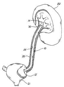
2o Brief Description of the Drawin,~s
Fig. 1 is a schematic view of the stmt of the present invention directed to
pass
through a ureter between a kidney and a urinary bladder;
Fig. 2 is a side view of the stmt in a radially expanded condition;
25 Fig. 3 is a side view of the stmt in a radially compressed and
longitudinally
extended condition;
Fig. 4 is a side view of the stmt of the present invention showing an
introducer
assembly;
Fig. 5 is a cut-away view of the stmt positioned over an introducer assembly;
3o Fig. 6 is a cross-sectional view taken along the axis of both the stmt and
the
introducer assembly;
CA 02365892 2001-09-18
WO 00/66032 PCT/US00/11380
Fig. 7 is an enlarged view of the retention member of the stmt according to
the
present invention;
Fig. 8 is a view of one embodiment of the stmt of the present invention having
convoluted sections at opposing ends of the stmt body;
5 Fig. 9 is a view of one embodiment of the stmt of the present invention
having
convolutions along the length of the stmt body;
Fig. 10 is a view of a material suitable for the construction of the stmt;
Fig. 11 is a view of a forming tool or mandrel being used to form the stmt of
the
present invention;
1o Fig. 12 illustrates the use of a mandrel or forming tool and the use of
heat to set
the material of the stmt to a preferred embodiment;
Fig. 13 is a view of one embodiment of the stmt of the present invention
having a
severable mid-secti.~r~;
Fig. 14 is a view of one embodiment of the stmt having a tether at one end;
Fig. 15 is an end view of the stmt in an elongated condition within a body
passage or vessel;
Fig. 16 is an end view of the stmt in an expanded condition within a body
passage
or vessel;
Fig. 17 is an illustration of the forces applied outwardly from the axis of
the stmt
and against the wall structure of the body passage or vessel;
Fig. 18 is a cut-away view of the stmt within a body passage or vessel in an
expanded condition;
Fig. 19 illustrates the relative length to diameter feature in an expanded
condition
of the stmt;
Fig. 20 illustrates the relative length to diameter feature in an extended
condition
of the stent;
Fig. 21 illustrates the relative length to diameter feature in an intermediate
condition of the stmt;
Fig. 22 is a side elevation view of a stmt formed of filaments and provided
with
3o an impregnation or a coating in a further embodiment of the invention;
Fig. 23 is a radial cross-section view taken along lines 23-23 of Fig. 22 and
illustrating an embodiment wherein the stmt has a central lumen;
CA 02365892 2001-09-18
WO 00/66032 PCT/US00/11380
6
Fig. 24 is a radial cross-section view similar to Fig. 23 and illustrating an
embodiment wherein the stmt has no central lumen;
Fig. 25 is a side elevation view of an embodiment similar to that of Fig. 22
wherein the coating provides sufficient column strength to facilitate
insertion of the stmt;
Fig. 26 is a radial cross-section view taken along lines 26-26 of Fig. 25 and
illustrating the stmt to have a central lumen;
Fig. 27 is a radial cross-section view similar to Fig. 26 and illustrating the
stmt
with no central lumen;
Fig. 28 is a side elevation view similar to Fig. 22 wherein the impregnation
or
1o coating is bio-absorbable;
Fig. 29 is a radial cross-section view taken along lines 29-29 of Fig. 28 and
illustrating the stmt in a low-profile state prior to insertion;
Fig. 30 is a side-elevation view similar to Fig. 28 with the coating at least
partially
oblated or absorbed to permit expansion of the stmt to a high-profile state;
is Fig. 31 is a radial cross-section view taken along lines 31-31 of Fig. 30;
Fig. 32 is a side elevation view similar to Fig. 22 wherein the filaments of
the
stmt are disposed in a generally parallel, axial orientation;
Fig. 33 is a radial cross-section view taken along lines 33-33 of Fig. 32 and
illustrating the stmt to have a central lumen;
2o Fig. 34 is a radial cross-section view similar to Fig. 33 a~:~~l.
illustrating a stmt with
no central lumen;
Fig. 35 is a side elevation view similar to Fig. 22 wherein the filaments are
spiraled in a rope configuration;
Fig. 36 is a radial cross-section view taken along lines 36-36 of Fig. 35 and
25 illustrating the stmt with a central lumen;
Fig. 37 is a radial cross-section view similar to Fig. 36 and illustrating the
stmt
with no central lumen;
Fig. 38 is a perspective view of a helical stmt illustrated in a low-profile
state;
Fig. 39 is a perspective view of the helical stmt of Fig. 38 in a natural,
high-
3o profile state;
Fig. 40 is a perspective view of a stmt having coiled ends and illustrated in
a
stretch configuration;
CA 02365892 2001-09-18
WO 00/66032 PCT/US00/11380
7
Fig. 41 is a perspective view of the coil-end stmt of Fig. 40 illustrated in a
natural
configuration;
Fig. 42 is a side-elevation view of an additional embodiment of the invention
similar to that of Fig. 41;
Fig. 43 is a side-elevation view of a positioner adapted for use with the
embodiment of Fig. 42;
Fig. 44 is a side-elevation view of the stmt of Fig. 42 disposed for insertion
in a
low-profile state on the positioner of Fig. 43;
Figs. 45-47 illustrate steps in a preferred method for insertion of the stmt
of Fig. 42;
Fig. 45 is a schematic view illustrating insertion of the stmt combination of
Fig. 44 over a guidewire;
Fig. 46 is a schematic view illustrating the step of removing the positioner;
Fig. 47 is a schematic view of the ureter illustrating the step of removing
the
guidewire;
Fig. 48 is a side-elevation view illustrating the step of covering the stmt
and
positioner combination with an oversheath;
Fig. 49 is a side-elevation view illustrating the positioner stmt and
oversheath in
combination;
2o Fig. 50 is a side-elevation view of a further embodiment of the invention,
including radiopaque markers;
Fig. 51 is a side-elevation view of a further embodiment of the invention,
including a tether;
Fig. 52 is a side-elevation view of an additional embodiment, including a non-
mesh pigtail anchor;
Fig. 53 is a side-elevation view of a further embodiment having an anchor with
a
spherical shape;
Fig. 54 is a side-elevation view of a further embodiment having multiple
spherical
mesh anchors;
3o Fig. 55 is a side-elevation view of a further embodiment with a preferred
body
portion of the stmt;
CA 02365892 2001-09-18
WO 00/66032 PCT/US00/11380
8
Fig. 56 is a side-elevation view of a further embodiment, including the body
portion with a solid cylindrical element;
Fig. 57 is a side-elevation view of a further embodiment, including a mesh
anchor
and a non-mesh body portion;
Fig. 58 is a side-elevation view of a further embodiment disposed in situ and
having a body portion with an aforeshortened link;
Fig. 59 is a further embodiment of the invention having a filament tether; and
Fig. 60 is a side-elevation view of a further embodiment free of any
anchor portions.
Detailed Description of the Presently Preferred
Embodiments
Turning to Figure 1, a stmt or prosthesis 30 according to the presently
preferred
1 s embodiment is illustrated having a proximal tube end 32 and a distal tube
end 34. The stmt
body 36 is shown within a body passage or vessel 38, such as a ureter. The
stmt body 36
extends within the ureter 38 between a kidney 40 and a urinary bladder 42. The
stmt body 36 of
the present invention is sized and configured to exert a compressive force
against the interior
surface 45 of the body passage 38. In the presently preferred embodiment, the
stmt 30
2o comprises a retention member 48 at the distal tube end 34. The stmt 30 of
the embodiment
shown in Figure 1 comprises a ureteral stmt, which is adapted for developing
or maintaining a
patent lumen in the ureter 38 between the kidney 40 and the urinary bladder
42. The stmt 30
facilitates passage of fluid in, through, and around the stmt body 36 from the
kidney 40 to the
urinary bladder 42.
25 The stmt of the present invention preferably comprises a woven material,
which
can be elongated and contracted. Figure 2 is a side view of the stmt 30 in a
contracted, radially
expanded condition. The condition illustrated in Figure 2 corresponds to an
"at rest" or natural
condition of the stmt 30. The lumen of the stmt body 36 is fully developed
along the length of
the stmt body 36, narrowing only at the distal tube end 34. The retention
member 48, which
3o forms a cuff or enlargement sized and configured to engage a portion of an
organ or passage, has
an enlarged diameter in the natural condition shown in Figure 2. The retention
member 48
assists in maintaining the stmt 30 within the body passage 38, as illustrated
in Figure 1, for
example.
CA 02365892 2001-09-18
WO 00/66032 PCT/US00/11380
9
Figure 3 illustrates the stmt 30 in a stretched, radially compressed and
longitudinally extended condition. The stmt body 36 is preferably reduced in
diameter in order
to facilitate placement of the stmt 30 into a body passage 38. When the
stent.30 is stretched
along its axis, the diameters of the stmt body 36 and the retention member 48
are significantly
s reduced to facilitate a low-profile configuration for insertion into the
body passage 38. As
presently embodied, the stmt 30 is placed into the low-profile condition by
application of a
tensile force applied to both the proximal tube end 32 and the distal tube end
34.
As illustrated in Figure 4, a compression sleeve 60, having a proximal end 62
and
a distal end 64 (Figure 5), can be inserted into a lumen of the stmt 30. The
compression
1o sleeve 60 is preferably inserted into the lumen of the stmt 30, until the
distal end 64 of the
compression sleeve 60 contacts the distal tube end 34 of the stmt 30. After
this placement, the
proximal tube end 32 of the stmt 30 can be drawn proximally, relative to the
compression
sleeve 60, to thereuy facilitate elongation of the stmt 30. In other words,
since the distal end of
the compression sleeve 60 cannot pass through the narrow aperture of the
distal tube end 34,
15 movement of the proximal tube end 32 proximally will lengthen the stmt 30.
As the stmt 30
increases in length, the diameter of the stmt 30 decreases. The reduced
diameter of the stmt 30
facilitates a less-intrusive insertion of the assembly into a body passage 38.
A guidewire 70, having a proximal end 72 and a distal end 74, may be placed
within the compression sleeve 60. The guidewire 70 provides a means for
establishing a track,
2o so that the stmt 30 and compression sleeve 60 may be advanced along the
guidewire 70 to a
desired location within the body passage 38, with the stmt 30 in an elongated
configuration.
After the stmt 30 is moved to the desired location, the proximal tube end 32
of the stmt 30 is
released or relaxed, to thereby allow the proximal tube end 32 to move
distally, resulting in an
enlargement of the diameter of the stmt 30. According to the presently
preferred method of
25 insertion, the guidewire 70 is placed within the body passage 38, and the
stmt 30 is then placed
over the proximal end 72 of the guidewire 70. Next, the compression sleeve 60
is placed over
the proximal end 72 of the guidewire 70 and into the stmt body 36.
Figure 5 illustrates a cut-away view of the stmt 30 positioned over both the
compression sleeve 60 and the guidewire 70, and Figure 6 illustrates a cross-
sectional view of
30 the assembly shown in Figure 5. As illustrated in Figures S and 6, the
compression sleeve 60 fits
between the stmt 30 and the guidewire 70. The opening at the distal end 34 of
the stmt 30 does
not permit the distal end 64 of the compression sleeve 60 to pass through.
This configuration
CA 02365892 2001-09-18
WO 00/66032 PCT/US00/11380
permits the stmt 30 to be stretched lengthwise, as the proximal end 32 of the
stmt 30 is extended
proximally along the surface of the compression sleeve 60. At full extension,
the profile of the
stmt 30 exceeds the outside diameter of the compression sleeve 60 by the
thickness of the wall
of the stmt body 36. This extended/compressed relationship exists as long as a
holding force is
maintained between the proximal end 32 of the stmt 30 and the compression
sleeve 60. When
this force is removed, the stmt 30 assumes an "at rest" or expanded profile.
Figure 7 illustrates an enlarged view of the retention member 48 of the
presently
preferred embodiment. The retention member 48 preferably comprises an enlarged
diameter
capable of engaging a portion within a vessel or organ, to thereby prevent the
stmt 30 from
10 migrating or slipping from a desired position or location within the vessel
or organ. The distal
ring 81 of the retention member 48 is preferably sized and configured to
prevent the compression
sleeve 60 (Figure 5) from passing therethrough. The distal ring 81 preferably
comprises a
thermally fused or melted portion of material fibers 84 from which the stmt 30
is woven. The
distal ring 81, however, may be formed in other ways and/or comprise other
materials. In the
presently preferred embodiment, the retention member 48 comprises the shape of
a cone 87
having a small diameter portion 89 distally located from a large diameter
portion 92. The
retention member 48 preferably comprises a substantially folded lip section 95
and a
substantially folded angular portion 98 providing a transition between the
stmt body 36 and the
retention member 48.
2o Figures 8 and 9 illustrate stems 30 having series of con ;~c~l.utions 100,
102, and
104 formed along the stmt bodies 48. These convolutions 100, 102, 104 can
operate to add
strength to the retention members 48 and 107. The convolutions 100, 102, 104
also provide
additional strength to the stmt bodies 36 for resisting compression in much
the same way as
corrugated tubing resists kinking and compression. Additionally, the
convolutions 100, 102, 104
assist in providing traction within the lumen of a body passage 38 and are
sized and configured
to be reduced in profile in the same manner as the stmt body 36 by the
application of traction or
tension upon the stmt body 36.
As illustrated in Figure 10, the stmt 30 is formed from an initial woven
tubular
structure 111, which preferably comprises a thermoplastic material or mesh.
This construction
3o begins by weaving or braiding a plurality of individual or groups of
individual fibers or
elements 84 into a tubular stmt body 36. Desired characteristics may be
developed within this
construction for providing ratios of expansion to extension, as is known in
the art.
CA 02365892 2001-09-18
WO 00/66032 PCT/US00/11380
11
After the woven tubular structure 111 is generated, the woven tubular
structure 111 is placed onto a forming tool or mandrel 113 having a proximal
end 115 and a
distal end 117. The mandrel 113 serves as a form in setting the thermoplastic
material of the
woven tubular structure 111. In the presently preferred embodiment, the
forming tool 113
comprises a first diameter near the proximal end 115 and a second diameter
near the distal
end 117. The first diameter represents the desired maximum deployed or
expanded diameter of
the stmt body 36 when the stmt body 36 is within a body passage or vessel 38,
and the second
diameter corresponds to the diameter of a conventional guidewire 70 (Figure 6)
but compression
sleeve 60 (Figure 6).
to Alternatively, the stmt 30 can be formed of metal material such as Nitinol
(a
trademark of Raychem, Inc.) or a titanium. Nitinol is well-known for its
heatset properties which
would enable it to function in the manner previously discussed. Titanium has
excellent bio-
compatibility features which might make it a preferred material in a
particular environment.
The woven tubular structure 111 of the stmt 30 is folded proximally upon the
forming tool 113 to thereby form the retention member 48. As shown in Figure
12, the forming
tool 113 and the woven tubular structure 111 are next exposed to radiation 121
from a heat
source or an oven preferably at a temperature sufficient to set the material
of the woven tubular
structure 111 to the preferred condition. In the presently preferred
embodiment, the material
comprises a thermoplastic, such as a polyester or nylon, since these materials
allow for the
2o development of a permanent, thermally-set condition. Additionally, the
distal tube end 34 and
the distal ring 81 are preferably fused or melted to form a solid ring or
collar which provides
support for the compression sleeve 60. As a secondary operation, a proximal
portion 123 of the
stmt body 36 may be coated with an elastomeric material to thereby provide
stability at the
proximal portion 123.
Figure 13 illustrates a stmt 30 having a tether 130 attached or formed at the
proximal tube end 32 for assisting in the placement or the removal of the stmt
30 from a
body passage 38.
Figure 14 illustrates a stmt having a first retention member 48 and a second
retention member 136 located at an end opposite from the first retention
member 48. The stmt
3o having the two retention members 48, 136 may be used as is or,
alternatively, the stmt may be
cut at a preferred location 138 to form two individual stems 140 and 142.
CA 02365892 2001-09-18
WO 00/66032 PCT/US00/11380
12
Figure 15 illustrates an end view of the stmt 30 of the presently preferred
embodiment within a body passage 38. The stmt 30 is illustrated in an
extended, small diameter
condition over both the compression sleeve 60 and the guidewire 70. Figures 16
and 17 illustrate
the stmt 30 in a large-diameter relaxed state. The guidewire 70 and the
compression sleeve 60
may be removed at this time. The stmt body 36 exerts a constant outward
pressure 151 upon the
interior surface 45 of the body passage 38. This outwardly directed radial
pressure, along with
the naturally occurring tendency for the intimal tissue to move away from a
foreign body,
combines to enlarge and/or maintain the lumen of the body passage 20.
An enlarged view of a body passage 38 is provided in Figure 18 with a stmt 30
of
1o the presently preferred embodiment fully extended within the lumen of the
body passage 38.
The individual fibers or groups of fibers 84 are spaced apart to thereby allow
for the flow 155 of
fluid through and around the stmt body 36 as the stmt body 36 applies outward
pressure to the
interior surface 45 of the body passage 38.
The relationship between the length and the diameter of the stmt 30 of the
present
invention is illustrated in Figures 19-21. The stmt 30 in the "at rest" or
natural, relaxed
condition is illustrated in Figure 19 with a fully expanded, maximum diameter
172. Due to the
naturally occurring relationship of the fibers or elements 84 of a woven or
braided tubular
structure 111 (Figure 10), a change in length 170 will accompany any change in
diameter 172.
Conversely, any change in length 170 precipitates a commensurate change in
diameter 172. The
present invention harnesses this relationship to facilitate the placement,
maintenance, and
removal of the stmt 30. As presently embodied, the length 174 and the diameter
176 of the
retention member 48 change somewhat proportionally to changes in the length
170 and
diameter 172 of the stmt body 36.
With reference to Figure 20, as the stmt 30 is stretched or extended in length
180,
181, the diameters 182 of the stmt body 36 and the diameter 186 of the
retention member 48 are
both reduced. Upon removal or relaxation of the stretching or extending force,
the stmt 30
attempts to assume an original "thermally set" or natural condition within the
body passage.
Accordingly, the length 190 and the diameter 192 increase from the length 180
and the
diameter 182 of Figure 20, as illustrated in Figure 21. Similarly, the length
191 and the
diameter 196 of the retention member 48 increase. The increased diameters 192,
196 exert
radially outwardly directed forces upon any resistive structure. As the
diameters 192, 196
CA 02365892 2001-09-18
WO 00/66032 PCT/US00/11380
13
increase, the lumen within the body passage 38 will also increase, thereby
facilitating further
increases in the diameters 192, 196.
The intimal tissue of the body passage 38 responds to the presence of the
braided
material by moving away from the stmt 30. Thus, the lumen of the body passage
38 enlarges in
response to the presence of the stmt 30. As the lumen enlarges, the self
expanding stmt 30
follows the inner surface of the body passage 38 and continues to expand.
This, in turn,
stimulates further enlargement of the lumen of the body passage 38. This
expansion-response of
the stmt 30 and body passage 38 continues until a maximum lumen diameter is
achieved.
The expansion-response reaction of the body passage 38 is believed to be a
to reaction to the members of the braided material and the motion of these
members within the
body passage 38, especially when the body passage comprises a ureter. The
expansion-response
reaction may also be attributed generally to a foreign body reaction within
the body passage 38.
In the particular case: of a ureter, it is believed that the irritation from
the braided or woven
members causes this response.
15 A further embodiment of the invention is illustrated in Figure 22 wherein
elements of similar structure are designated by the same reference numeral
followed by the lower
case letter "a." Thus, stmt 30a includes a plurality of fibers or filaments
84a which extend
generally along an axis 200 between proximal end 32a and distal end 34a.
The filaments 84a may be oriented to provide the stmt 30a with a central
20 lumen 203, best illustrated in Figure 23. Alternatively, the stmt 30a may
be formed with a
generally solid configuration, free of any central lumen, as illustrated in
Figure 24. In
combination these filaments 84a provide the stmt 30a with a generally
cylindrical outer
surface 202. In the embodiment of Figure 22, the stmt 30a is also provided
with a material 204
which at least partially impregnates and/or coats the filaments 84a.
25 The filaments 84a, together with the material 204, can provide the stmt 30a
with a
variety of characteristics. For example, in Figure 22, the filaments 84a have
a generally rigid
configuration when coated or impregnated with the material 204. However, in
the absence of the
material 204, the filaments 84a may be more limp and flexible. Taking
advantage of these
characteristics of the filaments 84a, the material 204 can be chosen with bio-
absorbable
3o characteristics. When the material 204 is disposed relative to the
filaments 84a, the stmt 30a has
the generally rigid characteristics which facilitate its insertion into the
body passage or conduit.
However, once the stmt 30a is operatively disposed within the conduit, the
material 204 is at
CA 02365892 2001-09-18
WO 00/66032 PCT/US00/11380
14
least partially absorbed or otherwise removed, leaving the stmt 30a with the
generally flexible
characteristics and thereby facilitating the fluid-flow properties of the
stmt.
In the embodiment of Figure 25, elements of similar structure are designated
by
the same reference numerals followed by the lower case letter "b." In this
case, the material 204b
is coated on, impregnated into, or otherwise disposed relative to the
filaments 84b. As illustrated
in Figures 26 and 27, the stmt 30b may be provided with a central lumen 203b
or, alternatively,
provided with a generally solid structure, respectively. The material 204b is
of particular interest
in this embodiment as it is chosen to provide the stent 30b with a generally
fixed, predetermined
length and diameter. Nevertheless, the material 204b may be very flexible. In
this case, the stmt
1o facilitates fluid flow between its ends 32b and 34b in a "wicking" action.
Another embodiment is illustrated in Figure 28 wherein like elements of
structure
are designated by the same reference numeral followed by the lower case letter
"c." In this case,
the characteristics chosen for the filaments 84c and the material 204c are of
particular interest.
For example, the filaments 84c can be made from a material having expansion
characteristics
15 which cause the stmt 30c to automatically move from a low-profile state to
a high-profile state.
The material 204c can be chosen with bio-absorbable characteristics whereby
the stmt 30c is
maintained in its low-profile state in the presence of the material 204c, as
illustrated in Figure 28.
In this low-profile state, the stmt 30c may have a generally solid
configuration as illustrated in
the radial cross-section view of Figure 29.
2o In this embodiment, it is the properties of the material 2~,~=.~c which
initially hold
the filaments 84c in the low-profile state. However, after the stmt 30c is
inserted into the body
conduit, these bio-absorbable characteristics cause the material 204c to be
absorbed, ablated, or
otherwise at least partially removed from the filaments 84c. This permits the
filaments 84c to
expand to the high-profile state as illustrated in Figure 30. In this view,
and the radial cross-
25 section view of Figure 31, a dotted line 206 illustrates the material 204c
in a partially removed
state permitting automatic expansion of the filaments 84c. In this embodiment,
the bio-
absorbable material 204 includes polyglycolic acid.
It can be seen that in several of these embodiments, it is the combination of
characteristics present in the filaments 84 and the material 204 which are
relied on to provide the
3o stmt 10 with different properties facilitating insertion on the one hand
and operative disposition
on the other hand. For example, in the embodiment of Figure 28, the filaments
84c have first
characteristics, such as a low-profile, and second characteristics, such as a
high-profile.
CA 02365892 2001-09-18
WO 00/66032 PCT/US00/11380
Similarly, the material 204 has first characteristics, such as an integrous
coating on the outer
surface 202c, and second characteristics, such as a weakened or absorbable
coating. In
combination, the first characteristics of the material 204 facilitates the
first characteristics of the
filaments 84 while inhibiting the second characteristics of the filaments 84.
This facilitates
5 insertion of the stmt 10. When the stmt 10 is operatively disposed, the
second characteristics of
the material 204 facilitate the second characteristics of the filaments 84
while inhibiting the first
characteristics of the filaments 84. This provides the stmt 30c with the best
performance when
disposed at the operative site.
The embodiments of Figures 32, 35, and 38 include elements similar to those
t0 previously discussed which are designated by the same reference numerals
followed by the lower
case letters "d", "e", and "f', respectively. These embodiments are
illustrative of the fact that the
filaments 84 can be disposed in any relative configuration typically providing
the stmt 10 with
an elongate, cylindrical configuration. For example, the filaments 84c in
Figure 28 may be
woven whereas the filaments 84d in Figure 32 are generally straight and
parallel to the axis 200d.
15 These filaments 84d can be oriented to provide the stmt lOd with a central
lumen 203d as
illustrated in Figure 33, or a generally solid configuration as illustrated in
Figure 34. In this
embodiment, the material 204d is shown to be impregnated into the filaments
84d.
In a further orientation, illustrated in Figure 35, the filaments 84e are
spiraled in a
rope configuration. This embodiment may also be formed with a central lumen
203e as
2o illustrated in Figure 36, or a generally solid configuration as illustrated
in Figure 37.
A further embodiment providing a spiraled configuration is illustrated in
Figure 8
wherein the stmt 30f is formed as a helix or spring. With this configuration,
the stent 30f may
have a single element 84f or a polarity of elements each forming a helical
spring. Where
multiple springs are contemplated, the elements 84f may be disposed one within
the other and
may also be spiraled in different directions.
In Figure 38, the stmt 30f is illustrated in a low-profile state which is
achieved by
separating the ends 32f and 34f. This low-profile state facilitates insertion
of the stmt 30f.
When the ends 32f and 34f are released, the helix is free to return to its
normal high-profile state,
as illustrated in Figure 39. In this embodiment, the desired freedom of
movement of the
filament 84f between its ends 32f and 34f is facilitated by the convolutions
of the helical spring
which are free to move relative to each other. Coils 205 and 206 can be formed
at the ends 32f
and 34f as illustrated in Figure 39. These coils 205, 206, which automatically
form when the
CA 02365892 2001-09-18
WO 00/66032 PCT/US00/11380
16
stmt 30f is in its natural state, tend to anchor the stmt 30f in its operative
position. Of course,
when the stmt 30f is initially inserted, it is desirable that these coils 205
and 206 straighten along
the axis of the stmt as illustrated in Figure 38. In this stretched
configuration, the stmt 30f can
be easily inserted into the conduit and then released to form its untensioned,
natural state as
illustrated in Figure 39.
These same coils 205 and 206 can be formed in the embodiment illustrated in
Figures 40 and 41 wherein similar elements are designated by the same
reference numerals
followed by the lower-case letter "g". In this embodiment, the stmt 30g is
formed from braided
or woven elements 84g which extend between the stmt ends 32g and 34g. In this
embodiment,
1o the coils 2058 and 206g can be formed in the ends of the stmt 30g as
previously discussed.
These coils 205g and 206g can be axially oriented by tensioning the stmt 30g
as illustrated in
Figure 40. This facilitates insertion of the stmt 30g which returns to its
natural state as
illustrated in Figure 41 when tension is removed at the operative site.
The stmt 30g is further illustrated in Figure 42 to include a body portion 210
with
a proximal end 212 and a distal end 214. This body portion 210 has a tubular
configuration with
a diameter such as one-eighth inch to one-quarter inch. Extending from the
distal end 214, an
anchor portion 216 can be provided in a contiguous relationship with the body
portion 210. This
anchor portion 216 also has a tubular configuration and is connected at one of
its ends to the
distal end 214 and is provided at its other end with a constriction 218. The
anchor portion 216
2o may also have a tubular configuration and may be formed as an extension of
the mesh defining
the body portion 210. A similar anchor portion 221 can be coupled to the
proximal end 212, but
it is preferably formed without a constriction.
As in previous embodiments, the filaments forming the mesh of the stmt 30g can
be heatset so that, at rest, the stmt 30g tends toward the general shape
illustrated in Figure 42.
This shape includes the enlarged body portion 210, as well as the pigtail
configuration of the
anchor portions 216 and 221. With these heatset properties, the stmt 30g is
particularly adapted
for insertion using a positioner 223 such as that illustrated in Figure 43.
This positioner 223
preferably has the configuration of a tube with an interior lumen 225. The
positioner 223 can be
formed of flexible or semi-rigid material, and provided with a generally
straight, but bendable,
3o configuration.
In operation, the positioner 223 is inserted into the anchor portion 221 at
the
proximal end of the stmt 30g. It is moved through the stmt 30g until it abuts
the
CA 02365892 2001-09-18
WO 00/66032 PCT/US00/11380
17
constriction 218 at the end of the anchor portion 216, as illustrated in
Figure 44. In this
configuration, the stmt 30g is maintained in a generally straight
configuration and stretched to a
low-profile state facilitating insertion.
Operative disposition of the stmt 30g is best described with reference to
Figures 45-47. In these figures, the ureter 38g is illustrated between the
bladder 42g and the
kidney 40g. Initially, a guidewire 230 can be introduced through the bladder
42g and into the
kidney 40g. With the positioner 223 operatively disposed in the stmt 30g, as
illustrated in
Figure 44, this combination can be introduced over the guidewire 230g, as
illustrated in
Figure 45. In accordance with this method, the guidewire tends to guide the
positioner 223 and
1o stmt 30g through the tortuous path of the ureter 38g.
Once the stmt 30g is appropriately positioned with the body portion 210
disposed
in the ureter 30g, the positioner 223 can be withdrawn leaving the stmt 30g
and the
guidewire 230, as ii~ustrated in Figure 46. The guidewire 230 can then be
moved from the
stmt 30g leaving the stmt 30g operatively disposed with the body portion 210
in the ureter 38g,
the anchor portion 216 in the kidney 40g, and the anchor portion 221 in the
bladder 42g. In the
absence of either the positioner 223, or the guidewire 230, the heatset
characteristics will cause
the ends of the stmt 30g to curl or coil into a pigtail configuration, as
illustrated in Figure 47.
These same heatset characteristics will cause the body portion 210 of the stmt
30g to expand,
thereby irritating the walls of the ureter 38g and causing them to further
expand the diameter of
the ureter 38g.
As illustrated in Figure 48, a second pusher 235 can be provided to abut the
proximal end of the stmt which is mounted on the first positioner 223. The
second
positioner 235 can aid in releasing the stmt 30g from the first positioner 223
as it is withdrawn
through the stmt.
An oversheath 236, but illustrated in Figure 49, can be provided to cover the
combination of the positioner 223 and stmt 30g. When operatively disposed, the
oversheath 236
covers at least a portion of the stmt 30g, as illustrated in Figure 49. The
placement of
radiopaque markers 238 and 241 on the stmt 30g and sheath 236, respectively,
can facilitate
maintenance of this operative disposition. When the oversheath 236 is in
place, the mesh
3o configuration of the stmt 30g is replaced with a smooth outer surface of
the oversheath 236 to
facilitate introduction of the stmt into the ureter 38g.
CA 02365892 2001-09-18
WO 00/66032 PCT/US00/11380
18
Other radiopaque markers can be provided on a stmt 30g, as illustrated in
Figure 50. In addition to the marker 238 at the end of the distal anchor
portion 216, a similar
radiopaque marker 243 can be provided at the end of the proximal anchor
portion 221. A
verification marker 245 can be provided along the distal anchor portion 216 in
proximity to the
body portion 210. Since the mesh of the stmt 30g is generally not visible
under fluoroscopy,
movement of the marker 238 into proximity with the verification marker 245
will provide an
indication that the loop, coil, or pigtail of the anchor portion 216 has
formed.
Figure 51 illustrates a further embodiment of the stmt wherein elements of
similar
structure are designated by the same reference numerals followed by the lower
case letter "h". In
to this particular embodiment, there is no anchor portion 221, but rather a
generally straight tether
which is attached to the proximal end of the body portion 210h. In those case
where an anchor is
not required in the bladder 42g, the tether 245 will merely provide a
connection to the body
portion 210 to ultimately facilitate removal of the stmt 30h.
A further embodiment of the stmt 30i is illustrated in Figure 52 wherein the
anchor portion 2161 is formed from a material such as a silicon, urethane, or
other elastomer, but
is not provided with the mesh configuration. Where the anchor portion 216i is
not formed
integral with the body portion 210i, these elements 2161 and 210i must be
coupled at a
junction 247 by other means such as an adhesive or a mechanical interlock. In
this embodiment
of Figure 52, the junction 247 can be formed with the restriction 218i so that
the positioner, such
2o as the positioner 223 of Figure 24, extends only to this junction 247. :are
this case, the
guidewire 230i is relied on to straighten the anchor 216i during insertion.
The positioner 223i
functions to push the anchor portions 216i, and to pull the remainder of the
stmt 30i distal of the
junction 247.
Further embodiments of the invention are illustrated in the side-elevation
views of
Figures 53, 54, 55, 56, 57, 58, 59, and 60. In these views, elements similar
to those previously
discussed are designated by the same reference numerals followed by the lower-
case letters j, k,
l, m, n, o, p, and q, respectively. For example, in the embodiment of Figure
53, the stmt is
designated by the reference numeral 30j. In this embodiment, the body portion
210j and the
tether 245j can be similar to those previously discussed. A distal anchor 250
can be heatset in
the general configuration of a sphere 252 having a diameter such as one-half
inch to one inch in
certain preferred embodiments. The sphere 252 can be formed of any of the
materials previously
CA 02365892 2001-09-18
WO 00/66032 PCT/US00/11380
19
discussed, but in a preferred embodiment is formed of a mesh material which is
integral with the
mesh of the body portion 210j.
Since most of the patient discomfort associated with stems results from the
anchors in the bladder 42 and kidney 40, the spherical anchor 250 offers
considerable advantage
to this embodiment of the invention. The only contact with the kidney in this
case is along a
hemispherical surface 254 which contacts the body portion 210j. This advantage
is achieved
without sacrificing the advantages of previous embodiments which provide for
use of a
positioner, such as the positioner 223 of Figure 24. Tensioning the stmt 30j
on such a positioner
causes the sphere 252 to collapse to a cylindrical, low-profile configuration
facilitating insertion.
1o Upon removal of the positioner 223 and guidewire 230 (Figures 46 and 47),
the heatset mesh
automatically expands to form the spherical anchor 250.
The embodiment of Figure 54 illustrates that the stmt 30k can be formed not
only
with the distal spherical anchor 250k, but also a proximal spherical anchor
256.
In the embodiment of Figure 55, the stmt 301 includes pigtail anchors 2161 and
2211 of the type previously discussed. In this embodiment, the body portion
2101 differs from
the generally cylindrical configuration previously discussed. In this case,
the body portion 2101
includes a central portion 261 which is heatset to a generally cylindrical
configuration. The body
portion 2101 also includes tapered portions 263 and 265 which are disposed at
opposite ends of
the central portion 261. The tapered portion 263 is connected between the
central portion 261
2o and the distal anchor 2161, while the proximal tapered portion 265 is
connected between the
central portion 261 and the proximal end 2211. The distal taper 263 in this
embodiment is
provided with a relatively large taper angle making this portion 263
relatively short compared to
the proximal tapered portion 265 where the taper angle is relatively small. In
many of the other
aspects of the stmt 301, features are similar to those previously discussed
which provide for low-
profile insertion using a positioner, such as the positioner 223 of Figure 24.
A further embodiment of the stmt is illustrated in Figure 56 and designated by
the
reference numeral 30m. This embodiment includes the mesh pigtail 216m, as well
as a mesh
body portion 210m with tapered portions 263m and 265m. In this embodiment, the
body
portion 210m also includes a cylindrical portion 267 which is formed of a
solid material and
3o joined to the mesh material of the tapered portion 265m at a junction 269.
The cylindrical
portion 267 can be formed of silicone, urethane, or other elastomer. This
material can be joined
CA 02365892 2001-09-18
WO 00/66032 PCT/US00/11380
to the mesh at the junction 269 by adhesive or by a mechanical, heatset
interlock between the
fibers of the mesh and the solid material of the cylinder 267.
The embodiment of Figure 57 is similar to that of Figure 56 in that it
includes the
cylinder 267n and proximal anchor 221n. In this embodiment, the mesh body
portion 210n has
5 been eliminated, but the distal mesh spherical anchor 252 has been retained.
The stmt 30o illustrated in Figure 58 combines the spherical mesh anchor 2500
of
the. Figure 53 embodiment, as well as the body portion 210o and tether 245o
associated with the
Figure 51 embodiment. In this case, it is noted that the body portion 210 has
a length which is
shorter than the length of the ureter 38. Realizing that the incision is made
in the upper portions
10 of the ureter 380, and that the features of the stmt 30o are most
appreciated in the vicinity of the
incision, the body portion 2100 of this embodiment is limited to that region.
In a preferred
embodiment, the shortened length of the body portion 210o is about one-half
the length of the
ureter 380. Only the tether 245o extends through the proximal end of the
ureter 38o and into the
bladder 420. The stmt 30p illustrated in Figure 59 is similar to that
illustrated in Figure 58,
15 except that the tether 2480 is formed as a solid shaft, string, or filament
270.
A further embodiment of the invention is illustrated in Figure 60, where the
stmt 30q is free of any anchors such as the distal anchor 216 or proximal
anchor 221 of the
embodiments previously discussed. This embodiment can still be formed of a
mesh material and
provided with a body portion 210q terminating in a distal taper 272 and a
proximal taper 274. At
2o the distal end, the constriction 218q can be formed to facilitate insertion
with a positioner, such
as the positioner 223 of Figure 24.
It can be seen from the foregoing discussion that various embodiments of this
concept include at least one filament which is formed from a relatively strong
material such as
polyester. While this material may be strong and somewhat rigid, the stmt 30
is provided with
relatively soft characteristics due to the configuration applied to the
filaments 84. Movement of
the filaments 84 between the ends 32 and 34 of the stmt 30 is desired not only
to facilitate this
soft characteristic, but also to "irritate" the wall of the conduit. This
causes the conduit wall to
move away from the stmt 30 thereby increasing the patency of the conduit. In
some cases, the
stmt 30 is provided with characteristics to naturally move toward a larger
diameter. With these
3o properties, the stmt 30 effectively chases the wall radially outwardly to
further increase the
patency of the conduit.
CA 02365892 2001-09-18
WO 00/66032 PCT/US00/11380
21
Between the ends 32 and 34 of the stmt 30, the elements 84 are free to move
relative to each other between a low-profile state facilitating insertion and
a high-profile state
facilitating conduit patency. This relative movement of the elements 84 not
only facilitates the
soft characteristics preferred for the stmt 30, but also results in the
desired irntation of the
conduit wall.
Although it is contemplated that most embodiments of the stmt 30 will include
elements 84 formed of the same material, this may not always be the case. In
some instances, it
may be desirable to form the elements 84 from different materials to provide
the overall stmt 30
with properties representative of each of the materials. For example, some of
the elements 84
1o may be formed from a polyester material providing the stmt with a
relatively high tensile
strength. Other elements may be formed of an absorbent material which can be
saturated, for
example, with an antibiotic, an anesthetic, an analgesic, a material to
control encrustation, a
radiopaque material, ~3r any other material having medical characteristics.
The impregnation or coating of the elements 84 with drugs or chemicals offers
~5 particular advantages. For example, some procedures require such chemicals
or drugs to be
administered at a specific site within a body passage. When these drugs or
chemicals are
administered systemically, there can be concomitant and adverse side-effects.
When it is
desirable to administer medications, drugs, or chemicals, particularly those
that are highly
concentrated or powerful, a system for localizing the effect to a specific
site can be particularly
2o advantageous in avoiding the side-effects of systemic administration. To
this end, an
intraluminal device for local administration of the medications, drugs, or
chemicals is
contemplated by the present invention.
More specifically, a stmt 30 having properties for absorbing and subsequently
delivering or releasing a chemical or a drug is foreseen. When the stmt 30 is
provided with a
25 woven or braided tubular structure, it can be inserted into a body passage
for the purpose of
increasing patency of that passage. The stmt can be constructed solely of mono-
filament fibers
or a rigid polymer, as previously discussed. These fibers are generally non-
absorbent.
However, in an alternate embodiment, at least one of the elements can be
formed of cotton,
dacron, or other absorbent material. These absorbent elements can be woven
with the mono-
3o filaments elements, in a predetermined ratio facilitating delivery of an
absorbed chemical, drug,
or medication. The stmt can then be soaked, wiped, or doped with the selective
chemical or
combination of chemicals or drugs. The absorbent elements may be formed as a
yarn and
CA 02365892 2001-09-18
WO 00/66032 PCT/US00/11380
22
provided with various properties including alternative rates of absorption or
take-up of the
chemical, as well as alternative rates of release or delivery of the chemical.
This may be
accomplished by blending various fibers within a single yarn element or by
controlling the
density of the weave or the chemical or mechanical treatment of the surface of
the yarn element.
The releasing element may also be made of an absorbable material that releases
the chemical or drug as the element desolves in body fluids. The agents may be
time-released or
bolused, depending on the properties of the fiber elements. The agents to be
released or
administered can be compounded so that a single woven or braided element
contains a variety of
agents to be delivered at defined rates and dosages over different times. Many
other
1o combinations of elements and materials will be apparent to provide the stmt
with selective
characteristics desirable in a particular operative setting.
Although exemplary embodiments of the invention have been shown and
described, many other changes, modifications, and substitutions will now be
apparent to those of
ordinary skill in the art, without necessarily departing from the spirit and
scope of this invention
15 as set forth in the following claims.