Note: Descriptions are shown in the official language in which they were submitted.
CA 02366676 2001-09-21
WO 00/56208 PCT/US00/07435
BIOPSY NEEDLE INSTRUMENT
TECHNICAL FIELD
The present invention relates in general to the
field of medical biopsy instruments, and more
particularly, to such instruments for use in fine needle
biopsy of human or animal tissue for medical diagnostics
and the like.
BACKGROUND ART
Biopsy instruments are often used to obtain
tissue samples for microscopic examination to test for
malignancy or other diseases and abnormalities.
Generally, biopsies may be guided by either stereotactic
means, CAT scan or ultrasound means. Image-guided biopsy
procedures are particularly useful for non-surgical
diagnosis of benign and malignant masses. The biopsy
itself may be either a core biopsy or a fine needle
aspiration biopsy. For example, an instrument for
performing percutaneous biopsy procedures and collection
of soft tissue is disclosed in Ritchant, et al., U.S.
Patent No. 5,649,547.
Other currently used biopsy instruments and
methods include those disclosed in Siczek, et al., U.S.
Patent No. 5,415,169 and Assa, U.S. Patent No. 5,240,011.
Siczek, et al. and Assa each disclose a motorized biopsy
needle positioner employed in a mammographic needle
biopsy system for receiving coordinate information
representative of an identified point of interest within
the patient's captive breast under examination and
automatically positioning a biopsy needle in accordance
with the coordinate information to permit insertion of
the biopsy needle to the identified point of interest.
CA 02366676 2001-09-21
WO 00/56208 PCT/US00/07435
-2-
Additionally, Clement, et al., U.S. Patent No.
5,368,045 discloses a handheld biopsy needle instrument
employing combined stylets and cannulas capable of taking
multiple specimens while the other hand is free to
manipulate an ultrasound probe. The stylet and cannulas
are spring loaded, which upon firing, will penetrate the
tissue for obtaining a biopsy specimen. A similar biopsy
instrument having a plurality of stylets and cannulas
which can be controlled independently for capturing a
plurality of discreet specimens at a controlled depth is
disclosed in Chin, et al., U.S. Patent No. 5,415,182.
See also Akerfeldt, U.S. Patent No. 4,944,308.
Fine needle aspiration biopsy is often
performed on a potentially malignant mass for
confirmation of diagnosis prior to surgery, on more than
one mass where multi-focal or multi-centric malignant
disease is suspected, on a suspected benign lesion such
as a fibroadenoma, where there is ambivalence about
follow-up versus excision, or on an ultrasound imaged
structure with features unlike a simple cyst. Among the
benefits of fine needle aspiration when compared with
other biopsy procedures are that it is less invasive,
requires no incision, causes minimal discomfort, takes
less time and costs considerably less. A discussion of
fine needle aspiration is disclosed in the article Fine
Needle Aspiration, Kathleen M. Harris, M.D., FACR, pp.
101-105.
Suction and capillary methods of aspiration
have been successful on the breast. For suction
aspiration, a syringe in a resting position is attached
to a sampling needle. Suction is created by pulling the
plunger of the syringe. In the capillary method, a
syringe is not used and suction is not applied. With
both methods, up to the present time the sampling needle
is manually moved back and forth rapidly by the physician
within the area to be studied. The needle is further
CA 02366676 2001-09-21
WO 00/56208 - 3 - PCT/US00/07435
angled in multiple directions to sample a cone-shaped
area within the area to be studied. In the suction
method, the suction should be maintained until material
is visible in the plastic needle hub, or for a minimum of
twenty up-and-down motions in varying directions. This
method is described further in Interventional Breast
Procedures, edited by D. David Dershaw, pp. 91, 94 and
95. A similar technique is described in General
Ultrasound, Ed., Carol A. Mittelstaedt, M.D., pg. 18.
l0 The technique is also described in Interventional Breast
Ultrasonography, Ellen B. Mendelson, M.D., pp. 57-76.
Another similar technique is that discussed in Thyroid
and Parathyroid, pg. 107.
Until now, and as described in the foregoing
references, fine needle aspiration biopsies have been
performed manually. Such a procedure involves manually
thrusting a needle alone or a needle attached to a
syringe, with or without suction. The procedure is
generally random in that the depth of the thrusts, number
of thrusts, the area covered and the force used are done
in a very haphazard way. For example, one thrust could
be 5 millimeters, while another could be 2 millimeters
and so forth.
A significant limitation with random depth is
that when a lesion is very small in diameter, there are
occasions where none or a few of the thrusts obtain the
necessary tissue sample. One of the thrusts may be
directed to a lesion, but may bypass the lesion
completely as a result of a lack of consistent direction
of the thrusts. Random depth results in a significant
amount of fine needle aspiration biopsies retrieving an
insufficient amount of tissue with which to do an
appropriate diagnostic evaluation. If the number of
thrusts is limited, this compounds the problem further
and increases the chances of missing the lesion.
CA 02366676 2001-09-21
WO 00/56208 PCT/tJS00/07435
-4-
Another limitation of the prior method is lack
of significant thrusting energy. The force behind the
thrust may be variable, and many may be insufficient
enough to pierce the outer margins of certain lesions,
especially fibroadenomas. The needle can potentially
bounce off the fibroadenoma or push it aside rather than
pierce the outer margin and obtain the necessary tissue.
Many fibroadenomas are currently surgically
excised without any attempt to perform a fine needle
biopsy. The cost of excisional biopsies are multiple
times the cost of a fine needle aspiration biopsy.
Significant medical financial resources could be saved by
performing fine needle aspiration biopsies instead of
excisional biopsies. Providing an improved method and an
automated biopsy instrument for performing fine needle
aspiration biopsies would reduce the need for excisional
biopsies together with their inherent risks.
There is disclosed in Defter, Jr., et al., U.S.
Patent Nos. 5,060,658 and 4,989,614 a medical instrument
for fine needle aspiration biopsies of the prostate only.
The biopsy instrument includes a needle having an opening
which can be occluded by a stylet during both the
penetration and withdrawal stage of an aspiration cycle
during the biopsy procedure. After penetration of the
target tissue, the needle is reciprocated a predetermined
number of times as determined by the desired cytological
sample yield. During the reciprocating procedure, the
needle opening remains unoccluded by withdrawal of the
stylet. Tissue sample is collected in a syringe under
vacuum. After sufficient tissue sample has been
collected, the stylet is returned to its forward
position, thereby occluding the needle opening prior to
withdrawal of the needle from the patient. The biopsy
instrument is opened in order to remove the syringe
containing the collected tissue sample for cytological
analysis.
CA 02366676 2001-09-21
WO 00/56208 - 5 - PCT/US00/07435
Naslund, U.S. Patent No. 4,605,011 discloses a
biopsy instrument for taking samples of cells of small
tumors using fine needle puncturing techniques. The
biopsy instrument includes a hand grip having a syringe
provided with a removable cannula. The cannula is
connected to a motor which is operative for driving the
cannula in an oscillating, recipricatory motion. The
motor is constructed as an electromagnet having pole
elements, which when energized, cause reciprocal motion
of a pole element which is coupled to the cannula. The
cannula is connected to a container which is placed under
vacuum for drawing a tissue sample from the cannula
during the biopsy procedure. This instrument is not used
without suction.
Patipa, et al., U.S. Patent No. 4,644,952
discloses a surgical operating instrument provided with a
needle which can be reciprocated by means of a cam and
cam follower arrangement. The needle is attached to one
end of a shaft, the other end supporting a laterally
extending cam follower. The cam follower is captured
interiorly within a cam between two opposing cam
surfaces. The cam is rotated by a motor thereby
effecting reciprocal motion of the needle. T1-iere is no
stated use for the instrument disclosed in Patipa, et al.
The instruments disclosed in Defter, Jr., et
al., Naslund and Patipa, et al., although effecting
reciprocal motion of the needle or cannula, have designs
which provide disadvantages in fine needle biopsy
procedures. For example, in certain cases the disclosed
designs are complicated and therefore expensive to
manufacture, do not provide accurate control of the
reciprocal motion and thrust force required of fine
needle biopsy procedures, are bulky or cumbersome in size
making the instrument difficult to handle during the
biopsy procedure, require the use of a stylet, or are not
suitable for vacuum collection of a tissue sample.
CA 02366676 2001-09-21
WO 00/56208 PCT/US00/07435
-6-
Similar disadvantages are known from a medical instrument
which effects reciprocal motion of a needle by a rotating
cam and spring arrangement. The cam is operative for
advancing the needle in a forward direction, the return
motion being effected by a compression spring.
There is accordingly the need for improvements
in fine needle biopsy instruments which provide
reciprocal and/or rotational motion of the needle to
collect tissue samples for medical diagnostics in an
accurate and efficient manner, while being suitable for
use in various environments such as hospitals and the
like.
DISCLOSURE OF THE INVENTION
The present invention broadly addresses the
need for improved quality and completeness of technique,
as well as an improved instrument for obtaining tissue
samples through fine needle biopsy.
The present invention involves the use of fine
needle biopsy techniques with a biopsy needle instrument
that may be programmed to provide a predetermined depth
and number of thrusts, a predetermined thrust cycle, a
predetermined pattern and/or area to be covered, and a
predetermined force of thrust. By manually changing
slightly the angle of the device with the needle,
multiple areas of the tumor can be sampled in a very
short period of time. The needle or syringe is attached
to a small handheld device, which can be driven by a
small electric motor or hydraulic fluid, e.g., compressed
air and the like. The needle can move in a "jackhammer"
type fashion to implement the programmed settings for
depth, number, cycles and force of thrusts. The force
behind each thrust could be constant and of sufficient
magnitude to pierce the outer margin of a small lesion
such as a fibroadenoma rather than pushing them aside
because of insufficient force. The device can be used
with or without suction for aspiration of the tissue
CA 02366676 2001-09-21
WO 00/56208 _ 7 - PCT/US00/07435
sample. Since all the functions of the instrument can be
predetermined and preprogrammed, the physician can start
the procedure, focus on the ultrasound monitor and then
position the needle in juxtaposition to the lesion. The
invention also incorporates a safety mechanism or
"deadman switch" to prevent accidental initiation of the
reciprocal action of the needle prior to the actual
biopsy.
The fine needle aspiration biopsy instrument in
accordance with the present invention generally includes
a powered handpiece, a biopsy needle to be inserted into
the handpiece, an internal programmable controller or
remote programmable computer for controlling the
instrument, a power source for operation of the
instrument and a suction source. As will be understood
from a further description of the present invention, the
suction connection is an optional feature.
The instrument to which the biopsy needle is
attached is operative to provide at least one, and
preferably two motions to the biopsy needle.
Specifically, the instrument incorporates a jack-hammer
type motion that causes a reciprocal thrusting motion of
the biopsy needle into the tissue to be biopsied, and
optionally, a rotary motion of the biopsy needle which
will produce a cutting effect.
The power source is operative for providing the
necessary power for operating the instrument to affect
the reciprocal and/or rotary type motion of the biopsy
needle by means of, for example, an electric or pneumatic
operated motor for operation of a reciprocating/rotating
assembly as disclosed pursuant to the present invention.
In addition to the thrusting or reciprocal motion, the
biopsy needle may also be rotated or manipulated about an
orbital pattern as opposed to rotation along its
longitudinal axis, which is also contemplated pursuant to
the present invention. Further in this regard, a
CA 02366676 2001-09-21
WO 00/56208 PCT/US00/07435
_g_
suitable cam assembly or other such mechanism can be
inserted into the handpiece to affect orbital rotation of
the biopsy needle in a predetermined pattern, for
example, oval, circular, random, zig-zag, rectangular and
the like. In use, the thrusting action of the biopsy
needle will orbit such that the pattern of specimens
taken of the tissue sample will correspond to the
predetermined pattern defined by the cam assembly or
other such mechanism in the instrument. It is therefore
possible for the instrument to sample the tissue at a
plurality of random or predetermined locations to ensure
that the area from which specimens are to be taken is
adequately sampled.
The programmable controller or computer may be
set according to the desired parameters either before or
after insertion of the needle into the patient. When the
physician is ready for the sample to be taken, he or she
may activate the instrument by turning a switch that
controls the power source, e.g., electricity or hydraulic
source. As the sample is being taken, the physician is
free to focus on the ultrasound monitor which will
demonstrate the lesion together with the needle within
it. By focusing on the monitor, this ensures that the
tissue extracted is from the lesion itself and not from
the surrounding tissues.
A programmable device for use in association
with the instrument permits programming of the depth of
thrusts, the number of thrusts per unit of time, the area
or pattern of thrusts, the force of the thrusts, as well
as other variable options to specifically select desired
parameters. A programmable device may be provided within
the handpiece itself or may be remote therefrom such as
using a programmable computer.
By way of one illustrative example, the biopsy
needle used for fine needle aspiration may range from 20
gauge to 25 gauge, having a 4.0 mm stroke length, a zig
CA 02366676 2001-09-21
WO 00/56208 PCT/US00/07435
_g-
zag area pattern, e.g., 2-6mm travel between thrusts and
10-20 strokes per second for 5 seconds. The biopsy
needle may be connected to the handpiece using a special
T-connector which is described hereinafter.
It can be appreciated from the foregoing
description of the biopsy needle instrument in accordance
with the present invention, that the physician can
program the instrument to accommodate any specific tissue
or lesion to be biopsied with a number of variable
parameters to ensure that sufficient samples of tissue
for biopsy are obtained. Once the specimen has been
obtained, with or without suction, into the biopsy
needle, the specimen can be extracted into a jar of
preservative fluid or onto a slide for analysis.
In accordance with one embodiment of the
present invention there is described a medical instrument
comprising a housing having an opening at one end
thereof; a first shaft within the housing for reciprocal
motion, the first shaft having a front section and a rear
section, the front section of the shaft extending
adjacent the opening in the housing; a cam assembly
within the housing, the cam assembly comprising first and
second cam followers arranged in spaced apart
relationship on the first shaft, a cam arranged between
the first and second cam followers mounted on a rotatable
second shaft, the cam having outwardly facing first and
second cam profiles respectively engaging an opposing one
of the first and second cam followers, whereby the cam
assembly upon rotation of the cam converting rotating
motion of the second shaft to reciprocal motion of the
first shaft.
In accordance with another embodiment of the
present invention there is described a medical instrument
comprising a housing having an opening at one end
thereof; a reciprocating shaft within the housing, the
reciprocating shaft having a front section and a rear
CA 02366676 2001-09-21
WO 00/56208 PCT/US00/07435
-10-
section, the front section of the shaft extending
outwardly through the opening; a cam assembly within the
housing operatively coupled to the reciprocating shaft,
the cam assembly comprising a cam having first and second
spaced apart outwardly facing cam profiles, a first cam
follower on one side of the cam in engagement with the
first cam profile, and a second cam follower on the other
side of the cam in engagement with the second cam
profile; and a motor operatively coupled to the cam for
rotational movement of the cam, whereby the engagement of
the first and second cam profiles with the first and
second cam followers during rotation of the cam causes
reciprocal movement of the reciprocating shaft.
In accordance with another embodiment of the
present invention there is described a medical instrument
comprising a housing having an opening at one end
thereof; a reciprocating shaft along a first axis within
the housing, the reciprocating shaft having a front
section and a rear section, the front section of the
shaft extending outwardly through the opening; a cam
assembly within the housing comprising a cam having first
and second spaced apart cam profiles, a first cam
follower on one side of the cam in engagement with the
first cam profile, and a second cam follower on the other
side of the cam in engagement with the second cam
profile; and a motor on a second axis within the housing
operatively coupled to the cam for rotational movement of
the cam, the second axis offset from the first axis,
whereby rotation of the cam causes reciprocal movement of
the reciprocating shaft.
In accordance with another embodiment of the
present invention there is described a medical instrument
comprising a housing having an opening at one end
thereof; a shaft within the housing for reciprocal
motion, the shaft having a front section and a rear
section, the front section of the shaft extending
CA 02366676 2001-09-21
WO 00/56208 PCT/US00/07435
-11-
outwardly through the opening of the housing; a motor
within the housing; and a cam assembly within the housing
comprising a cam operationally coupled to the motor for
rotational movement of the cam, the cam having a track
and a cam follower fixed to the housing and received
within the track; whereby receipt of the cam within the
track during rotation of the cam by the motor causes
reciprocal movement of the shaft.
BRIEF DESCRIPTION OF THE DRAWINGS
The above description, as well as further
objects, features and advantages of the present invention
will be more fully understood with reference to the
following detailed description of a biopsy needle
instrument, when taken in conjunction with the
accompanying drawings wherein:
Fig. 1 is a perspective view of a needle biopsy
instrument constructed in accordance with one embodiment
of the present invention;
Fig. 2 is an exploded perspective view of the
needle biopsy instrument showing its component parts
including a cam assembly in operative assembled
relationship;
Fig. 2A is a perspective view of a pair of pins
designed as cam followers in accordance with one
embodiment of the present invention;
Figs. 3A and 3B are front elevational views
showing the cam assembly in sequential operative
positions for effecting reciprocal motion of the needle
carrying shaft;
Fig. 4 is a diagrammatic illustration of a
needle biopsy instrument constructed in accordance with
another embodiment of the present invention;
Fig. 5 is a cross-sectional view taken along
line 5-5 in Fig. 4 showing a coupling arrangement;
CA 02366676 2001-09-21
WO 00/56208 -12 - PCT/US00/07435
Fig. 6 is a cross-sectional view showing a
coupling arrangement constructed in accordance with
another embodiment of the present invention;
Fig. 7 is a perspective view of a reciprocating
assembly for use in the needle biopsy instrument
constructed in accordance with another embodiment of the
present invention;
Fig. 8 is a perspective view of a reciprocating
assembly for use in the needle biopsy instrument
constructed in accordance with still another embodiment
of the present invention;
Fig. 9 is a perspective view of a reciprocating
assembly for use in the needle biopsy instrument
constructed in accordance with yet still another
embodiment of the present invention;
Fig. 10 is a diagrammatic illustration of a
needle biopsy instrument constructed in accordance with
another embodiment of the present invention;
Fig. 11 is a diagrammatic illustration of a
needle biopsy instrument constructed in accordance with
still another embodiment of the present invention;
Fig. 12 is a schematic illustration of one
embodiment of an electronic control circuit for operation
of the needle biopsy instrument;
Fig. 13 is a perspective view of the needle
biopsy instrument connected to a vacuum source for
collecting tissue samples during the biopsy procedure;
Fig. 14 is a graph illustrating the needle
travel displacement for one revolution of the cam
constructed in accordance with one embodiment of the
present invention;
Fig. 15 is a profile of a cam constructed in
accordance with another embodiment of the present
invention;
Fig. 16 is a graph illustrating the needle
travel displacement for one revolution of the cam as
CA 02366676 2001-09-21
WO 00/56208 PCT/US00/07435
-13-
shown in Fig. 15 in accordance with another embodiment of
the present invention;
Fig. 17 is a graph illustrating the needle
travel displacement for one revolution of a cam
constructed in accordance with another embodiment of the
present invention;
Fig. 18 is a graph illustrating the needle
travel displacement for one revolution of a cam
constructed in accordance with still another embodiment
of the present invention;
Fig. 19 is a graph illustrating the needle
travel displacement for one revolution of the cam
constructed in accordance with yet still another
embodiment of the present invention;
Fig. 20 is a partial cross-sectional view
showing the profile of a cam constructed in accordance
with another embodiment of the present inventio::;
Fig. 21 is a front elevational view showing a
cam assembly constructed in accordance with another
embodiment of the present invention;
Fig. 22 is a front elevational view showing a
cam assembly constructed in accordance with another
embodiment of the present invention;
Fig. 23 is a front elevational view showing a
cam assembly constructed in accordance with another
embodiment of the present invention;
Fig. 24 is a front elevational view showing a
cam assembly constructed in accordance with another
embodiment of the present invention;
Fig. 25 is a front elevational view showing a
cam assembly constructed in accordance with another
embodiment of the present invention;
Fig. 26 is a front elevational view showing a
cam assembly constructed in accordance with another
embodiment of the present invention;
CA 02366676 2001-09-21
WO 00/56208 -14 - PCT/I1S00/07435
Fig. 27 is a front elevational view showing a
cam assembly constructed in accordance with another
embodiment of the present invention;
Fig. 28 is a front elevational view showing a
cam assembly constructed in accordance with another
embodiment of the present invention;
Fig. 29 is a front elevational view showing a
cam assembly constructed in accordance with another
embodiment of the present invention; and
Fig. 30 is a front elevational view showing a
cam assembly constructed in accordance with another
embodiment of the present invention.
DETAILED DESCRIPTION OF THE PREFERRED EMBODIMENTS
One of the most dreaded diseases in the world
today is breast cancer. In this country alone, there are
over 200,000 new cases diagnosed every year, and there
are approximately 45,000-50,000 deaths per year. The
optimum chance for survival depends upon early detection,
diagnosis and treatment. The best method of detection is
mammography. Diagnosis depends upon biopsy, and
treatment consists mainly of surgery, chemotherapy and
radiation therapy.
There are four methods of biopsy surgical
excision, stereotactic large core biopsy, large core
biopsy "guns" and fine needle biopsy with or without
suction.
There are approximately 1-1.2 million breast
biopsy procedures performed per year in this country
alone. Approximately 80-900 of them turn out to be
benign. With this in mind, it should be the goal of any
biopsy procedure to provide as accurate a diagnosis as
possible. In addition, it should be as minimally
traumatic to the patient as possible, and as least
expensive as possible. The biopsy procedure that
addresses these matters the best is fine needle biopsy.
Surgical excision means a surgical procedure with
CA 02366676 2001-09-21
WO 00/56208 PCT/US00/07435
-15-
anesthesia, skin incisions, and patient morbidity. It is
the most expensive of the biopsy procedures.
Stereotactic and large core biopsy gun procedures utilize
large needles, some as large as 11 gauge, a~ well as
anesthesia, skin incisions and patient morbidity.
Fine needle biopsy, with or without suction,
can provide an accurate diagnosis. It is almost
completely atraumatic with very little, if any, patient
morbidity. It does not require anesthesia. There is no
skin incision. It takes only one needle insertion
through the skin. The needle size ranges between 20-25
gauge. The procedure is very rapid, 5 to 10 minutes at
most. It is the least expensive of the biopsy
procedures, and the diagnosis should be available the day
following the procedure. As an example, a woman could
have a diagnostic or screening mammogram on a certain
day. If a lesion is found, it can be biopsied the same
day, and she can have an answer the following day. At
the present time, she may have to wait weeks between the
mammogram and the answer to a biopsy procedure.
At the present time, FNA or fine needle
aspiration biopsies are performed manually. This
involves the manual thrusting of a needle alone or a
needle attached to a syringe with or without suction.
This is a random procedure in that the depth of the
thrusts and the area to be biopsied are done in a very
haphazard way. For example, one thrust could be 8 mm,
another 1.5 cm, another 4 mm and another 1.5 mm. The
lesion may be only 5-6 mm in diameter, and it is possible
that only 20-300 or even less of the thrusts may actually
obtain tissue. The biggest deficiency of fine needle
biopsy as it exists up to now is the lack of sufficient
tissue extracted. Therefore, lack of consistent
direction and depth is a major deficiency of the present
procedure. A second problem up to now is the lack of
consistent and sufficient thrusting force. The wide
CA 02366676 2001-09-21
WO 00/56208 PCT1US00/07435
-16-
variability in thrusting force could lead to the
inability of the needle to pierce the outer margins of
certain lesions such as fibroadenomas. The needle may
bounce off of the fibroadenoma or push it aside and, as
such, no tissue may be extracted.
The purpose of the proposed biopsy device is to
perform fine needle biopsies with a programmable device
whereby the depth of the thrusts are pre-determined and
controlled. In addition, the force behind each thrust is
constant and sufficient to pierce the outer margins of
certain lesions such as fibroadenomas rather than pushing
them aside. Other options would include a rotatory
motion of the needle to produce a cutting effect as well
as a pre-programmed area pattern to be biopsied such as a
circular or zigzag pattern.
The needle or syringe would be attached to a
small hand-held device. The device would function
similar to a jackhammer, producing rapid oscillatory
thrusts of the needle. Multiple thrusts would be
accomplished with pre-programmed depth settings and
possible pre-programmed patterned areas. For example, a
series of 10-20 thrusts could be performed directed at
one point, or a series of 20 thrusts or even 50 thrusts
could be directed in a circular pattern. The thrusts
could be programmed to travel 2 mm, 4 mm, 6 mm, whichever
one chooses. There may be suction or no suction.
A general description of the actual procedure
is as follows. A mass in the breast is identified by
ultrasound. A fine needle attached to the device is now
introduced into the breast under ultrasound guidance.
The needle is advanced to the lesion, and the tip of the
needle pierces the outer rim of the lesion. The device
is now activated, and a series of 10-20 thrusts/second is
accomplished for 2-3 seconds. The device is now angled
slightly while still in the lesion, and the device is
activated again for 2-3 seconds. This can be done 4-5
CA 02366676 2001-09-21
WO 00/56208 -1 ~ - PCT/US00/07435
times so that the whole lesion is biopsied. The needle
and device are then removed from the breast. The tissue
is extracted from the needle, put on a slide and sent to
the pathologist or cytologist for interpretation. The
whole procedure should take no more than 5-10 minutes.
There is no incision, no anesthesia, and no morbidity to
speak of.
At the present time, stereotactic biopsies are
performed in a pre-programmed direction and depth, by
only one thrust is made at a time with removal of
multiple large cores. The original stereotactic unit
costs approximately $225,000-$250,000. One needs a
dedicated room, technicians and nurses. It also utilizes
X-rays to localize the lesion. Biopsy guns use large
core needles, and anesthesia is necessary. As stated
already, only one thrust is accomplished at each "firing"
of the gun.
As stated in many articles on needle biopsy
procedures, any solid lesion that could be visualized
with ultrasound should be biopsied with a fine needle.
Calcifications, a possible sign of malignancy, cannot be
seen adequately with ultrasound and should therefore be
biopsied with large-gauge biopsy devices.
Many fibroadenomas, which are benign lesions,
are now surgically excised without any attempt at needle
biopsy. The cost of an excisional biopsy is many times
the cost of a fine needle biopsy. Millions of medical
dollars could be saved performing fine needle biopsies
instead of excisional biopsies. This could be
accomplished if fine needle biopsies are made reliable.
An automated fine needle biopsy device will greatly
enhance the reliability of fine needle biopsies.
The most common application will be for breast
lesions, but it should be understood that the device can
be used wherever fine needle biopsies are now performed,
CA 02366676 2001-09-21
WO 00/56208 PCT/US00/07435
-18-
including chest, abdomen, pelvis, neck (thyroid), axilla,
etc. It can also be utilized in veterinary medicine.
In summary, the most common deficiency with
fine needle biopsy up to the present is insufficient
tissue extraction. The automated fine needle biopsy
device as described pursuant to the present invention
will correct that problem. The procedure is rapid, cost
effective, almost completely without morbidity, and when
adequate tissue is obtained, a diagnosis will be possible
l0 in virtually every case.
There is no similar device in use today for
doing fine needle biopsies. Stereotactic and large-core
biopsy guns have many drawbacks as outlined. By
automating the fine needle biopsy procedure that is now
done manually, it is felt that the shortcomings of this
procedure as performed up to now will be corrected. As a
result of this, the reliability of this procedure can be
assured.
In describing the preferred embodiments of the
subject matter illustrated and to be described with
respect to the drawings, specific terminology will be
utilized for the sake of clarity. However, the invention
is not intended to be limited to the specific terms so
selected and is to be understood that each specific term
includes all technical equivalents which operate in a
similar manner to accomplish a similar purpose.
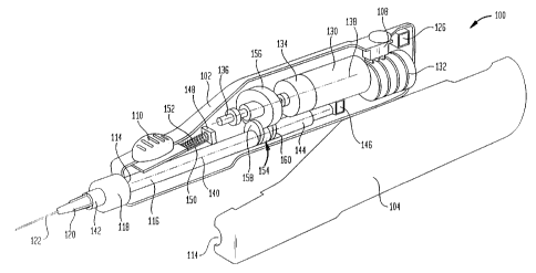
Referring now to the drawings, where like
reference numerals represent like elements, there is
shown in Fig. 1 a perspective view of a needle biopsy
instrument designated generally by reference numeral 100.
The instrument 100 is constructed from an elongated
housing 102 having a right half 104 and a left half 106.
Extending outwardly through the housing 102 at one end
thereof is a master on/off switch 108. In a similar
manner, a "deadman" switch 110 extends outwardly from the
housing 102 at the other end thereof. The forward end
CA 02366676 2001-09-21
WO 00/56208 -19 _ PCT/US00/07435
112 of the housing 102 is provided with an opening 114
through which there extends an elongated shaft 116. The
opening 114 is covered by a flexible boot 118 through
which the shaft 116 extends. The boot 118 can be
constructed of suitable polymer materials .well known in
the medical instrument art. A coupling device 120 is
attached to the end of the shaft 116 for releasably
securing a needle 122 thereto. In the preferred
embodiment, the coupling device 120 is integrally formed
as one unit with the needle 122 for attachment to the
instrument 100. In the preferred embodiment, the needle
122 which will be used for fine needle biopsy procedures,
will preferably have a size in the range of about 20-25
gauge. However, other size needles may be used with the
instrument 100 of the present invention. The instrument
100 may be connected to a remote computer 124 and/or be
provided with an internal programmable microprocessor 126
for operation of the instrument 100. The microprocessor
126 can be connected to the computer 124 using any
similar data link 128.
Referring to Fig. 2, the instrument 100
includes a motor 130 which may be electric or hydraulic.
In the case of hydraulic, the motor 130 may be driven by
an air or liquid feed supply (not shown) which can be
external to the housing 102 or provided internally by
means of, for example, a compressed air source. In the
illustrated embodiment, the motor 130 is in the nature of
an electric motor which is powered by a battery source
132. The battery source 132 may be in the nature of
rechargeable batteries, or conventional disposable
batteries. Also, the power source can be household AC
voltage or DC voltage through use of a converter. In
either event, the battery source 132 is operative of the
motor 130. The motor 130 is of known design in the
medical instruments field, for example, those having rpm
in the range of about 200-2000, which can provide 20
CA 02366676 2001-09-21
WO 00/56208 PCT/US00/07435
-20-
strokes in the range of 0.6-6 seconds. It is to be
understood that the foregoing particulars of the motor
130 are by way of example only, and other rpm's and
stroke frequencies may be incorporated into the
instrument 100 in accordance with the present invention.
The motor 130 may include a gear box 134 to
provide the desired rotational speed and torque for use
in the instrument 100. The motor 130 via the gear box
134, is operative for rotation of a shaft 136 coupled
thereto. The motor shaft 136 is rotated along its
longitudinal axis 138.
Shaft 116 extends longitudinally through the
housing 102 underlying motor shaft 136 and a portion of
the motor 130. The shaft 116 has its longitudinal axis
140 arranged parallel to the longitudinal axis 138 of
motor shaft 136. Shaft 116 is slidably mounted within
the housing 102 using any suitable means, such as bearing
supports (not shown), molded portions of the housing 102
and the like. The shaft 116 may have a rectangular cross
section along all or a portion thereof to preclude its
rotation within the housing 102, or a circular cross
section throughout where rotation of the shaft is desired
during operation of the instrument 100. A front section
142 of the shaft 116 extends outwardly through opening
114 to which the coupling device 120 is attached. A rear
section 144 of the shaft extends underlying the shaft 136
where it terminates adjacent a reciprocating shaft
positioning switch 146. Other switches within the
instrument 100 include a momentary actuator switch 148
which is coupled to the deadman switch 110 by means of a
push rod 150 and compression spring 152. A discussion of
the deadman switch 110, positioning switch 146 and
momentary actuator switch 148 will be described
hereinafter.
A cam assembly 154 is positioned within the
housing 102, coupling shaft 136 to shaft 116 at the rear
CA 02366676 2001-09-21
WO 00/56208 - 21- PCT/US00/07435
section 144. As further shown in Figs. 3A and 3B, the
cam assembly 154 includes a cam 156 and first and second
cam followers 158, 160. The cam 156 is constructed in
the nature of a cylindrical body 162 having spaced apart
surfaces defining outwardly facing first and second cam
profiles 164, 166. The body 162 of the cam 156 is
mounted to shaft 136 by means of a cylindrical member
168. From the foregoing description, rotation of shaft
136 by means of motor 130 and gear box 134 will cause cam
156 to rotate about axis 138.
The first and second cam followers 158, 160 are
mounted to the shaft 116. By way of example, each of the
cam followers 158, 160 are in the nature of flat disks or
preferably elongated cylindrical pins 161, see Fig. 2A
which project upwardly in a generally radial direction
from the shaft 116 toward cam 156. The spaced apart cam
followers 158, 160 define an opening 170 therebetween
which is sized to receive a peripheral portion of the cam
156. Cam follower 158 is operative for engagement with
the first cam profile 164, while the second cam follower
160 is operative for engagement with the second cam
profile 166. It is to be understood that the cam
followers 158, 160, can be any other shaped body which
extends outwardly from shaft 116 for engagement with the
first and second cam profiles 164, 166. In this regard,
the cam followers 158, 160 can be separately mounted
elements or integrally formed with the shaft 116.
As shown in Figs. 3A and 3B, upon rotation of
the cam 156 by operation of motor 130, the shaft 116 will
be caused to reciprocate as the cam followers 158, 160
ride in engagement with the first and second cam profiles
164, 166. By altering the first and second cam profiles
164, 166, various movements can be effected with respect
to the shaft 116. For example, the stroke length of the
shaft 116 can be changed by changing the angular
relationship between the longitudinal axis 172 of the cam
CA 02366676 2001-09-21
WO 00/56208 PCT/US00/07435
-22-
156 with respect to its rotational axis 138. In this
regard, the greater the angle between axes 138, 172, the
greater the stroke length will be produced on the shaft
116. A maximum stroke length in the range of about 1 cm
is contemplated for the instrument 100. However, it is
to be understood that other stroke lengths can be used in
biopsy needle instruments 100 in accordance with the
present invention.
Referring now to Fig. 12, there is illustrated
a schematic drawing of one electronic control circuit for
operation of the instrument 100, the bold circuit lines
representing the dynamic brake circuit for shaft
positioning. One side of the on/off switch 108 is
connected to the positive terminal of battery source 132.
The other side of the on/off switch 108 is connected to
terminal 174 on motor 130 and to terminal 176 on the
positioning switch 146. Terminal 176 is a normally open
position of the positioning switch 146. The negative
side of the battery source 132 is connected to terminal
178 on the positioning switch 146 and to terminal 180 on
the momentary actuator switch 148. Terminal 178
corresponds to a normally closed position on the
positioning switch 146, while terminal 180 corresponds to
a normally open position of the momentary actuator switch
148. Closed terminal 182 on the positioning switch 146
is connected to normally closed terminal 184 on the
momentary actuator switch 148. Terminal 186 of the motor
130 is connected to terminal 188 on the momentary
actuator switch 148, corresponding to a closed position.
The momentary actuator switch 148 is coupled to the
deadman switch 110 by means of push rod 150 and
compression spring 152.
In operation, the deadman switch 110 is closed
by depressing same manually so as to cause push rod 150
to close the connection between terminals 180, 188. At
the same time, the operator having actuated the on/off
CA 02366676 2001-09-21
WO 00/56208 PCT/US00/07435
-23-
switch 108 will allow power from battery source 132 to be
fed to the motor 130 for its operation. In the event of
release of the deadman switch 110, compression spring 152
will urge push rod 150 away from engagement with the
deadman switch 148 to open the connection between
terminals 180, 188. However, power to the motor 130 is
still provided after release of the deadman switch 110,
through positioning switch 146, until the shaft 16 is in
a "home" position.
The positioning switch 146 is positioned within
the housing rearwardly of the shaft 116. The positioning
switch 146 has an actuating lever 190. In the event of a
malfunction of the instrument 100, whereby the stroke
length of the shaft 116 is outside a predetermined
acceptable range, the shaft will engage lever 190 so as
to open the positioning switch 146. Normally, the
positioning switch 146 is in a closed position providing
electrical continuity between terminals 178, 182 so as to
close the circuit upon actuation of the momentary
actuator switch 148 by means of the deadman switch 110.
In the event that the positioning switch 146 is activated
by movement of lever 190, the positioning switch will
open thereby disconnecting power to the motor 130. The
positioning switch 146 thereby functions as a safety
switch to preclude injury to a patient. In this regard,
the positioning switch 146 provides a home position for
the shaft 116 to ensure that the first thrust of the
shaft is outward away from the instrument 100, as opposed
to being retracted within the instrument. As can be
appreciated by the foregoing description, actuation of
the motor 130 will effect rotation of cam 156 to cause
reciprocal motion of the shaft 116 as the cam followers
158, 160 engage the first and second cam profiles 164,
166. In the event that the deadman switch 110 is
inactivated by releasing same, and that activation of the
positioning switch 146 occurs, the motor 130 will stop
CA 02366676 2001-09-21
WO 00/56208 - 2 4 - PCT/US00/07435
operation. Thus, both the deadman switch 110 and the
positioning switch 146 control the motor 130. In order
for the motor 130 to shut off, the deadman switch 110
must be released and the positioning switch 146 must be
actuated.
The instrument 100 may operate in a manual
mode, in an on and off fashion, with continued
reciprocation of the shaft 116. The instrument 100 may
also be operated under programmed control according to
the desired parameters selected by the physician. For
example, by programming the instrument 100, this permits
predetermination of the number of thrusts, the number of
thrusts per unit of time, as well as other variable
options to specifically select desired parameters. The
programmable aspect of the instrument 100 may be achieved
by means of a programmed external computer 124 and/or an
internal microprocessor 126. In addition, the computer
124 and/or microprocessor 126 may store critical patient
data as well as other diagnostic information.
Referring now to Figs. 4-6, another embodiment
of a needle biopsy instrument 192 will now be described
wherein like reference numerals represent like elements.
A cam 194 is constructed from an elongated body 196
having a front section 198 and a rear section 200. The
front section 198 is provided with a circumferential
opening which forms a cam track 202 between adjacent
sidewalls 204, 206 of the opening. The axis 208 is
arranged at an angle to the longitudinal axis 138 of
shaft 136 about which the cam rotates. In other words,
the cam profile formed by sidewalls 204, 206 and hence
the cam track 202, is arranged at an angle to its axis of
rotation. A cam follower 210 is attached to the housing
102 and extends into the cam track 202. The cam follower
210 may be constructed as a projection or pin from the
housing 102 having its free end captured within the
opening forming the cam track 202.
CA 02366676 2001-09-21
WO 00/56208 _ 2 5 - PCT/US00/07435
The rear section 200 of the cam 194 is provided
with elongated internal bore 212. The bore 212 is sized
and configured to slidingly receive a coupling 214 which
is attached to shaft 136. The coupling 214 and bore 212
are provided with other than a circular shape, such as
square, triangular, polygonal, oval or the like such that
rotation of the coupling will effect rotation of the cam
194. In this regard, upon rotation of the coupling 214
by means of the motor 130, the rotary motion will be
transmitted to effect rotation of the cam 194. As the
cam follower 210 is captured within the cam track 202,
rotation of the cam 194 will cause reciprocal motion of
the cam. This reciprocal motion is transmitted to the
needle 122 which is attached to coupling device 120. The
coupling device 120 is attached to shaft 116 which is
supported on a support member 216. To prevent rotation
of the support member 216, and hence the needle 122, the
support member is maintained in contact with cam 194 by
an intervening bearing 218. The bearing 218 will permit
rotational motion of the cam 194, while facilitating the
prevention of rotational motion of the support member
216. In this regard, the support member 216 and adjacent
housing 102 will be provided with a guide pin and linear
track arrangement as to be generally described with
respect to the Fig. 6 embodiment. This, in turn, will
prevent the support member 216 from rotating, while at
the same time, permitting its reciprocal movement.
In an alternate embodiment as shown in Fig. 6,
the coupling 220 may be in the nature of a cylindrical
body which transmits rotational motion to the cam 194 by
means of an elongated key 222. The bore 212 in the cam
194 will also be of cylindrical shape. The key 222 is
received within an elongated opening 224 within the
coupling 220. As a result, the coupling 220 can slide
longitudinally within the bore 212, while transmitting
CA 02366676 2001-09-21
WO 00/56208 - 2 6 - PCT/US00/07435
rotation of the coupling to the cam 194 as a result of
the interlocking key 222.
Referring now to Figs. 7-9, there will be
described alternative assemblies for use in the
instrument 100 for effecting reciprocal motion of the
needle 122. As shown in Fig. 7, motor 130 is coupled to
a first beveled gear 226 which is meshed with a second
beveled gear 228. The second beveled gear 228 is
supported on a plate 230 which is coupled to a push rod
232 attached to a peripheral portion of the plate. The
push rod 232, in turn, is connected to one end of the
shaft 116. By rotation of the first and second beveled
gears 226, 228, the push rod 232 will effect reciprocal
motion of shaft 116. The shaft 116 slides freely within
a stationary sleeve 233 which is supported within the
housing 102.
Referring now to Fig. 8, the shaft 136 is
provided with a continuous helical groove 234 or gear.
The shaft 136 is received within a bore (not shown)
extending within one end of the shaft 116. The end of
the shaft 116 is provided with suitable means for
tracking within the helical groove 234 or engagement with
the gear to effect reciprocal motion of the shaft. Shaft
116 is slidingly received within a stationary sleeve 236
which is provided with outwardly extending elongated
projections 238. The projections 238 are captured within
a corresponding portion of the housing 102 to prevent
rotation of the stationary sleeve 236. The shaft 116 is
provided with similar shaped side projections 24o which
are slidingly received within the interior opening formed
by side projections 238 formed within sleeve 236. Based
upon this arrangement, shaft 116 will reciprocate freely
within sleeve 236 while being precluded from rotation by
the presence of the side projections 240.
Turning now to Fig. 9, a pair of C-shaped cam
members 242, 244 are respectively attached to shafts 136,
CA 02366676 2001-09-21
WO 00/56208 PCT/US00/07435
-27-
116. The C-shaped cams 242, 244 have respective cam
surfaces 246, 248 which are held in contact with each
other when in assembled relationship by means of, for
example, a spring (not shown). By rotation of the C-
shaped cam 242, its cam surface 246 will track the cam
surface 248 on C-shaped cam 244 causing reciprocal motion
of shaft 116.
As thus far described, the biopsy needle
instrument of the present invention provides reciprocal
motion to the attached needle 122. It may also be
desirable that the needle 122 be simultaneously rotated
during its reciprocal motion. Turning to Fig. 10, a
needle biopsy instrument 250 of similar construction to
instrument 192 as shown in Fig. 4 is illustrated. The
instrument 250 provides both reciprocal and rotational
motion of shaft 116. In the instrument 250, the shaft
116 is attached to the front section 198 of the cam 194.
As previously described with respect to the instrument
192 of Fig . 4 , the shaft 116 was separated from the cam
194 by means of bearing 218. By direct connection,
rotation of the cam 194 will effect rotation of shaft
116, and hence needle 122, while at the same time,
providing reciprocal motion. Accordingly, it is to be
understood that instrument 192 provides reciprocal motion
only, while instrument 250 provides both reciprocal and
rotary motion.
Referring now to Fig. 11, a needle biopsy
instrument 252 in accordance with another emb:,~diment of
the present invention will now be described which
provides both rotary and reciprocal motion to the needle
122. A drive gear 252 is coupled to the shaft 136 of the
motor 130. As shown, the rotational axis 138 of the
drive gear 252 is arranged parallel to, and spaced apart,
from the rotational and reciprocal axis 140 of shaft 116.
Shaft 116 is attached centrally to cam 254. Cam 254 is
constructed from a body 256 having two outwardly facing
CA 02366676 2001-09-21
WO 00/56208 PCT/US00/07435
-28-
first and second cam profiles 258, 260. The peripheral
edge of the cam 254 is received within an opening 262
formed between two spaced apart pins or cam followers
264, 266. The cam followers 264, 266 are fixedly mounted
to an interior portion of the housing 102. A gear 268 is
attached circumferentially about cam 254. The cam 254 is
positioned such that the gear 268 is arranged in meshed
engagement with drive gear 252. As shown, the rotational
axis of the gear 268 is arranged parallel to the
rotational axis of drive gear 252. Rotation of drive
gear 252 will, in turn, effect rotation of gear 268 and
cam 256, and hence, shaft 116. As the cam 256 is
rotated, its engagement with cam followers 264, 266 will
also cause the cam 256 to reciprocate thereby
reciprocating shaft 116 and needle 122. The reciprocal
motion of the cam 254 is accommodated by gear 268 sliding
in meshed engagement with the drive gear 252. If
rotational motion of the shaft 116 is not desired, the
shaft can be supported by the cam 256 using a bearing 218
in a similar arrangement as shown in the instrument 192
illustrated in Fig. 4.
The biopsy needle instrument of the present
invention provides for reciprocal and/or rotary motion of
a needle under programmed control during the biopsy
procedure. It may be desirable to couple the biopsy
needle instrument with a source of vacuum for aspiration
of the tissue sample into needle 122. By way of example,
as shown in Fig. 13, a T-connector 270 is attached
between the shaft 116 and coupling device 120 which is
formed as part of the needle 122. The near end 271 of
the T-connector 270 which is attached to the instrument
100 is closed off. Branch 272 of the T-connector 270 is
connected to a conventional syringe 274 by means of
flexible tubing 276 having an on/off valve 277. While a
tissue sample is being collected in the needle 122,
plunger 278 can be withdrawn from within the syringe 274
CA 02366676 2001-09-21
WO 00/56208 PCT/US00/07435
-29-
to create vacuum within the T-shaped connector 270, which
vacuum is maintained by closing on/off valve 277. This,
in turn, will draw the tissue sample into the needle 122
which is now under vacuum. After the predetermined
sampling cycle is completed, the vacuum is released by
opening the on/off valve 277, the needle 122 is removed
from the patient's body and the tissue sample can then be
dispensed from the need by means of advancing the plunger
278 of the syringe 274.
l0 Referring to Fig. 14, there is graphically
illustrated displacement or thrust distance of the needle
122 in relationship to one revolution of cam 156, 254 or
cam track 202. As shown, the maximum extended travel or
displacement of the needle 122 occurs at 180° of rotation
of the cam or cam track. By altering the angular
relationship between cam axis 172 and its rotational axis
which corresponds to axis 138, see Fig. 3A, the travel
distance of the needle 122 can be changed. This is
represented by the solid line and dashed line curves in
Fig. 14.
Another embodiment of a cam 279 is shown in Fig
15. The cam 279 has one segment 280 extending 180°
having its axis 282 perpendicular to axis 138. Another
equal segment 283 has its axis 284 at an angle to axis
138. The cam 279 provides the needle travel distance
profile as shown in Fig. 16. The travel distance
provides a dwell period of 90° before and after movement
of the needle 122 during rotation of the cam 279.
Using the foregoing modifications and
variations of the cam profiles, various combinations of
these cam profiles can produce various motion of the
needle 122. As shown in Fig. 17, there is initially
provided a dwell period followed by a high velocity
extension of the needle 122, followed by a slow
retraction of the needle into the instrument housing 102.
In Fig. 18, a similar travel of the needle 122 is
CA 02366676 2001-09-21
WO 00/56208 PCT/US00/07435
-30-
produced, but without a dwell period. As shown in Fig.
19, the needle 122 will have an initial low velocity
extension, followed by a high velocity retraction of the
needle into the housing 102. From the foregoing, it
should be understood that almost any profile can be
achieved with reasonable ramp angles. It is to be noted
that the higher the ramp angle, which produces higher
needle velocities, there is required more torque from the
motor 130. This effect can be dampened by the use of a
flywheel.
Turning to Fig. 20, there is illustrated in
cross-section a cam 300 constructed in accordance with
another embodiment of the present invention. The cam 300
includes a body 301 which forms a pair of spaced apart
outwardly facing cam surfaces 302, 304 for z~espective
engagement with pins 161 which are attached to the shaft
116.
Referring to Fig. 21, cam 306 is provided with
a circumscribing recessed portion 308 which is bound by a
pair of spaced apart sloping cam surfaces 310, 312. A
pair of spaced apart pins 161 attached to shaft 116
extend upwardly into the recessed portion 308 for
respective engagement with the cam surfaces 310, 312. As
shown in Fig. 22, a single cam follower 314 may be
received within the recessed portion 308. The cam
follower 314 has outwardly facing spaced apart surfaces
316, 318 for respective engagement with cam surfaces 310,
312. As shown in Fig. 23, the cam 306 is provided with
outwardly facing spaced apart sloping cam surfaces 320,
322 for respective engagement with spaced apart pins 161.
Referring to Figs. 24-28, various modifications
of the drive and cam assembly as shown in Fig. 11 will
now be described. In each of these embodiments, the cam
assembly will be operative to effect both rotary and
reciprocal motion of shaft 116, and hence, needle 122
which is attached thereto. As shown in Fig. 24, the cam
CA 02366676 2001-09-21
WO 00/56208 PCT/US00/07435
-31-
assembly 324 includes a gear 268 to which there is
attached on one side thereof a cam body 256. The cam
body is provided with a circumscribing recessed portion
326 defining a pair of spaced apart sloping cam surfaces
328, 330. A cam follower 314 attached to the housing 102
extends into the recessed portion 326 for engagement with
the cam surfaces 328, 330.
Turning to Fig. 25, the cam assembly 331
includes gear 268 provided with cam bodies 332, 334
supported on either side of the gear. A circumscribing
recessed portion 335 defined by the diameter of gear 268
and larger diameters of the cam bodies 332, 334, also
define a pair of spaced apart sloping cam surfaces 336,
337. In accordance with this arrangement, drive gear 252
by having a peripheral portion received within the
recessed portion 335 functions as a cam follower, as well
as effecting rotation of gear 268 as a result of the
meshed engagement therewith.
Turning to Fig. 26, the cam assembly 338
includes a cam body 339 having a circumscribing recessed
portion 340. The cam body 339 is attached to one surface
342 of drive gear 252. The recessed portion 340 defines
a cam surface 344 opposing surface 342 of the drive gear
252 which functions as a second cam surface. A cam
follower 346 attached on one side to gear 268, and
supporting on its other side shaft 116, has its
peripheral portion 348 received within the recessed
portion 340. The drive gear 252 is maintained in meshed
engagement with gear 268.
Turning to Fig. 27, the cam assembly 350
includes drive gear 252 provided with cam bodies 352, 354
supported on either side of the drive gear. A
circumscribing recessed portion 356 defined by the
diameter of the drive gear 252 and the larger diameters
of the cam bodies 352, 354, also define a pair of spaced
apart sloping cam surfaces 358, 360. In accordance with
CA 02366676 2001-09-21
WO 00/56208 - 3 2 - PCT/US00/07435
this arrangement, gear 268 by having a peripheral portion
received within the recessed portion 356 functions as a
cam follower, as well as effecting rotation of shaft 116
as a result of the meshed engagement with the drive gear
252.
Turning to Fig. 28, the cam assembly 362
includes a cam follower 264 attached to one side of drive
gear 252. A cam body 366 having a circumscribing
recessed portion 368 is attached to one surface 370 of
gear 268. The recessed portion 368 defines a cam surface
372 opposing surface 370 of gear 268 which functions as a
second cam surface. The cam follower 364 has a
peripheral portion 374 received within the recessed
portion 368 for engagement with the cam surfaces 370,
372. The drive gear 252 is maintained in meshed
engagement with gear 268 for rotation and reciprocal
motion of shaft 116 which is attached to the cam body
366.
Referring to Figs. 29 and 30, the cam
assemblies are operative for effecting only reciprocal
motion of shaft 116, and hence the needle 122. Referring
to Fig. 29, the cam assembly 376 includes a cylindrical
body 378 having an internal bore 380 opening at one end
thereof. The other end is closed by wall 382 from which
there extends shaft 116. The bore 380 is circular in
shape so as to rotatably receive a circular shaped cam
body 384 which is attached to shaft 136 for rotation by
means of motor 130. The cam body 384 has a
circumscribing recessed portion 386 defining a pair of
spaced apart sloping cam surfaces 388, 390. A cam
follower 392 in the nature of a pin is attached to the
body 378 and extends inwardly so as to be captured within
the recessed portion 386.
By rotation of cam body 384, the cylindrical
body 378 will reciprocate within the instrument 100. To
prevent rotation of the cy7.indrical body 378, at least
CA 02366676 2001-09-21
WO 00/56208 - 3 3 _ PCT/US00/07435
one, and preferably a pair of opposing pins 394 extend
outwardly from the body 378. The pins 394 are received
within longitudinal slots (not shown) formed within the
housing 102. In an alternate embodiment, the slots can
be helical in nature, which will impart rotary motion to
the cylindrical body 378, and hence to the needle 122.
Referring to Fig. 30, the cam body 384 is
provided with a pair of spaced apart regions of reduced
diameter so as to form an outwardly extending
circumscribing cam 396 forming a pair of spaced apart cam
surfaces 398, 400. The cam surfaces 398, 400 are
respectively engaged by pins 392 extending from the
cylindrical body 378.
Although the invention herein has been
described with reference to particular embodiments, it is
to be understood that the embodiments are merely
illustrative of the principles and application of the
present invention. For example, by suitable means such
as cams and other mechanical assemblies known in the art,
the end of the reciprocal shaft 116, and hence the needle
122, can be made to orbit or follow a zigzag or other
predetermined path during the thrust of the needle as
thus far described. It is therefore to be understood
that numerous modifications may be made to the
embodiments and that other arrangements may be devised
without departing from the spirit and scope of the
present invention as defined by the claims.
CA 02366676 2001-09-21
WO 00/56208 PCT/US00/07435
-34-
INDUSTRIAL APPLICABILITY
+ The present invention can be applied in the
medical field for performing fine needle biopsies of
human or animal tissue for diagnosis.