Note: Descriptions are shown in the official language in which they were submitted.
CA 02366918 2001-08-31
WO 00/52194 PCT/US00/05889
METHODS FOR DETECTING MEMBRANE DERIVED CASPASE
ACTIVITY AND MODULATORS THEREOF
TECHNICAL FIELD
The present invention relates generally to methods for detecting
membrane derived caspase activity and modulators thereof, and more
particularly to
novel cell-free screening assays for identifying inhibitors and enhancers of
membrane
derived caspase activity.
BACKGROUND OF THE INVENTION
Tissue homeostasis is maintained by the process of apoptosisthat is,
the normal physiological process of programmed cell death. Changes to the
apoptotic
pathway that prevent or delay normal cell turnover are often as important in
the
pathogenesis of diseases as are abnormalities in the regulation of the cell
cycle. Like
cell division, which is controlled through complex interactions between cell
cycle
regulatory proteins, apoptosis is similarly regulated under normal
circumstances by the
interaction of gene products that either function to prevent or induce cell
death.
Since apoptosis functions in maintaining tissue homeostasis in a range of
physiological processes, such as embryonic development, immune cell regulation
and
normal cellular turnover, the dysfunction or loss of regulated apoptosis can
lead to a
variety of pathological disease states. For example, the loss of apoptosis can
lead to the
accumulation of self reactive lymphocytes associated with many autoimmune
diseases.
Inappropriate loss or inhibition of apoptosis can also lead to the
accumulation of virally
infected cells and hyperproliferative cells, such as neoplastic or tumor
cells. Similarly,
the inappropriate activation of apoptosis can contribute to a variety of
pathological
disease states including, for example, acquired immunodeficiency syndrome
(AIDS),
neurodegenerative diseases and ischemic injury.
Although apoptosis is mediated by diverse signals and complex
interactions of cellular gene products, the results of these interactions
ultimately feed
into a cell death pathway that is evolutionarily conserved between humans and
CA 02366918 2001-08-31
WO 00/52194 PCT/US00/05889
2
invertebrates. The pathway, itself, is a cascade of proteolytic events
analogous to that
of the blood coagulation cascade.
Several gene families and products that modulate the apoptotic process
have now been identified. One family is the aspartate-specific cysteine
proteases
S ("caspases"). The caspase Ced-3, identified in C. elegans, is required for
programmed
cell death during development of the roundworm C. elegans. Ced-3 homologues as
well as other caspases have been characterized. The human caspase family
includes, for
example, Ced-3, human ICE (interleukin-1-13 converting enzyme) (caspase-1),
ICH-1
(caspase-2), CPP32 (caspase-3), ICEre,II (caspase-4), ICEre,III (caspase-5),
Mch2
(caspase-6), ICE-LAP3 (casepase-7), MchS (caspase-8), ICE-LAP6 (caspase-9),
Mch4
(caspase-10), caspase-1 l, caspase-12, caspase-13, caspase-14, and others.
The caspase family of cysteine proteases are essential effectors of the
apoptotic process (Yuan et al., Cell 75:641-652, 1993; Alnemri et al., Cell
87:171,
1996; Cohen, Biochem. 326:1-16, 1997; Miller, Semin. Imrnunol 9:35-49, 1997;
Salvesen and Dixit, Cell 91:443-446, 1997). Caspases are synthesized as
inactive
zymogens, which are activated by proteolytic processing to yield large (~18
kDa) and
small (~12 kDa) subunits that associate to form active enzymes (Thornberry et
al.,
Nature 396:768-774, 1992; Nicholson et crl., Nature 376:37-43, 1995; Stennicke
and
Salvesen, J. Biol. Chem. 272:25719-25723, 1997). Diverse apoptotic stimuli
cause the
activation of specific caspases which then initiate a protease cascade by
proteolytically
processing additional caspases (Srinivasula et al., Proc. Natl. Acad. Sci. USA
93:14486-
14491, 1996; Yu et al., Cancer Res. 58:402-408, 1998). Once activated, these
downstream (executioner) caspases kill cells by cleaving specific molecular
targets that
are essential for cell viability or by activating pro-apoptotic factors (Liu
et al., Cell
89:175-184, 1997; Enari et al., Nature 391:43-50, 1998; Salvesen and Dixit,
Cell
91:443-446, 1997). Although caspases have been generally shown to be cytosolic
proteins (Miller et al., ,I. Biol. Chem. 268:18062-18069, 1993; Nicholson et
al., Nature
376:37-43, 1995; Li et al., J. Biol. Chem. 272:30299-30305, 1997),
immunochemical
studies have suggested that in some instances, caspases might also be
associated with
the nucleus or plasma membrane (Singer et al., J. Exp. Med. 182:1447-1459,
1995;
CA 02366918 2001-08-31
W4 00/52194 PCT/US00/05889
3
Krajewski et al., Blood 89:3817-3825, 1997; Posmantur et al., J. Neurochem.
68:2328-
2337, 1997). Recently published data has also indicated an association of
certain
caspases with mitochondria and endoplasmic reticulum (Mancini et al., J. Cell
Biol.
140:1485-1495, 1998; Chandler et al., J. Biol. Chem. 273:10815-10818, 1998).
The Bcl-2 family constitutes another key set of regulators of the
apoptotic pathway. These proteins can function to modulate apoptosis in a wide
variety
of cell systems (Oltvai and Korsmeyer, Cell 79:189-192, 1994; Reed, Nature
387:773-
776, 1997). Bcl-2 family proteins contain one to four conserved domains,
designated
BH 1-BH4, and most family members contain a carboxyl-terminal transmembrane
anchor sequence which allows them to be associated with cellular membranes
including
the outer membrane of the mitochondria, the nuclear envelope and the
endoplasmic
reticulum (Reed, Nature 387:773-776, 1997; Krajewski et al., Cancer Res.
53:4701-
4714, 1993; Yang et al., J. Cell. Biol. 128:1173-1184, 1995; Lithgow et al.,
Cell
Growth Differ 3:411-417, 1994). The over-expression of Bcl-2 has been shown to
inhibit the activation of cytoplasmic caspases following apoptoic stimuli in
several cell
systems (Armstrong et al., J. Biol. Chem. 271:16850-16855, 1996; Chinnaiyan et
al., J.
Biol. Chem. 271:4573-4576, 1996; Boulakia et al., Oncogene 12:29-36, 1996;
Srinivasan et al., J. Neurosci. 16:5654-60, 1996). Moreover, previous work has
demonstrated that Bcl-2 inhibits the onset of apoptosis, but once apoptosis is
initiated,
Bcl-2 does not impede the process (McCarthy et al., J. Cell Biol. 136:215-217,
1997).
However, it remains unclear how the membrane bound Bcl-2 exerts control over
the
soluble cytoplasmic caspases. Further, no suitable methods exist for studying
membrane bound Bcl-2 and its effects on caspase activity in a cell free
manner.
The identification of compounds that modulate the apoptotic pathway via
enhancement or inhibition of membrane derived caspase activity has been
hindered by
the lack of such methods. Available methods are limited by the lack of
specificity,
efficiency, and/or utilization of whole cells or cytoplasmic extracts thereof.
For
example, most anti-cancer drugs are screened for their ability to kill cells
and therefore
will identify compounds that induce both necrosis or apoptosis. In addition,
many other
assay techniques focus on studying the inhibition or enhancement of caspase
enzymes
WO 00/52194 PCT/US00/05889
4
located further into the cascade. Therefore, there exists a need in the art
for methods of
identifying compounds that not only inhibit or enhance cell death, but also
compounds
that modulate the initiation of the apoptotic cascade. The present invention
fulfills this
need, while further providing other related advantages.
S The foregoing characteristics, and others which shall be described in
greater detail below, make the methodologies described herein particularly
attractive for
drug discovery applications.
SUMMARY OF THE INVENTION
The present invention generally provides methods for detecting
membrane derived caspase activity and methods for identifying modulators
thereof. In
one aspect, the invention provides a method for identifying membrane derived
caspase
activity, that includes, incubating a membrane fraction comprising heavy or
nuclear
membranes under conditions and for a time sufficient to allow for the
evolution of
caspase activity, and subsequently detecting caspase activity.
In another aspect, the present invention provides a method for
identifying an inhibitor of the activity of a membrane derived caspase, that
includes,
contacting a membrane fraction with a caspase substrate in the presence and
absence of
at least one candidate inhibitor; and comparing the levels of caspase
substrate turnover,
and therefrom identifying an inhibitor of the activity of a membrane derived
caspase.
In yet another aspect, the present invention provides a method for
identifying an enhancer of the activity of a membrane derived caspase, that
includes,
contacting a membrane fraction with a caspase substrate in the presence and
absence of
at least one candidate enhancer; and comparing the levels of caspase substrate
turnover,
and therefrom identifying an enhancer of the activity of a membrane derived
caspase.
A further aspect of the present invention is a method for identifying an
inhibitor or enhancer of the evolution of caspase processing within a membrane
fraction, that includes, contacting a membrane fraction with at least one
candidate
inhibitor or candidate enhancer; and detecting the presence of large and small
caspase
subunits, and therefrom determining the level of caspase processing, wherein a
decrease
CA 02366918 2001-08-31
WO 00/52194 PCT/US00/05889
in processing indicates the presence of a caspase processing inhibitor, and
wherein an
increase in processing indicates the presence of a caspase processing
enhancer.
In other embodiments, the present invention provides a method of
identifying a compound that modulates membrane fraction derived caspase
activity, that
S includes, incubating a membrane fraction, an inhibitor of apoptosis, and a
caspase
substrate in the presence and absence of at least one candidate compound under
conditions and for a time sufficient to allow for the evolution of caspase
activity; and
comparing the levels of caspase substrate turnover, thereby identifying a
compound that
modulates membrane derived caspase activity.
In other embodiments, inhibitors and enhancers of the activity of a
membrane derived caspase that are identified by the various methods are
provided.
In the various embodiments, caspase activity is detected by measuring
substrate turnover or caspase processing. In other embodiments, substrate
turnover is
measured by time course or endpoint analysis. In further embodiments, the
membrane
fraction comprises heavy or nuclear membranes. In yet further embodiments, the
membrane fraction is derived from cells expressing an anti-apoptotic
polypeptide. In
even further embodiments, the membrane fraction is derived from non-apoptotic
cells.
These and other aspects of the present invention will become evident
upon reference to the following detailed description and attached drawings. In
addition,
the various references set forth below that describe in more detail certain
procedures or
compositions (e.g., plasmids, etc.), and are therefore incorporated by
reference in their
entirety.
BRIEF DESCRIPTION OF THE DRAWINGS
Figure 1 is a scanned image of an immunoblot representing SDS-PAGE
analysis of subcellular fractions from 697-neo and 697-Bcl-2 cells using
antibodies to
PARP, cytochrome oxidase (subunit IV), D4GDI and Bcl-2. Nuc = nuclear
fraction,
HM = heavy membrane fraction, LM = light membrane fraction, S 100 = cytosolic
fraction. Arrows indicate the specific immunoreactive band.
CA 02366918 2001-08-31
CA 02366918 2001-08-31
WO 00/52194 PCT/US00/05889
6
Figures 2A-D are histograms of caspase substrate cleavage activity in
subcellular fractions.
Figures 3A and 3B are graphs representing membrane-associated
procaspase-3 spontaneous activation. Figure 3A illustrates the spontaneous
activation
of caspase activity in heavy membrane from 697-neo and 697-Bcl-2 cells as a
function
of DEVD-amc turnover. Figure 3B illustrates the generation of soluble caspase
activity
from membranes as a function of DEVD-amc turnover.
Figure 4 is a scanned image of an immunoblot representing SDS-PAGE
analysis of heavy membrane and cytosolic fractions from 697-neo and 697-Bcl-2
cells,
probed with an anti-caspase-3 polyclonal antibody. The arrowheads indicate the
migration of protein size markers (Rainbow Markers, Novex); the arrow
indicates
procaspase-3. HM = heavy membrane fractions; S 100 = cytosolic fraction.
Figure 5 is a graph illustrating activation of membrane associated
DEVD-amc cleavage activity by exogenous caspase-1.
Figures 6A and 6B are graphical representations of DEVD-amc cleavage
activity in 697-neo and 697-Bcl-2 cells in the presence and absence of
cytochrome c.
Figure 6A illustrates the caspase activity present in the heavy membrane
fraction.
Figure 6B illustrates the caspase activity present in the cytoplasmic
fraction.
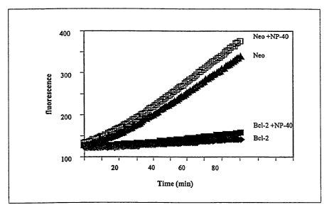
Figures 7A, 7B, and 7C are graphs representing the effects of
permeabilizing detergents on membrane-associated caspase activity. Figure 7A
is a
graph demonstrating the effects of NP-40 on spontaneous and induced caspase
activities
in neo-membranes. Figure 7B is a graph illustrating the effect of NP-40 on
spontaneous
caspase activation in Bcl-2 and neo-membranes. Figure 7C is a graph depicting
NP-40-
dependent and independent activation of procaspase-3 by granzyme B treatment
of
mitochondria) enriched fractions.
DETAILED DESCRIPTION OF THE INVENTION
As noted above, the present invention is generally directed to methods of
detecting and modulating membrane derived caspase activity. One application of
the
CA 02366918 2001-08-31
WO 00/52194 PCT/US00/05889
7
disclosed invention is in the identification of inhibitors or enhancers of
apoptosis. In
simple terms, the use of such a novel cell-free assay system provides a means
for
identifying compounds which promote or inhibit programmed cell death at a
critical
initiation point (i.e., membranes). Another aspect of the subject invention is
the ability
of the disclosed assay system to investigate the effects of membranes derived
from cells
over-expressing apoptotic pathway proteins, such as those of the bcl-2 family.
As described herein, a preferred assay system utilizes heavy or nuclear
membranes for detecting membrane derived caspase activity and/or for
identifying
compounds that modulate caspase activity, directly or indirectly, in a cell-
free system.
Therefore, by using such membrane systems, control points upstream of the
cytoplasmic apoptotic pathway can be effectively assayed and modulators
thereof may
be identified.
The assay methods of the present invention are particularly useful for
drug discovery, in part by use of high throughput methodologies. Accordingly,
by
utilizing the cell-free assay system of the present invention, identification
of compounds
that affect evolution of caspase activity from the membrane fraction is
rapidly achieved.
Prior to setting forth the invention, it may be helpful to an understanding
thereof to set forth definitions of certain terms that will be used
hereinafter.
As used herein, a "caspase" refers to a cysteine protease that specifically
cleaves proteins after Asp residues. Caspases are initially expressed as
zymogens, in
which a large subunit is N-terminal to a small subunit. Caspases are generally
activated
by cleavage at internal Asp residues. These proteins have been identified in
many
eukaryotes, including C. elegans, Drosophila, mouse, and human. Currently,
there are
at least 14 known caspase genes, named caspase-1 through caspase-14. Table 1
recites
ten human caspases along with their alternative names.
Caspase Alternative name
Caspase-1ICE
Caspase-2ICH-1
Caspase-3CPP32, Yama, apopain
Caspase-4ICEre,II; TX, ICH-2
Caspase-5ICEre,III; TY
CA 02366918 2001-08-31
WO 00/52194 PCT/US00/05889
8
Caspase-6 Mch2
Caspase-7 Mch3, ICE-LAP3, CMH-1
Caspase-8 FLICE; MACH; MchS
Caspase-9 ICE-LAP6; Mch6
Caspase-10I Mch4, FLICE-2
Within the context of this invention, it should be understood that a
caspase includes wild-type protein sequences, as well as other variants
(including
alleles) of the native protein sequence. Briefly, such variants may result
from natural
polymorphisms or may be synthesized by recombinant methodology, and differ
from
wild-type protein by one or more amino acid substitutions, insertions,
deletions, or the
like. Typically, when engineered, amino acid substitutions will be
conservative, i.e.,
substitution of amino acids within groups of polar, non-polar, aromatic,
charged, etc.
amino acids. In the region of homology to the native sequence, variants should
preferably have at least 90% amino acid sequence identity, and within certain
embodiments, greater than 92%, 95%, or 97% identity. Such amino acid sequence
identity may be determined by standard methodologies, including use of the
National
Center for Biotechnology Information BLAST search methodology available at
www.ncbi.nlm.nih.gov. The identity methodologies preferred are those described
in
U.S. Patent 5,691,179 and Altschul et al., Nucleic Acids Res. 25:3389-3402,
1997 all of
which are incorporated herein by reference. If Gapped BLAST 2.0 is utilized,
then it is
utilized with default settings.
As will be appreciated by those skilled in the art, a nucleotide sequence
encoding a caspase or variant may differ from the known native sequences, due
to
codon degeneracies, nucleotide polymorphisms, or amino acid differences. In
other
embodiments, variants should preferably hybridize to the native nucleotide
sequence at
conditions of normal stringency, which is approximately 25-30°C below
Tm of the
native duplex (e.g., SX SSPE, 0.5% SDS, SX Denhardt's solution, 50% formamide,
at
42°C or equivalent conditions; see generally, Sambrook et al. Molecular
Cloning: A
Laboratory Manual, 2nd ed., Cold Spring Harbor Press, 1987; Ausubel et al.,
Current
Protocols in Molecular Biology, Greene Publishing, 1987).
CA 02366918 2001-08-31
WO 00/52194 PCT/US00/05889
9
An "isolated nucleic acid molecule" refers to a polynucleotide molecule
in the form of a separate fragment or as a component of a larger nucleic acid
construct,
that has been separated from its source cell (including the chromosome it
normally
resides in) at least once in a substantially pure form. Nucleic acid molecules
may be
comprised of a wide variety of nucleotides, including DNA, RNA, nucleotide
analogues, or some combination of these.
A "membrane fraction", as used herein, refers to a subcellular fraction of
a eukaryotic cell comprising cellular membranes. In particular, the term
"heavy
membranes", as used herein, refers to a subcellular fraction substantially
free of nuclear
and light membranes, wherein one of the predominant components is
mitochondria.
A "stimulator of apoptosis" or "pro-apoptotic agent", as used herein
refers to an agent that increases the specific apoptotic activity of a cell.
Illustrative
examples of such stimulus are deprivation of a growth factor, Fas ligand, anti-
Fas
antibody, staurosporine, ultraviolet irradiation, gamma irradiation, tumor
necrosis
factor, and others well known in the art. Accordingly, a stimulator of
apoptosis is an
agent that increases the molecular activity of caspase molecules either
directly or
indirectly. In addition, a stimulator of apoptosis can be a polypeptide that
is capable of
increasing or inducing the apoptotic activity of a cell. Such polypeptides
include those
that directly regulate the apoptotic pathway such as Bax, Bad, Bcl-xS, Bak,
Bik, and
active caspases as well as those that indirectly regulate the pathway.
An "inhibitor of apoptosis" or "anti-apoptotic agent", as used herein
refers to an agent that decreases the apoptotic activity of a cell when
compared to
control agents. Illustrative examples of such anti-apoptotic agents include
small
molecules, fmk, p35, crmA, Bcl-2, Bcl-X~, Mcl-l, E1B-19K from adenovirus, as
well
as antagonists of pro-apoptotic agents (e.g., antisense, ribozymes,
antibodies, etc.).
Accordingly, an inhibitor of apoptosis is an agent that decreases the
molecular activity
of caspase molecules either directly or indirectly.
An "apoptotic pathway protein", as used herein refers to a protein
involved in the cell death pathway. Illustrative examples include Bcl-2, Bcl-
X5, Bcl-XL,
Bik, Bak, Bax, Bad, caspase molecules, Apaf 1, cytochrome c, and the like.
CA 02366918 2001-08-31
WO 00/52194 PCT/US00/05889
"Evolution of caspase activity", as used herein, refers to the increasing of
detectable levels of caspase protease activity over a time period. Such
evolution may be
evidenced by detectable increases in substrate turnover (e.g., fluorogenic
substrates)
and/or detectable increases in caspase processing.
5 "Membrane derived caspase activity", as used herein, refers to caspase
activity that is released from or associated with heavy or nuclear membranes.
A. MEMBRANE PREPARATIONS
Membrane preparations within the context of the present invention may
be derived from a variety of cell types or sources. Typically, for ease of
handling, the
10 cells utilized will be a eukaryotic cell line or other culturable cell
type. However, cells
can also be derived from tissues and other non-cultured sources. One of
ordinary skill
in the art would readily appreciate that the assays of the present invention
are not
dependent upon the exact source or type of cell from which membrane fractions
are
prepared.
Subcellular fractionation has been a basic research tool in cell biology
for the last 30 years. Accordingly, those of ordinary skill in the art are
familiar with
various techniques for such fractionation. Typically, subcellular
fractionation
comprises two basic steps, 1) homogenization and 2) separation. Homogenization
in its
ideal form allows particulate organelles such as the nucleus, mitochondria,
lysosomes,
and peroxisomes to remain intact. A variety of homogenization techniques are
known,
such as Dounce homogenizers (glass/glass), Potter-Elvehjem homogenizers
(glass/teflon), repeated pipetting, passage through small gauge needle, and
the like.
Exemplary techniques are described in detail by Hanns et al., Proc. Natl.
Acad. Sci.
USA 77:6139-6143 1980, Darte et al., J. Exp. Med. 157:1208-1228, 1983, and
Balch et
al., Cell 39:405-416, 1984.
Separation of subcellular fractions is traditionally performed using
density gradients. While sucrose gradients are the most widely used, many
other
alternatives are available (e.g., Ficoll, Percoll, Metrizamide, and Nycodenz)
(see
Methods in Enzymology Vol. 31, Part A (Flescher and Packer eds.), 1974). In
addition,
CA 02366918 2001-08-31
WO 00/52194 PCT/US00/05889
11
a number of alternative methods have been developed for isolation of various
components, including density modification, free flow electrophoresis, and
immuno-
isolation (see Cell free Analysis of Membrane Traffic, pp. 35-127, (Moue et
al.
eds.)(1988)). Moreover, a variety of references are available which detail a
multitude of
fractionation techniques, for example, see Methods in Enzymology Vol. 31, Part
A
(Flescher and Packer eds.), 1974; Partition of Cell Particles and
Macromolecules:
Separation and purification of Biomolecules, Cell Organelles, Membranes, and
Cells
(Albertsson, ed.), 1986; Martin et al., Eur. J. Clin. Inv. 13:49-56, 1983.
An exemplary method of cellular fractionation comprises suspending
cells in a hypotonic buffer in which a variety of protease inhibitors are
present (e.g.,
PMSF, leupeptin, pepstatin, aprotinin, EDTA, etc.). The samples are incubated
on ice,
then homogenized using a Dounce homogenizer. Following homogenization the
homogenate is centrifuged at 500 x g to separate nuclei. The nuclear pellet
can then be
washed and resuspended. The supernatant is then centrifuged at 14,000 x g for
30
minutes to pellet the heavy membranes. The 14,000 x g supernatant can then be
centrifuged at 100,000 x g for 30 minutes to yield a supernatant (cytoplasmic
fraction)
and a pellet (light membrane fraction). The pelleted fractions can then be
washed and
resuspended in the appropriate buffer for assaying.
B. SCREENING OF INHIBITORS AND ENHANCERS OF THE EVOLUTION
OF CASPASE ACTIVITY FROM A MEMBRANE FRACTION
1. Inhibitors and enhancers of membrane derived caspase activity
Candidate inhibitors and enhancers may be isolated or procured from a
variety of sources, such as bacteria, fungi, plants, parasites, libraries of
chemicals,
peptides or peptide derivatives and the like. Inhibitors and enhancers may be
also be
rationally designed, based on the protein structure determined from X-ray
crystallography (see, Mittl et al., J. Biol. Chem., 272:6539-6547, 1997). In
certain
embodiments, the inhibitor targets a specific caspase (e.g., membrane
associated
CA 02366918 2001-08-31
WO 00/52194 PCT/US00/05889
12
caspases). In other embodiments, the candidate inhibitor or enhancer
indirectly affects
the release/evolution of membrane derived caspase activity.
Without being held to a particular mechanism, the inhibitor may act by
preventing processing of a caspase, preventing caspase enzymatic activity, by
other
mechanisms, or by preventing liberation of the caspase from the membrane.
Accordingly, the inhibitor may act directly or indirectly. In one embodiment,
inhibitors
interfere in the processing of the caspase protein. In other embodiments, the
inhibitors
are small molecules. In yet another embodiment, inhibitors interact with Bcl-
2. In
other aspects, the inhibitors prevent apoptosis. Inhibitors should have a
minimum of
side effects and are preferably non-toxic. Inhibitors that can penetrate cells
are
preferred.
In addition, enhancers of caspase activity or expression are desirable in
certain circumstances. At times, increasing apoptosis will have a therapeutic
effect.
For example, tumors or cells that mediate autoimmune diseases are appropriate
cells for
destruction. Enhancers may increase the rate or efficiency of caspase
processing,
increase transcription or translation, increase caspase release/evolution from
the
membrane, or act through other mechanisms. As is apparent to one skilled in
the art,
many of the guidelines presented above apply to the design of enhancers as
well.
2. Screening Assay Formats
Screening assays for inhibitors and enhancers will vary according to the type
of
inhibitor or enhancer and the nature of the activity that is being affected.
In general,
assays, within the context of the present invention, are designed to evaluate
caspase
protein processing or caspase enzymatic activity as the result of caspase
activity that
evolves/derives from a membrane fraction. In any of the assays, a
statistically
significant increase or decrease compared to a proper control is indicative of
enhancement or inhibition. Moreover, it should be understood that detection of
membrane derived caspase activity may be by direct or indirect means. For
example, a
direct means is detecting membrane caspase substrate turnover, while an
indirect means
CA 02366918 2001-08-31
WO 00/52194 PCT/US00/05889
13
is detecting the processing or direct activity of a caspase activated by the
membrane
derived caspase.
In one embodiment, the assay utilizes membrane preparations from eukaryotic
cells. In this regard, any cell type may be used depending on the purpose of
the assay.
In certain embodiments, the membrane fraction comprises heavy membranes and/or
nuclear membranes. In one aspect, the membrane fraction is contacted or
contacted and
incubated in the presence or absence of a candidate inhibitor or enhancer and
the
substrate turnover or caspase-processing is measured. Substrate turnover or
caspase-
processing (cleavage of caspases into large and small subunits) can be
assessed by a
variety of methods known by those of skill in the art including, for example,
fluorescence spectroscopy, mass spectroscopy, HPLC, colorimetry (e.g., UV and
visible
spectroscopy), fluorography, radiography, gel electrophoresis, immuno-
blotting/immuno-affinity, chromatography, N-terminal peptide sequencing and
the like.
Moreover, one of ordinary skill in the art will recognize that incubation may
be carried
out at a variety of temperatures, depending on the kinetics to be studied. In
one
embodiment, the incubation temperature is from 20°C to 40°C. In
other embodiments,
the incubation temperature is from 25°C to 37°C.
One in vitro assay can be performed by examining the effect of a candidate
compound on the processing of a caspase (e.g., a pro-caspase or other protein
substrate
of a caspase) into two subunits. Briefly, a substrate (e.g., peptide, protein,
or peptide
mimetic) containing the enzyme recognition site of membrane derived caspase-3
is
utilized (e.g., DEVD), for example, when such a substrate is a protein or
peptide, the
substrate is in vitro translated or purified from a cell expression system.
This primary
product is contacted or contacted and incubated with the membrane fraction in
the
presence or absence of a candidate inhibitor or enhancer and assessed for
appearance of
the two subunits. To facilitate detection, typically, the protein or peptide
is labeled
during translation or via gene fusion prior to expression. If radiolabeled,
the two
subunits may be readily detected by autoradiography after gel electrophoresis.
One
skilled in the art will recognize that other methods of labeling and detection
may be
used alternatively.
CA 02366918 2001-08-31
WO 00/52194 PCT/US00/05889
14
An alternative in vitro assay is designed to measure cleavage of a caspase
substrate analog (e.g., Acetyl-DEVD-aminomethylcoumarin (amc), lamin, poly-
(ADP-
ribose)polymerase (PARP), and the like, a variety of which are commercially
available).
Substrate turnover (e.g., substrate hydrolysis) may be assayed using either
comparison
of timecourse (i.e., progress curve) assays (e.g., evolution of activity and
substrate
hydrolysis rate analysis via steady-state rate comparison) or endpoint
analysis (e.g.,
final fluorescence minus initial fluorescence). Briefly, in this assay the
membrane
fraction is incubated with a candidate inhibitor or enhancer along with the
caspase
substrate. Detection of cleaved substrate is performed by any one of a variety
of
standard methods. Generally, the substrate will be labeled and followed by an
appropriate detection means.
Typical substrates utilized within the context of the present invention
include those agents whose turnover measures, directly or indirectly, the
apoptotic
pathway and, in particular, the enzymatic activity of one or more caspase
molecules. In
this regard a variety of substrates such as labeled caspase molecules, lamin,
PARP and
caspase substrate analogues are known by those of skill in the art. Such
substrates are
also available commercially from such companies as Oncogene Research Products,
Cambridge, MA. Illustrative substrate analogues which are tagged with
fluorescent
markers include, ZEVD-amc (carbobenzoxy-Glu-Val-Asp-aminomethylcoumarin),
YVAD-amc (Acetyl-Tyr-Val-Ala-Asp-aminomethylcoumarin), and DEVD-amc
(Acetyl-Asp-Glu-Val-Asp-aminomethylcoumarin).
Moreover, any known enzymatic analysis can be used to follow the inhibitory or
enhancing ability of a candidate compound with regard to membrane derived
caspase
activity. It would be apparent to one of ordinary skill in the art that the
candidate
inhibitor or enhancer may be incubated with the cell prior to fractionation or
with the
membrane fraction after fractionation, but prior to detection. Moreover, the
candidate
inhibitor or enhancer may be contacted or contacted and incubated with the
membrane
fraction concurrently with a caspase substrate.
The assays briefly described herein may be used to identify an enhancer or
inhibitor that specifically affects membrane derived caspase activity. A
variety of
CA 02366918 2001-08-31
WO 00/52194 PCT/US00/05889
1S
methodologies exist that can be used to investigate the effect of a candidate
compound.
Such methodologies are those commonly used to analyze enzymatic reactions and
include, for example, SDS-PAGE, spectroscopy, HPLC analysis, autoradiography,
chemiluminescence, chromogenic reactions, and immunochemistry (e.g., blotting,
precipitating, etc.).
Furthermore, in other assay embodiments, eukaryotic promoters may be
utilized within a construct for delivering either inducible or constitutively
expressed
pro- or anti-apoptotic proteins to the cells from which membrane preparations
will be
derived. For example, cells can be transfected such that they overexpress the
anti-
apoptotic polypeptide Bcl-2, thereby providing cells wherein membrane
preparations
would have a higher level of Bcl-2, such that only enhancers of apoptosis
which were
capable of overcoming Bcl-2 inhibition would be detected. In this same regard,
such
cells could be treated with a stimulus of apoptosis such that the cell is
"poised" for
apoptosis prior to subfractionation. In such a method, treatment of the
membrane
1 S fraction with an apoptotic pathway enhancer results in significantly more
robust
activation rate than a comparable enhancer effect on non-poised cells.
In further embodiments, cells "poised" for cell death by delivery of an
apoptotic stimulator prior to subfractionation, may be created by treating
cells that do
not overexpress anti-apoptotic polypeptides, but which are fractionated prior
to
apoptosis. Such cells may be subfractionated and the membranes derived
therefrom
utilized for assaying candidate inhibitors and enhancers.
The methods described above for identification of inhibitors and
enhancers of apoptosis provides an alternative format for measuring apoptotic
activity,
in that a cell is treated so that it is "poised" for programmed cell death. In
this way the
2S cell has synthesized and/or activated all necessary components that are
required for
programmed cell death. All that is required is a stimulus to cause the cell to
extend past
its holding point and into apoptosis. Accordingly, an enhancer would cause the
cell to
progress into programmed cell death, while an inhibitor would delay or
suppress this
progress in the presence of an apoptotic stimulus.
CA 02366918 2001-08-31
WO 00/52194 PCT/US00/05889
16
The holding point which prevents the cell from proceeding into
programmed cell death can be the overexpression of a cell survival polypeptide
or
treatment of the cells with known apoptotic inhibitors. Cell survival
polypeptides are
characterized in that they exhibit the ability to prevent apoptosis when
expressed or
activated in a cell induced to undergo apoptosis. For example, in the absence
of a
functioning cell survival polypeptide, a cell treated with an apoptotic
enhancer (e.g., a
pro-apoptotic agent) will initiate or accelerate apoptosis. However, in the
presence of a
cell survival polypeptide, treatment with a pro-apoptotic agent/enhancer can
initiate the
programmed cell death pathway, but the cell will survive due to inhibition of
one or
more events along the pathway. Depending upon the point at which the cell
survival
polypeptide functions, the programmed cell death pathway can be inhibited
early or
relatively late within the execution of the cascade of events leading to
ultimate cell
death. Cell survival polypeptides and their encoding nucleic acids are well
known in
the art and include, for example, the Bcl-2 family of related proteins Bcl-2,
Bcl-XL,
Mcl-1, E1B-19K as well as inhibitors of the caspase activity such as p35, crmA
and the
dominant-negative forms of the caspases. These forms include, for example,
caspase's
with an inactivating mutation of the active site cysteine.
Overexpression of a cell survival polypeptide can be achieved using, for
example, recombinant methods known to those skilled in the art. Routine
procedures
for performing such recombinant expression methods are described in, for
example,
Sambrook et al., Molecular Cloning: A Laboratory Manual, Cold Spring Harbor
Laboratory, New York (1992), Greene Publishing Associates and Wiley-
Interscience,
New York, (1995). Such methods can be used to express stably or transiently a
cell
survival polypeptide at a level which is sufficient to prevent the induction
of apoptosis.
The nucleic acid molecule encoding the cell survival polypeptide can be
encoded by, for
example, a homologous nucleic acid derived from the same species or cell type,
or
alternatively, the nucleic acid molecule can be encoded by a heterologous
nucleic acid
derived from a different species or cell type. The source of the encoding
nucleic acid is
not important so long as the encoded cell survival polypeptide exhibits
apoptosis
inhibiting activity.
CA 02366918 2001-08-31
WO 00/52194 PCT/US00/05889
17
A level of expression of a cell survival polypeptide which is sufficient to
prevent the induction of apoptosis is known to those skilled in the art and
can also be
routinely determined by those skilled in the art. Expression vectors and
systems are
known and commercially available which provide for recombinant polypeptide
expression. It is a routine matter for one skilled in the art to choose a
vector or system
which will provide sufficient levels of expression in a particular host cell.
Alternatively, the expression level sufficient to prevent the induction of
apoptosis can
be routinely determined by expressing the cell survival polypeptide and then
measuring
whether the cell survives after treatment with a pro-apoptotic agent.
In addition to recombinant methods of over-expressing a cell survival
polypeptide, a cell can be used which inherently over-expresses a cell
survival
polypeptide. A specific example of a cell inherently over-expressing a cell
survival
polypeptide is the B cell lymphoma in which Bcl-2 was initially identified.
This
leukemia has a translocation of chromosome 14 to 18 causing high level
expression of
Bcl-2 and therefore cell survival. The leukemic phenotype is due to the
increased cell
survival. Other cell lines which inherently over-express a cell survival
polypeptide can
similarly be used in the methods of the invention.
The block from apoptosis due to over-expression of a cell survival
polypeptide and the treatment of the cells with a pro-apoptotic agent provides
antagonistic influences to the cell. In this way, the cells are essentially
poised for
programmed cell death. A pro-apoptotic agent can be a variety of different
insults to
the cell including, molecular, environmental and physical stimuli. As defined
previously, such stimuli are known to those skilled in the art and can be
characterized
by activating a molecule within the apoptotic pathway. Examples of pro-
apoptotic
agents include inducers such as deprivation of a growth factor, Fas ligand,
anti-Fas
antibody, staurosporine, Tumor Necrosis Factor, ultraviolet and gamma-
irradiation.
Thus, treatment of a cell over-expressing a cell survival polypeptide with a
pro-
apoptotic agent will prime the cell for apoptosis since both positive and
negative signals
provide balancing effects. One advantage of this priming is that all cell
death
components are available for apoptosis once a signal is received that
overcomes the
CA 02366918 2001-08-31
WO 00/52194 PCT/US00/05889
18
block of the cell survival polypeptide/anti-apoptotic agent. This allows for
the rapid
induction of apoptosis which can be use in screening for compounds that
possess
apoptosis inducing activity in the presence of Bcl-2 or Bcl-X~. Such cells are
particularly useful in screening for inhibitors of Bcl-2 or Bcl-XL,
respectively.
3. High throughput
The methods described herein are also amenable to high throughput
formats (e.g., a mufti-well format assay where large numbers of samples can be
screened rapidly and efficiently). For example, a 96-well format provides
practical
advantages since plates appropriate for manipulations and measuring devices
are
commercially available. Such procedures can be further automated to increase
further
the speed and efficiency of the method. These features, combined with the
specificity
of the method, allow for cell-free high throughput screening of candidate
inhibitors or
enhancers of caspase activity derived from membranes. For example, a library
of test
compounds can be administered to a plurality of membrane samples and then
assayed
for their ability to enhance or inhibit apoptosis. Identified compounds are
valuable for
both therapeutic and diagnostic purposes since they can allow for the
treatment and
detection of apoptotic mediated diseases. Such compounds are also valuable in
research
related to apoptotic mechanisms given that they can help deduce further
molecular
events and provide further specificity for the discovery and development of
future
compounds.
C. CASPASE AND APOPTOTIC PATI-IWAY GENES
As noted above, the invention provides assay methods relating to caspase
and other apoptotic pathway genes and gene products, and methods for the use
of the
genes and gene products. In particular, the invention provides assays that
detect
modulation of membrane derived caspase activity. Given the disclosure provided
herein, and the knowledge of those skilled in the art, an apoptotic pathway
protein
encoding gene can be isolated from a variety of cell types.
CA 02366918 2001-08-31
WO 00/52194 PCT/US00/05889
19
1. Isolation of apoptotic protein encoding genes
Apoptotic protein encoding genes may be isolated from either genomic DNA or
preferably cDNA. Isolation of apoptotic pathway genes from genomic DNA or cDNA
typically can proceed by, first, generating an appropriate DNA library through
S techniques for constructing libraries that are known in the art (see
Sambrook et al.,
Molecular Cloning: A Laboratory Manual, Cold Spring Harbor Press, 1989) or
purchased from commercial sources (e.g., Clontech, Palo Alto, CA). Briefly,
cDNA
libraries can be constructed in bacteriophage vectors (e.g., ~,ZAPII),
plasmids, or others,
which are suitable for screening, while genomic DNA libraries can be
constructed in
chromosomal vectors, such as YACs (yeast artificial chromosomes),
bacteriophage
vectors, such as ~,EMBL3, 7~gt10, cosmids, or plasmids.
In one embodiment known apoptotic protein gene sequences (caspase, Bcl-2,
Bcl-XS, Bcl-X~, Bik, Bad, Bax, etc.) may be utilized to design an
oligonucleotide
hybridization probe suitable for screening genomic or cDNA libraries.
Preferably, such
oligonucleotide probes are 20-30 bases in length. To facilitate hybridization
detection,
the oligonucleotide may be conveniently labeled, generally at the 5' end, with
a reporter
molecule, such as a radionuclide, (e.g., 32P), enzymatic label, protein label,
fluorescent
label, or biotin. Such libraries are then generally plated as phage or
colonies, depending
upon the vector used. Subsequently, a nitrocellulose or nylon membrane, to
which the
colonies or phage have been transferred, is probed to identify candidate
clones which
contain the apoptotic pathway gene. Such candidates may be verified as
containing the
target DNA by any of various means including, for example, DNA sequence
analysis or
hybridization with a second, non-overlapping probe.
Once a library is identified as containing an apoptotic protein gene, the gene
can
be isolated by amplification. Briefly, using a caspase gene as an
illustration, when
using cDNA library DNA as a template amplification primers are designed based
upon
known caspase gene sequences (see GenBank Accession Nos. X65019 (caspase-1),
U13021 (caspase-2), U13737 (caspase-3), U25804 (caspase-4), U28015 (caspase-
5),
U20536 (caspase-6), U37448 (caspase-7), U60520 (caspase-8), U56390 (caspase-
9),
U60519 (caspase-10), and sequences available in the art). Amplification of
cDNA
WO 00/52194 PCT/US00/05889
libraries made from cells with high caspase activity is preferred. Primers for
amplification are preferably derived from sequences in the 5' and 3'
untranslated region
in order to isolate a full-length cDNA. The primers preferably have a GC
content of
about 50% and contain restriction sites to facilitate cloning and do not have
self
5 complementary sequences nor do they contain complementary sequences at their
3' end
(to prevent primer-dimer formation). The primers are annealed to cDNA or
genomic
DNA and sufficient amplification cycles are performed to yield a product
readily
visualized by gel electrophoresis and staining. The amplified fragment is
purified and
inserted into a vector, such as 7~gt10 or pBS(M13+), and propagated.
Confirmation of
10 the nature of the fragment is obtained by DNA sequence analysis or
indirectly through
amino acid sequencing of the encoded protein.
Other methods may also be used to obtain the apoptotic pathway protein
encoding nucleic acid molecule. For example, a nucleic acid molecule encoding
caspase may be obtained from an expression library by screening with an
antibody or
15 antibodies reactive to caspase (see, Sambrook et al., Molecular Cloning: A
Laboratory
Manual, 2nd Ed., Cold Spring Harbor Laboratory Press, NY, 1987; Ausubel et
al.,
Current Protocols in Molecular Biology, Greene Publishing Associates and Wiley-
Interscience, NY, 1995).
Variants of apoptotic pathway protein genes may be engineered from
20 natural variants (e.g., polymorphisms, splice variants, mutants),
synthesized or
constructed. Many methods have been developed for generating mutants (see,
generally, Sambrook et al., supra; Ausubel, et al., supra, and the discussion
above).
Briefly, preferred methods for generating a few nucleotide substitutions
utilize an
oligonucleotide that spans the base or bases to be mutated and contains the
mutated base
or bases. The oligonucleotide is hybridized to complementary single stranded
nucleic
acid and second strand synthesis is primed from the oligonucleotide. The
double-
stranded nucleic acid is prepared for transformation into host cells,
typically E. coli, but
alternatively, other prokaryotes, yeast or other eukaryotes. Standard
screening and
vector growth protocols are used to identify mutant sequences and obtain high
yields.
CA 02366918 2001-08-31
CA 02366918 2001-08-31
WO 00/52194 PCT/US00/05889
21
Similarly, deletions and/or insertions of genes may be constructed by any
of a variety of known methods as discussed supra. For example, the gene can be
digested with restriction enzymes and relegated such that a sequence is
deleted or
relegated with additional sequences such that an insertion or large
substitution is made.
Other means of generating variant sequences may be employed with methods known
in
the art, for example those described in Sambrook et al. (supra) and Ausubel et
al.
(supra). Verification of variant sequences is typically accomplished by
restriction
enzyme mapping, sequence analysis, or probe hybridization.
D. VECTORS, HOST CELLS AND MEANS OF EXPRESSING AND PRODUCING PROTEIN
An apoptotic pathway protein may be expressed in a variety of host
organisms. In certain embodiments, the protein is produced in bacteria, such
as E. coli,
or mammalian cells (e.g., CHO and COS-7), for which many expression vectors
have
been developed and are available. Other suitable host organisms include other
bacterial
species, and eukaryotes, such as yeast (e.g., Saccharornyces cerevisiae), and
insect cells
(e.g., Sf~).
A DNA sequence encoding the protein is introduced into an expression
vector appropriate for the host. In certain embodiments, the protein can be is
inserted
into a vector such that a fusion protein is produced. As discussed above, the
sequence
may contain alternative codons for each amino acid with multiple codons. The
alternative codons can be chosen as "optimal" for the host species.
Restriction sites are
typically incorporated into the primer sequences and are chosen with regard to
the
cloning site of the vector. If necessary, translational initiation and
termination codons
can be engineered into the primer sequences.
At a minimum, the vector must contain a promoter sequence. As used
herein, a "promoter" refers to a nucleotide sequence that contains elements
that direct
the transcription of a linked gene and contains an RNA polymerase binding
site. More
typically, in eukaryotes, promoter sequences contain binding sites for other
transcriptional factors that control the rate and timing of gene expression.
Such sites
include TATA box, CAAT box, POU box, API binding site, and the like. Promoter
CA 02366918 2001-08-31
WO 00/52194 PCT/US00/05889
22
regions may also contain enhancer elements. When a promoter is linked to a
gene so as
to enable transcription of the gene, it is "operatively linked."
Other regulatory sequences may be included. Such sequences include a
transcription termination signal sequence, secretion signal sequence, origin
of
replication, selectable marker, and the like. The regulatory sequences are
operationally
associated with one another to allow transcription or translation.
The expression vectors used herein include a promoter designed for
expression of the proteins in a host cell (e.g., bacterial). Suitable
promoters are widely
available and are well known in the art. Inducible or constitutive promoters
are
preferred. Such promoters for expression in bacteria include promoters from
the T7
phage and other phages, such as T3, T5, and SP6, and the trp, lpp, and lccc
operons.
Hybrid promoters (see, U.S. Patent No. 4,551,433), such as lacVV, tczc, and
trc, may
also be used. Promoters for expression in eukaryotic cells include the P10 or
polyhedron gene promoter of baculovirus/insect cell expression systems (see,
e.g., U.S.
Patent Nos. 5,243,041, 5,242,687, 5,266,317, 4,745,051, and 5,169,784), MMTV
LTR,
CMV IE promoter, RSV LTR, SV40, metallothionein promoter (see, e.g., U.S.
Patent
No. 4,870,009) and the like.
The promoter controlling transcription of the apoptotic pathway protein
may itself be controlled by a repressor. In some systems, the promoter can be
derepressed by altering the physiological conditions of the cell, for example,
by the
addition of a molecule that competitively binds the repressor, or by altering
the
temperature of the growth media. Preferred repressor proteins include, but are
not
limited to the E. coli lacI repressor responsive to IPTG induction, the
temperature
sensitive ~,cI857 repressor, and the like.
In other preferred embodiments, the vector also includes a transcription
terminator sequence. A "transcription terminator region" has either a sequence
that
provides a signal that terminates transcription by the polymerase that
recognizes the
selected promoter and/or a signal sequence for polyadenylation.
Preferably, the vector is capable of replication in the host cells. Thus,
when the host cell is a bacterium, the vector preferably contains a bacterial
origin of
WO 00/52194 PCT/US00/05889
23
replication. Preferred bacterial origins of replication include the plSA,
pSC101, and col
E1 origins of replication, especially the on derived from pUC plasmids. In
yeast, ARS
or CEN sequences can be used to assure replication. A well-used system in
mammalian
cells is SV40 ori.
The plasmids also preferably include at least one selectable marker that
is functional in the host. A selectable marker gene includes any gene that
confers a
phenotype on the host that allows transformed cells to be identified and
selectively
grown. Suitable selectable marker genes for bacterial hosts include the
ampicillin
resistance gene (Amps, tetracycline resistance gene (Tc~ and the kanamycin
resistance
gene (Kan~. The kanamycin resistance gene is presently preferred. Suitable
markers
for eukaryotes usually require a complementary deficiency in the host (e.g.,
thymidine
kinase (tk) in tk- hosts). However, drug markers are also available (e.g.,
G418
resistance and hygromycin resistance).
One skilled in the art appreciates that there are a wide variety of suitable
vectors for expression in bacterial cells and which are readily obtainable.
Vectors such
as the pET series (Novagen, Madison, WI), the tac and trc series (Pharmacia,
Uppsala,
Sweden), pTTQl8 (Amersham International plc, England), pACYC 177, pGEX series,
and the like are suitable for expression of the protein. Baculovirus vectors,
such as
pBlueBac (see, e.g., U.S. Patent Nos. 5,278,050, 5,244,805, 5,243,041,
5,242,687,
5,266,317, 4,745,051, and 5,169,784; available from Invitrogen, San Diego) may
be
used for expression in insect cells, such as Spodoptera frugiperda sf~3 cells
(see, U.S.
Patent No. 4,745,051). The choice of a bacterial host for the expression of an
apoptotic
pathway protein is dictated in part by the vector. Commercially available
vectors are
paired with suitable hosts.
A wide variety of suitable vectors for expression in eukaryotic cells are
available. Such vectors include pCMVLacI, pXTl (Stratagene Cloning Systems, La
Jolla, CA); pCDNA series, pREP series, pEBVHis (Invitrogen, Carlsbad, CA). In
certain embodiments, the gene of interest is cloned into a gene targeting
vector, such as
pMClneo, a pOG series vector (Stratagene Cloning Systems).
CA 02366918 2001-08-31
WO 00/52194 PCT/US00/05889
24
The apoptotic pathway protein is isolated by standard methods, such as
affinity chromatography, size exclusion chromatography, metal ion
chromatography,
ionic exchange chromatography, HPLC, and other known protein isolation
methods.
(see generally Ausubel et czl. supra; Sambrook et czl. supra). An isolated
protein gives a
single band on SDS-PAGE when stained with Coomassie blue.
Apoptotic pathway proteins may be expressed as a hexa-his (His)6 fusion
protein and isolated by metal-containing chromatography, such as nickel-
coupled beads.
Briefly, a sequence encoding Hiss is linked to a DNA sequence encoding the
desired
protein. Although the HisG sequence can be positioned anywhere in the
molecule,
preferably it is linked at the 3' end immediately preceding the termination
codon. The
fusion may be constructed by any of a variety of methods.
E. USE OF INHIBITORS OR ENHANCERS
Inhibitors and enhancers may be used in the context of this invention to
exert control over the cell death process or cytokine activation (e.g., IL-l,
which is
activated by caspase-1). Thus, these inhibitors and enhancers will have
utility in
diseases characterized by either excessive or insufficient levels of
apoptosis. Inhibitors
of proteases have potential to treat the major neurodegenerative diseases:
stroke,
Parkinson's Disease, Alzheimer's Disease, and ALS. As well, caspase protease
inhibitors may be used to inhibit apoptosis in the heart following myocardial
infarction,
in the kidney following acute ischemia, and in diseases of the liver.
Enhancers of
caspase activity may be used in contexts when apoptosis or cytokine activation
are
desired. For example, inducing or increasing apoptosis in cancer cells or
aberrantly
proliferating cells may be effected by delivery of a caspase enhancer.
The inhibitors and enhancers may be further coupled with a targeting
moiety that binds a cell surface receptor specific to the cells.
Administration of
inhibitors or enhancers will generally follow established protocols. The
compounds
identified by the methods of the instant invention may be administered either
alone, or
as a pharmaceutical composition. Briefly, pharmaceutical compositions of the
present
invention may comprise one or more of the inhibitors or enhancers as described
herein,
CA 02366918 2001-08-31
CA 02366918 2001-08-31
WO 00/52194 PCT/US00/05889
in combination with one or more pharmaceutically or physiologically acceptable
carriers, diluents or excipients. Such compositions may comprise buffers such
as
neutral buffered saline, phosphate buffered saline and the like, carbohydrates
such as
glucose, mannose, sucrose or dextrans, mannitol, proteins, polypeptides or
amino acids
5 such as glycine, antioxidants, chelating agents such as EDTA or glutathione,
adjuvants
(e.g., aluminum hydroxide) and preservatives. In addition, pharmaceutical
compositions of the present invention may also contain one or more additional
active
ingredients.
Compositions identified by the methods of the present invention may be
10 formulated for the manner of administration indicated, including for
example, for oral,
nasal, venous, intracranial, intraperitoneal, subcutaneous, or intramuscular
administration. Within other embodiments of the invention, the compositions
described
herein may be administered as part of a sustained release implant. Within yet
other
embodiments, compositions of the present invention may be formulized as a
15 lyophilizate, utilizing appropriate excipients which provide stability as a
lyophilizate,
and subsequent to rehydration.
As noted above, pharmaceutical compositions also are provided by this
invention. These compositions may contain any of the above described
inhibitors,
enhancers, DNA molecules, vectors or host cells, along with a pharmaceutically
or
20 physiologically acceptable carrier, excipients or diluents. Generally, such
carriers
should be nontoxic to recipients at the dosages and concentrations employed.
Ordinarily, the preparation of such compositions entails combining the
therapeutic
agent with buffers, antioxidants such as ascorbic acid, low molecular weight
(less than
about 10 residues) polypeptides, proteins, amino acids, carbohydrates
including
25 glucose, sucrose or dextrins, chelating agents such as EDTA, glutathione
and other
stabilizers and excipients. Neutral buffered saline or saline mixed with
nonspecific
serum albumin are exemplary appropriate diluents.
In addition, the pharmaceutical compositions of the present invention
may be prepared for administration by a variety of different routes, including
for
example intraarticularly, intracranially, intradermally, intrahepatically,
intramuscularly,
CA 02366918 2001-08-31
WO 00/52194 PCT/US00/05889
26
intraocularly, intraperitoneally, intrathecally, intravenously, subcutaneously
or even
directly into a tumor. In addition, pharmaceutical compositions of the present
invention
may be placed within containers, along with packaging material which provides
instructions regarding the use of such pharmaceutical compositions. Generally,
such
instructions will include a tangible expression describing the reagent
concentration, as
well as within certain embodiments, relative amounts of excipient ingredients
or
diluents (e.g., water, saline or PBS) which may be necessary to reconstitute
the
pharmaceutical composition. Pharmaceutical compositions are useful for both
diagnostic or therapeutic purposes.
Pharmaceutical compositions of the present invention may be
administered in a manner appropriate to the disease to be treated (or
prevented). The
quantity and frequency of administration will be determined by such factors as
the
condition of the patient, and the type and severity of the patient's disease.
Dosages may
be determined most accurately during clinical trials. Patients may be
monitored for
therapeutic effectiveness by appropriate technology, including signs of
clinical
exacerbation, imaging and the like.
The following examples are offered by way of illustration, and not by
way of limitation.
CA 02366918 2001-08-31
WO 00/52194 PCT/US00/05889
27
EXAMPLES
EXAMPLE 1
CELL-LINES AND CELL CULTURE
S
697 human lymphoblastoid cells stably infected with a retroviral
expression construct containing Bcl-2 cDNA (697-Bcl-2 cells) or a control
neomycin
resistance gene (697-neo-cells) (Miyashita and Reed, 1993) (obtained from Dr.
John
Reed, Burnham Institute) were used in these studies. The cells were maintained
in
mid-log phase growth in RPMI 1640 medium (Irvine Scientific, Santa Ana, CA)
supplemented with 10 % fetal bovine serum ((FBS) Hyclone, Logan, UT), 0.2
mg/ml
G-418 (Gibco, Gaithersburg, MD) and 0.1 mg/ml penicillin/streptomycin (Irvine
Scientific). Murine dopaminergic MN9D cells (obtained from Dr. A. Heller,
University
of Chicago) were grown in DMEM medium (Irvine Scientific) supplemented with
10%
FBS, 2mM glutamine and 0.1 mg/ml penicillin/streptomycin. Mouse brain cortical
cells were prepared at E15 of gestation in Hank's buffered saline solution
(Irvine
Scientific) with 15 mM HEPES. The tissue was briefly dissociated with 0.1%
trypsin
and washed thoroughly with MEM medium supplemented with 10% FBS and 0.4
mg/ml DNase I (Sigma, St. Louis, MO), gently triturated and flash frozen.
EXAMPLE 2
SUB-CELLULAR FRACTIONAT10N
Frozen cell pellets containing ~ 109 cells were thawed and resuspended
in cold hypotonic buffer (10 mM Na-HEPES, 5 mM MgCh, 42 mM KCl, pH 7.4)
supplemented with 1 mM PMSF, 1 ~,g/ml leupeptin, 1 p,g/ml pepstatin A, 5 pg/ml
aprotinin, 0.1 mM EDTA, 0.1 mM EGTA and 5 mM DTT (Sigma) to a density of X1.5
x 10$ cells/ml. The samples were incubated on ice for 30 min at which time the
cells
were lysed using 30 - 40 strokes with a Dounce homogenizer. The sample was
centrifuged twice for 10 min at 500 x g, 4 °C to separate the nuclei.
The nuclear pellets
WO 00/52194 PCT/US00/05889
28
were then washed twice in the same buffer supplemented with 1.6M sucrose,
yielding
the nuclear fraction. The supernatant was then centrifuged at 14,000 x g for
30 min at 4
°C to pellet the heavy membranes. The heavy membranes were washed 3
times with
1.5 ml cold hypotonic buffer containing protease inhibitors and DTT. The
washed
membranes were resuspended in hypotonic buffer so that the total protein
concentration
was approximately 2 mg/ml, yielding the heavy membrane fraction, that was
either
flash frozen or used immediately for enzymatic measurements without freezing.
The
14,000 x g supernatant was centrifuged at 100,000 x g for 30 min at 4
°C, yielding a
supernatant (cytoplasmic fraction) and a pellet (light membrane fraction).
Protein
concentrations were measured using Protein Assay Kit II from BioRad with
bovine
serum albumin as the calibration standard. In some experiments, cell pellets
were lysed
as above, but without a freezing step. To test effects of cytochrome c on
caspase
activity, some samples were treated with 10 p.g/ml bovine cytochrome c (Sigma
Chemical, St. Louis, MO) throughout the entire isolation procedure. In some
experiments, mitochondria) fractions were prepared from lysed 697-neo and 697-
Bcl-2
cells by the rat liver mitochondria) methods of Mancini and collaborators
(Mancini, et
al., 1998) and used without freezing.
EXAMPLE 3
IMMUNOBLOTTING
Subcellular fractions (50 ~g protein per lane) were resolved by
SDS-PAGE on 12% or 16% gels (Novex, La Jolla, CA) and transferred to Immobilon
PVDF membranes (Millipore, Bedford, MA). Membranes were blocked in PBS/0.1%
Tween 20 (PBST) + 0.4 % casein (I-block, Tropix, Bedford, MA). Blots were
incubated in 1 ~g/ml primary antibody diluted in PBST/casein for 1 hour.
Following
three washes in PBST, blots were incubated for one hour in 1:15,000 dilutions
of
alkaline phosphatase conjugated goat antirabbit IgG or goat anti-mouse IgG
(Tropix) in
PBST/casein. Blots were then washed twice with PBST, twice in assay buffer (10
mM
CA 02366918 2001-08-31
CA 02366918 2001-08-31
WO 00/52194 PCT/US00/05889
29
diethanolamine, pH 10.0, 1 mM MgCl2) and then incubated in 250 p.M
chemiluminescent substrate CSPD (Tropix) in assay buffer and exposed to Biomax
film
(Kodak, Rochester, NY) overnight.
In some cases, following the secondary antibody incubations, the blots
were washed with l OmM Tris, pH9.5, 1 mM MgCI,. The blots were then incubated
for
30 minutes in 1.25 pg/ml DDAO phosphate (Amersham, Arlington Heights, IL)
dissolved in the Tris buffer. The blots were scanned using the STORM
fluorescence
imager (Molecular Dynamics, Sunnyvale, CA). The antibodies used were against
Bcl-2
(Transduction Labs, Lexington, KY), caspase-3 (Srinivasan, et cal., 1998),
cytochrome c
(Pharmingen, San Diego, CA), cytochrome oxidise, subunit IV (Molecular Probes,
Portland, OR), D4-GDP dissociation inhibitor (D4-GDI) (a gift of Dr. G.
Bokoch,
Scripps Research Institute, La Jolla, CA) and poly(ADP-ribose) polymerise
(PARP)
(Enzyme Systems, Livermore, CA).
EXAMPLE 4
ACTIVITY ASSAYS
Caspase activity was measured by mixing 50 pl of an
enzyme-containing fraction and 200 p l of 25 pM DEVD-amc
(Asp-Glu-Val-Asp-aminomethylcoumarin) substrate in ICE buffer (20 mM HEPES, 1
mM EDTA, 0.1 % CHAPS, 10 % sucrose, 5 mM DTT, pH 7.5) in duplicate Cytoplate
wells. Product formation was monitored by the increase in fluorescence (ex =
360 nm,
em = 460 nm) over 1 - 2 hours at 30 °C using the CytoFluor 4000 plate
reader
(Perceptive Biosystems, Framingham, MA). For kinetic studies, the substrate
concentration was varied in the range 1 - 100 pM. For inhibition studies the
enzyme
was pretreated with 150 pl inhibitor for 30 min at room temperature prior to
the
addition of 50 pl of SO pM substrate solution. Inhibitor ICS° values
were determined
using the equation:
WO 00/52194 PCT/fJS00/05889
OFL/4t = (OFL/Ot)o /(1+[I]/ICso)
OFL/4t is the observed initial rate of fluorescence change at inhibitor
concentration [I]
and (OFL/Ot)o is the initial rate fluorescence change for the uninhibited
enzyme.
S
EXAMPLE 5
ACTIVATION ASSAYS
10 Heavy membrane samples were diluted to 1 mg/ml in hypotonic buffer
or in 0.25 M sucrose, 10 mM MOPS, 2 mM EDTA, pH 7.4 (Mancini, et al., 1998)
containing 5 mM DTT with or without 1 % NP-40. Caspase activation was induced
by
adding either 60-160 ng/ml recombinant murine caspase-1 (in bacterial lysate),
2 yg/ml
of purified human granzyme B (Enzyme Systems Products, Livermore, CA) or
buffer,
15 and incubating the samples for 60 min at 30 °C or 37 °C.
After the activation period,
the heavy membrane pellet was removed from the sample by centrifugation for 10
min
at 14,000 x g at 4 °C. The DEVD-amc cleaving activities in the
resulting supernatants
were corrected for the activity of the exogenous enzymes. To examine the time
course
of spontaneous activation of caspase activity from membranes, 50 ql of heavy
20 membrane slurry containing 50-100 pg total protein was mixed with 200 ~l
hypotonic
buffer containing 25 p.M DEVD-amc substrate and 6 mM DTT in 96 well Cytofluor
plates and fluorescence was measured over time. At selected time points,
aliquots were
removed from some wells, centrifuged for 10 min at 14,000 x g to remove the
heavy
membranes and the supernatant was added back into the 96 well plate to measure
the
25 soluble DEVD-amc cleavage activity. In some experiments, subcellular
fractions were
treated with 1 pg/ml bovine cytochrome c (Sigma) and 50 yM dATP (New England
Biolabs, Beverly, MA) (final concentrations) for 40 min at 30 °C prior
to measurement
of caspase activity.
CA 02366918 2001-08-31
CA 02366918 2001-08-31
V1!O 00/52194 PCT/US00/05889
31
EXAMPLE 6
RECOMBINANT CA SPA SE PRODUCTION
BL21 (DE3) cells harboring a plasmid containing the cloned human
caspase-3 cDNA (Fernandes-Alnemri, et al., 1994) (provided by Dr. E. Alnemri,
Thomas Jefferson University) was ligated into the Bam HI/Xho I sites of pET2lb
(Novagen, Madison, WI) and were grown in one liter LB medium containing 0.1
mg/ml
ampicillin at 37 °C. When the culture density reached
A~°° = 1, IPTG (Sigma) was
added to a concentration of 1 mM and the culture was incubated at 25 °C
for three
hours. The cells were harvested by centrifugation at 2,000 x g for 15 min at 4
°C. The
cells were lysed using one freeze-thaw cycle in 100 ml Binding buffer (20 mM
TrisCl,
500 mM NaCI, S mM imidazole, 0.1 % triton X-100) with 0.1 mg/ml lysozyme. Cell
debris was removed from the sample by centrifugation at 20,000 x g, for 30 min
at 4 °C.
The lysed cells were treated just prior to centrifugation with MgCI, and DNase
I to
reduce viscosity. The supernatant was filtered through a 0.45 ~m syringe
filter and
loaded onto a 1 ml Ni+'- - charged HiTrap Chelating column (Amersham
Pharmacia,
Uppsala, Sweden) at a 1 ml/min flow rate. The column was washed at 1 ml/min
with
10 ml Binding buffer followed by 10 ml Binding Buffer containing 60 mM
imidazole.
The caspase-3 protein was eluted from the column using a 30 ml linear gradient
of
imidazole (60 - 500 mM).
Recombinant murine caspase-1 was expressed using BL21 (DE3) pLys S
cells harboring pET3ap30mICEFLAG plasmid (a generous gift of Drs. H.R. Horvitz
and Ding Xue, MIT) which contains the p30 caspase-1 cDNA inserted into the Nde
I/BamH I sites of the pET3a expression vector (Novagen). A three liter culture
was
grown at 37 °C in Induction medium (20 g/1 tryptone, 10 g/1 yeast
extract, 6 g/1 NaCI,
3g/1 Na,HP04, 1 g/1 KH~P04, 1 mM MgCl2, 0.1 mM CaClz, pH 7.4) containing 0.1
mg/ml ampicillin and 0.025 mg/ml chloramphenicol. When the culture reached a
density of A6°° = 1.0, IPTG was added to 1 mM and the culture
was shaken at 25 °C for
3 hours. The cells were collected by centrifugation at 2000 x g for 1 S min at
4 °C and
resuspended in 100 ml cold buffer containing 25 mM TrisCl, pH 8.0, 25 mM KC1,
0.1
CA 02366918 2001-08-31
WO 00/52194 PCT/US00/05889
32
triton X-100, and 0.1 mg/ml lysozyme (InovaTech, Abbottsford, B.C., Canada).
The
cells were lysed using one freeze/thaw cycle and lysate was clarified by
treating the
sample with 0.02 mg/ml DNase I, 0.5 mM MgCh (Sigma) for 60 min and then
centrifuging at 20,000 x g for 30 min at 4 °C to remove cell debris.
EXAMPLE 7
IN VITRO TRANSLATION OF CASPASES
35S-labeled caspases (wild-type) are obtained by in oit~°o translation
in the
presence of 3'S-methionine using a coupled transcription/translation system in
rabbit
reticulocyte lysate using TNT Kit (Promega) according to the manufacturer's
recommendations.
EXAMPLE 8
CHARACTERIZATION OF SUBCELLULAR FRACTIONS
Subcellular fractions were prepared from 697 cells stably infected with
retroviral constructs expressing either Bcl-2 cDNA or a neomycin resistance
gene
(697-Bcl-2 and 697-neo cells, respectively) (Miyashita and Reed, 1993).
Nuclear,
heavy membrane, light membrane, and cytosolic fractions were isolated from
these
cells, and characterized by Western blot analysis with antibodies specific for
proteins
with distinct known subcellular distributions, as in Example 3. Antibodies
used were
directed against cytochrome oxidase, specific for mitochondria) inner membrane
(Tzagoloff, 1982), poly(ADP-ribose) polymerase (PARP), specific for nuclei
(Bergen
1985), D4-GDP dissociation inhibitor (D4-GDI), specific for cytoplasm (Na, et
al.,
1996) and Bcl-2. The immunoblots were visualized on film by chemiluminescense,
except the cytochrome oxidase immunoblot which was visualized by
chemifluorescence.
CA 02366918 2001-08-31
WO 00/52194 PCT/US00/05889
33
As shown in,Figure 1, the mitochondria) marker was found almost
exclusively in the heavy membrane fraction, the nuclear marker only in the
nuclear
fraction, and the cytoplasmic marker only in the cytoplasmic fraction. Thus,
the
fractionation methods employed generated fractions with the expected
subcellular
distribution of marker proteins. Importantly, cytoplasmic contamination of the
nuclear
and membrane fractions could not be detected, and only minimal mitochondria)
contamination of nuclear fractions was detected (the diffuse D4-GDI reactive
band in
the nuclear fraction shown in Figure 1 is non-specific). Western analysis of
fractions
from 697-neo cells with an antibody to human Bcl-2 (Figure 1 ) demonstrated
strong
reactivity in nuclear and heavy membrane fractions, weaker reactivity in the
light
membrane fraction, and undetectable signal in cytoplasm, in accord with
previous
results (Krajewski, et al., 1993; Yang, et al., 1995; Lithgow, et cal., 1994).
Similar
analysis of fractions from 697-Bcl-2 cells showed significant overexpression.
EXAMPLE 9
SUBCELLULAR DISTRIBUTION OF CLEAVAGE ACTIVITY
Preliminary experiments indicated that caspase activity was associated
with membranes derived from unstimulated cells. To determine the subcellular
distribution of such caspases, caspase activity in the subcellular fractions
from 697-neo
cells was quantitated by incubating them with the substrate DEVD-amc, and
measuring
the increase in fluorescence over the subsequent 2 hours. DEVD-amc is a useful
substrate for all caspases characterized to date, with the exception of
caspase-2
(Talanian et al., 1997; and data not shown). While most of the DEVD-amc
cleavage
activity (~ 75%) was in the cytoplasmic fraction, a substantial amount of the
cleavage
activity was found in the nuclear, heavy membrane and light membrane fractions
(Figures 2A and 2C). DEVD-amc cleavage activity in subcellular fractions of
697 cells
transfected with neo control or Bcl-2 expression vectors were fractionated and
the
caspase activity of each subcellular fraction was assayed. The observed
cleavage
CA 02366918 2001-08-31
WO 00/52194 PCT/US00/05889
34
activity values in the histogram are normalized for constant number of cells
(Figure 2A
2B) or mg protein (Figure 2C-2D). The values listed for each column in A and B
indicate the percent of total cleavage activity present in each fraction. The
error bars in
Figures 2A-2D indicate the range of observed values for two independent 697
cell
preparations.
The major DEVD-amc cleaving activity in each fraction was indeed
caspase activity since it was potently blocked by specific caspase inhibitors
(Table I,
column 1 Example 13, and data not shown).
EXAMPLE 10
BCL-2 SUPPRESSION OF MEMBRANE-DERIVED CASPASE ACTIVITY
We next examined the effect of Bcl-2 on the caspase activities in the
various subcellular fractions. When subcellular fractions derived from 697-Bcl-
2 cells
were prepared and incubated with DEVD-amc substrate, substantially reduced
caspase
activity was observed in the nuclear and heavy membrane fractions compared
with
697-neo cells (Figure 2B). This Bcl-2 effect was evident when the caspase
activity was
measured on a per cell basis or per mg protein and resulted in an 80-90%
reduction in
caspase activity in these fractions (Figures 2B and 2D). The effect of Bcl-2
expression
on caspase activity in these fractions was specific, since little if any
suppression was
seen in the activities observed in the cytoplasmic or light membrane fractions
(Figures
2A-2D). These observations suggested that the membrane-associated caspase
activity
was not simply derived from a small percentage of apoptotic cells in the 697-
neo
cultures whose numbers were suppressed in the 697-Bcl-2 cultures. If that were
the
case, one would also have expect to see major differences in caspase
activities between
cytoplasmic fractions derived from 697-neo vs. 697-Bcl-2 cells. Indeed,
control
experiments demonstrated that when 697-neo cells were induced to undergo
apoptosis
by staurosporine treatment, the major increase in caspase activity was found
in the
cytoplasm (data not shown).
CA 02366918 2001-08-31
WO 00/52194 PCT/US00/05889
The ability of Bcl-2 to suppress membrane-associated caspase activity
was not limited to the 697 lymphoblastoid cells, since similar effects were
observed in
Jurkat T cells and FL5.12 cells (data not shown). Since the present data, as
well as
other published studies, have demonstrated that Bcl-2 protein is found
predominantly in
5 nuclear envelope and heavy membrane fractions (Figure 1; Krajewski et al.,
1993; Yang
et al., 1995), the present results were compatible with the possibility that
Bcl-2 might
act locally to regulate this membrane-derived caspase activity. In an effort
to begin
analyzing such mechanisms, we further characterized this membrane-derived, Bcl-
2-
suppressible caspase activity and focused our efforts on the heavy membrane
fraction.
EXAMPLE 11
SPONTANEOUS ACTIVATION AND MEMBRANE RELEASE OF MEMBRANE-DERIVED
CASPASE ACTIVITY
It was possible that the membrane associated caspase activity was due
either to an active membrane-bound enzyme, or alternatively, to the
spontaneous
activation and release of a soluble active enzyme. Therefore a set of
experiments was
designed to distinguish between these two possibilities. First, to freshly
prepared heavy
membranes derived from 697-neo cells (neo-membranes), hypotonic buffer and
DEVD-amc substrate at room temperature was immediately added, and the
emergence
of amc fluorescence over a 90 minute period (Fig. 3A) was measured. The DEVD-
amc
cleavage activity of was measured by adding 20 ~g of freshly prepared
membranes into
hypotonic buffer containing 20 ~M DEVD-amc (final concentration). The
evolution of
amc product was measured by the change in fluorescence (ex = 360 nm, em = 460
nm)
at room temperature. The data demonstrate that there is little detectable
fluorescence
change over the first 15 minutes of incubation, but after this lag period, the
rate of amc
production increases markedly (Figure 3A). These results indicated that the
freshly
prepared membranes did not contain active caspase, but that activation
occurred
spontaneously during the incubation period. To assess whether this newly
activated
CA 02366918 2001-08-31
WO 00/52194 PCT/US00/05889
36
caspase was soluble or membrane bound, membranes were incubated for different
periods of time (0 to 90 minutes), following which the samples were
centrifuged for 10
minutes at 14,000 x g at 10° C and the resulting supernatants were
assayed for caspase
activity with DEVD-amc substrate. These data demonstrated that very little
caspase
activity was present in the supernatant initially, but that soluble caspase
activity
appeared thereafter (Figure 3B). Quantitative analysis of these data
demonstrated that
for each supernatant, fluorescence increased linearly, indicating that once
released from
the membranes, no further activation occurred. Furthermore, the slopes of
these curves
(Figure 3B) approximate the instantaneous slopes of the corresponding time
points in
the progress curve for the heavy membrane slurry (Figure 3A). Therefore, all
of the
caspase-3 activity can be accounted for in the supernatant fraction,
indicating that all
active enzyme had been released from the membranes. In contrast to the
neo-membranes, membranes derived from the 697-Bcl-2 cells (Bcl-2-membranes)
failed
to generate significant DEVD-amc cleaving activity (Figure 3A).
EXAMPLE 12
PROCASPASE-3 PRESENCE IN HEAVY MEMBRANES
The lack of DEVD-amc cleaving activity in the Bcl-2-membranes could
be due either to the absence of activatable procaspase or suppression of
procaspase
activation. To distinguish between these alternatives, first, Western blot
analysis was
performed on the membrane and cytosolic fractions with antibodies specific for
caspase-3 (Example 3), since the measured DEVD-amc cleavage activity is in
fact due
to caspase-3 (see below). The results (Figure 4) demonstrate the presence of a
caspase-3 reactive band that is of similar intensity in both the neo-membranes
and
Bcl-2-membranes, and that is approximately the size expected for the
procaspase
zymogen. Interestingly, the electrophoretic mobility of the membrane-derived
bands
was slightly slower than that of cytoplasmic procaspase-3.
CA 02366918 2001-08-31
WO 00/52194 PCT/US00/05889
37
To further demonstrate the presence of procaspase-3 in both neo- and
Bcl-2-membranes, we attempted to activate these fractions by treatment with
exogenous
caspase-1, since procaspases can be activated by proteolytic cleavage at
aspartic acid
residues between their large and small subunits (Srinivasula et al., Proc.
Natl. Acad.
Sci. USA 93:14486-14491, 1996; Stennicke and Salvesen, J. Biol. Chem.
272:25719-
25723, 1997; Salvesen and Dixit, Cell 91:443-446, 1997). As we have shown
above,
membranes derived from Bcl-2 cells showed almost no caspase activity when
measured
under our standard conditions. However, treatment of the Bcl-2-membranes with
caspase-1 caused a robust induction of enzymatic activity (Figure 5). In this
experiment, heavy membrane fractions (containing 50 ~g total protein) from 697-
Bcl-2
and 697-neo cells were re-suspended and treated with murine caspase-1 for one
hour at
room temperature. Following centrifugation, the DEVD-amc cleavage activity of
the
resulting supernatant was measured. The DEVD-amc cleavage activity of caspase-
1
treated samples was corrected for exogenous caspase-1 activity by subtracting
the
fluorescence of control samples containing only caspase-1 from the observed
fluorescence. The error bars in Figure 5 represent the standard deviation of
the
observed values in 3 independent experiments. The neo-membranes were also
activated
by exogenous caspase-1. But importantly, following activation, the resulting
caspase
activities from the Bcl-2- and neo-membranes were always similar, within a
factor of
two (Figure 5). Together with the procaspase-3 immunoblot data, this supports
the
conclusion that comparable levels of procaspase-3 are present in neo- and
Bcl-2-membranes.
Caspase-1 treatment of membranes not only activated the endogenous
caspase activity, but also released it from the membranes, since the activity
remained in
the supernatant when the membranes were removed by centrifugation (Figure 5).
This
induction and release were due to the proteolytic activity of caspase-1, since
the
caspase-1 activation could be completely blocked by 200 nM acYVAD-aldehyde
which
inhibits caspase-l, but not the membrane caspase, at this concentration (data
not shown).
These results indicate that both neo- and Bcl-2-expressing cells contain
similar amounts
of a membrane-associated inactive procaspase that can be activated by caspase-
1.
CA 02366918 2001-08-31
WO 00/52194 PCT/US00/05889
38
However, without exogenous caspase treatment, only membranes derived from the
neo-expressing cells demonstrated spontaneous caspase activation.
S EXAMPLE 13
CHARACTERIZATION OF INDUCED AND SPONTANEOUS CASPASE ACTIVITIES
The membrane-derived caspase activities were further characterized by
measuring the inhibition of DEVD-amc cleavage by several peptide aldehyde
inhibitors
(Table I). The IC;o values for the inhibition of DEVD-amc activity derived
from
activated Bcl-2-membranes are quite similar to those for the inhibition of the
activity
derived from neo-membranes, suggesting that caspase-1 activates the same
procaspase
in both membrane preparations. Furthermore, these IC;o values are similar to
those for
the spontaneously activated DEVD-amc activity derived from neo-membranes,
suggesting that the spontaneous and caspase-I-induced activities derive from
the same
caspase. In all cases, the inhibition data fit well to a simple competitive
inhibition
curve, suggesting that each DEVD-amc activity arose from a single caspase
rather than
a mixture of enzymes. The observed IC;~ values for the membrane associated
caspases
are very similar to those for purified fully-processed recombinant human
caspase-3.
Kinetic measurements also indicate that K", values for hydrolysis of DEVD-amc
by the
membrane-derived caspases ( I 0 ~M) are similar to that observed with fully
processed
caspase-3 (Nicholson et al., Nature 376:37-43, 1995). N-terminal microsequence
analysis of activated, affinity purified heavy membrane caspase confirms that
this
enzyme is indeed human caspase-3.
WO 00/52194 PCT/US00/05889
39
Table I: Heavy membrane (HM) derived caspases from various cell
types and recombinant human caspase-3: Inhibition by peptide aldehydes.
S
IC50 (nM)
697-neo HM 697-neo HM 697 Bcl-2 HM cortical cell HM MN9D HM
inhibitor (spontaneous (caspase-1 (caspase-1 (caspase-1 (caspase-1 r-caspase-3
activity) treated) treated) treated) treated) (His)6
DEVD-ald 2.3 2.8 1.3 1.0 0.72 1.0
DFLD-ald 3.4 4.5 3.6 2.3 2.5 1.5
YVAD-ald >10,000 >10,000 >10,000 >10,000 >10,000 >10,000
To determine if the presence of membrane-associated caspase activity is
a general property of mammalian cells, the DEVD-amc cleavage activity in heavy
membranes from two other cell sources was measured: mouse E 15 primary brain
cortical cells and the mouse dopaminergic MN9D cell line (Choi et al.,
Neurobiology
89:8943-8947, 1992). Heavy membrane fractions were prepared using identical
procedures to those used for the 697 cells and were activated with caspase-1.
These
fractions contained a membrane-associated caspase activity with similar
cleavage
activities per mg protein as observed in 697 cells (data not shown) and that
was blocked
by caspase inhibitors with a similar potency to that observed with fractions
derived
from 697 cells or with recombinant caspase-3 (Table I). Accordingly, the
existence of
membrane-derived caspase activity is not specific to 697 cells, but appears to
be a more
general phenomenon.
EXAMPLE 14
EXOGENOUS CYTOCHROME C AND MEMBRANE ASSOCIATED PROCASPASE-3
Several recent reports have shown that the release of cytochrome c from
mitochondria can cause the activation of cytoplasmic caspase-3 (Liu et al.,
Cell 86:147-
157, 1996; Li et al., Cell 91:479-489, 1997). Other reports have demonstrated
that
cytochrome c is released from mitochondria following apoptotic insults and
that Bcl-2
CA 02366918 2001-08-31
CA 02366918 2001-08-31
WO 00/52194 PCT/US00/05889
can inhibit that release (Kluck et al., Science 275:1132-1136, 1997; Yang et
al., Science
275:1129-1132, 1997). Thus, it was possible that the difference observed
between
caspase activities in heavy membranes from Bcl-2- and neo-expressing cells
simply
reflected inhibition by Bcl-2 of cytochrome c release during preparation of
the heavy
5 membrane fractions or during subsequent incubation of these fractions. To
investigate
this possibility, cell fractionation was performed in the presence of
exogenous
cytochrome c and measured whether this influenced caspase activation. If the
Bcl-2-membranes had low caspase activity because of a Bcl-2 effect on
cytochrome c
sequestration, then the addition of exogenous cytochrome c during membrane
10 fractionation should increase the caspase activity derived from those
membranes to the
levels seen in membranes from neo-cells. Accordingly, during the fractionation
procedure for heavy membranes from neo- and Bcl-2-expressing cells, following
Dounce homogenization, the sample was split into two fractions. One fraction
was
processed with standard buffers, while to the other fraction 10 ~g/ml of
bovine
15 cytochrome c was added, and 10 ~g/ml to the buffers used to suspend and
wash the
heavy membranes. This concentration of cytochrome c was chosen since it
represents
the estimated total amount of cytochrome c present in the starting cell
pellets (Li et cal.,
J. Biol. Chem. 272:30299-30305, 1997). Finally, these membranes were
resuspended in
1 pg/ml cytochrome c plus 50 ~M dATP, incubated, and then assayed for DEVD-amc
20 cleaving activity. Aliquots of the cytochrome c-treated heavy membranes and
cytoplasmic fractions were then incubated with hypotonic buffer containing 50
p.M
dATP/1 ~g/ml cytochrome c for 40 min at 30 °C, while the membranes and
cytoplasmic
samples that had not been treated with cytochrome c were incubated only with
buffer.
Each sample was then centrifuged, and DEVD-amc cleavage activity in the
supernatant
25 was measured. The data in Figure 6 represents three equivalent experiments
(Figure
6A). This activity was compared to that from our usual membrane preparations
prepared without cytochrome c, and incubated without cytochrome c or dATP.
The data demonstrate that inclusion of cytochrome c during membrane
fractionation and incubation has no effect on membrane-derived caspase
activity; the
30 activity in the membranes derived from Bcl-2-expressing cells remained low
compared
CA 02366918 2001-08-31
WO 00/52194 PCT/US00/05889
41
to the activity in the neo-membranes, and furthermore, there was also no
effect of
cytochrome c on the caspase activity derived from the neo-membranes (Figure
6A).
Although the cytochrome c treatments did not activate the membrane-associated
caspase, the enzyme could still be activated by subsequent treatment with
exogenous
caspase-1 (data not shown). The lack of a cytochrome c effect on the
activation of the
membrane caspase was not due to an inactive preparation of cytochrome c, since
the
DEVD-amc cleavage activity of the cytoplasmic fractions from both neo and Bcl-
2 cells
were strongly activated by inclusion of cytochrome c during fractionation and
assay
(Figure 6B). Therefore, Bcl-2 expression suppresses the activation of the
membrane-associated procaspase-3, but this effect is not overcome by addition
of
exogenous cytochrome c. Furthermore, Bcl-2 overexpression did not affect the
ability
of cytochrome c to activate caspase-3 in cytoplasmic fractions.
EXAMPLE 15
RELEASE OF MEMBRANE-DERIVED CASPASE IS NOT VIA SIMPLE LEAKAGE FROM
ORGANELLES
A recent report described the presence of procaspase-3 in the
intermembrane space within mitochondria (Mancini et al., J. Cell Biol.
140:1485-1495,
1998). Thus, it was possible that this material could account for the
activatable caspase
activity that was measured in the mitochondria-containing heavy membrane
fractions.
Furthermore, it was possible that the spontaneous activity that was measured
in
membrane fractions from 697-neo cells was due to leakage of active caspase
from
mitochondria, and that mitochondria isolated from 697-Bcl-2 cells were simply
less
leaky (Yang et al., Science 275:1129-1132, 1997). However, several experiments
suggested that the activity measured was not due to leakage from mitochondria,
and that
the activity is distinct from that described by Mancini et al.,supra.
First, whether the addition of 1 % NP-40 to neo-membranes affected the
level of either spontaneous activity or the activity induced by caspase-1 or
granzyme B
CA 02366918 2001-08-31
WO 00/52194 PCT/US00/05889
42
was tested. It was reasoned that if procaspase and/or active caspase was
sequestered
within organelles, then enhanced activity would be measured in the presence of
NP-40.
Treatment with 1% NP-40 was sufficient to release almost all of the cytochrome
c
present in heavy membrane preparations (data not shown). Furthermore, it was
shown
by Mancini and colleagues that treatment of their mitochondria) preparations
with 1
NP-40 allowed granzyme B to cleave procaspase-3 whereas no cleavage was
observed
in the absence of detergent (Mancini et al., J. Cell Biol. 140:1485-1495,
1998).
However, the present results demonstrate that 1 % NP-40 had little effect
either on
spontaneous activity or the activity induced by treatment with caspase-1 or
granzyme B
(Figure 6A). In this experiment, 160 ~l of neo-membranes were diluted with 180
~l
hypotonic buffer and treated with 40 ~l 10 % NP-40 detergent or dH,O (final
vol = 380
~.1). The diluted membranes were activated by the addition of 20 ~I granzyme B
or
caspase-1 lysate or buffer, and incubated for 60 min at 30 °C.
Following activation, the
heavy membranes were removed by centrifugation and the DEVD-amc cleaving
activity
1 S of each sample was measured by adding 50 ~1 of each supernatant to 200 ~l
of 25 ~M
DEVD-amc substrate in ICE buffer (Figure 7A).
Next, to analyze whether membrane preparations from 697-Bcl-2 cells
may have low spontaneous activity due to enhanced sequestration of a caspase,
we
added DEVD-amc to Bcl-2- and neo-membrane preparations, incubated them in
buffer
alone or buffer plus 1 % NP-40, and measured the appearance of fluorescence.
In this
experiment, the effect of NP-40 on the progress curve for heavy membrane
catalyzed
DEVD-amc hydrolysis was measured by adding 50 ~1 freshly prepared neo- or
Bcl-2-membranes to 200 ~1 25 uM DEVD-amc in hypotonic buffer pH 7.5
(containing
4 mM DTT) with or without 1 % NP-40 detergent. The results indicate that 1 %
NP-40
had only a minor effect on the magnitude or rate of fluorescence increase.
Preparations
derived from 697-Bcl-2 cells had low activity regardless of whether 1 % NP-40
was
present, demonstrating that this low level of activity was not due to
sequestration of an
active caspase.
Lastly, mitochondria) fractions from 697-neo and 697-Bcl-2 cells were
prepared using the methods described by Mancini et al (1998) to more directly
assess
WO 00/52194 PCT/US00/05889
43
the relationship between our results and their published data. In this
experiment, diluted
membranes, with or without 1 % NP-40, were activated by the addition of
granzyme B
or buffer for 60 min, centrifuged, and assayed for DEVD-amc cleavage activity
as
described in Figure 7A. As shown in Figure 7C, fractions from both 697-neo and
697-Bcl-2 made by these methods have granzyme B-activatable caspase activity
in the
absence of NP-40. However, in the presence of 1% NP-40, granzyme B treatment
yielded enhanced caspase activity. Thus, under these conditions, granzyme B
generates
caspase activity in both NP-40 independent and dependent manners.
From the foregoing it will be appreciated that, although specific
embodiments of the invention have been described herein for purposes of
illustration,
various modifications may be made without deviating from the spirit and scope
of the
invention. Accordingly, the invention is not limited except as by the appended
claims.
CA 02366918 2001-08-31