Note: Descriptions are shown in the official language in which they were submitted.
CA 02367560 2002-O1-14
A METHOD FOR DELIVERING MEDICATION INTO AN ARTERIAL WALL
FOR PREVENTION OF RESTENOSIS
FIELD OF THE INVENTION
The present invention pertains generally to a method for treating the
vessel of a patient. More specifically, the present invention pertains to a
medical method for treating a vessel of a patient's cardiovascular system by
injecting a fluid directly into the vessel wall. The present invention is
particularly, but not exclusively, useful for preventing a restenosis by
releasing
a medicament at several predetermined locations within the vessel wall to
circumferentially disperse the medicament in the vessel wall.
BACKGROUND OF THE INVENTION
Angioplasty is a widely used procedure for treating a stenosis within a
body vessel such as a human artery. During an angioplasty procedure, a
medical catheter having an inflatable balloon attached to a catheter shaft is
advanced within the lumen of the body vessel until the balloon is adjacent to
the stenosis. Next, the balloon is~inflated causing the stenosis to compress
into the vessel wall and the lumen of the vessel to dilate.
Although the angioplasty procedure is generally successful in dilating
the lumen of the vessel and thereby allowing increased blood flow through the
vessel, often times a restenosis occurs soon after the angioplasty procedure.
It is widely recognized that the bodies response (inflammation) to tissue
damage that occurs during the angioplasty procedure contributes to the
restenosis. Several medicaments are known to be efficacious in the
1
CA 02367560 2002-O1-14
r'" 4
v
prevention of a restenosis if properly delivered near the site of the
inflammation.
Heretofore, a number of devices have been suggested for use in
conjunction with an angioplasty procedure to obviate a restenosis. For
example, one such device utilizes a balloon to position a plurality of
apertures
against the vessel wall near the stenosis. After positioning the apertures, a
medicament is released from the apertures, where the medicament contacts
the endothelium layer of the vessel. Unfortunately, use of the aperture device
generally results in an insufficient amount of medicament being delivered to
the target area because a large portion of the released medicament does not
penetrate the vessel wall, but rather, is washed away into the blood stream.
Further, due to the toxic nature of some of the medicaments used in this
procedure, the large portion of medicament entering the bloodstream can
cause adverse health effects to the patient.
Also heretofore, devices capable of penetrating the wall of a vessel
with a dispenser and releasing a medicament within the vessel wall have
been disclosed. For example, U.S. Patent No. 5,713,863, filed on January
11, 1996 and entitled "Catheter With Fluid Medication Dispensers" and which
is assigned to the same assignee of the present invention; discloses such a
device.
It is to be appreciated that the use of devices with expanding members
and penetrating dispensers will cause some trauma to the vessel wall.
Specifically, as indicated above, dilation of the vessel lumen with a balloon
or
other expanding member is generally known to cause tissue injury to the
vessel wall. Further, penetration of the vessel wall with a dispenser will
certainly cause some injury to vessel wall tissue. Finally, the release of a
medicament within the vessel wall will also cause some injury to the tissue of
the vessel wall.
These various forms of tissue injury will trigger an inflammation
response. As indicated above, this inflammation response is widely
recognized to contribute to the restenosis of the vessel. It is also known
that
2
CA 02367560 2002-O1-14
w r
this inflammation response will cause localized changes near the injured
tissue including increased permeability and increased blood flow. This
localized increase in blood flow and permeability will generally increase the
dispersion rate of medicaments released near an injury in a vessel wall.
For a medicament to be effective in preventing a restenosis it must be
delivered to a prescribed area and in a prescribed dosage. As indicated
above, the size, shape and location of the prescribed treatment area is
dependent on the amount and location of tissue injury. On the other hand,
the amount of tissue injury is dependent on a number of factors including the
size of the balloon, the number of penetrating dispensers and the amount of
medicament released. Further, the dispersion rate of the medicament will be
affected by the amount of inflammation, the type of medicament, and the
amount of medicament released. Consequently, all of these factors must be
considered when determining the arrangement of the dispensers and the
amount of medicament to be released at each dispenser that will result in a
uniform dispersion of medication at the prescribed treatment area.
In light of the above, it is an object of the present invention to provide a
method useful for preventing a restenosis caused by trauma to vessel tissue
from an intravascular procedure. It is another object of the present invention
to provide a method for preventing a restenosis in a vessel by delivering a
medicament at predetermined locations within the vessel wall for dispersion
into a prescribed shape that takes advantage of the increased medicinal
dispersion rate due to the localized inflammation created by the procedure. It
is yet another object of the present invention to prevent a restenosis by
delivering a medicament at predetermined locations within a vessel wall to
create a circumferential dispersion of the medicament within the vessel wall
near a stenosis. Another object of the present invention is to safely deliver
dangerous medicaments into a vessel wall while minimizing the amount of
medicament which is washed away into the blood stream. Still another object
of the present invention is to provide a method for treating a vessel which is
easy to perform, safe, relatively simple, and inexpensive to perform.
3
CA 02367560 2002-O1-14
,A i
SUMMARY OF THE PREFERRED EMBODIMENTS
The present invention is directed to a method for preventing a
restenosis from occurring near the site of an intervascular catheter procedure
such as a balloon angioplasty procedure. In accordance with the present
method, the restenosis is prevented by medicating a prescribed treatment
area within the vessel wall near the site of the angioplasty procedure. Far
the
present method, a medicament known to prevent restenosis is delivered at
predetermined locations within the vessel wall and allowed to subsequently
disperse thereby medicating the prescribed treatment area. The delivery of
the medicament can be accomplished either during the angioplasty procedure
or shortly thereafter.
In accordance with the present method, first, the shape, size and
location of the treatment area to be medicated is prescribed. For purposes of
the present invention, the treatment area is generally a circumferentially
shaped volume (or annulus) within the vessel wall near the site of the
catheter
procedure. For angioplasty procedures that dilate the lumen of the vessel
near an existing stenosis, the present method contemplates medication of an
annulus near the treated stenosis having a annulus length of approximately
the size of the stenosis. Further, the prescribed annulus is preferably wholly
contained within a particular vessel layer. For example, in the case of an
arterial vessel, the particular vessel layer may be the intima or the media.
Next, the delivery locations, delivery rates and delivery amounts are
calculated after considering the dispersion rate of the medicament and the
various factors that affect the dispersion rate such as the effect of
inflammation. Once the delivery locations, rates and amounts are
determined, the arrangement and size of the medicament dispensers can be
determined and used to configure a catheter for delivering the medicament.
To deliver the medicament in accordance with the present method, a
catheter with an expanding member, such as a balloon, is advanced along a
catheter shaft within the lumen of a body vessel until the expanding member
4
CA 02367560 2002-O1-14
t
is located adjacent to the prescribed treatment area. A plurality of
dispensers
are mounted on the expanding member and an extracorporeal mechanism for
pumping a medicinal fluid to the dispensers through a lumen in the catheter is
provided. Importantly, in order to medicate an annulus within the vessel wall
as contemplated by the present method, all of the dispensers are positioned
on the expanding member in a plane oriented substantially perpendicular to
the axis of the catheter shaft.
Once the expanding member is positioned adjacent to the treatment
area, it can be activated to force the dispensers into the vessel wall. By the
proper design and dimension of the expanding~member and dispensers, the
dispensers can be made to penetrate to the prescribed vessel layer. Once
the dispensers have penetrated the vessel wall to the proper depth, a
medicament can be selectively pumped through each dispenser for release at
the predetermined locations. Preferably, the dispensers create a plurality of
equally spaced localized medicinal deliveries which subsequently disperse to
substantially medicate an annulus within the vessel wall. Simultaneously, the
expanding member, which may be a balloon, can dilate the lumen of the
vessel, thereby producing results similar to the balloon angioplasty procedure
described above.
As provided below, the expanding member selectively and accurately
controls the movement of the dispensers, and the medicament source
selectively provides a pressurized supply of medicament to the dispensers.
Thus, the expanding member mechanism which causes the dispensers to
penetrate the vessel wall operates independently from the extracorporeal
mechanism for pumping the medicinal fluid to the dispensers, thereby
allowing greater freedom in medicinal delivery.
For the method of the present invention, the expanding member may
include a balloon which is expandable from a contracted, first configuration
to
an expanded, second configuration. Preferably, the dispensers extend
radially from the balloon and move with the balloon between the first
configuration and the second configuration. This structure allows the
5
CA 02367560 2002-O1-14
r ,
dispensers to penetrate into a prescribed target vessel layer such as the
intima or media for selective release of a medicament when the balloon is at
the second configuration. When properly designed, this structure allows both
the depth of penetration of the dispensers into the vessel wall and the force
used to penetrate the vessel wall to be precisely controlled.
Further, for the method of the present invention, at least one fluid
passageway provides for fluid communication between the medicament
source and the dispensers. For example, the fluid passageway can include a
flexible tubular sleeve which substantially encompasses and encloses at least
a portion of an outer surface of the balloon. The medicament source can also
include an extracorporeal fluid pump which is in fluid communication with the
fluid passageway for selectively providing a pressurized supply of
medicament from the medicament source to the dispensers.
Each dispenser can be a substantially tubular protrusion having an
attachment end and a penetrating section for penetrating the wall of the
vessel. The attachment end includes a base plate which mounts directly onto
the tubular sleeve. In some of the devices disclosed herein for use in the
present method, an open edge defines the penetrating section of the
dispenser. In alternative devices useful for the present method and disclosed
herein, each dispenser can include a porous section or an opening through
the dispenser wall which defines the penetrating section.
Depending upon the medicament and the desired treatment, the
medicament can be released while the dispenser penetrates the treatment
area or there can be a time delay between the dispenser penetration and the
release of the medicament from the dispensers.
An alternative structure for the expanding member may include a multi
lumen catheter, a grommet, a plurality of flexible tubes which connect the
grommet to the catheter and a dispenser secured to each of the flexible tubes.
The grommet is movable relative to the catheter to reposition the flexible
tubes
near the vessel wall.
6
CA 02367560 2002-O1-14
y
. '~ r
Various medicaments can be used in the method of the present
invention depending on the needs of the individual patient. As indicated
above, a medicament suitable for the treatment of a stenosis or disease de
novo, inhibiting a restenosis by minimizing the effects of a previous
intravascular procedure and/or inhibiting a stenosis in a vessel may be used.
For example, to inhibit a restenosis, the medicament may contain an anti-
proliferative agent which inhibits the proliferation of smooth muscle cell
growth in a vessel under certain pathological conditions. Further,
medicaments which selectively kill rapidly dividing cells can also be used to
inhibit the proliferation of smooth tissue growth. Other suitable medicaments
can include anti-proliferative agents such as methotrexate, prednisone,
adriamycin, mitomycinc, protein synthesis inhibitors, toxin fragments such as
pseudomonas, exotoxin (PE) or Ricin A (RA) Toxin, and radioactive isotopes
such as "'Indium, g°Yttrium, 6'Gallium, 99"'Tc(Technetium 99},
Z°SThallium,
and 32P(Phosphorous 32) radiopharmaceutical. Alternatively, a medicament
which stimulates the production of collateral vessels can be delivered to the
target area by the present method. This provides preventative treatment for
the patient by creating new collateral vessels in the event the original
vessel
develops a stenosis. A medicament which includes an angiogenis factor can
be utilized for this purpose.
In order to decrease the amount of medicament washed away into the
blood stream, a portion of the medicament could precipitate at approximately
the vessel pH level of the vessel. Typically, the vessel pH is approximately
7.
Thus, a medicament having a pH level of less than approximately 6 or greater
than approximately 8 can be utilized. After the medicament is dispensed into
the wall of the vessel, the medicament pH level approaches 7 and a portion of
the medicament precipitates. For these purposes, the fluid can include a
precipitator, an active component attached to or included within the
precipitator and a carrier component which carries the precipitator and the
active component. The precipitator precipitates in the wall of the vessel
while
the carrier component gets washed away into the blood stream. Because the
7
CA 02367560 2002-O1-14
r
active component is attached to or included within the precipitator, the
active
component of the fluid remains in the vessel wall. This minimizes the amount
of the active component of the fluid medicament which is washed away into
the blood stream. For these purposes, the active component of the
medicament, for example, can include an anti-proliferative agent as discussed
above. Alternatively, the precipitator and active component, for example, can
include a radionuclide or radiopharmaceutical precipitate, such as gold
colloidal, i.e. '98Au and '99Au, and/or an inorganic precipitate.
Additionally, the active component of the medicament can be designed
to have a slow, time-release formulation so that the active component is
released to the vessel wall over an extended period of time. Stated another
way, the active component can biodegrade slowly over a period of time to
gradually release the active component of the medicament into the vessel
wall. A biodegradable polymer could be used to provide a control release
formulation to the active component.
Alternatively, the medicament could include a binder secured to the
active component of the medicament. The binder binds, attaches or
crosslinks to at least a portion of the wall of the vessel. The binder can
include a ligand which binds to a portion of the vessel wall such as collagen
or
the smooth muscle cell component of the vessel wall. This ensures that the
bulk of the active component of the medicament remains in the vessel wall
and minimizes the amount of the active component of the medicament which
is washed away into the blood stream. Examples of ligands binding to the
vessel wall components include PDGF receptors, adhesive molecules
including, but not limited to certain molecules of the integrin family and
receptors on activated platelets such as thrombin receptors. Alternatively,
for
example, phosphors tridentite which binds to collagen can be utilized.
Further, a binder that has a direct affinity to form ionic bonds, covalent
bonds
or Van der Waal attractions to the wall of the vessel or some component
thereof can be used in the method of the present invention.
8
CA 02367560 2002-O1-14
i
Further, a medicament for performing gene therapy on the vessel wall
can be used. For example, the medicament could include either retroviral,
adenoviral vectors or Adenovirus Associated Vectors (AAV) carrying the
appropriate DNA payload for appropriate gene switching. The method of the
present invention also allows for the use of medicaments which genetically
alter the specific treatment site of the vessel without effecting the rest of
the
body. Additionally, the method of the present invention may be used to inject
radioactive isotopes directly into the vessel wall.
BRIEF DESCRIPTION OF THE DRAWINGS
The novel features of this invention, as well as the invention itself, both
as to its structure and its operation will be best understood from the
accompanying drawings, taken in conjunction with the accompanying
description, in which:
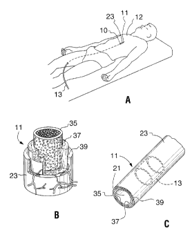
Fig. 1A is a perspective view of a patient with a device positioned in an
artery of the patient in accordance with the method of the present invention;
Fig. 1 B is a perspective view of a portion of an artery of a patient
showing the intima, media and adventitia layers;
Fig. 1 C is a perspective view of a portion of an artery of a patient
showing a circumferential dispersment of a medicament (in phantom) in
accordance with the method of the present invention;
Fig. 2 is a perspective view of a device suitable for use in the method
of the present invention;
Fig. 3A is a cross-sectional view of the device of Fig. 2 as seen along
line 3-3 in Fig. 2, positioned in an artery of a patient;
Fig. 3B is a cross-sectional view of an artery showing a dispenser
positioned for release of a fluid medicament in the media layer of the artery;
Fig. 4A is a perspective view of a first embodiment for a dispenser
suitable for use in the present invention;
9
CA 02367560 2002-O1-14
Fig. 4B is a perspective view of a second embodiment for a dispenser
suitable for use in the present invention;
Fig. 5A is a side plan view of a third embodiment of a dispenser
suitable for use in the present invention;
Fig. 5B is a side plan view of a fourth embodiment of a dispenser
suitable for use in the present invention;
Fig. 5C is a side plan view of a fifth embodiment of a dispenser
suitable for use in the present invention;
Fig. 6 is a perspective view of another embodiment of a device suitable
for use in the present invention;
Fig. 7 is a cross-sectional view of the device shown in Fig. 6 as seen
along line 7-7 in Fig. 6;
Fig. 8 is a perspective view of yet another embodiment of a device
suitable for use in the present invention;
Fig. 9 is a cross-sectional view of the device of Fig. 8 shown in a
retracted configuration, as seen along line 9-9 in Fig. 8;
Fig. 10 is a cross-sectional view of the device of Fig. 8 shown in an
expanded configuration, as seen along the line 9-9 in Fig. 8;
Fig. 11 is a cross-sectional view of the device of Fig. 8 positioned in the
blood vessel of a patient;
Fig. 12A is a longitudinal cross-sectional view of a portion of the vessel
and a device prior to a dispenser penetrating the vessel wall;
Fig. 12B is a longitudinal cross-sectional view of a portion of the vessel
and a portion of the device after a dispenser penetrates the vessel wall;
Fig. 12C is an axial cross-sectional view of the vessel and the device
illustrating the dispensers penetrating the vessel wall;
Fig. 12D illustrates a longitudinal crass-sectional view of the intima
layer of the vessel wall after the fluid medicament has been injected into the
vessel wall;
CA 02367560 2002-O1-14
'4
Fig. 12E is an axial cross-sectional view illustrating the intima layer of
the vessel wall after the fluid medicament has been injected into the vessel
wail;
Fig. 12F is a longitudinal cross-sectional view of a portion of the intima
layer of the vessel and the device illustrating the fluid medicament after
dispersion in the vessel wall;
Fig. 12G is an axial cross-sectional view of the intima layer of the
vessel and the device illustrating the fluid medicament after dispersion in
the
vessel wall;
Fig. 13A is a longitudinal cross sectional view of the vessel and a
device illustrating a fluid medicament containing a radioactive isotope being
injected into the vessel wall;
Fig. 13B is a longitudinal cross sectional view of a portion of the vessel
and the device after a fluid medicament containing a radioactive isotope is
injected into the vessel wall;
Fig. 14A is a longitudinal cross-sectional view of a portion of the vessel
and the device after a fluid medicament containing a precipitant is injected
into the vessel wall;
Fig. 14B is a longitudinal cross-sectional view of a portion of the vessel
and the device after a portion of an injected fluid medicament precipitates
within the vessel wall;
Fig. 15A is a longitudinal cross-sectional view of a portion of the vessel
and the device after a fluid medicament with a binder has been injected into
the vessel wall;
Fig. 15B is a longitudinal cross=sectional view of a portion of the vessel
and the device showing the binder of an injected medicament binding to a
portion of the vessel wall;
Fig. 16A is a longitudinal cross-sectional view of a portion of a vessel
and device illustrating the cell genes of the vessel prior to penetration of
the
vessel with the dispenser;
11
CA 02367560 2002-O1-14
wr
Fig. 16B is a longitudinal cross-sectional view of a portion of a vessel
and device illustrating the vessel after a fluid medicament that includes a
virus
gene is injected into the wall of the vessel by the device; and
Fig. 16C is a longitudinal cross-sectional view of a portion of the vessel
and device illustrating the vessel wall after the injected virus genes have
attacked and replaced the cell genes.
DESCRIPTION OF THE PREFERRED EMBODIMENTS
Referring initially to Fig. 1A, a device 10 for injecting a fluid
medicament 13 into a wall 23 of a living blood vessel 11 in accordance with
the method of the present invention is shown positioned in an upper body,
blood vessel 11 of a patient 12. It is to be appreciated that the present
method can be used in arteries and other vessels throughout the body of the
patient 12. Fig. 1 B shows the wall 23 of an arterial blood vessel 11 having
three layers of importance for the present invention, the intima 35, the media
37 and the adventitia 39. As shown in Fig. 1 C, the intima 35 surrounds the
lumen 21 of the blood vessel 11. Importantly, as provided in detail below, the
device 10 when used in accordance with the method provided herein, allows
for a substantially circumferential dispersion of the fluid medicament 13
within
the wall 23 of the blood vessel 11, as shown in Fig. 1 C. Further, in
accordance with the present method, a circumferential dispersion of fluid
medicament 13 can be made within one of the layers 35, 37, 39 of wall 23 of
the blood vessel 11.
Referring to Figs. 2 and 3A, a first version of a device 10 suitable for
the method of the present invention includes a multi-lumen catheter 14, an
expanding member 15 mounted thereon, a tubular sleeve 18 and a plurality of
dispensers 20. Although Figs. 2 and 3A show the expanding member 15 as
an inflatable balloon 16, any expanding member known in the art may be
used. The balloon 16 is inflatable and deflatable between a first,
substantially
deflated configuration and a second, substantially expanded configuration.
12
CA 02367560 2002-O1-14
a
The balloon 16, while in the second configuration, can be anywhere from
partially inflated to fully inflated depending upon the size of the blood
vessel
11. The balloon 16 and tubular sleeve 18 can be made of a number of
materials including polyethylene terephthalate (PET). As shown in Fig. 2, the
tubular balloon 16 defines a longitudinal axis 17.
Further, Fig. 2 indicates that the tubular sleeve 18 surrounds a
substantial portion of the balloon 16, and that a plurality of dispensers 20
are
mounted onto the tubular sleeve 18. Of these, the number of dispensers 20
illustrated is only exemplary. Importantly for the present method, all
dispensers 20 are positioned in a single plane 19 that, as shown, is oriented
substantially normal to the longitudinal axis 17. Also, it is preferable for
the
present method that the dispensers 20 be equally spaced around the axis 17.
A more complete appreciation of the structural cooperation between
the balloon 16, the tubular sleeve 18 and the dispensers 20 is provided by
Fig. 3A wherein, it will be seen that a distal end 22 of tubular sleeve 18 is
attached directly to an outer surface 25 of balloon 16. By cross-referencing
Figs. 2 and 3A it can be seen that the tubular sleeve 18 substantially
surrounds and encloses the balloon 16 and that a proximal end 24 of tubular
sleeve 18 extends proximally from and beyond the balloon 16 over catheter
14. The tubular sleeve 18 cooperates with the outer surface 25 of the balloon
16 to define a portion of a fluid passageway 26. The proximal end 24 can be
connected to an outer lumen 27 (not shown in Fig. 3A) of the catheter 14 to
complete the fluid passageway 26.
Fig. 3A further shows that the distal end 28 of balloon 16 is affixed to
the catheter 14, and that the proximal end 30 of the balloon 16 attaches onto
the catheter 14 to create an inflation chamber 32 in the interior of the
balloon
16. A balloon port 34 provides fluid access into the inflation chamber 32. For
purposes of the present invention, the balloon port 34 can be connected in
fluid communication with a balloon lumen (not shown) of the catheter 14. Fig.
3A also shows that catheter 14 is formed with an inner lumen 36 which is
dimensioned to receive a guidewire 38 therethrough.
13
CA 02367560 2002-O1-14
a
As discussed previously, the wall 23 of the blood vessel 11 includes
multiple layers. To facilitate the present discussion, some of the layers,
namely, the intima layer 35, the media layer 37, and the adventitia layer 39
are illustrated in Fig. 1 B and again in Fig. 3B. Importantly, when the device
10 is used in accordance with the present method, the depth of penetration of
each dispenser 20 can be precisely controlled by controlling the length 41
(shown in Fig. 5A) of each dispenser 20. In accordance with the method of
the present invention, the dispensers 20 extend a length 41 of between
approximately 0.005 inches and approximately 0.02 inches from the tubular
sleeve 18 when the balloon 16 is inflated. However, those skilled in the
pertinent art will recognize that these distances are merely exemplary. Thus,
the device 10 is able to deliver the fluid medicament 13 to a desired, target
layer in the wall 23 of the blood vessel 11. For example, as illustrated in
Fig.
3B, the dispenser 20 penetrates through the intima layer 35 and precisely
delivers the fluid medicament 13 to the media layer 37, i.e. the target layer
in
this example. It is to be appreciated that a shorter dispenser 20 could be
utilized to deliver the fluid medicament 13 to the intima layer 35.
Additionally,
in accordance with the method of the present invention, the device 10 can be
used to simultaneously release the fluid medicament 13 within a target layer
and dilate the lumen 21 of the blood vessel 11.
Referring now to Fig. 4A, each dispenser 20 includes a base plate 40
and a tubular protrusion 42 having an attachment end 44 and a penetrating
section 46. Further, it is seen that the attachment end 44 of the tubular
protrusion 42 affixes to and is an integral part of the base plate 40.
Preferably, the dispenser 20 is made of nickel and the tubular protrusion 42
is
formed by punching out the base plate 40. in the dispenser embodiment
illustrated in Fig. 4A, the penetrating section 46 is defined by an opening
which is opposite the base plate 40. The tubular protrusion 42 defines a fluid
channel 48 which extends through the dispenser 20. The penetrating section
46 of the dispenser 20 shown in Fig. 4A is substantially annular shaped.
14
CA 02367560 2002-O1-14
Fig. 4B shows another embodiment of the dispenser 20. In this
embodiment, each tubular protrusion 42 is substantially conical shaped as
shown in Fig. 4B. Like the embodiment shown in Fig. 4A, the dispenser 20
shown in Fig. 4B is preferably made of nickel and is formed with a fluid
channel 48 which extends through the dispenser 20.
Figs. 5A, 5B and 5C illustrate additional, alternative embodiments of
the dispenser 20. In the embodiments illustrated in Figs. 5A, 5B and 5C, the
tubular protrusion 42 is substantially conical shaped. However, in Fig. 5A,
the
penetrating section 46 is defined by an opening which extends through the
side of the tubular protrusion 42. Somewhat similarly, in Fig. 5B, the
penetrating section 46 is defined by a pair of openings which extend through
the side of each tubular protrusion 42. This feature inhibits plugging of the
penetrating section 46 during insertion into the wall 23 of the blood vessel
11.
In Fig. 5C, the tubular protrusion 42 is made of a porous material. Thus, the
porous material defines the penetrating section 46 of each dispenser 20. In
the embodiment shown in Fig. 5C, the fluid medicament 13 is forced through
the pores 49 of the porous tubular protrusion 42.
Referring now to Fig. 3A, the dispensers 20 are mounted on the
tubular sleeve 18 so that the fluid channel 48 of each respective dispenser 20
is aligned with a hole 52 in the tubular sleeve 18. This is done to establish
fluid communication between the particular dispenser 20 and the fluid
passageway 26. As a practical matter, it may be preferable in the
construction of the device 10 to first mount the dispenser 20 on the tubular
sleeve 18, which can be done in any manner well known in the pertinent art,
such as by bonding, and then pierce a hole 52 in the tubular sleeve 18
through the dispenser 20.
An alternative structure for a device 10 suitable for use in the present
method is shown in Fig. 6. As shown, the alternative device 10 includes a
multi-lumen catheter 14 formed to accommodate a guidewire 38, a balloon 16,
a plurality of dispensers 20 and a plurality of tubular channels 64 mounted on
the outer surface 25 of the balloon 16. Each tubular channel 64 has a smaller
CA 02367560 2002-O1-14
diameter than the balloon 16 and is positioned to be substantially parallel
with a
longitudinal axis 65 of the balloon 16.
Fig. 6 further shows that mounted on the surface of each tubular
channel 64 is a dispenser 20. The dispensers 20 are positioned on the surface
of tubular channel 64 so that when balloon 16 is inflated, the dispensers 20
move outwardly from the longitudinal axis 65 in a radial direction.
Importantly
for the present method, afl dispensers 20 are positioned in a single plane 67
that is oriented substantially normal to the longitudinal axis 65 of the
balloon
16. Further, it is preferable for the present method that the dispensers 20 be
equally spaced around the longitudinal axis 65.
Referring now to Fig. 7, the cross-sectional view of the alternative device
10 shows the tubular channel 64 in more detail. More specifically, a distal
end
66 of tubular channel 64 is sealed to create a portion of the fluid passageway
26 which connects the dispensers 20 to the fluid source 60. Referring to Figs.
6 and 7, it is to be appreciated that the proximal end 68 of the tubular
channel
64 is in fluid communication with the outer lumen 27 of the catheter 14. In
turn,
the outer lumen 27 is connected in fluid communication with the fluid pump 58
and the fluid medicament source 60.
Still referring to Fig. 7, the dispensers 20 are shown mounted on the
surface of the tubular channel 64. As Fig. 7 further shows in detail, a base
plate 40 of a dispenser 20 is mounted on the tubular channel 64 over a
corresponding hole 70. From this view, it can be appreciated that any number
of tubular channels 64 could be mounted on the external surface of balloon 16.
Fig. 8 shows yet another version of a device 10 suitable for the method
of the present invention. In this version of the device 10, the expanding
member 15 includes a mufti-lumen catheter 80 and a grommet 82. Both the
mufti-lumen catheter 80 and the grommet 82 are disposed about the same
longitudinal axis 97 with the grommet 82 positioned distally, and separated
from, the distal end 88 of the mufti-lumen catheter 80.
A mechanism is provided to move the grommet 82 translationally along
the longitudinal axis 97. For example, referring to Fig. 8, a push-pull wire
84, is
16
CA 02367560 2002-O1-14
shown connected to the grommet 82. The push-pull wire 84 extends through
one of the lumens of the mufti-lumen catheter 80 allowing the push-pull wire
84
to move translationally in line with the longitudinal axis 97. The
translational
movement of the push-pull wire 84 causes the grommet 82 to undergo a similar
translational displacement. Further, this version of the device 10 can be used
in combination with the guidewire 38, as shown in Fig. 8. Specifically, the
push-
pull wire 84 may be formed with an internal lumen, allowing the catheter 80
and
push-pull wire 84 to pass over the guidewire 38.
In the version of the device 10 shown in Fig. 8, a plurality of hollow,
flexible tubes 86 are attached between the grommet 82 and the mufti-lumen
catheter 80. Each of the flexible tubes 86 includes a distal end 88, a
proximal
end 90 and a central region 92. The proximal end 90 of each tube 86 is joined
to the mufti-lumen catheter 80. The distal end 88 of each tube 86 is joined to
the grommet 82. Preferably, the tubes 86 are distributed radially around the
mufti-lumen catheter 80 and grommet 82 in a manner substantially as shown in
Fig. 8.
Referring now to Figs. 9-11, it may be seen that each flexible tube 86 is
formed with a lumen 94. The lumen 94 of each flexible tube 86 passes through
the mufti-lumen catheter 80 allowing fluid medicament 13 to be passed through
mufti-lumen catheter 80 and into flexible tubes 86. The lumen 94 of each
flexible tube 86 passes separately through mufti-lumen catheter 80 allowing a
different fluid medicament 13 to be passed into each flexible tube 86.
Alternatively, the lumen 94 of each flexible tube 86 may be attached to one or
more common lumens within the mufti-lumen catheter 80.
Referring to Figs. 8 and 9, it is shown that a dispenser 20 is attached to
the central region 92 of each flexible tube 86. Each flexible tube 86 is
formed
with a hole 96 which correspond to a respective dispenser 20. Functionally,
each hole 96 connects the fluid channel 48 of a respective dispenser 20 to
lumen 94 allowing the fluid pump 58 to pump fluid medicaments 13 from the
fluid source 60 into lumen 94 to be expelled through the dispensers 20.
Importantly for the present method, all dispensers 20 are positioned in a
17
CA 02367560 2002-O1-14
single plane 95 oriented normal to the longitudinal axis 97 defined by the
expanding member 15. Further, it is preferable for the present method that
the dispensers 20 are equally spaced around the longitudinal axis 97.
Referring now to Figs. 9 and 10, it is shown that the device 10 is
movable between the first, contracted configuration (shown in Fig. 9) and the
second, expanded configuration (shown in Fig. 10). Specifically, it may be
seen
that the grommet 82 and the multi-lumen catheter 80 are distanced by a first
separation distance 98. The device 10 shown in Fig. 9 also has a first overall
width designated 100. In comparison, the grommet 82 and the multi-lumen
catheter 80 shown in Fig. 10 are distanced by a second separation distance
102 which is smaller than the first separation distance 98 of Fig. 9. The
device
10, shown in Fig. 10 also has a second overall width 104 which is greater than
the first overall width 100 shown in Fig. 9.
The movement between the first, contracted configuration shown in Fig.
9 and the second, expanded configuration shown in Fig. 10 is accomplished by
the translational movement of the grommet 82 along the longitudinal axis 97.
Specifically, as the push-pull wire 84 causes the grommet 82 to move towards
the multi-lumen catheter 80, each of the flexible tubes 86 bows outwardly away
from the longitudinal axis 97. In this manner, the push-pull wire 84 may be
used to move the grommet 82 translationally to cause the flexible tubes 86 to
alternately bow, as seen in Fig. 10, and straighten, as seen in Fig. 9. In
some
cases, it will be preferable to fabricate the flexible tubes 86 from a
resilient
material and shape the flexible tubes 86 to be initially biased in either a
bowed
or straight configuration.
Figs. 12A-12F show the process whereby the fluid medicament 13 is
pumped from each dispenser 20 into the intima layer 35 of an exemplary
blood vessel 11 and then allowed to disperse. Figs. 12A-12F further show
that the fluid medicament 13 can be pumped into a target layer, in this case
the intima 35, and allowed to disperse until a circumferential dispersion of
fluid medicament 13 is achieved. First, as shown in Fig. 12A, the dispenser
20 is positioned adjacent to the target area of the blood vessel 11. Next, as
18
CA 02367560 2002-O1-14
shown in Figs. 12B and 12C, the expanding member 15 is expanded, forcing
the dispenser 20 to penetrate the target layer (in this case, the intima 35).
Preferably, as illustrated in Fig. 12C, the dispensers 20 are
circumferentially
spaced to create a plurality of spaced apart medicinal deliveries 106. Figs.
12D and 12E show the medicinal deliveries 106 which are confined to the
intima layer 35. Figs. 12F and 12G show the subsequent dispersion of the
fluid medicament 13 around a circumference of the wall 23 of the blood
vessel 11, creating a circumferential dispersion. The pumping rate required
to achieve the desired circumferential dispersion depends upon the viscosity
of the fluid medicament 13 utilized. Typically, between approximately 400
microliters and 700 microliters of the fluid medicament 13 is dispensed during
a period of between approximately five and forty-five seconds to create the
desired medicinal delivery 106 that will result in a circumferential
dispersion.
However, it should be recognized that the amounts and time frames provided
herein are merely exemplary. ft is also to be appreciated that the medicinal
dispersion rate will be affected by the body's response (inflammation) to the
tissue injury caused by the present method.
Further, the spacing required to create a plurality of spaced apart
medicinal deliveries 106 which subsequently disperse the fluid medicament
13 along the treatment area 54 will also vary according to the fluid
medicament 13 utilized. It is contemplated for the present method that the
dispensers 20 are to be spaced a circumferential distance 108 of between
approximately 1 millimeter and 6 millimeters, roughly 70 degrees and 140
degrees apart.
The composition of the fluid medicament 13 to be injected into the wall
23 of the blood vessel 11 depends upon the treatment being performed and
the physical characteristics of the patient 12. More specifically, the fluid
medicament 13 can be designed to treat a stenosis or disease de novo,
inhibit a restenosis by minimizing the effects of a previous intravascular
procedure and/or inhibit a stenosis in a blood vessel 11. For example, to
inhibit a restenosis, the fluid medicament 13 can contain anti-proliferative
19
CA 02367560 2002-O1-14
agents which inhibit the proliferation of smooth muscle cell growth in the
vessel in certain pathological conditions. These fluids selectively kill
rapidly
dividing cells and can be utilized to inhibit the proliferation of smooth
tissue
growth. Suitable fluids can include anti-proliferative agents such as
methotrexate, prednisone, adriamycin, mitomycinc, protein synthesis
inhibitors, toxin fragments such as pseudomonas, exotoxin (PE) or Ricin A
(RA) Toxin, and radioactive isotopes 112 such as "'Indium, 9°Yttrium,
6'Gallium, 99'"Tc (Technetium 99), 2°SThallium, and 32P (Phosphorous
32)
radiopharmaceutical. It is believed that the present method is uniquely suited
to safely deliver toxic fluid medicaments 13 into the wall 23 of the blood
vessel 11 while minimizing the amount of fluid medicament 13 which is
washed away into the blood stream.
Alternatively, for example, a fluid medicament 13 which stimulates the
production of collateral vessels can be delivered by the present method.
These fluid medicaments 13 provide preventative treatment for the patient 12
by creating new collateral vessels in the event the original blood vessel 11
develops a stenosis. A fluid medicament 13 which includes an angiogenis
factor can be utilized for this purpose.
Figs. 13A and 13B, illustrate the delivery and dispersion of a fluid
medicament 13 that includes a radioactive isotope 112 which can reduce and
inhibit tissue and/or cell growth of the wall 23 of the blood vessel 11.
Because the radioactive isotopes 112 are injected directly in the wall 23 of
the
blood vessel 11 and are symmetrically injected around the circumference of
the wall 23 of the blood vessel 11, relatively low energy radioactive isotopes
112 having a relatively short half -life can be utilized. These relatively low
energy radioactive isotopes 112 should cause minimal trauma to the patient
12. The present method provided herein is uniquely suited to safely deliver a
radioactive isotope 112 to only the treatment area 54 of the wall 23 of the
blood vessel 11, while minimizing the amount of radioactive isotope 112
which is washed away into the blood stream. Additionally, the radioactive
isotope 112 can be encapsulated within a suitable carrier such as amino-
CA 02367560 2002-O1-14
mannose modified liposome, which is rapidly absorbed into the smooth
muscle cells of the intima layer 35.
The exact dose of radiation to be delivered to the wall 23 of the blood
vessel 11 can be varied to suit the needs of the patient 12. It is presently
believed that a tissue absorbed dose of between approximately 8-40 Gray will
be utilized to inhibit restenosis. The exact amount of fluid medicament 13
and type of fluid medicament 13 injected into the wall 23 of the blood vessel
11, can be varied to account for fluid medicament 13 washed into the blood
stream and/or account for the active life of the fluid medicament 13.
Referring to Figs. 14A and 14B, it is shown that a precipitation process
can be used to minimize the amount of fluid medicament 13 which is washed
away into the blood stream. Specifically, a portion of the fluid medicament 13
can be precipitated at approximately the pH level of the wall 23 of the blood
vessel 11. Typically, the vessel pH is approximately 7. A fluid medicament
13 containing a precipitator 114, and having a fluid pH level of less than
approximately 6 or greater than approximately 8 can be utilized. After the
fluid medicament 13 and precipitator 114 are dispensed into the wall 23 of the
blood vessel 11, the fluid medicament pH level wilt approach 7, and a portion
of the fluid medicament 13 may precipitate. For this embodiment, the fluid
medicament 13 could include a precipitator 114, an active component 115
attached to or incorporated within the precipitator 114 and a carrier
component 117 which carries the precipitator 114 and the active component
115. The active component 115 is the portion of the fluid medicament 13
which is designed to treat the patient 12. In this example, the precipitator
114
could precipitate in the wall 23 of the blood vessel 11 while the carrier
component 117 gets washed away into the blood stream.
Because the active component 115 is attached to or incorporated
within the precipitator 114, this ensures that the bulk of the active
component
115 of the fluid medicament 13 remains in the wall 23 of the blood vessel 11
and minimizes the amount of the active component 115 of the fluid
medicament 13 which is washed away into the blood stream. In this
21
CA 02367560 2002-O1-14
embodiment, the active component 115 of the fluid medicament 13, for
example, can include an anti-proliferative agent as outlined above.
Alternatively, the precipitator 114 and the active component 115 can be a
radionuclide or radiopharmaceutical precipitate, such as gold colloidal, i.e.
'98Au and '99Au, and/or an inorganic precipitate such as organo-metallic
precipitate.
Additionally, the active component 115 of the fluid medicament 13 can
be designed to have a slow, time-release formulation so that active
component 115 is released to the wall 23 of the blood vessel 11 over an
extended period of time. Stated another way, the active component 115 can
biodegrade slowly over a period of time to release the active component of
fluid medicament 13 into the wall 23 of the blood vessel 11 over an extended
period of time. A biodegradable polymer may be used to provide a control
release formulation to the active component 115.
Alternatively, referring to Figs. 15A and 15B, the fluid medicament 13
may include a binder 116, the active component 115 and the carrier
component 117. The binder 116 is secured to the active component 115 of
the fluid medicament 13. The binder 116 is adapted to bind, attach andlor
crosslink to at least a portion of the wall 23 of the blood vessel 11. For
example, the binder 116 could include a ligand which binds to a portion of the
wall 23 of the blood vessel 11 such as collagen or the smooth muscle cell
component of the wall 23 of the blood vessel 11. Because the binder 116 is
secured to the active component 115, this ensures that the bulk of the active
component 115 of the fluid medicament 13 remains in the wall 23 of the blood
vessel 11 and minimizes the amount of the active component 115 of the fluid
medicament 13 which is washed away into the blood stream. Examples of
ligands capable of binding to the arterial wall components include PDGF
receptors, adhesive molecules including, but not limited to certain molecules
of the integrin family, and receptors on activated platelets such as thrombin
receptors. Another suitable type of ligand is sold under the name
CERETEC~ by Amersham located in Arlington Heights, Illinois. Alternatively,
22
CA 02367560 2002-O1-14
for example, phosphors tridentite which binds to collagen can be utilized. In
yet an alternative embodiment, the binder 116 can have a direct affinity to
form ionic bonds, covalent bonds or Van der Waal attractions with the wall 23
of the blood vessel 11 or some component thereof.
Alternatively, as illustrated in Figs. 16A-16C, the fluid medicament 13
can be used for gene therapy on the wall 23 of the blood vessel 11. In this
embodiment, the fluid medicament 13 can include a suitable viral vector 118
which is adapted to infect a cell 120 and replace, modulate, inhibit or
enhance
one of the cell genes 122 within the cell 120. For example, the fluid
medicament 13 could include a retroviral, adenoviral vectors or Adenovirus
Associated Vectors (AAV) carrying the appropriate DNA payload for
appropriate gene switching. Alternatively, for example, naked DNA or
polycation-condensed DNA could be utilized for gene therapy. The method of
the present invention allows for the use of fluid medicaments 13 which
genetically alter the treatment area 54 of the wall 23 of the blood vessel 11
without effecting the rest of the body.
Still other fluid medicaments 13 which could be utilized with the
method of the present invention include antibodies such as receptor site
monoclonal antibodies, a toxic agent such as saponin, a genetic material
such as DNA, a cellular material such as endothelial cells and/or
medicaments such as heparin. The examples provided herein are merely
examples of fluid medicaments 13 which may be useful with the present
invention. Those skilled in the art will recognize that additional fluid
medicaments 13 will be developed as medical technology improves.
Additionally, those skilled in the art will recognize that the present
invention
can be utilized for applications other than inhibiting a restenosis. For
example, with extended dispensers 20, the method of the present invention
could deliver fluid medicaments 13 from the blood vessel 11 to specific
organs.
23
CA 02367560 2002-O1-14
II~C~ATtflA1
An example of the operation of the balloon 16 version of the expanding
member 15 can best be visualized with initial reference to Figs. 1-3. First,
the
guidewire 38 is positioned into the blood vessel 11 of the patient 12. This is
done to establish a mechanical pathway through the blood vessel 11 to the
treatment area 54 where the fluid medicament 13 is to be released.
Next, the balloon 16, which is attached to the catheter 14, is moved
over the guidewire 38 to the treatment area 54. The balloon 16 is at its first
configuration during movement over the guidewire 38 in the blood vessel 11.
Once the balloon 16 is properly positioned proximate the treatment area 54,
an inflator 56 is activated to inflate the balloon 16 to its second
configuration.
As shown in Fig. 2, the inflator 56 is connected to the proximal
(extracorporeal) end 29 of the catheter 14.
Referring back to Figs. 3A and 3B, it will be appreciated that, as the
balloon 16 is inflated, the expanding balloon 16 urges against the tubular
sleeve 18 and causes the tubular sleeve 18 to likewise expand.
Consequently, the dispensers 20 mounted on the tubular sleeve 18 move
radially from the longitudinal axis 17 and embed into the treatment area 54.
Further, the balloon 16 can be used to simultaneously dilate the lumen 21 of
the blood vessel 11.
With the dispensers 20 embedded into the treatment area 54, the fluid
pump 58 shown in Fig. 2 is activated to pump a fluid medicament 13 from the
fluid medicament source 60 into the fluid passageway 26. Importantly, this
pumping action also causes any fluid medicament 13 which has already been
pumped into the fluid passageway 26 to be expelled through the fluid
channels 48 of dispensers 20 and into the tissue of treatment area 54.
Alternatively, the fluid pump 58 could be activated prior to embedding
the dispensers 20 into the wall 23 of the blood vessel 11 and a valve 62 could
be used to prevent the flow of fluid medicament 13 until the dispensers 20 are
embedded in the treatment area 54. The valve 62 can then be opened when
24
CA 02367560 2002-O1-14
. ~.
the dispensers 20 penetrate into the treatment area 54 so that injection
occurs substantially simultaneously with the embedding of the dispensers 20
in the treatment area 54. Alternatively, the injection of the fluid medicament
13 could happen after a time delay by waiting to open the valve 62 for at
least
about one second to about twenty seconds. Further, one or more fluid
medicaments 13 can be released at different time intervals in the wall 23 of
the blood vessel 11.
After the fluid medicament 13 from the fluid medicament source 60 has
been dispensed into the treatment area 54, the balloon 16 can be deflated to
the first configuration by reversing the inflator 56. This action will cause
the
balloon 16 to collapse and withdraw the dispensers 20 from the treatment
area 54. The entire device 10 can then be withdrawn from the patient 12 over
the guidewire 38.
The embodiment shown in Fig. fi utilizes a plurality of individual, tubular
channels 64. With this embodiment, it is possible to either maintain fluid
communication with, or fluid isolation between, each tubular channel 64. For
example, fluid communication between each tubular channel 64 can be
established by fluidly connecting each tubular channel 64 together within one
outer lumen 27 of the catheter 14 so that each tubular channel 64 is supplied
fluid medicament 13 from the same fluid pump 58. Alternatively, fluid
isolation
may be maintained between each tubular channel 64 by providing each tubular
channel 64 with a corresponding and independent outer lumen 27 and
establishing its own fluid connection to a corresponding and independent fluid
pump 58. Consequently, it is possible to inject a variety of alternate fluid
v5 medicaments 13 simultaneously by using a plurality of tubular channels 64
which are each connected to a separate fluid pump 58.
While the particular Method for Delivering Medication Into an Arterial
Wall for Prevention of Restenosis as herein shown and disclosed in detail is
fully capable of obtaining the objects and providing the advantages herein
before stated, it is to be understood that it is merely illustrative of the
presently preferred embodiments of the invention and that no limitations are
CA 02367560 2002-O1-14
intended to the details of the construction or design herein shown other than
as defined in the appended claims.
26