Note: Descriptions are shown in the official language in which they were submitted.
CA 02367599 2001-09-17
WO 00/54705 PCT/USOO/06643
APPARATUS AND METHOD FOR FIXATION
OF OSTEOPOROTIC BONE
This invention relates to a novel surgical apparatus for use in osteoplasty
and
other methods of injecting materials into a subject for medical purposes.
Particularly,
the present invention relates to the surgical treatment of traumatic,
pathogenic, or
osteoporotic bone conditions of the human and other animal body systems and
more
particularly, to a novel apparatus and method for injection of a material into
a lesion
of a vertebral body or other bony structure.
BACKGROUND
Lesions within the bone can result from osteoporosis, tumor, or other
pathogenic causes. Most common among the elderly population is the
degenerative
effect of osteoporosis, particularly the female elderly. Osteoporosis is
mediated at
least in part by genetic defects and a fall in circulating estrogen levels.
Although
calcium replacement therapy can have some beneficial effects, the larger doses
of
calcium involved have other less helpful consequences and accordingly, the
prognosis
for those with bone demineralization is not particularly good. Of great
concern is the
fact that every year in the United States there occurs approximately 1.2
million bone
failures due to osteoporosis. Vertebral compression failures are a major
orthopedic
health concern of the elderly due to the long term debilitating nature of the
injury.
Historically, osteoporotic vertebral body compression failures have been
treated with bed rest, analgesics, and intravenous hydration during the first
week after
onset of the problem. These steps are followed by the prescription of a soft
or finn
spinal corset, depending upon the physician's preference. In most cases the
corset is
CA 02367599 2001-09-17
WO 00/54705 PCT/US00/06643
not worn because the patient suffers much discomfort and oftentimes greater
discomfort than that due to the failure of the vertebral body. In any case,
this
conventional approach required extensive hospitalization and bed rest, which
often
results in very limited success, chronic pain, and further osteoporosis with
worsening
conditions of the vertebral body. The costs associated with such extended
hospitalization and the negative effect on the general health of the patient
from such
prolonged inactivity should be avoided if possible.
Traditional surgical techniques employed to alleviate vertebral compression
failures can involve major invasive surgical techniques with all of the
possible
negative consequences. Such techniques have typically required prolonged
patient
recuperation and unfortunately have met with limited success in alleviating
pain and
returning the patient to a normal life style.
More recently efforts have been made to develop surgical techniques for repair
of vertebral compression failures of osteoporotic bone by using conventional
instruments in a transpedicular approach to penetrate the vertebral body,
including a
standard syringe, and then inject a flowable synthetic bone material or bone
cement
directly into the vertebral body through the syringe. This technique of
vertebroplasty
requires that the physician take the utmost care to avoid damage to the spinal
cord
when drilling through the narrow dimensions of the pedicle of the vertebrae.
To
avoid potentially catastrophic results physicians practicing conventional
vertebroplasty require the use of CAT scanning, biplane fluoroscopy, magnetic
resonance imaging, or other imaging devices to ensure the proper alignment of
the
instruments, which bore through and are passed through the narrow pedicle. The
availability of CAT scanning or sophisticated biplane fluoroscopy in surgical
procedures is limited due to the additional cost associated with equipping
surgical
2
CA 02367599 2001-09-17
WO 00/54705 PCTIUSOO/06643
suites with the necessary equipment. Further, to protect against accidental
damage to
the spinal cord during the conventional transpedicular approach to the
vertebral body,
the patient is typically placed in a restraining device and stereotaxic
procedures are
used to guide the physician's drill and cannula through the pedicle. Due to
the
extraordinary care and precision required in conventional vertebroplasty, the
time
needed to complete the surgery and the cost associated with the procedure can
be
extensive. Further, general anesthetic is not recommended due to the close
proximity
of the physician's instruments to the spinal cord and the associated need to
conununicate with the patient. This requirement, however, also causes concern
of
movement of the patient during the surgery; movement which could have serious
consequences should the spinal cord be damaged as a result. Scholten et al. in
U.S.
Patents 4,969,888 and 5,108,404 teaches the conventional surgical technique of
vertebroplasty with the additional step of employing a balloon as an expansion
device
within the body of the vertebrae to compact the osteoporotic cancellous bone
away
from the center and against the walls of the vertebral body. This additional
step to
conventional vertebroplasty, taught by Scholten et al., is intended to provide
additional space within the vertebral body to accept the flowable bone cement
through
the needle (syringe). While the conventional vertebroplasty technique using
conventional surgical apparatus has the distinct disadvantage of drilling
through the
pedicle with the potential risk of damage to the spinal colunln, this
additional balloon
expander employed in the process of Scholten et al., provides an additional
disadvantage by compressing the naturally present internal matrix of the
osteoporotic
vertebra against the wall of the vertebral body. Absent this natural matrix,
the
injection of bone cement into the cavity created by the compressing step
results in the
formation of an unstructured bolus of bone cement in the center of the
vertebral body.
3
CA 02367599 2001-09-17
WO 00/54705 PCT/US00/06643
Because of the compression of cancellous bone, which as a result lines the
walls of
the vertebra, the bone cement which is infused into the vertebral body does
not make
a strong, direct, bonding contact with the vertebral wall, thus resulting in a
potentially
weaker post-surgery vertebral body.
There is, therefore, a great need for a surgical technique and associated
instrumentation by which osteoporotic bone can be safely, expeditiously and
efficiently treated. There is a particular need for a vertebroplasty procedure
and
associated instrumentation which provide a safer, faster procedure that
ultimately
results in a repair to the osteoporotic vertebral body wherein the injected
material
does not disturb the natural matrix of the cancellous bone, which along with
direct
contact to the vertebral wall provides a strong, composite matrix. The present
invention provides an apparatus and a method of percutaneous bone failure
fixation,
which satisfies these needs.
SUMMARY OF THE INVENTION
The process and apparatus of the present invention can be generally used to
perform osteoplasty, that is the introduction of any injectable material into
any of the
bones or tissues of the body. The present invention is particularly suitable
for
injecting materials into bones which have or are susceptible to compression
failure
due to lesions within cancellous bone. More particularly, this invention
relates to a
method and apparatus, involving the injection of materials for the fixation of
lesions
or failures of bones, particularly as a result of osteoporosis, tumor, other
pathogenic
conditions or trauma. The invention is especially suitable for use in the
vertebroplasty
procedures, such as, the fixation or prevention of vertebral body compression
failures,
although the instrumentation and methods of the present invention can be used
for a
4
CA 02367599 2001-09-17
WO 00/54705 PCTIUSOO/06643
wide variety of osteoplasty procedures, such as, failures or lesions in nones
throughout the body.
An object of the present invention is to provide an apparatus, which is useful
for the surgical procedure of safely introducing a material into a lesion or
space within
or around a bone or tissue.
Another object of the present invention is to provide a surgical method for
safely introducing an injectable material into a lesion or space within or
around a bone
or tissue.
More particularly, it is an object of the present invention to provide an
apparatus, which is sized and configured to safely contact or breach the
cortical bone
and establish an introducing channel through the apparatus and through the
cortical
bone into the cancellous bone through which a material can be introduced. The
material introduced into the interior of the bone can be any biocompatible or
therapeutic materials, such as, for example, antibiotics, whole cellular
implants,
natural products of cells, recombinant nucleic products, protein products of
recombinant cells, allograft or autograft bone, bone cement products as are
well
known in the art (such as polymethylmethacrylate and the like), or any other
flowable
material useful for therapeutic, prosthetic, or bone strengthening purposes.
Another object of the present invention to provide an apparatus, which is
sized
and configured to be used by a physician to safely introduce a material into
the
cancellous bone of a vertebral body. ln the surgical procedure of the present
invention the apparatus can introduced by direct vision, open or
percutaneously,
laproscopically, thorascopically, or by open surgical procedures. The
apparatus can
be introduced into the vertebral body by a variety of approaches, to include,
for
example, postero-lateral and lateral and/or bilateral percutaneous approaches
and a
5
CA 02367599 2001-09-17
WO 00/54705 PCT/[7S00/06643
transpedicular approach. Such introduction of the apparatus can be
accomplished
with or without the conventional requirement for CAT scanning or sophisticated
biplane fluoroscopy and further can be performed safely using general or local
anesthetic. No irrigation, evacuation, or use of cancellous bone expanders is
required
for the successful use of the apparatus to introduce the material into the
interior of the
vertebral body.
Additionally, an object of the present invention is to provide a modular
pedicle
finder, which facilitates the placement of an instrument for penetrating the
pedicle of
a vertebra.
BRIEF DESCRIPTION OF THE DRAWINGS
The invention will now be described, by way of illustration only, with
reference to the accompanying drawings.
FIG. I is an isometric view of the components of the one embodiment of the
apparatus of the present invention.
FIG. 2 is an isometric view of the assembled Guide wire and Aligning
Cannulae of the present invention.
FIG. 3 is an isometric view of the assembled Delivery cannulae and Plunger of
the present invention.
FIG. 4 depicts the present invention equipped with an optional syringe system.
FIG. 5 is a depiction of a guide wire that can be used in the present
invention
having a Luer lock for providing a fluid tight attachment to an infusion
device or
syringe.
6
CA 02367599 2001-09-17
WO 00/54705 PCTIUSOO/06643
FIG. 6 is a depiction of a delivery cannulae that can be used in the present
invention, which is configured to be capable of receiving the guide wire shown
in
FIG. 5.
FIG. 7 is a depiction of the assembled guide wire and delivery cannulae shown
in FIGs. 5 and 6.
FIG. 8 is a depiction of a handle configured to be capable of removable
attachment to the Luer lock of the guide wire shown in FIG. 5 or the cannulae
shown
in FIG. 6.
FIG's. 9A-C are detail views of the handle shown in FIG. 8.
FIG. 9D is a depiction of an embodiment of the handle shown in FIG. 8 which is
configured with a removable proximal end for purposes of exposing the proximal
end
of the guide wire for ease in movement, insertion, and extraction from the
delivery
cannulae. FIG. 9E-F shows examples of some of the alternative end attachments,
which can be employed with the handle shown in FIG. 9D. FIGS. 9H-G depict a
cannulated T-handle which can be used with the present invention. FIG. 91 is a
partial
sectional view of an alternative embodiment of the present invention employing
a
handle having a removable proximal end, which acts an extended impact surface.
FIG's. 1 0A-B are cross-sectional side (1 0A) and end (l OB) views of the
plunger shown in FIG. 1 OC, which can be used with the apparatus of the
present
invention. FIG. l OC is a depiction of the plunger assembly, which includes
the
handle shown in FIG's. l0A-B. FIGS. l OD-J are various views of an alternative
embodiment of a plunger that can be used with an embodiment of the present
invention employing a threaded plunger and cannulae.
FIG. 11A is a depiction of a hand operated plunger actuator which can be used
with the apparatus of the present invention. FIG. 11B is a depiction of a type
of
7
CA 02367599 2001-09-17
WO 00/54705 PCTIUSOO/06643
syringe which can be used to contain a material for use in the metnoo oi ine
preseni
invention, the syringe being an example of the type syringe which can be used
with
the hand operated plunger actuator shown in FIG. 11 A. Unlike other plunger
actuators, this plunger actuator of the present invention allows for
controlled
injection down to I cc of material per squeeze by the operator. FIGS. 11 C-E
are
depictions of an alternative multilumen-type cannulae which can be used to
contain
more than one material for simultaneous or sequential injection in the method
of the
present invention.
FIG. 12A is a depiction of an application of the method of the present
invention, which employs a flexible cannulae for delivery of a material into
the bone
material of a joint, such as, for example into the acetabulum.
FIG. 12B is an enlarged cross-sectional depiction of the flexible cannulae
shown in FIG. 12A showing an example of a mechanism which can be employed to
steer the flexible cannulae. The plunger technology depicted in FIG. 10
maintains a
flexible shaft for delivery through the flexible lumen of the flexible
cannulae.
FIG's. 13A-B show a specialized impact forceps, which can be used with the
device of the present invention for purpose of facilitating the entry of the
device into
the bone.
FIG. 14 and FIG. 15 are depictions of a conventional prior art method of
vertebroplasty. FIG 14. shows a transpedicular approach to the vertebral body.
FIG.
15 shows the deep penetration of the vertebral body using a transpedicular
approach.
FIG. 16 is a depiction of the apparatus of the present invention positioned
relative to a sectional view of a vertebral body during operation of the
method of the
preferred embodiment of the present invention.
8
CA 02367599 2001-09-17
WO 00/54705 PCT/US00/06643
FIG. 17 is a depiction of a first alternative embodiment of the method of the
present invention showing a bilateral approach to the vertebra. Such a
bilateral
approach would preferably be done in order of first one side and then the
other,
although the figure depicts both steps simultaneously.
FIG. 18 is a depiction of a second alternative embodiment of the method of the
present invention in which the cancellous bone is penetrated with minimal
disruption
of the cancellous bone to pennit more extensive infusion of the injectable
material.
DETAILED DESCRIPTION
The apparatus and method of the present invention can be adapted for use in
the introduction of any material into any bone that contains a lesion or
sufficient
porosity to acceptthe materials. The employment of the apparatus and surgical
procedure of the present invention in vertebroplasty; particularly to treat
vertebral
compression failures which result from osteoporotic conditions is herein
described
below as illustrative of the present invention.
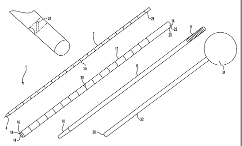
The following description of the device of the present invention relates to
FIG'S. 1-3. The apparatus of the present invention is an intraosseous
injection device
generally shown at 1. One object of the present invention is to use the
injection
device 1 in a surgical procedure for the safe, effective introduction of
materials into a
lesion within a bone, whereby the procedure includes the introduction of a
first guide
wire 2 having a tapered end 4 for effectively breaching the dense compact
bone, for
example, the cortical bone of the vertebra. An aligning cannulae 6 is
configured and
sized to easily pass over the first guide wire 2 and when passed down the
shaft of the
guide wire 2 serves as a soft tissue protective sleeve from the point of entry
of the
apparatus into the body to the contact point at the exterior surface of the
bone being
9
CA 02367599 2001-09-17
WO 00/54705 PCT/US00/06643
treated. The aligning cannulae 6 has a blunt first end 8 which has a textured
surface
to facilitate handling and a tapered second end 10 which during operation of
the
instrument is brought into contact with the bone being treated.
A delivery cannulae 12, which is sized and configured to easily pass over the
aligning cannulae 6 is inserted over the aligning cannulae 6 for purpose of
providing a
material conduit 14 through which the injectable material can be introduced
into the
bone being treated. The delivery cannulae 12 is configured at the delivery
cannulae
distal end 16 to have a securing edge 18 which serves to hold the delivery
cannulae 12
in place on the outer surface of the bone being treated. The delivery cannulae
proximal end 20 is configured to have a handle retention member 22, which
serves to
releasably secure a handle member 24 to the delivery cannulae 12. The handle
member 24 can be used for insertion of the delivery cannulae 12 over the
aligning
cannulae 6 and for improving the grip of the user when placing the securing
edge 18
of the delivery cannulae 12 firmly into position on the outer surface of the
bone being
treated. The removable handle member 24 also can be useful at a later step of
the
surgical procedure for providing a secure grip, which may be necessary to
disengage
the delivery cannulae 12 from the surface of the bone prior to extracting the
device 1
from the body of the patient. The surface of the delivery cannulae can be
provided
with graduated indicia 30 which provide depth of penetration information
during
insertion by the user.
The guide wire 2 can be provided with graduated guide wire indicia 26 which
extend from the tapered end 4 to the more proximal guide wire blunt end 28.
The
guide wire indicia 26 provides a means by which the user can easily determine
the
depth of insertion of the guide wire 2 into the patient during the surgical
procedure of
the present invention.
CA 02367599 2001-09-17
WO 00/54705 PCT/US00/06643
A plunger member 32 can be provided with an ergonomically contigured
gripping member 34 at a first end which is used by the user to exert pressure
on the
plunger member 32 as it snuggly passes through the material conduit 14 of the
delivery cannulae 12. The second end of the plunger member 32 is configured to
have a blunt smooth tip 36. The fit of the plunger member 32 within the
material
conduit 14 of the delivery cannulae 12 is such that easy sliding engagement of
the
plunger is permitted without allowing the passage of the injectable material
proximally past the blunt smooth tip 36. Further, the plunger member 32 is
sized
diametrically to provide a fit within the material conduit 14 so as to permit
the release
of air proximally past the plunger while maintaining the PSI of the injected
material
as the plunger forces the material distally through the outer cannulae and
into the
subject. The user can, upon exerting force against the gripping member 34,
displace
the plunger member 32 through the length of the material conduit 14 of the
delivery
cannulae 12 and, in doing so, displace any preloaded injectable material out
of the
distal end of the material conduit 14, through the breach formed by the
tapered end 4
of the guide wire 2 and into the interior of the bone being treated.
Alternatively, the movement of the material through the material conduit 14
and into the cancellous bone of the vertebrae could be accomplished by means
of a
syringe system, generally shown in FIG. 4, at 38. The syringe system of the
present
invention can include a fluid connector 40, such as, for example, a
conventional Luer
lock, a bayonet fitting, a hydraulic quick disconnect fitting, or any other
fluid tight
fitting as is well known in the art. The fluid connector 40, which would be
attached
to the delivery cannulae 12 and in fluid tight communication with the material
conduit
14 can be attached directly to a syringe 42, to a syringe via a flexible
conduit 44, or
alternatively to an automated infusion device as is well know in the art (not
shown).
11
CA 02367599 2001-09-17
WO 00/54705 PCT/US00/06643
The syringe system 42 can be provided with a syringe plunger tip 42a, which
can
include one or multiple sealing rings diametrically sized to slidably move
within the
syringe 42 in a manner conventional to syringes but with one or more air
passages
42b to allow the proximal flow of air past the plunger tip 42a while the
plunger tip
42a forces the material distally through and out of the syringe 42a. The air
passages
42b are sized to permit the flow of air but not the flow of the injectable
material in a
proximal direction within the syringe 42. Further, the air passages 42b can be
arranged on one or more than one annular rings 42c on the plunger tip 42a.
When
multiple air passages 42b are arranged on multiple annular rings 42c, it is
preferred
that the air passages 42b through one annular ring 42c are offset from the air
passages
42b from an adjacent annular ring 42c. The fluid connector 40 can be attached
to the
delivery cannulae 12 in approximate alignment to the longitudinal axis of the
delivery
cannulae 12, at right angles to the longitudinal axis of the delivery cannulae
12, or at
any position or any angular arrangement to the delivery cannulae 12, which
will
permit fluid flow through the connector into the material conduit 14.
In the process of the present invention, the mixing of the injectable
material,
such as bone cement, could be accomplished within the syringe system.
Another alternative mode of operation would permit the movement of the
plunger can be automated by attachment of an electro-mechanical or pneumo-
mechanical servo mechanism which would be under control of the physician.
Without departing from the concept of the present invention presented in
Figures 1-4, alternative embodiments of the intraosseous injection device and
peripheral elements as shown in FIG's 5-12B can be provided for use in the
method
of the present invention.
12
CA 02367599 2001-09-17
WO 00/54705 PCTIUSOO/06643
As best shown in FIG. 5, a locking guide wire 46, having an attached
longitudinally aligned male Luer lock 48 and female Luer lock 50 can be
provided for
use with a corresponding alternative delivery cannulae 52, the locking guide
wire
having corresponding guide wire connectors 54. FIG. 7 shows the alternative
delivery
cannulae 52 assembled with the locking guide wire 46. FIG. 8 shows a locking
guide
wire handle 56, which can be secured to the locking guide wire by the Luer
lock 48.
As best shown in FIG's. 9A-C, the locking guide wire handle 56 defines a
longitudinal lumen 58, which is sized and configured to permit passage of the
locking
guide wire 46 as well as the larger cross dimension diameter of the delivery
cannulae
52. The guide wire handle 56 can be provided with a view slot 60, which may be
equipped with a magnifying or non-magnifying clear cover (not shown). The
viewing
slot 60 is sized and configured in the guide wire handle 56 to permit the user
to view
the graduated guide wire indicia 26 during operation of the present invention.
The
ability to view the guide wire indicia 26 during operation of the present
invention
provides a safety feature, which permits the operator to know the depth of
insertion of
the subsequently positioned aligning cannulae and/or outer cannulae. The guide
wire
handle 56 can define a first clearance hole 62, which provides cross access to
the
longitudinal lumen 58 and has an orifice diameter sized and configured to
correspond
to the guide wire 46 and can be used to help drive the aligning cannulae into
position.
The guide wire handle 56 can be similarly configured to define a second
clearance
hole 66, which serves much the same function as the first clearance hole with
the
exception that the second clearance hole is sized and configured to assist in
the
insertion of the large delivery cannulae 52. The impact connector element 64
can be
provided in cross-sectional diameters, which correspond to either the first
clearance
hole 62 or the second clearance hole 66. The handle distal end 68 can be
provided
13
CA 02367599 2001-09-17
WO 00/54705
PCT/USOO/06643
with a handle Luer connector 70 which corresponds to connectors 54 of the
alternative delivery cannulae 52, thus providing a secure, quickly released
connection
between the guidewire handle 56 and the alternative delivery cannulae 52. An
enlarged cross-sectional view of the handle Luer connector 70 is shown in FIG.
9B.
Although the Luer type connection disclosed in detail is the preferred means
of
providing the handle connection described above, it is within the concept of
the
present invention to provide the handle connection using any known connection
means, such as, for example, other threaded connections, snap-fit connections,
cotter-
pin connections, friction connections, and the like.
The locking guide wire 46 in combination with the attached guide wire handle
56 and the alternative delivery cannulae 52provides a very effective modular
pedicle
finder which can be used to facilitate the location and penetration of the
pedicle of a
vertebra. The advantageous use of the alternative delivery cannulae 52 in
combination with such a modular pedicle finder provides the user with a device
accessing the vertebral body by a transpedicular approach far superior to that
known
in the art. The positioning and direction of insertion of the guide wire 2, or
locking
guide wire 46 can be facilitated by using image guidance means such as
fluoroscopy,
CAT scan, MRI or the like. Stereotactic methods and the employment of
registration
diodes can also be employed to provide accuracy in guide wire insertion when
the
process of the invention is practiced from any approach to the vertebral body,
including the use of the locking guide wire 46 to perform a transpedicular
approach to
the vertebral body. It is also within the concept of the present invention to
employ
robotic systems to control the accuracy of the insertion of the device.
As best shown in FIG. 9D, one alternative embodiment of the guide wire
handle 56 can be provided with a removable proximal end 72. The removable
14
WO 00/54705 CA 02367599 2001-09-17
PCT/US00/06643
proximal end 72 permits the user to expose the proximal end of the guiae wirc
,u,
ease in movement, insertion, and extraction from the delivery cannulae. The
removable proximal end 72 of the guide wire handle 56 can be releasably
secured to
the guide wire handle 56 by any known releasble connection means, such as, for
example, threaded connections, snap-fit connections, cotter-pin connections,
friction
connections, and the like. FIG's. 9E-F show examples of some of the altemative
end
attachments which can be employed with the alternative embodiment of the guide
wire handle shown in FIG. 9D. Any configuration for the removable proximal end
72
that provides a gripping surface for the user is within the concept of the
present
invention. Preferred altetnative embodiments of the removable proximal end 72
are
the spherical or oval gripping surface 76 (FIG. 9E) and the T-handle form 78
(FIG.
9F). Alternative handles which can be used with the present invention includes
the
cannulated T-handle shown in FIGS. 9G-H. FIG. 91 provides a partial sectional
view
of one embodiment of the present invention utilizing another option for the
removable
proximal end 72, that of a removable impact extension member 72a. This
optional
member enables the user to attach an impact surface which surrounds and
protects the
guide wire if impacting the device is necessary during operation.
FIG'S I OA-C show details of an altemative plunger assembly 80 which can
have a removable gripping member 82, which is secured by a removable lock pin
84
or similar securing member. The alternative plunger assembly 80 with the
gripping
member 82 removed can be configured to an automated impelling means (not
shown)
much like automated infusion devices, which are known in the art. With the
alternative plunger assembly 80 so configured, the degree of pressure applied
to the
plunger assembly in moving the material through the material conduit can be
automatically controlled by the user to avoid over pressurizing the material
into the
WO 00/54705 CA 02367599 2001-09-17
PCT/USOO/06643
spaces within the bone. The plunger assembly can be manufactured with a locK
pin
84, which is not removable. So configured, the plunger assembly would
essentially
be that of the earlier described unitary plunger member 32.
FIGS. I OD-J provide depictions of alternative embodiments of the present
invention, which can use a standard threaded plunger and cannulae (FIGS. IOD-
E) or,
as shown in FIGS. I OF-G a long-threaded or optional mixing-tip plunger. Such
embodiments of the present invention provide a controlled insertion of the
plunger
and an inherent resistance to any back pressure from the material being
injected
through the device. FIGS. 10H-J depict alternative handles which can be used
with
any of the earlier described embodiments of the present invention;
particularly those
shown in FIGS. I OD-G. The swivel ball gripping member 82a can be used to
provide
ease of movement of the plunger; particularly one of the threaded plungers
depicted in
FIGS. 10D-G.
FIG 11A shows a hand operated plunger actuator 86, which can be used to
assist in the impelling of the material through the material conduit 14 of the
present
invention. FIG. 1 IB shows a type of syringe 42 which can be used to contain
the
material for use in the method of the present invention, the syringe being an
example
of the type syringe which can be used with the hand operated plunger actuator
shown
in FIG. I 1A. Other impelling devices can also be used to assist in the
movement of
the material into the material conduit 14 without departing from the concept
of the
present invention.
The present invention also contemplates the use of an intraosseous injection
device similar to the embodiments described above with the alternative
modification
of providing lumens which incorporate rifling along the bore of the lumen
which can
be of assistance to the user in enabling the ease of material insertion and
allowing the
16
CA 02367599 2001-09-17
WO 00/54705
PCT/US00/06643
escape of air or other fluids of less consistency than that of the material
being infused
into the body. The tolerances between the plunger assembly 32 or 80 and the
sides of
the material conduit 14 are such that the material is easily forced through
the conduit
without loss of the material around the plunger, yet air or other light
consistency
fluids within the material conduit 14 are allowed to pass away from the body
around
the plunger to freely escape.
It is also within the concept of the present invention to provide an
intraosseous
injection device which has multiple lumens for passage of the material into
the body,
thus allowing for the possibility of mixing of material components at the time
of
injection. A multi-lumen device 116 such as that shown in FIGS. 11C-E can be
used
in a variety of situations, to include, for example, when it is desirable to
withhold
mixing of injectable material components as long as possible prior to
injecting the
mixed components into a subject. As best shown in FIG. 11E, the device can be
provided with a separate plunger 11 8a, 11 8b for each lumen; the plungers
being
configured such that they can be operated independently or can be operated
together
by apply pressure to the overriding handle of one of the plungers 11 8a.
FIG. 12A shows an application of the method of the present invention, which
employs a flexible delivery cannulae 88 for delivery of a material into the
bone
material of a joint, such as, for example into the acetabulum 90. A sealing
washer 92
can be provided to assist in maintaining the delivery cannulae 88 in place at
the point
of entry into the bone. FIG. 12B is an enlarged cross-sectional depiction of
the
flexible cannulae shown in FIG. 12A showing an example of a mechanism which
can
be employed to steer the flexible delivery cannulae 88. FIG 12B depicts a
steering
wire system 94, which employs at least two steering wires 96, one end of each
steering wire being attached at the delivery cannulae distal end 98 in
opposition one to
17
CA 02367599 2001-09-17
WO 00/54705 PCTIUSOO/06643
the other and the other end of the respective steering wires being attached in
opposition one to the other to a rotary reel control 100 located adjacent to
the Luer
lock of the delivery cannulae. The steering wire system 94 described herein
and
shown in FIG. 12B is provided as an example of a steering system which can be
used
in the present invention. It is, however, within the concept of the present
invention to
employ any of the known means of producing a steerable catheter.
Also provided is a specialized impact forceps 102, as shown in FIG's. 13A-B.
The specialized impact forceps can be used in conjunction with the device of
the
present invention for purpose of facilitating the entry of the device into the
bone. The
impact forceps 102, are operated by a user much like surgical forceps known in
the
art. A hinge member 104 connects the opposing halves 106a and 106b of the
forceps
allowing the halves 106a and 106b to be closed tightly together. A forceps
lock 108
allows the halves 106a and 106b to be locked into a closed position. Unique to
the
specialized forceps of the present invention is a first groove 110 and a
second groove
112 found in the end of the forceps which is tightly closed when the forceps
is in the
closed and locked position. The first groove I 10 is sized and configured to
securely
grasp the guide wire element 2, which is sized to fit the first clearance hole
62 of the
guide wire handle. The second groove 112 is sized and configured to securely
grasp
an impact connector element 64, which is sized to fit the second clearance
hole 66 of
the guide wire handle. The forcepsl02 can have a striking plate 114, which is
configured to receive driving blows from an operator using a mallet, hammer,
spring-
loaded driver, or other impacting device. In combination, the forceps 102 and
the first
clearance hole 62 can be used to facilitate driving the guide wire 46 into
position in
the bone. Similarly, the forceps 102 and the second clearance hole 66 can be
used to
facilitate driving the delivery cannulae into position.
18
CA 02367599 2001-09-17
WO 00/54705 PCT/USOO/06643
In its most general form, the surgical procedure of the present invention
includes the step of the physician, by tactile sensation, recognizing the
appropriate
back-pressure on the plunger gripping member and thereafter ceasing the manual
introduction of injectable material into the cancellous bone. It is, however,
within the
scope of the present invention to provide a back-pressure sensor attached to
the device
I such that when the preselected back-pressure on the plunger member is
reached, the
physician is apprised of the situation and introduction of material can be
discontinued.
It is further, within the scope of the present invention for the alternative
embodiment
which prov.ides for automatic infusion of the biomaterial through the device
1, to
provide a processor which receives a back-pressure signal at a preselected
back-
pressure and in turn transmits a pressure cut-off signal to the automatic
infusion
system.
The injection device of the present invention can be fabricated from any of a
variety of materials, which are compatible for use as surgical instruments.
Examples
of such materials include metallic materials and non-metallic materials, which
are
suitable for use in surgical instrument manufacturing processes. Metallic
materials
can include, for example, surgical instrument grade stainless steel and alloys
thereof,
anodized aluminum and alloys thereof, and titanium and alloys thereof to
include
nickel-titanium. Non-metallic materials can include, for example,
thermoplastics,
ceramic materials, carbon fiber materials, composite materials, and the like.
It is within the scope of the present invention to provide a kit, which
includes
the injection device disclosed above. The kit could also include some or all
of the
alternative features discussed herein, to include the injectable material.
Such a kit
could be provided in an appropriate packaging, which could be designed for
autoclaving or other means of sterilization.
19
WO 00/54705 CA 02367599 2001-09-17
PCT/US00/06643
In operation, the user can insert the guide wire 2 using a posterior lateral
approach to the vertebral body. This can be safely done with the patient under
general
or local anesthetic.
The surgical procedure of the present invention can be performed by direct
vision, open or percutaneously, laproscopically, thorascopicaIly, or by open
surgical
procedures. Performance of the surgery percutaneously is preferred. A very
important feature of the present invention is the ability to perform the
surgical
procedure percutaneously by a posterior-lateral approach in addition to the
transpedicular approach. The use of a posterio-lateral approach is preferred
over the
transpedicular approach because the physician can quickly, effectively and,
most
importantly, safely perform a vertebroplasty without bringing any instruments
within
close proximity to the spinal cord. Alternatively, the method of the present
invention
can be performed using a transpedicular approach with the limited bone
penetration
and accuracy of employment aspects of the present invention providing improved
safety over conventional transpedicular approaches.
The surgical procedure is also easily adapted to be performed on any vertebrae
from T3 down, which also represents a major expansion of applicability over
the
convention methods used.
Additionally, the procedure has been shown to be useful in fixing vertebral
bodies which have tumors to the extent that the tumors have not caused the
formation
of holes in the compact bone of the vertebrae adjacent to the spinal cord.
Of major importance is the very limited degree of penetration of the guide
wire 2 through the compact bone of the vertebrae. Unlike conventional
vertebroplasty, which requires CAT scanning to precisely control drilling
using a
conventional vertebroplasty apparatus through the pedicle (see FIG'S 14 and
15), the
WO 00/54705 CA 02367599 2001-09-17
PCT/US00/06643
present invention can be more efficiently, and more quickly accomplished being
aided
only by the use of fluoroscopy. FIG. 14, shows the angle relative to the
spinal
column for transpedicular approaches using the conventional vertebroplasty
apparatus
and the conventional procedure of deeply penetrating into the cancellous bone
of the
vertebral body. The preferred posterior-lateral approach to the vertebra by
the guide
wire 2 and the penetration of the tapered end, which need only penetrate the
compact
cortical bone of the vertebral body, results in the cancellous bone of the
vertebra
being left in tact. In the alternative transpedicular approach of the present
invention
the transpedicular approach angle is similar to conventional methods, however,
the
improved control of depth of penetration of the apparatus of the present
invention
provides greater accuracy and therefore greater safety over conventional
apparatus
and methods. It is well known in the art, as evidenced by the discussion in
Gray's
Anatomy, 38'h Ed. (1995) at page 427 and 454, that the relatively thin-walled
exterior
compact bone derives powerful support from the trabeculae of cancellous bone
located within. Conventional vertebroplasty drills through and penetrates well
into
the cancellous bone of the vertebrae (see FIG. 15), thus severely disrupting
the natural
internal reinforcing structure of the vertebra. In the preferred embodiment of
the
present invention the guide wire 2 does not penetrate through the cancellous
bone and
therefore does not radically disrupt the trabeculae of the cancellous bone..
The result
is that when the bone cement is introduced through the material conduit 14 of
the
delivery cannulae 12, it flows into the naturally porous configuration of the
intact
cancellous bone thus taking advantage of, not replacing, the natural internal
supporting trabeculae structure of the vertebra.
As depicted in FIG. 16, In a first embodiment of the process of the present
invention the vertebra are infused with bone cement using an entry port on one
side
21
CA 02367599 2001-09-17
WO 00/54705
PCT/USOO/06643
only of the vertebra. This unilateral infusion process does not completely
fill the
porous structure of the natural matrix of the cancellous bone; but fills it
sufficiently on
one side to fully support the failed vertebra.
As depicted in FIG. 17, in an alternative embodiment of the process of the
present invention the surgery can be done as a bilateral procedure by first
infusing the
failed vertebra from one side and then repeating the entire process from the
opposite
side of the vertebra. By such a bilateral approach, it is possible for the
physician, if he
desires, to substantially fill all of the porous structure of the cancellous
bone of the
vertebra.
As depicted in FIG. 18, a further altemative embodiment of the process of the
present invention could include the step of extending the guide wire 2 further
into the
cancellous bone of the vertebra and thus positioning the material conduit 14
of the
delivery cannulae 12 more central to the cancellous bone portion of the
vertebrae. As
the porous structure of the cancellous bone is infused with bone cement using
this
alternative process, the delivery cannulae 12 can be slowly withdrawn from the
cancellous bone structure while continuing to infuse the bone with bone
cement. The
result would be a substantially filled vertebrae using a unilateral process.
lt should be known that while the surgical process of the present invention
described above is particularly appropriate to provide fixation of vertebral
compression failures due to osteoporosis, tumor or other pathogenic bone
conditions,
the process can also be used in cases of trauma induced compression failures.
Further, it is possible that the process could be used as a preventive or
protective
measure that could conceivably be used for patients, which present themselves
as
being extremely likely to suffer vertebral compression failures.
22