Note: Descriptions are shown in the official language in which they were submitted.
CA 02368156 2001-09-14
WO 00/42417 PCT/US99/30863
AN APPARATUS AND METHOD FOR THE RAPID SPECTRAL RESOLUTION
OF CONFOCAL IMAGES
This invention was made with partial Govenunent support under Federal Grant
No.
N00014-97-1-071 awarded by the Office of Naval Research. The Govenunent has
certain
rights in this invention.
BACKGROUND OF THE INVENTION
Field of the Invention:
i 0 The present invention relates to a new confocal scanning beam microscope
and
method for spectrally resolving a confocal image in a significantly short
time.
Background of the Prior Art:
The past decade has seen confocal microscopy emerge as a common tool in many
areas of basic and applied science. A confocal microscope sequentially
illuminates spots
or locations of a sample that are confocal to a pinhole. By scanning the
sample, typically
in a raster pattern, a complete image is formed. Its main benefit over
traditional light
microscopy is the capability to resolve a two-dimensional slice (hereinafter
referred to as
the "sample plane") of a three-dimensional structure without the need to
physically
2 0 section the sample under investigation. A basic point scanning confocal
microscope is
disclosed by United States Patent No. 3.013,467 of Minsky, which is hereby
incorporated
herein by reference.
Confocal microscopy has been applied principally in biological and medical
2 5 sciences. Simultaneous labeling of biological materials by specific
fluorescent dyes has
become a common tool for locating these materials within tissues and cells.
Multiple
dyes are often employed, each labeling a distinct molecule or cellular region.
This class
of application has required fairly sophisticated multichannel detection
techniques in
which each channel is made sensitive to the presence of each different dye by
means of
3 0 filtering. Detection of fluorescence from these dyes is usually
accomplished using as
-1-
CA 02368156 2001-09-14
WO 00/42417 PCT/US99/30863
many photomultiplier tubes (PMTS) as dyes. The fluorescence from the sample
(the
signal) is split and the resulting split signals are filtered and detected.
This method
frequently suffers from ''cross talk": the fluorescence from one dye label
overlaps
spectrally with that of another, thus reducing the ability to distinguish
between separately
., labeled regions. Furthermore, autotluorescence, the native fluorescence of
the sample in
the absence of dye labels, often reduces the image contrast.
An alternate approach is to spectrally resolve each pixel of the confocal
image,
which allows even dyes with very similar spectra to be distinguished from one
another
~ 0 and from the autofluorescent background. This approach offers the
possibility of detailed
exploration of light emitting microscopic environments by analysis of the
position and
bandwidth of particular materials in a spectrally resolved sample. Besides
basic research
applications, this detection technique may be applied to medical diagnosis.
For example,
spectroscopic differences have been observed between healthy and cancerous
tissue, and
15 these differences can be detected by spectrally resolved imaging
techniques. One
significant disadvantage of current confocal microscopes when used to obtain
spectrally
resolved images of tissue samples, for example, is that the time it takes to
produce the
spectrally resolved image (referred to hereinafter as "acquisition time") is
prohibitively
long.
Fig. 1 illustrates the basic components of a typical prior art confocal
microscope
20 and having added thereto spectral dispersion device 60. Confocal microscope
20
includes laser 22 that emits a collimated light beam 24 that is expanded by
beam
expander 23. Light beam 24 is projected onto first scan mirror 30. First scan
mirror 30, in
2 5 this example, is driven to oscillate between two angles by computer
control of voltages
applied to first scan mirror 30, causing light beam 24 to be scanned
vertically, along the y
axis. Intermediate placed lenses 34, 36, having focal length f, form a unitary
telescope
that directs vertically scanned light beam 24 onto second scan mirror 38,
which scans
beam 24 horizontally to form a light beam moving in a raster pattern. As with
first scan
-2-
CA 02368156 2001-09-14
WO 00/42417 PCT/US99/30863
mirror 30, second scan mirror 38 is driven to oscillate between two angles by
computer
control of voltages applied to second scan mirror 38. A raster refers to a
scan pattern in
which the sample is scanned by the laser beam from side to side in horizontal
lines and
from top to bottom. In .Fig. 1. scan mirror 30 (the Y-axis scan mirror) is set
to oscillate
much slower than second scan mirror 38 (the X-axis scan mirror). The resulting
raster
beam moves fast horizontally and slow vertically, and is relayed by a second
set of
intermediate lenses 40, 42, each having focal length f and forming a second
unitary
telescope. This arrangement directs the recollimated beam to the entrance
aperture 48 of
microscope objective 46, such that the angle of light beam hitting aperture 48
varies over
time, thereby continuously scanning the sample plane 50 in a tightly focused
raster
pattern. The light, emitted, reflected, or scattered from the sample (the
signal beam)
retraces the path of the excitation beam through the microscope objective 46,
scan mirrors
38, 30 and intermediate optics 42, 40. 36, and 34. On the retrace path, the
signal beam is
partially descanned by second scan mirror 38 (horizontal motion is removed)
and then
1 S fully descanned by first scan mirror 30 (vertical motion is removed). The
collimated and
fully descanned signal beam from sample plane ~0 is then reflected by a
dichroic
beamsplitter ~2 of detection arm ~ 1 of confocal microscope 20, and focused by
lens 49
through pinhole 53 that rejects the light emitted, reflected, or scattered
from that part of
the sample not in the plane of focus of the objective, and passes light to
detector ~4 only
2 0 from that part of the sample that is in the plane of focus, i.e., sample
plane ~0. Detector
54 is typically a CCD camera. Pinhole 53 is critical, because it gives the
confocal
microscope its sectioning capability. The light that passes through pinhole ~3
is detected
and recorded as a function of the angles of the scan mirrors over time to
create an image.
A spectral dispersion device 60 as shown in Fig. 1, such as a grating, may be
placed
2 5 between pinhole 53 and detector 54. Using the current technology, it is
therefore possible
to spectrally resolve each pixel, one pixel at a time, along a raster pattern.
It is
understood in the art that the position of focus on the sample is directly
related to the
position of both scan mirrors. Figure 1 illustrates a confocal microscope of a
type similar
to one manufactured by Kaiser Optical Systems, Inc., Model No. HiRes532.
-3-
CA 02368156 2001-09-14
WO 00/42417 PCT/US99/30863
United States Patent No. x,192.980 of Dixon et al. discloses a scanning
optical
microscope for spectrally resolving light reflected. emitted, or scattered
from a sample.
Dixon et al. recognizes. that the diffraction limited spot at the specimen
acts like the
entrance aperture of an integrated monochromator. The confocal microscope of
Dixon et
al. therefore acts as the entrance aperture and the first collimating optic of
a scanning
monochromator. A diffraction grating, lens, and pinhole complete the
monochromator.
One obvious problem with the Dixon et al. design, and the design of other
confocal
microscope devices currently used in the art, is that it takes a substantial
amount of time
to acquire a full spectrally resolved image. For every pixel (smallest image
unit at a
particular resolution) of the image to be acquired, a full grating scan is
required. A small
(but not atypical) confocal image is on the order of 200 X 200, or X0.000
pixels. For
example, in Dixon et al. and for other confocal microscopes in the art,
building a
complete spectrally resolved image requires the following summarized steps: (
1 ) position
both scan mirrors so that the scan beam focal point is on one spot
(corresponding to one
image pixel) of sample plane ~0: (2) open the shutter attached to the CCD
camera; (3)
wait long enough to get the spectrum of light corresponding to that pixel: (4)
close the
shutter: and (5) move the scan mirrors to position the light bealIl OIl the
point on the
sample plane corresponding to the next image pixel and repeat process until
all points of
2 0 the sample plane 50 corresponding to all of the image pixels have been
spectrally
resolved. Even if a spectral scan of one pixel takes as little time as one
second (an
unusually short time for a spectral
scan with the low light levels available from confocal microscopy), one small
spectrally
resolved image would take over 10 hours.
Now referring briefly to Fig. 2, which illustrates a confocal microscope
arrangement also known in the art, whereby the image is non-descanned when
detected.
In other words, the scanned image is projected directly onto a two-dimensional
detector
array 212. To accomplish this, detection arm 202 is placed between microscope
objective
-4-
CA 02368156 2001-09-14
WO 00/42417 PCT/US99/30863
204, and scan mirror 206, i.e., dichroic beam splitter 208 reflects fully
scanned light beam
201 to lens 210, which focuses light to detector 212. Placing detection arm
202 in a
position enabling it to receive the signal beam -prior to its descanning has
the benefit of
extremely rapid image acquisition: however. sectioning capability is not
possible with
one photon excitation due to the absence of a pinhole to reject light
originating li-om
points not in the focal plane. Therefore, in order to retain the sectioning
capability of the
microscope, mufti-photon excitation must be used.
United States Patent No. 5.504.336 of Noguchi is a spectrofluorometric
apparatus
for obtaining spectral image information from a sample. However. the Noguchi
invention
is not used in connection with a confocal microscope or other type microscope
and
appears to be used in connection with laser interferometric detecting and
ranging
(LIDAR).
An important application of the present invention is as a detection method for
DNA sequencing. A typical DNA sequencing technique generates DNA fragment
strands
of various lengths from a template that is the strand to be sequenced. The
polymerization
reactions of the fragments are terminated by the incorporation of a dideoxy
analog of each
of the four bases into each strand fra~~ment thus leading to a mixture of
strand fragments
2 0 of all possible lengths. The strand fragment replica mixture is separated
by
electrophoresis along a gel microcapillary into discrete bands in accordance
with strand
replica molecular weight.
One way that detection and analysis of gel bands can be accomplished is by
using
2 5 radioisotope labeled DNA. The radioactive gel slabs containing the
separated DNA
fragments are exposed overnight to film. After development of the x-ray film,
the
sequence or size of the DNA separated fragments are read directly from the
images on the
film. The disadvantage of autoradiographic detection is the time required to
expose and
develop the film, and the use of hazardous environmentally harmful materials.
CA 02368156 2001-09-14
WO 00/42417 PCT/US99/30863
An alternative to radioisotope labeling of DNA, is fluorescently labeling the
DNA. (L.M. Smith, J.Z. Sanders, R.J. Kaiser, P Hughs, C.Dodd, C.R. Connell, C.
Heiner. S.B.H. Kent. and L.E. hood. .~'crtz~re vol. 321. pp 674-679) The
present
detection apparatuses and methods for detection and analysis of fluorescentlv
labeled
DNA employ a set of four filters that are selected to pass light emitting from
four dyes
used to label the four bases of strands of DNA. A desirable set of four dye
labels should
have very well separated emission spectra, but should have nearly overlapping
absorption
spectra so that they can all be successfully excited with the same laser line.
Such criteria
(non-overlapping emission spectra coupled with well-overlapped absorption
spectra) are
extremely difficult to achieve due to the nature of the relation between
absorption and
emission. This presents severe limitations on the choice of the set of dye
labels. The
apparatus and method of the present invention described below broadens the set
of dye
labels available to researchers by using other spectral characteristics, such
as bandwidth
and the spectral center of mass to serve as unique identifiers of spectrally
overlapping dye
sets.
Mathies et al. in U.S. Patent No. 5.091,652 disclose a confocal microscope and
method for detecting and analyzing fluorescently labeled DNA separated using a
plurality
2 0 of gel filled microcapillaries. However. the confocal microscope apparatus
of Mathies et
al. requires the traditional use of four dyes having well separated emission
spectra. and
further must read and analyze the fluorescence emitted in bands traveling
within each
capillary one capillary at a time.
With the foregoing in mind, it becomes a general object of the present
invention
to provide a scanning beam confocal microscope apparatus and method to reduce
the
acquisition time to spectrally resolve an entire confocal image.
-6-
CA 02368156 2001-09-14
WO 00/42417 PCT/US99/30863
It is another object of the present invention to provide a scanning beam
confocal
microscope apparatus and method to project light from a region of the sample
plane
corresponding to at least two image pixels along one axis of a two dimensional
detector
array. while using a spectrometer to disperse the spectra of the region's
composite pixels
along the other axis of the detector array.
It is yet another object of the present invention to provide an apparatus and
method to reduce the acquisition time to spectrally resolve an entire confocal
image using
a partially point-descanned spectral imaging confocal microscope.
It is an object of the present invention to provide an apparatus and method to
reduce the acquisition time to spectrally resolve a confocal image using a
direct
projection line-scan spectral imaging confocal microscope.
It is another object of the present invention to provide an apparatus and
method
for use in the rapid detection and acquisition of fluorescence emitted from
fluorescent
labeled samples being separated by microcapillarv electrophoresis.
SUMMARY OF THE INVENTION
2 0 The apparatus and method of the present invention is a unique optical
configuration of a confocal microscope, which preserves the confocal
microscope's
important sectioning capability while offering good spatial and spectral
resolution with a
greatly reduced image acquisition time over previous inventions. The present
invention
simultaneously detects and acquires a region of the sample plane of a sample
and projects
2 5 the region's image along one axis of a two-dimensional detector array
while using a
spectrometer to disperse the spectra of the region's composite pixels along
the other axis.
The sample plane of the present invention is therefore comprised of or divided
into a
plurality of regions, each region comprised of at least two points in the
sample plane
corresponding to at least two image pixels. Each region of the sample plane
scanned is
_7_
CA 02368156 2001-09-14
WO 00/42417 PCT/US99/30863
found along the fast axis of the scan, and is further defined as that portion
of the scan that
occurs between shuttering events. Each region of the sample plane scanned is
projected
onto the vertical entrance slit of a spectrometer and subsequently mapped onto
the
vertical axis of the detector array. 1n imaging spectrometer. such as the
Holospec. f,~l .8
from Kaiser Optical Systems. Inc., may be used to spectrally resolve the light
emitted.
reflected. or scattered from the sample. Alternatively, a Fourier transform
interferometer
may be used to spectrally resolve the light emitted, reflected, or scattered
from the
sample. An image of the entire sample falling in the sample plane is obtained
by the
invention once all regions are scanned, mapped. and spectrally resolved.
BRIEF DESCRIPTION OF THE DRAWINGS
FIG. 1 is a schematic diagram of a standard point-focused. scanning confocal
microscope fully descanned spectrally resolved detection of the prior art.
FIG. ? is schematic diagram of a standard line scanning confocal microscope
showing non-descanned detection of the prior art.
FIG. 3 is a schematic diagram of the first embodiment of the present
invention. in
particular, the point-focused, partially descanning confocal microscope of the
present
2 0 invention.
FIG. 4 is a schematic diagram of the second embodiment of the present
invention.
in particular. the line-scanning confocal microscope of the present invention.
2 5 FIG. ~ is a schematic diagram of a third embodiment of the invention which
is a
modification of the second embodiment wherein a Fourier transform
interferometer is
used to spectrally resolve the fluorescence.
_g_
CA 02368156 2001-09-14
WO 00/42417 PCT/US99/30863
FIG. 6A is a schematic diagram illustrating the point-focused, scanning
confocal
microscope of the present invention applied as detection means for
microcapillary
electrophoretic separations.
FIG. 6B is a graph illustrating a spectrally resolved line scanned across a
plurality
of microcapillaries to form a detected image on a detector array of the
present invention.
FIG. 7 is a schematic diagram illustrating the line-focused. scanning confocal
microscope of the present invention applied as a detection means for
microcapillary
electrophoretic separations.
DETAILED DESCRIPTION OF PREFERRED EMBODIMENTS
OF THE INVENTION
The apparatus and method of the present invention preserves confocal
microscope
sectioning capability while offering good spatial and spectral resolution with
a greatly
reduced acquisition time over previous inventions. In summary. the present
invention
simultaneously acquires light from multiple points of a region of the sample
plane and
projects this light along one axis of a two-dimensional detector array while
using a
2 0 spectrometer to disperse the spectra of the light from that region along
the other axis. The
spectrometer slit rejects out-of plane fluorescence, thus preserving some of
the sectioning
ability of the confocal technique.
A first embodiment of the invention, as illustrated in Fig. 3, uses two scan
mirrors
2 5 to project image regions or lines onto a spectrometer slit, which each are
then spectrally
dispersed and imaged onto a detector array. A second embodiment of the
invention as
illustrated in Fig. 4 uses one scan mirror and a cylindrical lens to similarly
focus and
project image regions onto a detector array. Variations of these designs. such
as use of a
prism spectrometer instead are possible provided they preserve the spatial
information
-9-
CA 02368156 2001-09-14
WO 00/42417 PCT/US99/30863
contained in the image region. In all embodiments of the present invention,
light from at
least two points in a region of the sample plane corresponding to at least two
image pixels
is projected along one axis of a two-dimensional detector array while a
spectrometer
disperses the spectra of light from the region's composite points along the
other axis of
the detector array. The beam scanning variety of the confocal microscope is
critical to
this instrument; a stage scanning variety is not possible.
Fig. 3 shows a point-focused, partially descanned spectral imaging confocal
microscope 300 of the present invention. Beam expander 302 expands and
collimates a
beam of light from laser 304. Light beam 305 is then deflected vertically by a
static
mirror 308 and directed to first scan mirror 310, which scans light beam 305
in the
vertical plane. Scan mirror 310 is driven at a high rate of speed compared to
second scan
mirror 306. The vertical axis is also referred to as the fast axis of the
scan. In the
preferred embodiment of the present invention, scan mirrors 310 and 306 are
galvanomirrors Model No. 6810P, manufactured by Cambridge Technology, Inc.. it
being
understood. of course, that any similar galvanomirror or beam deflection optic
can be
used. First scan mirror 310 directs light beam 305 through first unitary
telescope 309
which is comprised of lenses 312 and 314, which can be replaced by elliptical
or
spherical mirrors for a reflective geometry. The distance between lens 312 and
scan
2 0 mirror 310 is f the focal length of lens 312. In the case of the confocal
microscope and
optics illustrated in Fig. 3, ,f = 10 cm. The distance between lenses 312 and
314 is 2f,
twice their focal length or 20 cm. The distance between lens 314 and second
scan mirror
306 is f, the focal length of lens 314. This arrangement centers the
recollimated beam on
second scan mirror 306. The second scan mirror 306 scans light beam 30~ in the
2 5 horizontal axis, at a speed slower than first scan mirror 310 scans in the
vertical axis. In
this example of the invention, the horizontal axis is also referred to as the
slow axis.
Still referring to Fig. 3, a second unitary telescope 320 comprised of lenses
322
and 324 (lenses 322 and 324 are each of focal length f and are separated by a
distance 2f~,
-10-
CA 02368156 2001-09-14
WO 00/42417 PCT/US99/30863
centers the recollimated light beam 305 on the entrance aperture of a high
numeric
aperture (NA), infinity corrected microscope objective 326. In the preferred
embodiment.
microscope objective 326 is a 100X oil immersion Plan-Apochromat from Carl
Zeiss,
Inc. Lens 322 is placed a distance f from scan mirror 306. and lens 324 is
placed a
distance f from the objective 326. An intermediate static mirror 328 deflects
the beam
vertically in order to obtain the inverted microscope geometry.
The action of scan mirrors 310, 306 results in a raster pattern scan of the
now
tightly focused beam across the sample plane 330 of the sample. Light
reflected. emitted,
or scattered from the sample is collected by the objective 326, and passed
back through
second unitary telescope 320. which centers the collimated fully scanned
signal on second
scan mirror 306. Second scan mirror 306 reflects and descans the horizontal or
slow axis
of the fully scanned signal. resulting in partially descanned light beam
containing image
information from a region of the sample containing at least two pixels. In the
preferred
embodiment, the region is comprised of a line of points in the sample plane
(corresponding to a line of image pixels) that are illuminated by the action
of the fast axis.
As stated above, the number of pixels acquired between immediately consecutive
shuttering events defines the region. The image line is then reflected from a
dichroic
beamsplitter 334, which is chosen to reflect the signal from the sample, and
pass the laser
light. The image line is focused by lens 336 onto entrance slit 338 of imaging
2 0 spectrometer 340, such as the Holospec. fi'1.8 from Kaiser Optical
Systems. Inc. Lens
336 should be chosen such that,f': cf matches the_f number of the
spectrometer, where,f" is
the focal length of lens 336 and d is the diameter of the entrance aperture of
objective
326. This ensures maximum light throughput to detector array 342 while
preserving
spectral resolution. A two-dimensional detector array 342 such as the TEA/CCD
512 B
2 5 from Princeton Instruments is located at the focus of spectrometer 340 as
shown in Fig. 3.
Referring again to Fig. 3, spectrometer slit 338 serves the dual purpose of
defracting the light for the spectrometer and rejecting out-of plane
fluorescence for
sectioning. The image line is spectrally dispersed by spectrometer 340.
Detector array 342
-11-
CA 02368156 2001-09-14
WO 00/42417 PCT/US99/30863
is placed at the focus of spectrometer 340. A single photo-frame of detector
342 consists
of each pixel corresponding to each point in the region scanned by the fast
axis mirror
along the vertical axis and the light from each-point in the region is
spectrally resolved
along the horizontal axis. Thus each readout or frame of the array contains
many spectra
corresponding to a region of the image. (Recall that a region is comprised of
points in the
sample corresponding to at least two image pixels.) Such a frame is collected
for each
resolvable position of the slow axis to form a complete two-spatial-
dimensional image.
Such an image is in fact three-dimensional where two dimensions are spatial
and the third
is spectral. The sectioning capability of this microscope can be taken
advantage of in the
usual way by adjusting the distance between the sample and the objective (Z-
axis
resolution). Thus, four-dimensional (three spatial. one spectral) imaging is
possible. In
summary. the present invention acquires at least two pixels of the image data
comprising
a line or region of the sample plane simultaneously and records the image of
the region on
a first axis of detector array 342 and spectrally resolves each pixel of the
region along a
second axis of detector array 342, instead of acquiring spectra pixel by
pixel, as required
by the apparatuses and methods currently known in the art.
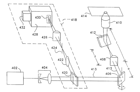
Fig. 4 shows the direct projection line scanning geometry of the confocal
microscope of the present invention. Detection arm 418 of Figure 4 is similar
in function
2 0 to the detection arm illustrated in Fig. 3. Fig. 4 illustrates a laser 402
emitting laser light
to beam expander 404 which is then incident on a galvanomirror or similar beam
deflection device such as scan mirror 406 that scans light beam 413
horizontally. The
scanned beam 413 passes through a cylindrical lens 408 placed a distance o
from scan
mirror 406. Lens 408 focuses light beam 413 to a vertical line a distance f
from
cylindrical lens 408. where f is the focal length of lens 408. Objective 410
is placed a
distance i from cylindrical lens 408, where o and i are chosen such that 1 /f
= ( I to + 1 /i).
A static mirror 412 is placed between lens 408 and objective 410 in the light
beam 413
path to deflect the light beam 413 upward, allowing an inverted microscope
arrangement.
-12-
CA 02368156 2001-09-14
WO 00/42417 PCT/US99/30863
This optical layout results in a tightly focused line of laser light that is
scanned in a
direction perpendicular to this line across sample plane 414 of the sample.
Still referring to Fig. 4. detection arm 418 is comprised of dichroic beam
sputter
420. lens 422, pinhole 424. and cylindrical lens 426. Spectrometer 428 is
positioned after
cylindrical lens 426 and includes a spectrometer slit 430 and detector 432. In
operation.
light from the sample is collected by objective 410 and redirected through
cylindrical lens
408, which collimates it. The collimated signal is descanned by scan mirror
406 and
reflected by a dichroic beamsplitter 420. The beam is then focused by lens 422
through
pinhole 424, which gives the confocal microscope embodiment of the invention
illustrated in Fig. 4 full sectioning capability. The light from the sample
following pinhole
424 now contains image information from a region (the line referred to above)
of sample
plane 414. Cylindrical lens 426 focuses this image region of sample plane onto
spectrometer entrance slit 430 of an imaging spectrometer 428. Detector array
432 is
placed at the focus of spectrometer 428 to acquire the data. A single
detection frame
consists of an image region, preferably a line, along the vertical axis of
detector array 432
and the dispersed spectrum of each pixel of the image region along the
horizontal axis of
detector array 432. A two-spatial- dimensional image is obtained by collecting
a frame of
data for each position of the beam deflector 406. Four-dimensional imaging
(three spatial
2 0 and one spectral) is possible as before. The lens and pinhole elements 422
and 424 can
be removed for diminished axial resolution (sectioning ability) and improved
sensitivity.
although some axial resolution is retained by virtue of the spectrometer slit.
Removing
the lens and pinhole elements 422 and 424 would be acceptable when using mufti-
photon
excitation, due to the sectioning ability that is already inherent in that
technique. If multi-
2 S photon excitation is employed. the slit may be unnecessary for good axial
resolution.
although the spectral resolution would suffer.
Fig. 5 illustrates the third embodiment of the invention showing detection arm
510
in detail for direct projection line-scanning geometry of the microscope. In
this
-13-
CA 02368156 2001-09-14
WO 00/42417 PCT/US99/30863
configuration, a collimated, expanded beam of light 514 from laser 512 enters
microscope
apparatus 516 from laser 512. Microscope apparatus ~ 16 has an identical
optical layout to
that of Fig. 4. The collimated emission from the sample is reflected from a
dichroic
beamsplitter 518. Spectral resolution is obtained by detection arm 510 via
spatially
resolved Fourier transform interferometry as described below. The collimated
emission
520 that is reflected from the dichroic beamsplitter 518 contains image
information along
its vertical axis as discussed in the description of Fig. 4. Now, however this
signal beam
is incident on beamsplitter X22, which passes half of the beam to a static
mirror X24, and
reflects half of the beam to a movable mirror 526. The mirrors 524 and 526
retroreflect
the beams, directing them along paths parallel to their incoming paths back
toward
beamsplitter 522, where they are recombined. The recombined beam is focused by
lens
530 through pinhole 532. which gives the full sectioning capability that is
the attractive
feature of the confocal microscope. Next, the recombined beam is focused onto
a
vertically oriented one-dimensional detector array X34 by cylindrical lens
536. As with
the previous embodiments described above. each pixel on the one-dimensional
detector
array X34 is confocal to a point on the focal plane of the objective of the
sample, thus an
entire image line or region is detected. An interferogram is recorded for each
pixel on the
detector by monitoring the pattern of constructive and destructive
interference of the light
originating from each corresponding point in the sample. This interferogram is
generated
2 0 by moving mirror 526 (moving in a back and forth direction as indicated by
bi-directional
arrow AR), and the spectrum of each pixel is obtained by Fourier
transformation of said
interferogram as is known in the art. The Fourier transform interferometric
detection is
an attractive alternative to the dispersive techniques described previously
due to its
extreme sensitivity. A cylindrical lens and a slit can replace the lens and
pinhole
2 5 elements 530, 532 for reduced sectioning capability but increased
sensitivity.
Alternatively, these elements may be eliminated altogether for maximum
sensitivity if
sectioning ability is not required or if multi-photon excitation with its
intrinsic sectioning
ability is employed.
-14-
CA 02368156 2001-09-14
WO 00/42417 PCT/US99/30863
Figure 6A shows the point-scan spectral imaging confocal microscope of the
present invention used to detect and analyze electrophoretic separation of
samples
simultaneously along a plurality of microcapillaries or lanes. Laser beam 604
from laser
602 is expanded by beam expander 606 and deflected vertically by a static
mirror 608 to
scan mirror 610 that scans the beam in the vertical plane. Lens 612 with focal
length f is
placed a distance_f= 10 cm. from scan mirror 610. Lens 614, also with focal
length f, is
placed a distance 2f from lens 612. The entrance aperture of an objective 616
is also
placed a distance f from lens 614. Static mirror 618 is placed between lens
614 and
objective 616 in order to deflect the beam upwards for an inverted microscope
geometry.
The action of scan mirror 610 is to scan the light beam 604 perpendicular to
and across an
array of microcapillary tubes 619. Fluorescence from the fluorescent labels of
the
sequenced fragments flowing through the microcapillaries 619 is collected and
collimated
by objective 616. This fluorescence is deflected by dichroic beamsplitter 620
and
incident on relay lens 622. The fluorescence is focused by lens 622 to a scan
line onto the
slit 624 of an imaging spectrometer 626 that disperses the spectra of every
point in the
scan line. A two-dimensional detector array 628 is placed at the focus of the
spectrometer
626. The sensitivity of this design can be improved at the cost of decreased
spectral
resolution by the removal of the spectrometer slit 624 because axial
resolution is
unimportant to this experiment.
Figure 6B is a graphic representation of a single frame of data obtained from
scanning across a plurality of microcapillaries using the confocal microscope
of the
present invention illustrated in Fig. 6A. The vertical axis is the
microcapillary number;
each of the microcapillaries detected in a scan has a corresponding spectrum
that appears
2 5 horizontally on the detector array. The fluorophores present in
microcapillaries 1 and 3
are the same, and those in microcapillaries 2 and ~ are the same due to the
identical
nature of their spectra. In this example, the benefits of spectral resolution
are clear.
There is significant spectral overlap between the spectra of fluorophores
present in
microcapillaries 4 and ~. This spectral overlap may prohibit the use of these
dyes in the
-15-
CA 02368156 2001-09-14
WO 00/42417 PCT/US99/30863
detection methods of the prior art for microcapillary electrophoresis
detection because
their identities may be confused. However, the spectral resolution of the
present
invention allows for clear differentiation between these fluorophores based on
their
distinct spectra.
J
Fig. 7 shows the line-focus spectral imaging confocal microscope of the
present
invention used to detect and analyze microcapillary electrophoretic separation
of samples
simultaneously along a plurality of microcapillaries or lanes. Laser beam 704
from laser
702 is expanded by a beam expander 706 and is directed to cylindrical lens
708.
i 0 Cylindrical lens 708 focuses beam 704 to a vertical line a distance f from
lens 708. where
f is the focal length of lens 708. Beam 704 is deflected vertically by a
static mirror 710
into a microscope objective 712. The effect of the combined actions of the
cylindrical
lens 708 and the objective 712 is to tightly focus the beam into a line
perpendicular to and
across microcapillary array 714. Fluorescence from the fluorescent labels of
the
15 sequenced fragments flowing through microcapillaries 714 is collected by
the objective
712. This fluorescence is directed back through cylindrical lens 708 and is
thus
collimated. The collimated fluorescence is deflected by dichroic beamspitter
716 and
focused by cylindrical lens 718 onto slit 720 of imaging spectrometer 722.
Spectrometer
722 disperses the spectra of every point in the line. A two-dimensional
detector array 724
2 0 is placed at the focus of the spectrometer 722. A frame of data is shown
in FIG. 6B as
already described above. Again. sensitivity can be improved while spectral
resolution
sacrificed by removal of spectrometer slit 720. In an alternative detection
technique.
Fourier transform interferometry can also be used for spectra resolution as
also discussed
above.
In summary, all embodiments described above project light originating from a
region of the sample plane of a sample comprised of at least two points
corresponding to
the same number of image pixels along one axis of a two-dimensional detector
array
while a spectrometer spectrally resolves the image's composite pixels along
the other axis
-16-
CA 02368156 2001-09-14
WO 00/42417 PCT/US99/30863
of the detector array. A region is identified as the points scanned between
immediately
consecutive shutter events of the detector. The significant advantage of the
present
invention is that an entire image is spectrally resolved in a significantly
reduced amount
of time compared to presently available technology. By way of further
explanation, the
acquisition time of a CCD camera can be represented by the equation t~,~~. =
te,~P_ + treadout
+ tsi",t~e~, where t~,~p. is the exposure time (the time the signal is
incident on the detector
array), treadout 1S the time to digitize and store the data, and tshutter is
the time required to
open and close the shutter. The quantities t~~p and trea~out are the same in
the present
invention as they are for the confocal microscopes currently known in the art,
but tn,utter is
significantly different. For example, if an image were comprised of 40,000
pixel (200 x
200 pixels), currently known spectrally resolving confocal microscopes would
require
40,000 shuttering events (tsi,u~,e~) to create a spectrally resolved image.
However, in
significant contrast, tsnut«~ for the present invention would only require 200
shuttering
events (200 times smaller) to create the same spectrally resolved image. The
advantage of
the present invention is the significant reduction of shuttering events, which
reduces
shuttering time such that the image acquisition time is significantly reduced.
The method
and apparatus of the present invention may be used to rapidly obtain images of
numerous
different sample types. including but not limited to biomolecules, DNA,
cellular systems,
isolated cells, organelles, antibodies and other proteins. encapsulated
molecules, biochips,
2 0 polymers. ligand crystals, solgels, and electrophoretic gels.
While the invention has thus been described with reference to specific
embodiments thereof, it will be appreciated that numerous variations,
modifications. and
embodiments are possible. and accordiny~ly, all such variations,
modifications. and
2 5 embodiments are to be regarded as being within the spirit of the
invention.
-17-
CA 02368156 2001-09-14
WO 00/42417 PCT/US99/30863
BEST MODE
The best mode of configuring a confocal scanning beam microscope of the line-
scanning type according to the present invention comprises:
a) A support for supporting a sample to be obser~~ed and measured:
b) an illumination source producing a light beam along a path toward said
sample;
c) means for focusing the light beam on a prescribed sample plane of said
sample;
d) a first scan mirror for scanning said light beam fast in a first axis and
directing
said light beam to a second scan mirror, wherein a second scan mirror scans
said
light beam slowly in a second axis to form a raster pattern on said sample
plane
of the sample with said first axis projected onto a light receiving slit and
partially descans the light reflected, scattered, or emitted form at least one
region
of said sample plane;
e) means for simultaneously acquiring at least two points of said
predetermined
scan pattern on the sample plane. wherein said points include a region of the
sample represented by at least two image pixels;
f) a detection arm placed in the path of said light reflected, scattered, or
emitted
from said region on said sample plane of said sample for receiving said
partially
descanned light and focusing it upon said light receiving slit, comprising
means
for resolving the spectra of said light from each said region including a
light
2 0 receiving slit and further comprising a two-dimensional detector having
first and
second axes;
g) means for focusing the light from said region reflected, scattered, or
emitted
from said sample plane of said sample to said light receiving opening of said
means for resolving the spectra of the light from said region; and
2 5 h) region and means for simultaneously detecting the spectra of said light
of said
region.
-18-
CA 02368156 2001-09-14
WO 00/42417 PCT/US99/30863
INDUSTRIAL APPLICABILITY
The confocal microscope of the invention provides direct and indirect
industrial
opportunity. As a direct use. the confocal microscope serves as a highly
effective
laboratory analytic tool for the biological and medical fields. The invention
improves
multichannel detection techniques for cell and DNA investigations. As an
indirect
industrial application, new pharmaceuticals and new surgical equipment and
methods are
likely to become available through the information garnered by use of this
innovative
laboratory instrument.
-19-