Note: Descriptions are shown in the official language in which they were submitted.
CA 02368200 2001-10-02
WO 00/59560 PCT/US99/30145
IMPLANTABLE VENTRICULAR ASSIST DEVICE
Background of the Invention
Field of the Invention
The present invention relates to devices and associated methods for
pumping fluids, for example, blood. More particularly, the present invention
relates to implantable ventricular assist devices (VADs) that are utilized to
replace the function of either the right ventricle or the left ventricle, or
both, of
the heart. The ventricular assist devices of the present invention include
certain
features that relate in the art to electric pulsatile devices.
Description of the Related Art
Four hundred thousand new cases of congestive heart failure are
diagnosed in the United States annually, a number which will only rise in the
foreseeable future with the aging of the baby-boom generation. According to
the Framingham Heart Study, the five-year mortality rate for patients with
congestive heart failure was 75 percent in men and 62 percent in women.
Standard medical and surgical therapies benefit only a small percentage of
patients with ventricular dysfunction. Potential cardiac transplant recipients
with hemodynamic instability may receive temporary mechanical circulatory
support, such as an implantable blood pump, as a bridge to cardiac
transplantation. Moreover, estimates in the field suggest that 17,000 to
66,000
patients each year in the United States may benefit from a permanent
implantable blood pump.
The ventricular assist device (VAD) is a blood pump designed to assist
or replace the function of either ventricle, or both ventricles, of the heart.
A
right ventricular assist device (RVAD) supports pulmonary circulation by
receiving or withdrawing blood from the right ventricle and returning it to
the
pulmonary artery. A left ventricular assist device (LVAD) supports systemic
perfusion by receiving or withdrawing blood from the left ventricle (or left
atrium) and returning it to the aorta. A biventricular assist device (BVAD)
CA 02368200 2001-10-02
WO 00/59560 PCTIUS99/30145
2
supports both ventricles of the heart. Ventricular assist devices may be
either
implantable or extracorporeal, with implantable VADs positioned
intracorporeally in the anterior abdominal wall or within a body cavity (other
than the pericardium) and with extracorporeal VADs located paracorporeally,
along the patient's anterior abdominal wall, or externally at the patient's
bedside.
The first ventricular assist devices attempted to mimic the pulsatile flow
of the natural left ventricle by utilizing flexible chambers with volumes
approximately equal to the volume of the respective ventricle being assisted.
The typical volume of blood expelled by the left ventricle of an adult is
between
70-90 ml, but may range from 40-120 ml. The chambers are expanded and
contracted, much like a natural ventricle, to alternately receive and expel
blood.
One way valves at the inlet and outlet ports of the chambers ensured one way
flow therethrough.
So-called "pulsatile pumps" may include one or a pair of driven plates
for alternately squeezing and expanding flexible chambers. The flexible
chambers typically comprise biocompatible segmented polyurethane bags or
sacs. The blood sac and drive mechanism are mounted inside a compact
housing that is typically implanted in the patient's abdomen. A controller,
backup battery, and main battery pack are electrically connected to the drive
mechanism. Even the most basic drive mechanisms of the prior art are
relatively complex and expensive, and typically incorporate some type of
mechanical cam, linkage, or bearing arrangement subject to wear.
Because of the varying volume of the blood sac within the rigid
encapsulation housing of pulsatile pumps, accommodation must be made for the
air displaced thereby. Some devices utilize a percutaneous tube vented to the
atmosphere, which is a simple approach but has the disadvantage of a skin
penetration and associated infection risk. Another approach, proposed for
fully-
implantable VAD systems, is to use a volume compensator. This is a flexible
chamber, implanted in the thoracic cavity adjacent to the lungs and
communicating with the air space within the housing and outside the blood sac
CA 02368200 2001-10-02
WO 00/59560 PCT/US99/30145
3
via an interconnecting tube. As the blood sac expands with incoming blood, air
is displaced from the housing to the volume compensator. Conversely,
expulsion of blood from the blood sac creates a negative pressure within the
housing and pulls air from the volume compensator. While eliminating the
infection risk of the percutaneous vent, the volume compensator poses certain
challenges: increased system complexity, an additional implanted component
and potential site of infection, maintaining long-term compliance of the
implanted volume compensator sac, problems associated with gas diffusion in
or out of the enclosed volume, and problems associated with changes in ambient
1 o pressure, such as experienced during a plane flight.
One example of an electric pulsatile blood pump is the Novacor N 100
left ventricular assist system (Novacor Division, Baxter Healthcare
Corporation,
Oakland, California). This system contains a single polyurethane blood sac
with a nominal stroke volume of 70 ml that is compressed by dual
symmetrically opposed pusher plates in synchronization with the natural left
ventricle contraction. The pusher plates are actuated by a spring-decoupled
solenoid energy converter. The blood pump and energy converter are contained
within a housing that is implanted in the patient's abdomen. The N 100 is a
tethered system employing a percutaneous vent tube carrying power and control
wires.
Biventricular heart assist devices employ two pumps, with the input of
each connected to separate pumping chambers of the heart. For instance, U.S.
Patent No. 4,468,177 to Strimling discloses a diaphragm pump having a piston
that is moveable within a chamber to reduce the volume of a chamber on one
side while simultaneously enlarging the volume of a second chamber on the
opposite side. Another patent to Strimling, U.S. Patent No. 4,547,911,
discloses
an implantable heart pump having two pusher plates driven synchronously
between two variable-volume chambers at a multiple of the natural rhythm of
the heart. Each of the Strimling devices may function either as a BVAD heart
pump with each chamber communicating separately with a respective ventricle
of the heart, or as a single ventricle-assist pump wherein the two chambers
are
CA 02368200 2001-10-02
WO 00/59560 PCT/US99/30145
4
connected in series with a shunt therebetween.
In recent years there has been increased study into the potential of using
rotary pumps (centrifugal or axial) for ventricular assist. These pumps employ
fast-moving impellers to impart forward flow to the blood. The impellers are
either supported by bearings or are magnetically levitated. A significant
advantage of rotary pumps is their relatively compact size and low cost. In
addition, the pressure difference maintained by the impeller eliminates the
need
for one-way valves as in pulsatile pumps. Finally, no venting or volume
compensator is necessary.
The use of rotary pumps has generated a significant amount of interest in
this field, but as yet many drawbacks prevent general acceptance. For
instance,
bearing-supported impellers usually require lubrication that must be
absolutely
kept out of contact with the blood, thus requiring seals that remain highly
effective for extended periods. In some designs, the bearings are within the
pump housing in contact with blood, which is then used as the lubricating
fluid
and may be subject to degradation. In addition, the heat generated by some
bearing configurations may adversely affect the blood. Some designs eschew
bearings altogether and instead utilize magnetically levitated impellers.
However, these are relatively complex and sometimes unstable. A safety issue
with rotary pumps is their non-occlusive character which provides a shunt path
for blood regurgitation if the impeller is not rotating. That is, the one-way
valves in pulsatile pumps ensure a uni-directional pathway through which blood
is propelled, and prevent regurgitation from the arterial vessel if the device
shuts
off or fails. The natural ventricle can thus function as a back-up perfusion
system, bypassing the pump circuit. If the impeller in a rotary pump stops,
however, a flow path is created permitting blood from the arterial vessel to
be
shunted through the pump, thus seriously impairing the back-up capability of
the natural ventricle. To prevent this situation, a one-way valve or occluder
of
some sort must be provided at the rotary pump outflow. A still further issue
with rotary pumps, as yet to be resolved, is the efficacy of the continuous
flow
of blood provided thereby. There are studies on both sides which either favor
CA 02368200 2007-05-23
pulsatile flow, or at least suggest no negative side effects from continuous
flow.
In view of the foregoing, there is an ongoing need in the art to improve
upon conventional ventricular assist devices. For example, reductions in size
and the elimination of the volume compensator would be advantageous to
5 facilitate full implantation of a device. In addition, pulsatile flow
without the
disadvantages of conventional devices is a goal. Further, a device that is low
in
cost but does not have the disadvantages of rotary pumps would be
advantageous for long-term use. Accordingly, there remains a need in the art
for a small, efficient, atraumatic, and fully implantable ventricular assist
device
that overcomes the deficiencies of conventional devices.
Summarv of the Invention
The present invention provides a pumping system for assisting the
ventricles of the heart. The pumping system of the invention has a relatively
small size and is free of many disadvantages inherent in conventional blood
pumps. In addition, the pumping system of the present invention can provide
pulsatile flow of varying degrees and duration, even up to continuous flow, in
a
small fully implantable and relatively mechanically simple device. Desirably,
the pumping system provides intermittent periods of substantially continuous
flow during systole. Accordingly, the present invention provides a pumping
system that is small, efficient, atraumatic, and fully implantable while
overcoming the deficiencies of conventional devices.
In one aspect, the present invention provides a ventricular assist
device comprising an implantable housing and a pair of variable-volume
chambers mounted therein, each of the chambers having an inlet port and an
outlet port with one-way valves. At least one ventricular outflow conduit is
adapted to be connected between the ventricle and the inlet ports. An
actuator comprises a first electromagnetic coil on one side of the pair of
chambers and a second electromagnetic coil on the opposite side of the
chambers. The actuator is arranged within the housing to alternately contract
one of the variable-volume chambers while expanding the other, and vice
versa, to provide a positive displacement pump. Preferably, the actuator
comprises a movable plate having a permanent magnet thereon, and a portion
of the housingis magnetically permeable so that the plate is unstable in
CA 02368200 2001-10-02
WO 00/59560 PCT/US99/30145
6
a central position between the two variable-volume chambers, and is biased
toward one or the other upon a slight displacement in that direction.
Furthermore, the device preferably includes electromagnetic coils mounted in
the housing and situated so as to generate a coil flux path through the
housing
and through a magnetically permeable portion of the movable plate. In this
manner, the movable plate functions as an armature and the coil flux displaces
the armature toward one of the two variable volume chambers depending on the
current direction through the coil. In addition, the coil flux preferably does
not
travel through the permanent magnet which might otherwis,- depolarize it.
According to one particular aspect of the invention, the pumping system
is configured as a ventricular assist device including a pump and cannulation
for
connecting the pump to the cardiovascular system of a patient. The pump
includes a pair of compressible chambers and electro-magnetic structure having
a frame, a pair of coils, and a plate. The coils are disposed in a spaced
relationship within the frame. The coils generate coil flux and define a pair
of
respective poles when electrically activated. The plate has an armature and a
magnet and is disposed within the frame such that the armature is between the
poles and the magnet is between the coils. The magnet generates bias flux.
Each chamber is disposed between the plate and one of the coils. Each of the
chambers has a volume substantially less than the ejection volume of the
ventricle, preferably about one-quarter of the ejection volume of the
ventricle.
For example, in an LVAD in accordance with the present invention, assuming
that the ejection volume of a typical left ventricle is about 80 ml, the
volume of
each chamber may be on the order of about 20 ml.
The present invention also provides a method of ventricular assist using
a ventricular assist pump including two pumping chambers, valved inlet and
outlet conduits for each chamber, and an actuator. The method includes
directing an inflow of blood from a single ventricle to both of the chambers,
and
actuating the pumping chambers with the actuator during a systolic phase of
the
assisted ventricle to alternately expel blood from one of the chambers while
drawing blood into the other of the chambers.
CA 02368200 2001-10-02
WO 00/59560 PCT/US99/30145
7
Another aspect of the invention is a method of ventricular assist using a
positive displacement pulsatile pump having two variable-volume chambers
each with a volume less than about one-half of the ejection volume of the
ventricle. The method includes implanting the pump in a patient so as to be in
fluid communication with the blood circulatory system, actuating the pump
during systole to provide substantially continuous flow output and propel the
ventricular ejection volume into an arterial vessel, and resting the pump
during a
diastolic phase of the assisted ventricle.
One of the advantages of the pumping system of the present invention is
that there is no need for a volume compensator or a compliance chamber.
Accordingly, the overall size and complexity of the system is substantially
reduced. It follows that system cost is reduced while system reliability and
patient acceptability are increased. In addition to the compliance chamber,
bearings and other blood-damaging components are eliminated. This feature is
advantageous not only in reducing complexity and cost but also in increasing
the long-term period for which the pumping system may be implanted in a
patient.
Another advantage of the present invention is that the pumping system
provides substantially pulsatile flow. As the volume of each chamber is, for
example, about one-quarter the ejection volume of the ventricle, a controller
of
the pumping system may cause the coils to stroke the plate about 4 times
during
one beat of the heart (i.e., during systole or ventricular contraction).
Accordingly, even employing chambers with a substantially reduced volume,
the pumping system is able to keep up with the normal ejection volume of the
ventricle. This pulsatile flow provided by the pump is substantially analogous
to the blood flow out of a healthy ventricle. Alternatively, the pumping
system
may be operated continuously to provide substantially uniform flow.
According to another aspect of the invention, the flux generated by the
coils follows a path including one of the poles, one of the gaps defined
between
the armature and the poles, the armature, the other gap, and the other pole
such
that the magnet is substantially free of the coil flux. One of the advantages
of
CA 02368200 2001-10-02
WO 00/59560 PCT/US99/30145
8
this feature of the invention is that the bias flux remains substantially
constant,
such that the magnet does not depolarize. Another advantage stemming from
the bias magnet not being depolarized is that the bias magnet may be made
relatively small, thereby further reducing the size and weight of the pump.
For
example, the pump, which may be substantially cylindrical, may have a
diameter less than about 100 millimeter.
Other aspects, features, and advantages of the present invention will
become apparent to those skilled in the art from a consideration of the
following
detailed description taken in conjunction with the accompanying drawings
1 o which illustrate, by way of example, the principles of the present
invention in
the context of a blood pump for left-ventricular assistance, but which are
equally relevant to blood pumps for assisting the right or both ventricles, or
in
general to other devices for pumping fluid.
Brief Description of the Drawing
FIG. 1 is a perspective view of a ventricular assist system of the present
invention connected to a heart of a patient (shown in phantom) for left
ventricular assist;
FIG. 2 is a perspective view of a blood pump of the ventricular assist
system in which variable-volume chambers operate in parallel;
FIG. 3 is a cross-sectional view of an exemplary blood pump of the
invention taken along line 3-3 of FIG. 2,
FIGS. 4A to 4F are schematic views of a preferred configuration of
pumping chambers of the present invention operating in parallel, particularly
illustrating sequential stages in which each stroke is an ejection stroke;
FIG. 5A is a schematic view of an exemplary drive structure of the
invention, illustrating a magnetic flux path generated by a bias magnet on an
armature (shown in an equilibrium position) and a coil flux path generated by
a
fixed electromagnetic coil;
FIG. 5B is a view similar to that of FIG. 5A, showing the armature
displaced to the right and being driven to the left;
CA 02368200 2001-10-02
WO 00/59560 PCT/US99/30145
9
FIG. 5C is a view similar to that of FIG. 5A, showing the armature
displaced to the left and being driven to the right;
FIG. 6 is a circuit diagram of a magnetostatic equivalent circuit for the
exemplary drive structure of the present invention;
FIG. 7 is a graph showing various force components of the drive
structure superimposed along a position axis;
FIGS. 8A to 8E are diagrammatic views illustrating phasing
relationships among (A) an electrocardiogram signal, (B) left-ventricular
pressure, (C) aortic pressure, (D) armature position, and (E) pump outlet;
FIG. 9 perspective view of an alternative blood pump of the present
invention in which variable-volume chambers operate in series; and
FIGS. l0A to l OF are schematic views of the blood pump of FIG. 9,
illustrating the variable-volume chambers of the pump operating in series.
Description of the Preferred Embodiments
Ventricular-Assist System
With reference first to FIG. 1, a living human host patient P is shown in
fragmentary front elevational view, and with parts of the patient's anatomy
shown
in phantom or removed solely for better illustration of the salient features
of the
present invention. A pumping portion 20 of a ventricular assist system 22 is
surgically implanted into the patient's abdominal cavity AC and connected to
the
heart H with cannulation. The cannulation includes an inlet conduit 24
communicating blood from the patient's left ventricle LV into the pumping
portion 20, and an outlet conduit 26 communicating blood from the pumping
portion 20 to the patient's aorta AO. As discussed in detail below, pumping
portion 20 includes a blood pump 28, an exemplary embodiment of which is
shown in FIG. 2.
For purposes of explanation and without limiting the scope of the
present invention, exemplary ventricular assist system 22 is illustrated
assisting
the left ventricle LV of the heart of the patient P. In addition to being
CA 02368200 2007-05-23
configurable as a left ventricular assist device (LVAD), the ventricular
assist
system 22 may also be configured to assist the right ventricle (RVAD), as well
as both ventricles (biventricular assistance, or BVAD). Therefore, as a
general
matter, and except in reference to the illustrated LVAD, the source of blood
for
5 the ventricular assist system 22 may be termed the "assisted ventricle,"
while the
destination of the pressurized blood will be designated the "arterial vessel."
Each of the conduits 24 and 26 include flexible segments 30 and 32
extending to the left ventricle LV and aorta AO, respectively. The inlet and
outlet conduits 24 and 26 are attached to the natural tissue of the ventricle
and
10 the arterial vessel by sutures to establish and maintain blood flow, and
may
include appropriate structure for this purpose such as a sewing ring 34 for
ventricular attachment. In any of the contemplated configurations of LVAD,
RVAD, or BVAD, the inlet conduits are anastomosed to the respective
ventricle, while the outlet conduits are anastomosed to the appropriate
arterial
vessel, which for left ventricular assist is typically the aorta AO and for
right
ventricular assist is typically the pulmonary artery. As will be explained
below,
the exemplary ventricular assist system 22 includes a single ventricular
anastomosis branching to two input ports in the pumping portion 20, but those
of
skill in the art will realize that the device will function with two separate
anastomoses. Likewise, the outlet conduit 26 is shown branched, but could
remain as two separate conduits with separate arterial anastomoses. Details of
the
conduits 24, 26 are shown and described in U.S. Patent No. 6,001,056.
With continued reference to FIG. 1, a power cable 38 extends from the
pumping portion 20 outwardly of the patient's body via an incision I to a
controller 40 and a power supply 42, such as a battery pack. Other means for
powering the ventricular assist system 22 are known which do not require a
cable
through the skin, and the present invention is not so limited. The blood pump
28
necessarily includes a rigid housing 44 outwardly formed of a biocompatible
coating such as a polymer or other suitable biocompatible material.
CA 02368200 2001-10-02
WO 00/59560 PCT/US99/30145
11
Positive-Displacement VAD
The pumping portion 20 of the system 22 is seen in greater detail in FIG. 2
connected to a branching structure of the conduits 24 and 26. More
particularly,
the flexible segment 30 connects to a bifurcated inlet conduit segment 50,
which
in turn connects to a pair of inlet branches 52a, 52b, each of which are
placed in
fluid communication with a separate inlet port 54a, 54b of the pumping portion
20. Likewise, the flexible segment 32 connects to a bifurcated outlet conduit
segment 56, which in turn connects to a pair of outlet branches 58a, 58b, each
of which are placed in fluid communication with a separate outlet port 60a,
60b
of the pumping portion 20. As will be described below, the pump 28 includes a
pair variable volume chambers operating in synchronization with one filling
with
blood, and the other expelling blood, thus resulting in a positive
displacement
pump. The branching structure of the conduits 24 and 26 enables this parallel
pumping action with the common inlet source (tube 30) and common outlet
destination (tube 32).
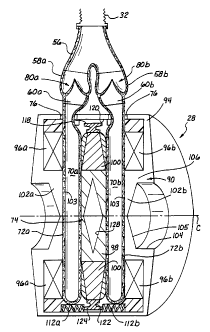
As seen in FIGS. 3 and 4A-4F, exemplary pump 28 includes a pair of
variable-volume chambers 70, for example, a left chamber 70a and a right
chamber 70b, each of which has a volume that is no more, and preferably less,
than half the ejection volume of the ventricle being assisted; for example, in
the
illustrated setup, the volume of each chamber 70 may be on the order of one-
quarter the volume of the left ventricle LV. In the illustrated embodiment,
the
chambers 70 are defined within the cavities of flexible sacs 72a, 72b that are
preferably configured as relatively flat disk-shaped bags. It should be noted
that
other sac configurations are possible within the understanding of one skilled
in
the art, and also that variable-volume chambers may be defined by structures
other than flexible sacs, such as piston-cylinder couples, moveable walls,
etc. A
number of features of the present invention can thus be transferred to other
fluid
propulsion arrangements, though the use of dual flexible sacs provides a
number
of significant advantages and is thus preferred.
The sacs 72a, 72b are disposed in parallel and spaced apart by an
CA 02368200 2001-10-02
WO 00/59560 PCT/US99/30145
12
actuator plate 74. The actuator plate 74 is preferably affixed to the inwardly-
facing flat surfaces of each sac 72 with, for example, adhesive. To accept and
pump the ejection volume of the ventricle in full with the reduced-volume
chambers 70, the blood pump 28 has a drive system that pumps the chambers 70
a plurality of times for each beat of the heart H and provides a substantially
continuous flow of blood during such pumping. The drive system displaces the
actuator plate 74 left and right to alternately compress each chamber 70. As
shown more clearly in the simplified schematics of FIG. 4, the chambers 70 are
connected in parallel so as to eject oxygenated blood into the arterial vessel
during each stroke of the plate 74. This feature of the invention not only
reduces the overall size of the blood pump 28 but also eliminates the need for
a
compliance chamber (or volume compensator), which will be discussed in detail
below.
The cross-section of FIG. 3 is taken across a midplane M of the
pumping portion 20 except for the area at the top of FIG. 3 which is taken
tangentially through the outlet conduit 26. That is, each of the inlet and
outlet
conduits 24, 26 extend generally tangentially from the cylindrical pumping
portion 20. The configuration of the outlet ports 60a, 60b is seen in FIG. 3
and,
though not apparent from the drawing, these also are disposed tangentially
with
respect to the disk-shaped sacs 72a, 72b. Likewise, the inlet ports 54a, 54b
are
tangential to the sacs 72a, 72b. The tangential orientation of the ports 54,
60 is
believed to most effectively fill and flush blood to and from the chambers 70.
The housing 44 includes appropriate inlet apertures (not shown) and outlet
apertures 76 for receiving the inlet ports 54a, 54b and the outlet ports 60a,
60b,
respectively. These apertures 76 are sealed about the ports 54, 60 to prevent
fluid seepage therebetween.
As seen in detail in FIG. 3, each of the outlet branches 58a and 58b
includes a pair of outlet valves 80a and 80b. Likewise, as seen in the
schematic
views of FIGS. 4A-4F, each of the inlet branches 52a, 52b, includes a pair of
inlet valves 82a and 82b. The valves 80, 82 enable the positive-displacement
pump to function as will be explained below. The valves 80, 82 are desirably
CA 02368200 2007-05-23
13
polymeric or xenograft tissue valves, such as porcine aortic valves, although
the
present invention is not so limited. Details of various aspects of tissue
valves
are shown and described in U.S. Patent No. 5,810,708, issued on September 21,
1998. The illustrated embodiment shows discrete branch segments 52, 58
disposed
between the bifurcated inlet conduit segment 50 and respective ports 54, 60
which is a convenient arrangement for tnounting of tissue valves.
Alternatively,
one or more of the bifurcated inlet conduit segment 50, branches 52, 58 and
even ports 54, 60, may be integrally formed of a suitable polymer, for
example,
with the valves also being formed therein of the same or a different material.
With further reference to FIG. 3, exemplary pump 28 of the present
invention may have a shoe 118 disposed between the bias magnet 100 and an
inner surface 120 of the frame 94. The shoe 118 is shaped somewhat like an I-
beam, with a narrow neck 122 and an outer rai1124. Without the narrow neck
122, and without the centering shear forces imposed on both sides by the
elastic
sacs 72, radial movement of the plate 74 would reduce the annular gap a (see
FIG. 5A) consequently increasing the radial magnetic force which tends to
displace the plate 74 over to that side, possibly into contact with the frame
94.
In operation of the present embodiment, if the plate 74 shifts radially close
to
the inner surface 120 on any side, the neck 122 saturates with magnetic flux
which limits the radial magnetic forces and thus halts further lateral
displacement of the plate 74 which might otherwise tend to occur. Eventually,
axial movement of the plate 74 and the centering shear force imposed by the
flexible sacs 72 couple to re-center the plate.
Also shown in the exemplary embodiment of FIG. 3, the armature 98
has a diamond-shaped hollow center, indicated by reference numeral 128, which
reduces the weight of the pump 28. More to the point, the hollow center 128
reduces the mass of the plate 74 which thus reduces the power (and battery
size)
needed to displace it, and in turn reduces the size of coils 96 required. The
entire device can thus be reduced in size to further facilitate successful
implantation.
CA 02368200 2001-10-02
WO 00/59560 PCT/US99/30145
14
Operation of Positive-Displacement VAD
As mentioned above, the drive system (a preferred embodiment of which
is described below) displaces the actuator plate 74 left and right to
alternately
compress each variable-volume chamber 70. In a first stage of operation in
FIG. 4A, the actuator plate 74 displaces to the right toward the right chamber
70b which is filled with oxygenated blood from the ventricle being assisted.
The plate 74 compresses the right chamber 70b and ejects blood through outlet
port 60b and outlet valve 80b and into the flexible outlet segment 32 for
delivery to the arterial vessel. The plate 74 helps pull a reduced pressure in
the
left chamber 70a which in turn receives blood from the flexible inlet segment
30
through the left inlet valve 82a and left inlet port 54a. The left outlet
valve 80a
prevents blood from entering the left chamber 70a from the flexible outlet
segment 32, and the right inlet valve 82b prevents blood from being ejected
into
the flexible inlet segment 30 when the plate 74 is moving to the right.
As the plate 74 continues to move to the right as shown in FIG. 4B, the
left chamber 70a expands, thereby receiving blood from the ventricle being
assisted. When the plate 74 has moved all the way to the right as shown in
FIG.
4C, the right chamber 70b is compressed to a minimum volume while the left
chamber 70a is expanded and filled with oxygenated blood from the assisted
ventricle. The chambers 70 are substantially compressed to eject a majority of
blood therein, but are not completely emptied to avoid contact between the
inner
surfaces of the sacs 72.
In the reverse sequence of FIGS. 4D-4F, the drive system displaces the
plate 74 to the left to compress the left chamber 70a and eject blood through
the
left outlet port 60a and the left outlet valve 80a and into the flexible
outlet
segment 32 for delivery to the arterial vessel. The plate 74 helps pull a
reduced
pressure in the right chamber 70b which in turn receives blood from the
flexible
inlet segment 30 through the right inlet valve 82b and right inlet port 54b.
When the plate 74 is moving to the left, the right outlet valve 80b prevents
blood from entering the right chamber 70b from flexible outlet segment 32, and
CA 02368200 2001-10-02
WO 00/59560 PCT/US99/30145
the left inlet valve 82a prevents blood from being ejected into the flexible
inlet
segment 30.
As the plate 74 continues to move to the left, the right chamber 70b
expands, thereby receiving blood from the assisted ventricle. When the plate
74
5 has moved all the way to the left as shown in FIG. 4F, the left chamber 70a
is
compressed to a minimum volume while the right chamber 70b is expanded and
filled with oxygenated blood from the assisted ventricle.
Preferred Electromagnetic Drive System
10 With reference to FIG. 3 and additional reference to the schematic view
of FIG. 5A, the drive system of the present invention preferably comprises a
substantially cylindrical electro-magnetic structure 90; accordingly, for the
purposes of this description, the electro-magnetic structure 90 has a center
axis
C and a midplane M. In addition to the moving plate 74 which forms an
15 armature, exemplary electro-magnetic structure 90 generally includes an
outer
frame 94 in which is mounted a pair of electrically-conductive coils 96,
including a first or left coi196a and a second or right coi196b. Exemplary
plate
74 functions as an armature and thus has a magnetically permeable portion 98
in
a radially central region and a surrounding bias magnet 100. The exemplary
bias magnet 100 is a permanent magnet which is radially polarized, and the
frame 94 includes a magnetically permeable portion situated so as to provide a
magnetic flux path (Db, as seen in FIG. 5A. The magnetic flux path (Db tends
to
create an instability of the plate 74 in a central position between the two
variable-volume chambers 70, so that the plate is biased toward one or the
other
variable-volume chamber upon a slight displacement in that direction.
As shown in FIG. 3, the coils 96, which may be configured as annular
rings, are disposed in a spaced relationship within the frame 94 on opposite
axial sides of the plate 74. The coils 96a and 96b are connected electrically
in
series and, when actuated, generate a magnetic flux defining a pair of poles
102,
including a first or left pole 102a and a second or right pole 102b. The
polarity
of the electric circuit through the coils 96 determines the magnetic flux
direction
CA 02368200 2001-10-02
WO 00/59560 PCT/US99/30145
16
as shown in FIGS. 5B and 5C and, thus, the physical influence on the armature
98 on the plate 74.
The plate 74 is disposed within the frame 94 such that the armature 98 is
positioned between the poles 102 and the bias magnet 100 is positioned betweer
the coils 96. When the armature 98 is centered between the poles 102 at the
midplane M, a gap g is defined on either side of the armature, as shown in
FIG.
5A. In addition, an annular gap a having a substantially constant radial
di:nension is defined between the radially outennost surface of the plate 74
and
an inner surface of the frame 94.
With reference to FIG. 3, each of the poles 102 defines an inwardly
facing surface 103, generally within the annular coils 96, disposed normal to
the
central axis C and facing the armature 98. The frame 94 includes a pair of
centrally-located, outwardly-facing cylindrical cavities 104 having tapered
floors 105 so that the poles 102 comprise annular regions 106 that transition
along the tapered floors 105 to the area of the inwardly facing surfaces 103.
In
this manner, the overall mass of the device is reduced which helps facilitate
patient acceptability and comfort.
The exemplary pump 28 may also include pairs of springs 112a and
112b radially disposed about a periphery of the chamber 70 and each disposed
to provide a compressive bias between the plate 74 and one side of the frame
94.
Only one pair of springs 112 is shown in the drawings because of the offset
cross-section taken along line 3-3 of FIG. 2. Although exemplary springs 112
are shown as helical compression springs, any spring configuration that
resists
displacement of the armature 98 away from the midplane M may be used, for
example, leaf springs, one large spring on each side of the chambers 70 such
as
a large diameter coil spring, and the elasticity of the chambers 70
themselves.
Desirably, the springs exert axisymmetric forces on both sides of the plate 74
tending to center the plate at the midpoint M. Accordingly, the combined
forces
of the springs 112, and to a lesser extent the forces exerted by the
elasticity of
walls of the sacs 72, oppose the force of the bias magnet 100 and tend to
maintain the plate 74 substantially equidistant from the poles 102 when the
coils
CA 02368200 2001-10-02
WO 00/59560 PCT/US99/30145
17
96 are not activated.
Electromagnetic Drive System Function
Radially polarized bias magnet 100 generates bias flux (Db that follows a
closed magnetic circuit including the frame 94, a respective one of the poles
102, a respective one of the gaps g, and the armature 98. Advantageously,
electro-magnetic flux (D, generated by the coils 96 does not travel a path
through
the bias magnet 100, but instead traverses around the outside of the frame 94
and through the poles 102, gap g, and armature 98; accordingly, the bias flux
(Db
remains substantially constant and predictable. As the bias flux (Db is
substantially constant, the bias magnet 100 is not subject to depolarization,
which is discussed in more detail below.
The armature 98 of exemplary plate 74 moves either right as shown in
FIG. 5B or left as shown in FIG. 5C by a distance indicated as x representing
a
displacement of a midplane P of the plate 74 from midplane M of the electro-
magnetic structure 90. With particular reference to FIG. 5B, when electrically
activated, the coils 96 generate coil flux (D, which follows a path including
the
frame 94, one of the poles 102a or 102b, one of the gaps (either [g + x] or [g
-
x]), the armature 98, the other gap, the other pole, and the frame.
As discussed in detail below, exemplary electro-magnetic structure 90 is
configured so that:
(a) the coil flux (Dc follows a substantially closed
path to make efficient use of the bias
magnet 100;
(b) the total bias flux (Db is substantially constant
to eliminate depolarization of the bias
magnet 100 which generates the bias flux;
(c) a relatively low magnetic field intensity (H)
over a relatively large area A of the poles
102 significantly reduces the need for
CA 02368200 2001-10-02
WO 00/59560 PCT/US99/30145
18
high-precision components; and
(d) energy conversion is linear to simplify
optimization and control.
With reference to FTG. 6, a magnetostatic equivalent circuit 114 of the
electro-magnetic structure 90 shown in FIG. 5B is illustrated. The
magnetostatic circuit 114 includes circuit elements equivalent to components
of
the electro-magnetic structure 90: left coil 96a', right coi196b', bias magnet
100', left reluctance RL, right reluctance RR, annular gap reluctance Ra, left
flux
(DL, right flux (DR, and bias flux (Db. Each of the coils 96' is represented
by a
number of turns N and current I. Exemplary electro-magnetic structure 90 is
configured so that the maximum values of the left and right fluxes OL or (DR
traversing between the armature 98 and the poles 102 of FIG. 5B (and the
connected parts of electro-magnetic structure 90) are below the magnetic
saturation level of the armature 98 and poles 102; accordingly, the
magnetostatic equivalent circuit 114 is linear. Also, exemplary electro-
magnetic structure 90 is preferentially configured so that fringing magnetic
fields located around the poles 102 and the annular gap a are insignificant;
therefore, the reluctances RL and RR are substantially proportional to the
gaps,
such as:
RL = (g + x)/A (1 a)
RR = (g - x)/A (1 b)
To assure this proportionality, the poles 102 preferably have a relativel_v
large area A. Accordingly, the magnetic flux density (B) is preferably on the
order of 0.5 tesla (T) for an exemplary blood pump embodiment. A magnetic
flux density of this magnitude is significantly less than the magnetic flux
density saturation (BSAT) of core material used in the armature 98 and the
poles
102. Therefore, exemplary plate 74 may have a hollow armature 98 (as shown
in FIG. 3) to reduce the size and weight of the overall electro-magnetic
structure
90.
As the system is linear, the superposition principle applies.
CA 02368200 2001-10-02
WO 00/59560 PCT/US99/30145
19
Accordingly, the bias fluxes 4)L and (DR and the coil flux 4),, may be
calculated
separately. The left and right bias fluxes cFL and (DR are calculated by
solving
the magnetostatic equivalent circuit 114 in FIG. 6 with no coil current (that
is,
NI = 0):
(DL - (Db(g - x) /2g (2a)
(DR = (Db(g + x) /2g (2b)
The magnetic energies in the left and right gaps can then be calculated
and combined to give the total bias energy (Wb) and force (Fb) due to the bias
magnet 100 for the case when the coil current is zero (i.e., I = 0):
Wb = (1)bZ(g2 - xz)/4 ogA (3)
Fb = aWlax = -(')bZx/4 ogA (4)
(where o is the permeability of free space, or 47t * 10"' in SI
units)
Accordingly, as the armature 98 moves to the right (i.e., toward right pole
102b), the bias flux (Db shifts from left to right, with the total flux (Db
remaining
constant. As energy W and force F vary with ~', the shift in the bias force Fb
is
marked. This phenomenon is illustrated in FIG. 5B. The shift in the bias force
Fb constitutes a negative spring (k) that can be used to balance the
elasticity of
the chambers 70, which will be discuss in detail below. As shown in Equation
3, the bias energy Wb is independent of coil current.
Exemplary bias magnet 100 is preferably made from a material having a
high energy density and a low marginal permeability, for example, rare earth
material such as samarium cobalt (SmCo) or neodymium iron (NdFe).
Accordingly, the bias magnet 100 as described above and as shown in circuit
114 of FIG. 6 is a source of flux. Therefore, the bias flux (Dh is constant in
the
bias magnet 100, and all of the flux (Dc generated by the coils 96 traverses
the
loop shown by the dashed lines in FIG. 5B, including the frame 94, the left
pole
102a, the left gap (g + x), the armature 98, the right gap (g - x), the right
pole
96b, and the frame 94. Accordingly, the coils 96 produce a magnetic field
intensity as:
CA 02368200 2001-10-02
WO 00/59560 PCTIUS99/30145
Hc = 2NI/L(g + x) + (g - x)] = NI/g (5)
and a magnetic flux as:
(Dr = ABc = oANI/g (6)
Energy of the left and right gaps may then be determined by using Equations 3
5 and 6 and adding the bias flux 4)b and the coil flux 0, contributions in the
gaps
g. The total energy WT has the bias component Wb represented by Equation 3
and the following coil self-inductance energy Wcc:
Wcc = q),:Zg/ oA (7)
The product energy tenns, that is, terms containing ((D. and (D,,) or (Oc and
(DR),
10 add to zero. This is a necessary consequence of the magnetic linearity.
Accordingly, there is no energy component dependent on the product of the two
fields. In addition to having a small magnitude, the self-inductance energy
Wcc
does not depend on displacement x or on flux q), which is also required by the
linearity assumption, such that the self-inductance energy Wcc does not
15 contribute to mechanical force. As such, movement of the armature 98 merely
shifts a portion of the gap g (i.e., displacement x) from side to side but
does not
change the total reluctance of the loop.
The force contributed by the coil current I is calculated from the total
energies in the left and right gaps (holding the left and right bias fluxes
(DL and
20 (DR constant):
Fc = aWcBlax = -NI(D/2g (8)
The coil force Fc is independent of displacement x and area A and is linear in
flux (D. Accordingly, the following total force F7 equation results from the
bias
force Fb and the coil force Fc respectively represented by Equations 4 and 8:
FT = Fb + Fc =-(DTZx/4 ogA - NI(D/2g (9)
It can be seen from Equation 9 that the effect of the coil current I is to
move bias
energy Wb side to side without affecting total energy WT except for the small
self-inductance energy Wcc=
Equation 9 enables wide design latitude through varying the flux (D and
the area A of the poles 102 as the area A does not contribute to the coil
force Fc.
CA 02368200 2001-10-02
WO 00/59560 PCTIUS99/30145
21
For example, it is desirable for the value of the flux 4) to be large as flux
directly determines the coil force Fc generated by a given coil current I. For
a
given coil geometry, force F is proportional to the product of number of turns
N
and coil current I (that is, F cc NI), and power dissipation PD15; is as
follows:
Ppss = hR oc (NI)2 (10)
Accordingly, efficiency may be improved by using a high flux (D and a modest
NI. To prevent the large flux (1) from developing too much bias force Fb
(which
is balanced by the elasticity of the chambers 70 and/or springs 112), the
poles
102 preferably have a relatively large area A. A large pole area A, in turn,
implies a low value of magnetic flux density B; accordingly, the effect of
fringing fields is minimized or substantially eliminated.
Electromagnetic Drive System - Forces
FIG. 7 illustrates the forces exerted on the plate 74 by the various
components of the electro-magnetic structure 90. The horizontal axis
corresponds to the centerline C of the pump 28, with the midplane M shown,
and the vertical axis represents forces on the plate 74, with a positive force
representing a force on the plate 74 to the right using the conventions
established herein. First, the force exerted by the bias magnet 100 is seen as
a
positive slope illustrating its unstable nature tending to displace the plate
74
away from the midplane M in all positions. Of course, the force created by the
bias magnet 100 flux is idealized, and there would normally be some fringing
loss. Secondly, the preferably equal and opposite force exerted on the plate
74
by the spring 112 (and perhaps in conjunction with the sac 72) is seen as a
negative slope, indicating that the spring force would tend to center the
plate 74.
Finally, there are two different forces associated with the coils 96. A force
tending to displace the plate 74 to the right is seen at the top of the graph
and
represents the coil current flow seen in FIG. 5B. In like manner, a force
tending
to displace the plate 74 to the left is seen at the bottom of the graph and
represents the coil current flow seen in FIG. 5C.
CA 02368200 2001-10-02
WO 00/59560 PCT/US99/30145
22
In the exemplary embodiment shown in FIG. 3, the springs 112 provide
all or a substantial portion of the force offsetting that of the bias magnet
100.
One of the advantages of incorporating the springs 112 into the embodiment of
the pump 28 shown in the drawings is that the force characteristics of the
springs 112 is more predictable and stable than that produced by the
elastomeric
material composing the chambers 70. Alternatively, the elasticity of the
chambers 70 may partly or completely offsets the force produced by the bias
magnet 100, and no springs would be used. In such a configuration, however,
the thickness of the sacs 72 would have to be increased beyond what is
currently
preferred for stress considerations.
The electro-magnetic structure 90 functions in a way as to reduce the
power needed for displacing the plate 74 and substantially eliminate the
possibility of depolarizing the magnet 100. More particularly, with reference
to
FIGS. 5B and 5C, the magnetic flux (Db travels a radially outward path and
then
splits to travel around the frame and back to the plate 74 essentially along
the
centerline C. In contrast, the coil flux (D, travels around a larger path,
always
going in the same direction along the radially outermost portion of the frame
94
for any one current flow direction. Therefore, the magnetic flux (Db opposes
the
coil flux (D, in the region of the frame in which they travel in opposite
directions, and supplements the coil flux cl), in the region of the frame in
which
they travel in the same direction. In FIG. 5B, for example, the magnetic flux
(Db
opposes the coil flux (D, on the right side of the diagram and supplements the
coil flux cD,, on the left side.
Looking at the structure in another way, and as illustrated in FIG. 7, the
force exerted on the plate 74 by the bias magnet 100 and the force by the
spring
112 cancel each other to leave the constant force generated by the coils.
Therefore, the force and pump pressure vary directly with the product NI
regardless of the displacement x of the armature 98; accordingly, the control
of
the pump is simplified. In addition, as both inductance and the effects of
inertia
are negligible in the relevant time domain, control of the pump 28 is further
CA 02368200 2001-10-02
WO 00/59560 PCT/US99/30145
23
simplified. For example, when the coils 96 are not energized, the pressures in
the chambers 70 equalize by means of the inlet of blood through the inlet
conduit. The difference between inlet and outlet pressures is proportional to
coil current I when the pump 28 is simultaneously filling and ejecting the
chambers 70, which is any time the plate 74 is moving.
Ventricular Assist System - Coordination with Heart
Referencing FIGS. 8A-8E and taking the foregoing intc consideration, it
is advantageous from an energy point of view to accept and pump blood ejected
by the assisted ventricle during systole (i.e., ventricular contraction) as
rapidly
as is consistent with fluid flow considerations, and to stop pumping during
diastole (i.e., ventricular dilation). In this discussion systole and diastole
correspond to inflow to and outflow from the ventricular assist system 22,
respectively, in the context of either left or right ventricular assist.
As shown in FIG. 8A, an exemplary electrocardiogram (ECG) records
the changing potentials of the electrical field imparted by the heart. To
briefly
explain the cycle of systole and diastole, the ECG signal shown in FIG. 8A
illustrates a series of points representing various muscle contractions within
the
heart. Generally, blood is received in the left ventricle and it fills during
the T-
Q period. Then, during the period Q-T, the left ventricle contracts and expels
blood into the aorta. Accordingly, the pressure diagram in FIG. 8C shows the
left ventricular pressure rapidly increasing during the spike indicated at R
on the
ECG signal. For the purposes of this description, ventricular systole may be
considered as occurring between the R and T points on the ECG wave.
FIGS. 8D and 8E illustrate and exemplary movement of the actuator 74
in and outflow pressure of the ventricular assist system 22, respectively, in
correlation with the signals shown above in FIGS. 8A-C. FIG. 8D illustrates
the
position, from left (L) to right (R), of the plate 74. When the heart enters
systole (e.g., at the beginning of the QRS complex of the ECG), the controller
40 activates the coils 96 to move the plate 74 to the right (R) to accept
blood
from the left ventricle into one of the chambers 70 (for example, the left
CA 02368200 2001-10-02
WO 00/59560 PCT/US99/30145
24
chamber 70a as shown in FIG. 4A). As the chambers 70 preferably have a
relatively small capacity or volume, for example, 20 ml, compared to the 80-m1
ejection volume of the left ventricle, the controller 40 repeatedly activates
the
coils 96 to move the plate 74 back and forth during systole to accept and pump
a
substantial portion or all of the blood entering the left ventricle from the
left
atrium.
For example, referencing FIG. 8E, if each chamber 70 has a volume of
about 20 ml, then each stroke of the plate 74 pumps about 20 ml. Accordingly,
to accommodate the typical 80-m1 capacity of the left ventricle, the
controller 40
may initiate four strokes of the plate 74, thereby pumping about 80 ml of
blood
total during systole. If the time required to complete each stroke (i.e.,
stroke
time) is on the order of about 40 msec, then a volume of about 80 ml may be
pumped in a typical 160-msec ejection time. Given this exemplary stroke cycle,
the pump 28 of the invention may have a weight of about 500 grams and may
pump about 6 liters/minute into a typical systemic pressure of about 100 mm Hg
while consuming about 5.5 watts of energy. This energy consumption is
significantly lower than conventional ventricular assist devices.
The sequence of strokes of the actuator 74 shown on the right side of
FIG. 8D results in four side-by-side pressure pulses as seen on the right in
FIG.
8E. These pressure pulses are slightly trapezoidal in shape given the slight
lag
time between movement of the actuator 74 and pressure change. However,
given the extremely short time duration for each stroke of the actuator,
pressure
pulses produced a nearly constant outflow of blood from the ventricular assist
system 22. Indeed, if the actuator 74 continued to move back and forth without
stopping, the outflow of the ventricular assist system 22 would be
approximately continuous. As it is, a short duration of approximately
continuous flow of blood is generated at the appropriate time to assist the
left
ventricle in perfusing the circulatory system of the patient (or for an RVAD,
to
assist the right ventricle in perfusing the pulmonary system of the patient).
The output from the system is represented by the aortic pressure shown
in FIG. 8B. As can be seen, the aortic pressure lags slightly behind
ventricular
CA 02368200 2001-10-02
WO 00/59560 PCT/iJS99/30145
pressure and is not quite as pronounced in terms of peaks. FIG. 8B shows small
discontinuities or steps in the rise of the aortic pressure, which correspond
to the
closely spaced pulses from the ventricular assist system 22.
5 Control of Ventricular Assist S sy tem
If the heart is performing at below capacity, the magnitude of the ECG
or pressure signals may not be as large is normal, but the timing of the
various
modes of operation will remain essentially the same. Therefore, the signals
sensed by either ECG, pressure sensors, flow sensor, or other such information
10 gathering device, can be used to stimulate the ventricular assist device of
the
present invention.
Because the ECG records electric impulses generated by the muscles of
the heart, this provides an indication of the frequency of the heart beat, and
relative timing of the systole and diastole phases. This information may then
be
15 used to control the ventricular assist device of the present invention, as
will be
described below. It will be understood, however, that various other means for
sensing changes within the heart representing the "normal" or reference beat
frequency are known.
The ECG provides an indirect indication of the relative amplitude of the
20 pressure and flow outputs, but other more direct measurements can be taken
and
should be used alone or in conjunction with the ECG signal in the control loop
of the present invention. For example, with reference to FIG. 2, exemplary
pumping system 20 may also include one or more sensors for sensing the
pressure in the assisted ventricle of the heart H of the patient. In this
regard, a
25 preload sensor 130 is preferably located upstream of the pump 28 on the
bifurcation 50. The sensor 130 is in communication with the controller 40 by,
for example, an electrical lead (not shown) incorporated in the cable 38. The
controller 40 may utilize pressure information provided by the sensor 130 to
determine when to activate the coils 96 to pump blood.
In a preferred embodiment, the ventricular pressure is monitored by the
sensor 130 and the coils 96 are activated when that sensed pressure exceeds a
CA 02368200 2001-10-02
WO 00/59560 PCT/US99/30145
26
preset threshold pressure. The coils 96 remain activated until the sensed
pressure decreases to below the threshold pressure. This rise and fall of
ventricular pressure theoretically corresponds to the systolic phase, and so
the
entire ejection volume of the ventricle enters and is propelled by the
ventricular
assist system 22. In a preferred mode of operation, the coils 96 are activated
to
maintain the ventricular pressure monitored by the sensor 130 within a preset
range.
The number of strokes of the actuator 74 during each systolic pumping
phase is determined by the volumetric outflow of each of the variable-volume
chambers 70. That is, the outflow of the ventricular assist system 22 during
any
one systolic phase is preferably about the normal outflow volume of the
assisted
ventricle. Therefore, if the volume of the chambers 70 is exactly half of that
of
the assisted ventricle, and given normal outflow of the ventricle, the
actuator 74
will only need to move back and forth once (one cycle, two strokes) to
generate
a total volumetric outflow equal to the assisted ventricle volume. Likewise,
if
the chamber volume is one-quarter the volume of the assisted ventricle,
actuator
74 will go through 2 cycles, or four strokes, as seen on the right of FIG. 8D.
To
generalize, the volume of the chambers 70 is desirably a fraction of the
volume
of the assisted ventricle, with a maximum of half the volume of the assisted
ventricle. When the total ventricular ejection has been pumped, the preload or
inlet pressure drops below the threshold value and the device stops pumping.
Of course, the ratio of volume of the chambers 70 to the actual
volumetric output of the assisted ventricle will not be a round fraction, but
given
an estimate of the ventricular output one can select the chamber volume so
that
the device will function optimally. That is, the volume of the chambers 70 is
selected based on an estimate of the ventricular output, and understanding of
an
optimum operating speed of the device. Broadly stated, the variable-volume
chambers 70 preferably each have a volume which is within the range of 1/8 to
1/2 of the volumetric output of the assisted ventricle. Theoretically, the
chambers 70 could be made smaller, which would accordingly make the entire
device smaller, but would also increase the speed of operation. Eventually,
flow
CA 02368200 2001-10-02
WO 00/59560 PCT/US99/30145
27
and valve wear considerations limit the chamber size. A preferred range of
chamber volume is between about 1/6 and 1/3 of the assisted ventricle volume,
with the most preferred volume being about 1/4 of the assisted ventricle
volume.
With this preferred volume, and given normal outflow of the ventricle, each
operation during the systolic pumping phase requires four strokes, or two
cycles, of the actuator 74.
Conceivably, a series of devices manufactured in accordance with
present invention and having different chamber volumes can be made available
and selected based on a predicted assisted ventricle volume. It should also be
apparent that a device with relatively small chamber volumes, such as 1/8 of
the
assisted ventricle volume, can be used in a wide range of patients at
differing
speeds, for example, to accommodate a wide range of ventricular outflows. As
will be appreciated by those of skill the art, the range of operating modes of
the
present invention greatly enhances the ability of the medical personnel to
customize the ventricular assist system.
Diastolic Operation
In a preferred mode of operation, as seen in FIGS. 8D and 8E, the
ventricular assist system 22 is actuated during the systolic phase, and rests
during the diastolic phase. Alternatively, the ventricular assist system 22
may
be actuated one more times during the diastolic phase to reduce the
possibility
of stasis within the respective inflow and outflow conduits and variable-
volume
chambers 70. One exemplary mode of operation is to displace the actuator 74
one stroke about halfway through the diastolic phase, although other
possibilities during the diastolic phase include a relatively consistent and
slow
movement of the actuator 74, or relatively rapid movement spaced out at a
slower frequency than the actuation frequency during the systolic phase.
Another possibility is to operate the system 22 at a first frequency during
the
systolic phase (the duration of which is based on the sensed inflow pressure),
and at a second lower frequency during the diastolic phase, with intermediate
modes of frequency ramp-up and ramp-down to avoid abrupt changes
CA 02368200 2001-10-02
WO 00/59560 PCTIUS99/30145
28
therebetween. In general, however, the present system operates during the
systolic pumping phase until the ventricular pressure falls below the
threshold
level, and does not operate or else operates only intermittently during the
diastolic phase.
Series-Displacement VAD
In addition to the parallel pumping relationship of the chambers 70
shown in FIG. 4, the pump 28 may be configured in accordance with the
alternative ventricular assist system 22' shown in FIGS. 9 and 10A-10F in
which the chambers 70 are connected in series. Many of the elements are
common to the first embodiment and will thus be numbered the same. As
before, the flexible inlet segment 30 of the inlet conduit 24 connects to, for
example, the left ventricle LV of the heart H (see FIG. 1) and the flexible
outlet
segment 32 of the outlet conduit 26 connects to, for example, the aorta AO. In
this embodiment, the flexible inlet segment 30 is only connected to the inlet
port
132 of one of the chambers (such as the left chamber 70a), and the flexible
outlet segment 32 is only connected to the outlet port 133 of the other
chamber
(such as the right chamber 70b).
According to the embodiment shown in FIG. 9, a transfer conduit 136 is
connected between the outlet port 135 of the chamber connected to the flexible
inlet segment 30 (the left chamber 70a) and to the inlet port 134 of the
chamber
connected to the flexible outlet segment 32 (the right chamber 70b). In
addition, a pair of valves are provided, including an inlet valve 138 disposed
at
the inlet port 134 of the chamber connected to the outlet conduit 26 and an
outlet valve 140 disposed at the outlet port 133 of the chamber connected to
the
outlet conduit 26.
In accordance with the series flow blood pump 28 exemplified in FIG. 9,
blood from the left ventricle is initially pumped to the left chamber 70a in
the
inlet conduit 24. The coils 96 are activated to move the plate 74 to the right
as
shown by the arrow in FIG. 10A, thereby ejecting blood received within the
right chamber 70b through the outlet port 133 and the outlet valve 140 and
into
CA 02368200 2001-10-02
WO 00/59560 PCT/US99/30145
29
the flexible outlet segment 32 for delivery to the aorta. During this ejection
stroke of the plate 74, the inlet valve 138 prevents blood from entering the
transfer conduit 136. In addition, the left chamber 70a is expanded, thereby
drawing oxygenated blood through the inlet conduit 24 from the left ventricle
LV into the left chamber as shown in FIG. l OB. At the end of the ejection
stroke as shown in FIG. l OC with the plate 74 positioned to the ri ght, the
left
chamber 70a is filled with oxygenated blood from the left ventricle, and the
right chamber 70b is compressed to a minimum volume.
The coils 96 are then activated to move the plate 74 to the left as shown
1 o by the arrows in FIGS. 10D and 10E, thereby drawing blood from the left
chamber 70a into the right chamber 70b via the transfer conduit 136. The
outlet
valve 140 prevents blood in the aorta or the outlet conduit 26 from being
drawing back into the right chamber 70b. In addition to left ventricular
pressure, the low pressure within the right chamber 70b caused by the
expansion of the chamber ensures that blood within the left chamber 70a enters
the right chamber 70b and is not ejected back into the inlet conduit 24. If
desired, an additional valve may be disposed at the inlet port 132 of the left
chamber 70a to also prevent blood from entering the inlet conduit 24. At the
end of the transfer stroke as shown in FIG. l OF with the plate 74 positioned
to
the left, the right chamber 70b is filled with oxygenated blood from the left
ventricle, and the left chamber 70a is compressed to a minimum volume. The
ejection stroke illustrated in FIGS. 1 0A-1 OC and the transfer stroke
illustrated
in FIGS. l OD-10F may repeated in accordance exemplary methodology of the
invention described above.
The series flow generated by the ventricular assist system 22' may be
used in many of the same modes of operation as described above, although the
continuity of the flow is not available. Control of the system 22' may be
based
on the input pressure sensed at sensor 142 as described above. One advantage
of the ventricular assist system 22' is the reduction of the number of valves
needed, from four to two. This in turn reduces the cost of the device.
CA 02368200 2001-10-02
WO 00/59560 PCT/US99/30145
Advantages of Present S sy tem
As will now be apparent to those of skill in the art, the present
ventricular assist system 22 provides regularly spaced and sustained pulses of
blood to the circulatory system of the patient using a pulsatile pump. This
5 represents a hybrid between existing pulsatile flow pumps, and rotary type
pumps. Although the present system enjoys the advantages of both types of
pumps, it suffers none of their primary disadvantages. In particular, the
superior
hemo-compatibility of pulsatile flow pumps is combined with the smaller size
and lower energy requirements of rotary type pumps. In addition, the system
10 eliminates the need for a vent or compliance chamber, and is thus fully
implantable if used with an inductive power transmission system. Further, the
present invention maintains a uni-directional pathway therethrough so that in
the event of stoppage or failure, regurgitation from the natural circulatory
system into the outlet of the device is precluded. Finally, operation
flexibility
15 inherent in the design greatly enhances the ability of medical personnel to
react
to changing physiological conditions of the patient. That is, as the rate of
the
heart beat speeds and slows, and the blood volume requirements fluctuate, the
present system is able to adapt and therefore more effectively support the
patient
to full recovery.
20 To facilitate implantability, it is preferable to minimize the overall size
of the blood pump 28. As such, and as indicated in FIG. 2, according to an
exemplary embodiment of the invention the substantially cylindrical pump 28
has a diameter D in general of less than 100 millimeters (mm) and preferably
less than about 70 mm. Additionally, the pump 28 has a width w in general of
25 less than about 60 mm and preferably less than about 50 mm. Accordingly,
such a small size enables the pump 28 to be implanted in a wide variety of
patients, even those patients of smaller stature.
In addition, the pump is reduced in weight from conventional pulsatile
pumps by about half. The present invention desirably weighs about 0.5
30 kilogram, which lessens the burden on the patient after implantation.
CA 02368200 2001-10-02
WO 00/59560 PCT/US99/30145
31
Conclusion
Those skilled in the art will understand that the preceding exemplary
embodiments of the present invention provide the foundation for numerous
zlternatives and modifications thereto. For example, the pumping system 20
may be configured to assist the right ventricle or both ventricles of the
heart. In
addition, rather than utilizing pressure information, the controller 40 may
determine when to activate the coils 96 by using the current magnitude through
the coils 96. That is, as stated above, for a given coil geometry, force F on
the
plate 74 is prc portional to the product of number of coil turns N and coil
current
1 o I. The difference between inlet and outlet pressures is proportional to
coil
current I when the pump 28 is simultaneously fillinQ and ejecting the chambers
70, which is any time the plate 74 is moving, and so the actual pressure
difference can be derived from knowledge of pump flow characteristics. One
can therefore use the magnitude of current flow in a feedback loop to signal
the
ventricular assist system 22 when to start and stop. Or, the input pressure
can
be used in conjunction with the coil current for control purposes. These and
other modifications are also within the scope of the present invention.