Note: Descriptions are shown in the official language in which they were submitted.
CA 02369018 2001-09-26
WO 99/49785 PCT/SE99/00521
Method and arrangement for taking up apertures
Technical field
The present invention pertains to a method for aligning an arrangement for
marking of directions for perforating together with a position determining
arrangement,
attached to a machine with a movable arm, in bone structures at surgery, and
to said
S arrangements.
Background art
Present methods regarding, for example, hip fracture surgery involves a great
deal of
craftsmanship. A patient with an injured leg is placed supine on a fracture
table and a
reduction maneuver is carried out. The foot of an injured leg is firmly fixed
in a specially built
shoe. Traction and rotation are applied until the fracture is reduced.
A fixation of an injured leg is normally so firm that no movement will take
place
unless a substantial force is applied to the leg. A mobile X-ray apparatus, a
"C-arm", with two
perpendicular radiographs, the anteroposterior (AP) and the lateral (Lat)
projections, checks
quality of reduction. The result of a reduction can only be judged through
said at least two
radiographs, and no correction for variations in hip rotation is possible.
Screws are inserted in order to fixate the fractured bone parts. The screws
are
introduced by a hand-held drill which opens up guide holes for the screws,
whereby a surgeon
has to judge the position of the drill from said two perpendicular radiographs
and manually
adjust the position of the drill in three dimensions, which is a very
difficult task.
Unfortunately, the insertion of screws very often has to be repeated. Repeated
trials
of insertion destroy the bone structure in the femoral neck.
To be able to check the quality after a performed hip fracture surgery a
method was
developed for determining the post surgery position of a fixating means. It
was introduced
by the inventor of the present invention in his thesis "Internal Fixation of
Femoral Neck
Fractures", Stockholm 1993, ISBN 91-628-0804-4. Nevertheless, the method has
only been
used for post-surgery quality checks and scientific statistics, and it has not
occurred to or
been obvious to any person skilled in the art to modify the method so it can
be used in
determining how and where to drill in a femoral bone fracture in order to
attach fixating
means and facilitate healing of said fracture in a best possible way.
Today, orthopedic surgery has promulgated towards sophisticated hi-tech
implants
being manually inserted through in-precise techniques. To manually insert
implants is a task
for a highly skilled orthopedic surgeon with, for example, 10 years of
training in the present
medical field.
SUBSTITUTE SHEET (RULE 26)
CA 02369018 2001-09-26
WO 99/49785 PCT/SE99/00521
2
For diagnosis groups, where a great number of injuries is accumulated, the
result of
performed surgery is less satisfactory than it could be. Hip fractures belong
to such a group,
whereby about 18.000 incidents/year occur in Sweden alone, 9.000 cervical and
9.000
pertrochanteric, to a cost of approximately SEK 1.4 billions.
Despite of the more than 100 different fixating methods developed for this
kind of
fractures, the result of performed surgery is relatively poor. As much as
approximately 3 S
of all cervical fractures do not heal, and 20% of them have to be re-operated
within a time
period of 1-2 years. For pertrochanteric fractures the same rates are 10 % and
4
respectively. Every re-surgery approximately costs SEK 185.000.
It is agreed with among surgeons and other experts that the main reason for
the high
percentage of re-surgery is an inadequately positioning of the fixating
screws, which hold the
fracture together during the following healing process, see "Fixation of
femoral neck
fractures: comparison of the Uppsala and Von Bahr screws." By Rehnberg &
Olerud, Acta
Orthop Scand 60, 1989, p. 579-584.
Considering the costs of SEK 185.000 for one re-surgery, a decrease in the
rate of
such surgery with 50 % would gain a save of SEK 160 millions in Sweden a year
in surgery
costs. A bigger Swedish Hospital would save approximately SEK 8 millions, not
to say what
is gained in relief for fractured patients.
A known arrangement to support surgery is the so-called ROBODOC~ Surgical
Assistant System. The ROBODOC~ robot is able to precisely prepare a femoral
channel for
placement of a cementless prosthesis.
Due to the manual surgery technique involved in surgery relating to bone
fractures
and judgements made from said radiographs in real time during surgery/surgical
treatment
without any tools for performing analysis, the X-ray radiation will be
unnecessary high for
patients and personnel serving during surgery.
From US-A-5,603,243 by Finley, an alignment apparatus for aligning X-ray
images is known. The apparatus comprises two elongate members in an orthogonal
configuration in relation to each other on a supporting framework. Within each
member
there are four predetermined axes with a plurality of balls mounted on each
axis. The balls
are preferably of different sizes or are designed to absorb different
quantities of X-ray
radiation, so that the images of the balls may be recognised individually on
an X-ray plate.
Since the orientation and spacing of the balls is known, it is possible to
determine the
CA 02369018 2001-09-26
WO 99/49785 PCT/SE99/00521
3
precise position and precise orientation of parts of a patient present within
an X-ray image
through calculation.
The support frame and the elongated members with axes comprising balls
provides
a fairly complicated apparatus with a lot of calculations for alignment of X-
ray images, thus
an alignment method or apparatus of simpler construction would be appreciated.
It would be an advantage therefore, to provide a method and arrangements that
can
aid a surgeon in preparing and supporting orthopedic surgery. Such a method
and
arrangements are set forth through attached independent claims. Specific
embodiments of
the invention are introduced through the attached dependent claims. Hence, the
method and
arrangements of the present invention and details thereof provides such
advantages.
Summary of the disclosed invention
The present invention aims to solve problems related to determining positions,
directions and distances in magnified X-ray images for bone structure surgery.
In order to solve said problems, the present invention sets forth a method for
aligning a means for marking of directions for perforating together with
position determining
means, attached to a machine with a movable arm, in bone structures at
surgery, comprising
the following steps:
attaching a marking pin to said means for marking;
activating said machine and move said means for marking to a defined start
position;
assigning said machine a first operation position changing its co-ordinate
system so that movement of the marking pin is, approximately, performed within
the cross-
section of a movable marking pin holder;
aligning said position determining means in relation to said means for
marking, said position determining means having at least four round elements;
positioning said position determining means vertical to a reference surface,
adjusted so that the marking pin points in a direction, which axis coincides
with each round
element when the machine moves a specified distance in a square pattern;
placing said position determining means adjacent to the part of the body where
the perforation is to be made;
placing an X-ray machine adjusted so that, when radiographs are taken, two
of said four round elements cover each other in two orthogonal projection
planes;
CA 02369018 2001-09-26
WO 99/49785 PCT/SE99/00521
4
determining a starting position, whereby said two covered round elements
represent the starting position for the machine;
digitizing said radiographs and using the distances between said other non
covered round elements in said radiographs, representing the magnification
factor, which is
calculated and displayed, whereby the magnification factor relates to the
movement of the
machine arm;
introducing said marking pin through skin and muscles to a position close to
the bone structure which is to be perforated;
measuring the distance the marking pin holder has to be moved, thereby giving
the machine a second operation position in accordance with the distance the
marking pin
holder has been moved, whereby the machine arm is able to move around this
second
position even if the pin and second cylinder is removed, thus all machine
movements can
take place outside a patients body, but the center of movements will still be
close to the
bone inside the patients body;
measuring the position of the Marking steel pin, and the lenght to a
predetermined marker on said marking pin out off said digitized radiographs,
calculating a
scale factor for the position of the marker;
marking a desired position for a perforation means in the bone on said
digitized radiographs;
comparing a desired position for said perforation means in the bone with the
actual position for said marking pin;
malting corrections for magnification and scale;
calculating distances and angles that said machine has to move its arm in
order
to align the marking pin with the perforation position in the bone;
automatic repositioning of the machine in accordance with said calculated
distances and angles; and
through said machine, performing a perforation of the bone.
In one embodiment the marking pin is changed to a perforator, held by a
similar pin holder, said perforator being suitable for drilling, screwing,
pinning, milling,
grinding or threading.
In another embodiment, a chosen perforator is advanced, by means of said pin
holder, so that it enters through the skin and muscles to a position close to
the bone.
CA 02369018 2001-09-26
WO 99/49785 PCT/SE99/00521
In a still further embodiment a checking procedure is performed by marking
and outlining the direction and position of the perforator and comparing it
with the marked
perforator position. If the trajectory of the perforator deviates more than a
specified distance
from the marked desired position, the positioning procedure is redone from the
present
position, considerably reducing the distances the machine has to move, and
therefore
reducing positioning errors.
Another embodiment of the present invention comprises that said pin holder is
slide-able, and that it is provided in different dimensions with different
sizes of a central
hole for fitting of marking pins and perforators.
In order to be able to accomplish the aims of the present invention two means
are part of the invention.
According to the invention a means for marking of directions and holding of
tools for perforation in bone structure surgery, for attachment to a machine
with a movable
arm is set forth. It comprises an outer casing with an aperture for holding an
inner casing, one
of said casings being slide-able in relation to said movable arm, said inner
casing having an
attachment for holding perforation means. It is attached to a turnable support
or a turnable and
tiltable support on said machine.
In one embodiment the slidable casing is electrically actuated to move back
and
forth.
Another embodiment comprises that one of said casings is revolving for
drilling,
grinding, milling or other movement used during perforation.
A still further embodiment encompasses that the attachment is able to fit a
machine for drilling, grinding, milling or other movement used during
perforation.
According to the invention a position determining means used for finding
directions for perforation in bone structure surgery, attached to a machine
with a movable arm
is set forth. It is provided with a first plate and a second plate, both
plates being substantially
transparent to X-ray radiation in an orthogonal direction, and mounted in an
orthogonal
configuration, said first plate being provided with four round elements, said
second plate
being mounted on a turnable and tiltable support for attachment to said
machine or initially
attached , whereby an X- ray magnification factor relating to two orthogonal X-
ray
radiographs, comprising the round elements, taken of the bone structure is
determined when
two of said round elements cover each other in both radiographs, said
magnification factor
CA 02369018 2001-09-26
WO 99/49785 PCT/SE99/00521
6
being determined by the distance between said two uncovered round elements and
dependent
on the movement of said machine arm.
In one embodiment of the position determining means according to the present
invention a turn-able support is mounted on a movable frame of said machine,
whereby it can
be placed in front of means for marking of directions and holding of tools for
perforation on
said machine.
In another embodiment of the invention the round elements are opaque to X-ray
radiation. The round elements are made out of materials opaque to X-ray
radiation such as
tantalum, lead, steel etc alloys of said materials.
In a preferred embodiment of the invention said round elements are placed in a
square configuration on said first plate.
Brief description of the drawings
For a more complete understanding of the present invention and for further
objectives and advantages thereof, reference may now be had to the following
description in
conjunction with the accompanying drawings, in which:
Fig. 1 schematically illustrates a femoral bone structure in perspective view,
indicating symbols for mathematical calculation in accordance with prior art;
Fig. 2 illustrates a cross-section of a dislocated hip fracture showing
symbols for
fixating means according to the present invention;
Fig. 3 illustrates a cross-section of a non-dislocated hip fracture displaying
symbols
for fixating means according to the present invention;
Fig. 4 illustrates a femoral bone structure from its anteroposterior
projection with
marked screw positions according to the present invention;
Fig. 5 illustrates a femoral bone structure from its lateral projection with
marked
screw positions according to the present invention;
Fig. 6 illustrates a side view elevation of an arrangement according to the
present
invention;
Fig. 7 illustrates a top plan view of an arrangement according to the present
invention;
Fig. 8 illustrates a front view elevation of an arrangement according to the
present
invention;
Fig. 9 illustrates a flow chart depicting steps taken in a method of the
present
invention;
CA 02369018 2001-09-26
WO 99/49785 PCT/SE99/00521
7
Fig. 10 illustrates a side view elevation of a casing with a tool holder on a
support
according to the present invention;
Fig. 11 illustrates a top plan view of a casing with a tool holder in
accordance with
Fig. 10;
Fig. 12 illustrates a side view elevation of a casing with a tool holder and
support
mounted on a robot arm according to the present invention; and
Fig. 13 illustrates a positioning determining means mounted on a support and
attached to a robot fundament or stand according to the present invention.
Detailed description of preferred embodiments
In order to accomplish a better determination of where to place fixating
means,
such as screws, pins, nails etc., on fractured bone parts, the present
invention introduces a
method specifically developed for the task. The method and arrangements herein
disclosed
for hip fractures are shown for purposes of illustration only, and are not
limiting of the
present invention. Hence, the method and means for perforation of bone
structures
according to the present invention can be used for any bone perforation, not
only does
related to hip fracture surgery.
The following description taken in conjunction with Fig. 1-9 is subject for a
co-
pending patent application, and Fig. 10-13 represent the present invention.
With the technique used in the present invention it is possible to achieve an
accuracy of ~1.0 mm when attaching fixating screws in hip fracture surgery,
and in addition
determining the screw length, screw fixating angle, and dislocation degree of
the fracture. A
method used in connection with the present invention provides means for
storing digitized
radiographs from performed surgery and patient records in a database for
quality checks and
scientific research.
In radiographs of femoral neck fractures in accordance with prior art, the
position
of a pin or screw in relation to a construed femoral neck axis can be
determined by
measuring the distances from a discretionary point to a construed femoral neck
axis, located
at the same distance from the femoral head center in both the AP and lateral
projections, to
the pin or screw. The exact position is obtained if the following criteria are
fulfilled:
1. The anterioposterior and lateral radioplates are parallel to the femoral
neck axis and
perpendicular to each other, and the anterioposterior radioplate is parallel
to the femoral
shaft axis.
CA 02369018 2001-09-26
WO 99/49785 PCT/SE99/00521
8
2. The directions of the central X-ray beams in the anterioposterior and
lateral projections
are perpendicular to the femoral neck axis.
3. The degrees of magnification in the anterioposterior and lateral
projections are equal.
A reversed method, i.e., it is determined where to place the pin or screw in
the
best possible way, is provided in connection with the present invention.
By using an image intensifier to adjust for the position of the femoral neck
in
space, the criteria nos. 1-3 can be fulfilled. This is time-consuming,
however, and
impracticable in routine examinations. In routine radiographs of internally
fixed femoral
neck fractures, the rotation of the hip varies in successive examinations and
also in
successive exposures, i.e. criteria nos. 1-3 above is not fulfilled. In order
to determine the
position of where to put pins/screws out off such radiographs, the rotated
projections must
be derotated to straight anterioposterior (AP) and lateral (Lat) projections.
This is intuitively
accomplished when a routine radiograph is interpreted, but this derotation is
subjective and
non-reproducible.
However, by determining the derotation angle omega (S2) the rotation of the
hip
can be compensated for in a reproducible way.
A femoral bone structure in perspective view marked up with symbols for
mathematical calculation is schematically illustrated in Fig. 1. A method used
for
determining where fixating means have been placed after a performed surgery in
hip
fractures, derived from Fig. 1. is prior art, as stated above. Such a method
was introduced
and used by the inventor of the present invention in his study "Internal
Fixation of Femoral
Neck Fractures", Stockholm 1993, ISBN 91-628-0804-4. Nevertheless, the method
has
been used only for post-surgery quality checks and scientific statistics, see
"Quality of
Reduction and Cortical Screw Support in Femoral Neck Fractures", by Stig
Lindequist and
Hans Tornkvist, Journal of Orthopaedic Trauma, Vol. 9, No. 3, pp. 215-221,
1995 Raven
Press Ltd, New York. A reversed method provided in connection with the present
invention
can be used in determining how and where to drill in a femoral bone fracture
in order to
facilitate healing of said fracture in a best possible way.
Despite the fact that the position of the fixation means in femoral neck
fractures is
considered to be of great importance for the outcome of a performed surgery,
no other
method of determining this position with a known degree of accuracy has been
found in the
literature.
CA 02369018 2001-09-26
WO 99/49785 PCT/SE99/00521
9
It is a known fact that fixating means, as for example screws, are to be
placed as
adjacent to the femoral neck bone structure (the cortex) 10 as possible, and
centered in the
femoral head 12 so that a fractured neck 10 can bear relatively heavy loads.
Loads of 1500
N is common. This should be considered along with elderly peoples deteriorated
content of
marrow inside the bone structure, which emphasis the importance of the screws
being
placed adjacent to the cortex.
"A Simple biplanar method of measuring femoral anteversion and neck-shaft
angle" by Ogata K and Goldsand EM, J Bone Joint Surg (Am), 1979, 61:846-51,
and
"Radiographic measurements of the femoral anteversion, Acta Orthop Scand,
1983; 54:
141-46 by Herrlin and Ekelund, describes how the anteversion angle 8 (not
shown) and the
neck-shaft angle 8 can be calculated from the measured cerovicofemoral angles
a and Vii,
and the inclination angle ~. According to their methods, the anteversion angle
0 is obtained
by an assumed denotation of the proximal end of the femur to zero degree of
anteversion in
the AP and Lat projections. This denotation takes place along the femoral
shaft axis 14 and
with the lateral radiograph plate positioned along the femoral shaft. However,
an assumed
denotation of the proximal end of the femur to zero degree of anteversion in
the AP and Lat
projections can also take place along the femoral neck axis 16. This angle of
denotation
around the femoral neck axis 16 is defined as S2. The relation between the
rotation angle of
the femoral neck S2, the projected cerovicofemoral angles a and ~3, the
inclination angle ~.,
the deviation angle K and the true femoral neck-shaft angle 8 can be expressed
as follows:
sin S2 = CD / AC = HI / AC
= tan /30 * (AB + AI)/tan b * AB
tan K = AI / AD and tan a = AD/AB, therefore
sin S2 = tan (30 * (AB + tan a * tan K * AB)/tan S * AB
sin S2 = tan Rio * ( 1 + tan a * tan K) / tan 8
tan 8 = AC / AB and cos S2 = AD / AC, therefore
tan S2 = tan po * tan x+ tan ~o / tan a
CA 02369018 2001-09-26
WO 99/49785 PCT/SE99/00521
where x is the angle of deviation of a central X-ray beam from a direction
perpendicular to
the femoral neck axis in the lateral projection, and where a = 8 ~ x
If a derotation by SZ degrees of the femoral neck 10 is assumed to occur, a
pin
5 placed along the femoral neck axis will also rotate S2 degrees. If the co-
ordinates for the pin
position in the digitized radiographs are to be X and Y, a matrix for
transformation of co-
ordinates in a right-angled co-ordinate system,
X' = Y * sin S2 + X * cos S2;
10 Y'=Y*eosS2
will give the corrected co-ordinates X' and Y' for a pin/screw position in the
straight AP
and Lat projections.
Fig. 1 further illustrates distances and angles OADG: Parallel to the film
plane 11
in the AP projection 15; OAEF: Parallel to the film plane 13 in the lateral
projection 17;
OB: Central axis of the femoral neck 10; BC: Central axis of the femoral shaft
18; a:
Cervicofemoral angel in the AP projection 1 S; Vii: Cervicofemoral angle in
the lateral
projection 17 when the central X-ray beams are perpendicular to the femoral
neck axis 16; Rio:
Cervico-femoral angle in the lateral projection 17 when the central X-ray
beams are not
perpendicular to the femoral neck axis 10; s: The angle of inclination of the
central X-ray
beams in the lateral projection 17; x: The angle of deviation of the central X-
ray beams in
the lateral projection 17 from a direction perpendicular to the femoral neck
axis 10; 8: True
femoral neck-shaft angles and S2: Femoral neck rotation angle.
The transformed co-ordinates X ' and Y ' in the femoral head 12 are plotted in
a
Cartesian co-ordinate system and circumscribed by a circle with the center
placed at the
origin of the co-ordinate system, and with the radius equal to that of the
femoral head 12 at
the measuring point. The obtained graph represents a cross-section of the
femoral head at the
level of the measuring point, as illustrated in Fig. 2 and Fig. 3 described
below.
For the femoral neck 10, the transformed co-ordinates X'and Y'are plotted in a
co-
ordinate system and circumscribed by cross-section graphs of the femoral neck
10.
CA 02369018 2001-09-26
WO 99/49785 PCT/SE99/00521
If series of radiographs are to be compared to each other, all measured
distances
are converted from mm to units of measurement by dividing the distance by the
diameter of
the femoral head 12 on the film in question. The values for pin co-ordinates
are expressed
as fractions of the femoral head diameter.
Provided with the present invention, a known method named PINTRACE ~ has
been adapted and developed, to suggest screw positions, in said femoral neck
axis 10 and
said femoral shaft axis 14 in the AP and Lat radiographs instead of only
analyzing already
applied screws. The known older method PINTRACE ~ was developed by the same
inventor as for the present invention and referred to in his thesis "Internal
Fixation of
Femoral Neck Fractures", Stockholm 1993, ISBN 91-628-0804-4.
In the original older method PIIVTRACE '~, the positions of inserted fixating
pins/screws are calculated and presented in constructed cross-sections of the
femoral neck
10 and head 12. An entirely new PINTRACE 2.0 ~ method was developed. In fact,
the
common part between the old known PINTRACE "~ and the new PINTRACE 2.0 ~
method
is that the configuration according to Fig. 1 is provided in order to make
necessary
calculations possible. The old PINTRACE ~ method is a sub-method to the method
provided together with the present invention where the shape of the femoral
neck 10 and
head 12 are determined, constructed and displayed on, for example, a computer
screen as
empty cross-section graphs. This allows a surgeon to place symbols for
fixating
pins/screws, or use displayed predefined positions for such symbols. It should
be
appreciated that placing symbols is a dynamic task, which varies among
patients,
considering the importance of placing pins/screws as adjacent to the cortex as
possible.
Positions of provided symbols are transferred to the digitized AP and Lat
radiographs and
overlaid on these in form of colored lines of varying thickness.
To assist a user in detecting incorrect measurements of the femoral neck
radii,
(PIN'TRACE 2.0"x), according to the present invention, uses a standard
neck/head ratio
(AP view = 0.70, lateral view = 0.57: "The proximal end of the femur" thesis
by
Backman, Karolinska Institutet, Stockholm 1957, and "Anatomy of the Femoral
Neck and
Head with comparative data from Caucasians and Hong Kong Chinese, Hoaglund and
Low,
Clin.Orthop 152: 10-16, 1980) for a preview calculation of the femoral neck AP
and lateral
radii. The user can change the calculated standard radii if they differ from
the measured
radii on the film in question.
CA 02369018 2001-09-26
WO 99/49785 PCT/SE99/00521
12
Fig. 2 illustrates a cross-section of a dislocated hip fracture displaying
symbols for
fixating means, here screws indicated by broken lines and filled circles,
provided in
connection with the present invention.
The cross-section view in Fig. 2 of caput 12 (femoral head) and collum femoris
10
(femoral neck) shows a dislocated hip fracture with two screws attached.
Filled black circles
20 represent the position of said screws in collum femoris 10, and filled
white circles
represent the position of screws in caput 12. Adjacent circles 24, 26 indicate
a specific
degree of uncertainty for marked up screw positions.
Further, Fig. 3 illustrates a cross section of a non-dislocated hip fracture
displaying
symbols for fixating means, here screws indicated by broken lines and filled
circles,
according to the presentinvention.
The cross-section view in Fig. 3 of caput 12 and collum femoris 10 shows a non-
dislocated hip fracture with two screws attached. Filled black circles 20
represent the
position of said screws in collum femoris 10, and filled white circles
represent the position
of screws in caput 12. Adjacent circles 24, 26 indicate a specific degree of
uncertainty for
marked up screw positions.
Now referring to Fig. 4 and Fig. 5.
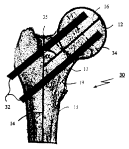
Fig. 4 illustrates a femoral bone structure 30 from its anteroposterior
projection 15
with two marked screws.
Fig. 5 illustrates a femoral bone structure 30 from its lateral projection 17
with two
marked screws 32.
In the AP and Lat radiograph projections 15, 17 the central axes 14, 16 of the
femoral neck 10 and shaft 18 are marked and the femoral neck-angles a and ~3
are
measured. The distance from the femoral neck axis 16 to a marker which
indicates where to
position a screw 32, when surgery is performed, at one measuring points in the
Lat view 17
is taken to represent the X co-ordinate and the distance in the AP view 15 the
Y co-ordinate
for the pin. Angles as shown in Fig. l, Fig. 4 and Fig. 5 are used to place
the femoral bone
structure 30 in space, thus finally indicating the direction for insertion of
screws 32.
Co-ordinates in the inferior or posterior halves of the femoral head 12 and
neck 10
are assigned negative values, see Fig. 2 and Fig. 3.
For the femoral neck 10, the point of intersection of the femoral head sphere
and
the femoral neck axis 16 is used as measuring point. The diameters 34, 35,
indicated by
CA 02369018 2001-09-26
WO 99/49785 PCT/SE99/00521
13
broken lines in figures 4 and 5, of the femoral head 12 and neck 10 are
determined at the
measuring points in both the AP and Lat projections 15, 17.
Since magnification factors in the AP and Lat projections often differ, all
measured
distances are adjusted by calculating the ratio of the greatest and the
smallest femoral head
diameter and then multiplying the distances in the projection with the
smallest diameter by
the ratio.
A method for determining, in three dimensions, where to position fixating
means in
a hip fracture by pre-surgery analysis of at least one anteroposterior and one
lateral digitized
radiograph of said fracture, is provided in connection with the present
invention. It comprises
the following steps:
determining a construed femoral shaft axis 14 out off said graphs from at
least two
midpoints on said shaft axis and drawing a line through said midpoints;
determining a construed femoral neck axis 16 from at least two midpoints on
said
neck axis and drawing a line through said midpoints;
determining femoral neck angles a, ~i
determining a femoral head diameter 34 out off said graphs by placing a circle
over
the perimeter of the femoral head 12;
scaling and rotating said radiographs to a predefined size and position, using
an angle
between the femoral shaft and an Y-axis in said digitized radiographs, and
said diameter 34 of
the femoral head 12;
determining the distance from said neck axis 16 and the center of said femoral
head,
representing a remaining displacement of the hip fracture which is implicitly
known from said
diameter 34;
measuring the height of the medial cortex 19 out offthe anteroposterior graph;
displaying said femoral neck angles a, (3;
displaying cross-sections (Fig. 2 and Fig. 3) of the femoral head 12 and neck
10 with
a predetermined degree of accuracy;
rotating said cross-sections of the femoral head 12 and neck 10 in order to
determine
a degree of derotation of the hip, imposed by the fracture,
displaying the degree S2 of hip rotation in said anteroposterior and lateral
radiographs; and
CA 02369018 2001-09-26
WO 99/49785 PCT/SE99/00521
14
in one embodiment using said steps in an arrangement in order to determine a
position, in
three dimensions for attachment of said fixating means 32 and to pre-adjust a
tool to work in
said position.
Femoral neck-/shaft-angels a, (3 are preferably displayed both as lines and as
numerical values in said digitized graphs, said lines are automatically re-
drawn if the value is
changed.
Lines are possible to determine with two midpoints, but it should be
understood that
a more exact line is obtained by drawing a regression line from at least three
midpoints on
said axes.
Symbols 20 for said fixating means are placed within the cross-section of the
femoral
neck 10 and a warning function is activated if said fixating means are placed
outside of the
femoral neck 10 in said digitized radiographs.
Fixating means 32 for attachment are automatically displayed in said graphs,
through
graphical means known per se, with relation to made measurements. Also, it is
possible to put
down symbols 20 for fixating means 32 in the digitized radiographs.
In accordance with the present invention, radiographs are analyzed before any
surgical treatment, and measured and computed values can be applied as control
input to an
arrangement 40 which accomplishes insertion guides, holes for example, for
bone fracture
fixating means 32, said arrangement 40 being described below.
Fig. 6 illustrates a side view elevation of an arrangement 40 provided in
connection
with the present invention;
Fig. 7 illustrates a top plan view of an arrangement in accordance with Fig.
7; and
Fig. 8 illustrates a front view elevation of an arrangement 40 in accordance
with Fig.
6 and 7.
The arrangement 40 according to the embodiment schematically shown in figures
6-8 is a robot on a mobile stand 42 attached with wheels 44 and adjustable
feet such as bars,
poles 46 or the like for stabilization when ever needed. Further equipment
attached, is a
control box 48 with a cable link 50 connected to an articulated robot arm 52
with servo or
stepper motors 54. A transformer 56 distributes power. The control box 48 is
adapted to be
connected to peripheral equipment such as a computer with I/O ports for
control and
communication, a display device, a printer, scanner, frame grabber, and other
known
computer equipment.
CA 02369018 2001-09-26
WO 99/49785 PCT/SE99/00521
Also, attached on the robot arm 52 is a tool holder 58, for example, used to
hold a
drilling-machine.
It is comprised in the present invention that said control box 48 for
controlling the
robot contains hardware devices, firmware devices and software controlled by a
processor,
5 each device known per se, but forming an unique entity for applications
according to the
present invention. Although, in this preferred embodiment of the invention,
only one
measurement means is described for performing measurements, it is appreciated
that
measurement means/function can be composed of multiple means, or integrated
into one or
more means/function as described below.
10 Thus, in one preferred embodiment comprising means and/or functions, such
as:
measurement means or function accomplishing measurement of the femoral shaft
axis 14 out off said graphs from at least two midpoints on said shaft axis,
calculating and
drawing a line through said midpoints. Further, the device or function
measures a construed
femoral neck axis 16 from at least two midpoints on said neck axis, 16,
calculating and
15 drawing a line through said midpoints. The femoral neck angles oc, (3 are
determined. Still
further, it measures the height of the cortex 19 out off said anteroposterior
graph. Also, the
means measures, calculates and determines the femoral head diameter 34 out off
said graphs
by placing a circle over the perimeter of the femoral head 12, and measures,
computes or
determines the distance from the femoral neck axis 16 to the center of said
femoral head 12,
representing a remaining displacement of the fracture, which is implicitly
known from said
femoral head diameter 34, whereby the means measures, calculates and
determines the
femoral neck diameter 35 out off said graphs by drawing a line, perpendicular
to the femoral
neck axis, at the intersection of the femoral head sphere and the central
femoral neck axis 16;
a scaling function scaling and rotating said radiographs to a predefined size
and
position, using an angle between the femoral shaft and an Y-axis in a display,
displaying co
ordinate axes together with said digitized radiographs, and said femoral head
diameter 34;
a display device for displaying data of interest to hip fracture surgery e.g.
digitized
radiographs, neck angles a, hip rotation S2, cross-sections (figures 2 and 3),
computed figures,
lines 16, 14, etc.
a function providing displays of said cross-sections of said femoral head 12
and
femoral neck 10;
CA 02369018 2001-09-26
WO 99/49785 PCT/SE99/00521
16
a driver for rotating said cross-sections of the femoral head 12 and neck 10
in order
to determine a degree of hip derotation between said head and neck, imposed by
the fracture;
said means
providing control input to a robot with stand means 46 for said robot, tool
means on
said robot for working in a direction given by said control input, and
distortion correcting
means (not shown) compensating for x-ray distortion.
Femoral neck angels a,a are displayed by said display as lines and numerical
values
in said digitized gaphs. The lines are automatically re-drawn, preferably
controlled through
software and/or graphic means, if the value is changed.
Symbols 20 for said fixating means are placed within the cross-section of the
femoral
neck and displayed by said display.
It is included a device or function that activates a warning function if
fixating means
32 are placed outside the femoral neck in said digitized radiographs.
Further, it is included that fixating means for attachment are automatically
displayed
in said digitized radiographs with relation to made measurements, by software.
Means, such as graphic drivers, can be provided for putting down symbols for
fixating means 20, 32 in the digitized radiographs of the present invention.
The method as herein described is preferably applied to control the robot in
using
its tool to work in the right direction, and prepare for insertion of fixating
means such as
screws, pins, nails, etc.
The distortion compensating means according to the present invention is
described
below.
Now referring to Fig. 9, which illustrates a flow chart depicting steps 900 to
980,
taken in the method of the present invention, which is applied as control
information to an
arrangement.
At step 900 AP and Lat radiographs from C-arm fluoroscopy are obtained. The
radiographs are digitized, rotated and scaled 910, by drivers for that
purpose, followed by
performing 920 necessary measurement operations, with means or functions
described above,
on the radiographs of a hip fracture.
Made measurements are resulting in construction 930, through a software, of
femoral
neck and head cross-section graphs, which are displayed on a screen. Displayed
cross-sections
are marked 940 with pin/screw markers, which are changeable through software.
An outlining
950 of the marked pin/screw positions in the digitized radiographs is thus
performed.
CA 02369018 2001-09-26
WO 99/49785 1 ~ PCT/SE99/00521
The steps 900-950 are applied to position 960 a robot to drill in said
positions. This is
followed up by a final check of determined positions. Eventually, during
surgery, a surgeon
manually inserts 980 pins/screws.
Accordingly it should be understood that the PINTRACE 2.0~ method or like
methods are adapted to the method described in accordance with the flow chart.
Henceforth the present invention is described in conjunction with Fig. 10-13.
The
following description is had to a preferred embodiment of the present
invention, but the
invention is not limited to this specific embodiment.
Fig. 10 illustrates a side view elevation of a casing with a tool holder on a
support
according to the present invention; and
Fig. 11 illustrates a top plan view of a casing with a tool holder in
accordance with
Fig. 10.
The arrangement 100 with a tool holder or drill guide system as depicted in
Fig. 10 and 11 comprises a support 110 to be mounted on a machine 40, like a
robot with
an articulated arm 52. It further comprises a means of attachment 112 for
support 110 and
an outer casing 114 with a slide-able inner casing 116 acting as a so called
end effector
holder or tool holder with a stop screw 118 for locking of the inner casing
114. A flanch
120, for forming of a stop position for the inner casing, is provided. Finally
an X-ray
marker pin 122 is hold by the inner casing. The marker 122 can as well be
changed to
another end effector such as a tool, for example, a tool for drilling,
screwing, pinning,
milling, grinding, threading etc, or even machines for the same purposes could
be attached
to the inner casing 116.
In a preferred embodiment, the support 112 is turnable and lock-able (not
shown).
Now referring to Fig. 12 which illustrates the arrangement according to Fig.
10 and 11 attached to an articulated robot-arm 52. The arrangement comprises
the same
components as in the two previous figures and a sliding arrangement 200 with
upper 124
and lower 126 slide-able plates. The lower slide-able plate 126 is attached to
an electrically
isolating plate 128, and working flanch 130 (platform 130) articulated
attached to the robot-
arm 52.
Sliding plates 124, 126 mounted on a robots working flanch 130 makes it
possible to move a part of the arrangement 100, manually or by electric means
in a forward
- backward direction. An electronic distance measuring apparatus is attached
to the sliding
CA 02369018 2001-09-26
WO 99/49785 PCT/SE99/00521
18
arrangement 200 and provides input to the robot about the distances the
sliding device has
moved.
The casing 114 is possibly a metallic cylinder and attached to the support
112.
Another casing 116 formed as a metallic cylinder fits into the first cylinder
114. It is
provided with a central hole through which pins or drills can be introduced.
The inner
cylinder 116 is available with different diameters of the central hole through
which different
end effectors such as drills, screws, pins etc can be introduced.
A small steel pin 122 with radiographic markings for each 25 mm at one end is
introduced through the central hole and serves as a measurement guide.
Also depicted in Fig. 12 is a sterilized cloth 132 for anti bacterial
protection
during preparations for surgery.
Now referring to Fig. 13 which shows a positioning determining arrangement 140
mounted on a support 150 and attached to a support connected to the robot
fundament or
stand 42 according to the present invention.
X-ray distortion is compensated for with a new inventive method and an
arrangement
140, 150 for that purpose.
X-ray distortion such as magnification arises from a diverging X-ray beam as
it is
emitted from an X-ray tube towards a radioplate. Hence by having a reference
at a
predetermined distance from the tube a magnification factor would be gained by
the ratio of
the distance measured on a radioplate with the known distance of the
reference.
The arrangement comprises, in a preferred embodiment, a plexiglass plate 152
with
four lead balls 154 attached and placed in a square pattern. While trans-
illuminating in the AP
and Lat projections in parallel with the ball pattern, two balls 154 are
adjusted to be placed in
the center of the radiation field so that they cover each other entirely.
Thus, through
measuring the distance between the two balls that are uncovered, an absolute
measure of the
distortion is determined (the magnification ratio) provided that a
predetermined distance
between the radiation tube and the plexiglass 152 plate is upheld. This
distance corresponds to
the working distance when distance determinations are made. A calculation in
percentage to
adjust the robot arm is finally applied.
Depicted in Fig. 13 is an open plexiglass box 140 with two plates 154, 156
attached
orthogonal to each other. Plate 154, in a vertical position in Fig. 13,
provided with four X-ray
opaque balls in a square pattern. The plates 154, 156 are mounted on a support
150, which is
attached to a fundament 42 for the robot. Four means of attachment are making
up the support
CA 02369018 2001-09-26
WO 99/49785 19 PCT/SE99/00521
150. A first means 158, here a rod, is mounted on a frame 160 of the robot
fundament 42,
which is movable back and forth in relation to the frame 160. A second means
162 is movable
around the rod 158, and attached to the rod 158. A third means 164 is attached
to an axis 166
which makes it movable in a vertical direction to said means 164. Finally, the
fourth means
168 is movable in relation to said third means 164 horizontal and slide-able
in all directions
possibly in 360 degrees if neccessary.
Said plates 152, 156 are of a material substantially transparent to X-ray
radiation and provided with 4 round elements in a square configuration. The
round elements
are made of X-ray opaque material such as tantalum, lead, steal etc. The
support 150 is
movable in three dimensions so it can be placed in front of robot 40 end
effector and, after
measuring, retracted so it does not disturbe the movement of a robot arm 52.
Specific steps 1)-3) are provided for control of position determining etc in
accordance with the present invention, Seth forth below:
1) A computer program designed to calculate the magnification factor from
measurements on the measuring template (position determining arrangement).
2) A computer program designed to calculate the desired movement of the
robot arm from input measurements through i) the position of the Alignment
system, ii) the
position of the Marking pin and iii) the marked position for the drill / screw
/ pin in the
bone.
3) A computer program for checking the final robot position.
Henceforth, a method used in the present invention is described.
A) Activate the robot arm and move to a defined start position with, for
example for drilling, the arrangement for marking 100 pointing forward and the
marking
pin 122 put in place and extending, for example, SOmm. The robot is assigned a
first start
tool Position, i.e. the robot adapts its co-ordinate system so that each
movement is carried
out around the tip of the marking pin 122. Said measuring template 140 is
placed in front of
the drill guide 100. Place the measuring template vertical to the floor and
adjust it so that
the marking pin 122 hits each round element 154 when the robot moves a
specified distance
in a square pattern.
B) Placing of the measuring template 140 adjacent to the part of a body where
the drilling / screwing is intended. A movable X-ray machine (C-arm) is placed
and
adjusted so that, when radiographs are taken, two out of the four round
elements 154 cover
CA 02369018 2001-09-26
WO 99/49785 PCT/SE99/00521
each other in the two orthogonal planes (AP and Lat projection). The position
for the two
covered round elements represent a starting position for the robot. Said two
radiographs are
digitized and fed into a computer and the distances between the two uncovered
round
elements, representing the magnification factor, is calculated and displayed
by a computer
5 program. The magnification factor relates to the movement of the robot arm.
C) Said thin steel-pin 122 (the marking pin) with radiographic markings for 25
mm on one end introduced through the central hole in the second cylinder so it
protrudes 50
mm. By advancing the sliding arrangement 200 the marking pin 122 penetrates
through the
skin and muscles to a position close to the bone which is to be drilled.
Distances the sliding
10 device has to move are measured manually or electronically and fed into a
robot 40
computer program. Now providing the robot a second tool position, start tool
position two,
according to the distance the sliding device has moved. Now letting the arm 52
move
around this second position even if the pin and second cylinder is removed,
meaning that all
robot movements can take place outside the patients body, but the center of
movements will
15 still be close to the bone inside the patients body.
D) Two radiographs (AP and Lat) are taken and digitized, and fed into a
computer. The position of the marking steel pin 122, and the length of the 25
mm marking,
are measured on the digitized radiographs. A scale factor for this position is
calculated.
E) A desired position for the drill / pin / screw in the bone is marked on the
20 digitized radiographs. The computer program now compares the desired
position for the
drill / pin / screw in the bone with the actual position for the marking pin.
After making
corrections for magnification (B) and Scale (D) the computer calculates the
distances and
angles the robot has to move in order to align the marking pin with the
desired drill / pin /
screw position in the bone. These distances and angles are fed into the robot
computer and
the robot repositions itself accordingly.
F) After the robot 40 has moved into the new position, the marking pin 122
and the second cylinder 116 is changed to similar devices, suitable for
drilling, screwing,
pinning etc. A chosen end effector is advanced by means of the sliding
arrangement 200 so
that it enters through the skin and muscles to a position close to the bone.
At this, AP and
Lat radiographs are taken and digitized. A computer program performs a
checking
procedure by marking and outlining the direction and position of the end
effector
(pin/screw/drill) and comparing it with the marked desired pin/screw drill
position. If the
CA 02369018 2001-09-26
WO 99/49785 PCT/SE99/00521
21
trajectory of the end effector deviates more than a specified distance from
the marked
desired position, the positioning procedure is redone from the present
position. This will
considerably reduce the distances the robot has to move and therefore reduce
the positioning
errors.
G) When the end effector is optimally positioned, the surgeon manually
introduces pins/screws or drills through second cylinders 116 with central
holes in different
sizes. A drilling machine can also be fixed to the sliding device and
automatically introduce
the end effectors. To ensure stability and safety, the robot arm is locked
during drilling
procedures, thus creating a stable platform for these procedures.
It is thus believed that the operation and construction of the present
invention will
be apparent from the foregoing description. While the method and arrangements
shown or
described have been characterized as being preferred, it will be obvious that
various
changes and modifications may be made therein without departing from the
spirit and scope
of the invention as defined in the following claims.