Note: Descriptions are shown in the official language in which they were submitted.
CA 02369793 2001-10-19
WO 00/62661 PCT/US00/10806
-1-
METHOD FOR IMPROVING CALIBRATION OF A BLOOD MONITORING INSTRUMENT
BACKGROUND OF THE INVENTION
The present invention relates to the detection and measurement of the
concentration of constituents of a solution or suspension using radiation,
preferably
near-infrared radiation. More particularly, methods have been developed for
the non-
invasive measurement of the concentration of constituents such as hemoglobin
and its
variants and derivatives, glucose, cholesterol and its combined forms, drugs
of abuse,
and other analytes of clinical and diagnostic significance. Because these
methods do not
require the withdrawal of blood in order to perform these measurements, they
are
particularly suitable for home testing of glucose levels in diabetics and of
urea or
creatinine levels in patients undergoing home dialysis. The present invention
provides a
method of calibrating these measurements to obtain an absolute concentration
without
the requirement of obtaining extensive calibration data for each subject.
In addition to home testing, development of non-invasive clinical testing
procedures has become an important goal, due to the widespread fear of AIDS
and other
diseases, such as hepatitis, which can be spread through the use of invasive
procedures.
In the published research, a major issue in the in vivo quantification of
blood
analyte concentrations is the problem of how to take the signals generated by
the
apparatus and create from those signals an absolute value for the constituent
concentration of interest. Current methods for the evaluation of concentration
levels
involve conversion of the signals to an estimated constituent concentration by
some
arbitrary algorithm using values generated by a contemporaneous set of
invasive
measurements from appropriately generated samples of blood or tissue. If the
concentrations estimated by the converted signals and the concentrations
estimated by
the invasive measurements are highly correlated, then the correlation thus
found is
CA 02369793 2001-10-19
WO 00/62661 PCT/US00/10806
-2-
accepted as a "calibration curve" for the constituent of interest. However,
the
calibration curve thus generated is not necessarily valid over a wider range
of subjects or
physiological conditions than the range used to generate the curve.
In cases where only a relative trend in the data is of interest an accurate
calibration is less critical and the foregoing method is adequate. However, in
many
cases, either calibration data are unavailable or a more accurate estimate of
the
constituent concentration is required. For these cases, a calibration method
applicable to
all subjects under all conditions is desirable.
A number of related publications suggest the use of water as an internal
standard.
Since water is an absorber in the near infra-red, the general approach is to
measure the
optical effect of water and to compare it with the optical activity of the
constituent of
interest. For example, Matcher et al. (Phys. Med. Biol., 38, 177, 1993)
discusses the use
of certain features of the water absorption spectrum to estimate the
"differential path
length" traveled by radiation in a scattering medium which includes water.
However,
their calculation for the concentration of water in the tissue studied (the
human forearm)
varies by approximately 12 % around the mean value. Other publications
(Documents
Geigy, 7th edition, 1970) indicate that, depending on the tissue of interest,
water
concentrations can vary between 60% and 90%.
Jobsis (US Patent 4,805,623) describes a method in which an unknown
concentration is estimated using the presence in the sample of an absorber
having a
known concentration. However, in the Jobsis disclosure, the absorber of known
concentration is water in tissue. Jobsis states that the variability is about
15%. Thus the
concentration of water is subject to the same lack of constancy as in the
disclosures by
Matcher et al. Jobsis does not discuss the use of any water concentration
having a level
sufficiently constant to employ as a universal calibration or reference level.
In fact,
Jobsis states that "the practice of the present invention depends strongly on
the
development of either a means of translating the results in terms of accepted
standards,
CA 02369793 2001-10-19
WO 00/62661 PCT/US00/10806
-3-
such as spectrophotometric data in clear solutions, or on the de novo
development of an
extensive data base where accepted standards are not relevant, i.e., in
heterogeneous
systems such as the brain."
Pologe (U.S. Patent 5,297,548) discloses the use of simultaneous measurement
on a common optical path using pulsatile signals to determine the relative
amounts of
the dominant absorbers: water, deoxyhemoglobin, and oxyhemoglobin. Pologe does
not
indicate the possible use of such an apparatus to generate a universal
calibration method
applicable across multiple subjects. In fact, Pologe indicates that
calibration of such an
apparatus is intended to be performed empirically.
Other workers, such as Carim et al. (US Patent 5,553,615) and Kuestner (US
Patent 5,377, 674), also disclose the use of optical measurements for
noninvasive
analysis in which one or more detectors are sensitive to wavelengths in which
water is
the primary absorbing species. However, neither of these disclosures attempts
to create
a universal calibration or reference level.
1 S As the above discussion suggests, the difficulty of in vivo calibration
problem
results from a combination of two factors. First, the physical pathlength over
which any
absorber is present in the tissue or blood is unknown and varies from person-
to-person.
Second, the intense scattering in tissue and its variation from person-to-
person causes
the unknown pathlength to be multiplied by an unknown factor that varies with
wavelength as well as with subject. A successful solution to this problem
requires
consideration of both of these issues.
Several patents from the laboratory of the present inventor disclose various
procedures which can assist in diminishing some sources of variability and
provide
better precision. These include United States Patent No. 5,334,287, which
describes the
basic procedure now known as Kromoscopy, and United States Patent No. 5,
434,412,
United States Patent No. 5,424,545, United States Patent No. 5,818,048 and
United
States Patent No. 5,672,875, all of which describe improvements and variants
on the
basic Kromscopic system and methods. The disclosures of all the above-
referenced
CA 02369793 2001-10-19
WO 00/62661 PCT/US00/10806
-4-
patents are incorporated herein by reference. While many of these patents
relate to
methods of improving sensitivity and precision of the assays, the biological
system is so
complex that additional modifications and processes are helpful.
SUMMARY OF THE INVENTION
The method of the present invention makes use of the physiological fact that
the
kidneys and their associated regulatory systems maintain a virtually constant
water
concentration in the blood. These regulatory systems maintain the osmotic
pressure
difference across the filtration systems of the kidney at a stable level and
thereby
provide the renal system with maximal control over the critical function of
solute
filtration.
As a result of this regulation, the water concentration in the blood, as
measured
by a variety of techniques, varies from approximately 830-860 grams per
milliliter of
blood, a variation of t1.8% around the average level. In contrast, the
concentration of
water in tissues can vary by as much as X20% around the average level. This
exceptionally high stability of blood water concentration can be used to
calculate
concentrations of other constituents in the blood.
In the present invention, this highly stable value for the concentration of
water in
blood is employed in a universal calibration scheme by combining optical
measurements
performed at two or more wavelengths in such a way as to eliminate the
dependence of
concentration on either the thickness of the body part, on the thickness of
the absorbing
regions within the body part, and on the scattering properties within the body
part.
This is accomplished, in a general sense, by employing several types of
normalization of the detection channel outputs. For each detection channel the
output
signals are scaled to fractional modulations by comparing the differential
output
produced by the cardiac pulse to the background output produced at diastole,
as
employed in pulse oximetry. In addition, the present invention contemplates
additional
normalization across multiple detection channels, which normalizes the
fractional
modulations in each detection channel to the relative amounts of water-
specific
CA 02369793 2001-10-19
WO 00/62661 PCT/US00/10806
-5-
information included in all the detection channels or in a specific subset of
said
detection channels. This normalization thereby allows expression of the
detection
channel responses in such a way that the water-specific information in the
resultant data
is maintained at a constant level, despite the effects of thickness, of
scattering, or of
changes in pulse magnitude. The non-water-specific information remaining in
the data
expressed in this way is then predominantly a function of the relative
absorptivities and
concentrations of other absorbing constituents in the arterial blood to that
of water, and
quantitative calibrations and measurements may be performed for such
constituents.
In one particularly useful embodiment, readings at two or more channels or
detectors containing primarily water-specific information are used separately
to provide
a means of normalizing the outputs from the other detection channels to
achieve
concentration measurements. In another embodiment, which is particularly
useful when
the analyte of interest has only absorption bands that overlap with those of
water, the
method provides normalization over the totality of the water-specific
information
available in all of the detection channels. This normalization method is
based, in part,
on the concept that the response from a series of detectors to a fractional
modulation in
arterial pulse can be understood and operated upon as a vector in an N
dimensional
space, where N is the number of simultaneous and spatially congruent detection
channels. Each of these vectors can be normalized using a scalar related to
the
responses of the detector channels to water in vitro. These forms of
calibration improve
accuracy, including both sensitivity and precision, of the requisite
measurements.
In all these embodiments, the initial step in obtaining this combination of
measurements is to express each individual optical measurement as the ratio of
the
difference between the transmission maximum, produced when the arterial blood
pressure is at its diastolic minimum level, and the transmission minimum,
produced
when the arterial blood pressure is at its systolic maximum level. Once the
optical
measurement has been so scaled to a fractional modulation in each detection
channel,
the measurements in the various optical channels can be combined in a number
of ways,
which will be detailed below.
CA 02369793 2001-10-19
WO 00/62661 PCT/US00/10806
-6-
Note that an accurate combination of the fractional modulations across optical
channels places a stringent set of requirements on the optical measurements.
First, since
the condition of the sampled body part varies throughout the cardiac cycle,
the
measurements must be made before there is any major physical change. For
example,
since the pulse normally takes about 1 second, taking 25 or more
measurements/second
will be in short enough time intervals that the physiological changes between
measurements are minor. Second, the accuracy of the result of the combination
of
measurements is maximized when there exists a common light path from the
source or
sources, through the body part, to each of the several detectors. Third, to
produce
accurate signal combinations, the effects of scattering on the optical
measurements must
be minimized by substantially eliminating light reaching the detectors with a
large angle
of incidence. In a preferred embodiment, light incident on the detectors at an
angle
greater than about 10 degrees is minimized.
When the above constraints are met, the resulting combination of measurements
minimizes adverse effects caused by the concentration of water in the arterial
blood, the
concentration of the other constituents of the arterial blood, the absorption
coefficient of
water, and the absorption coefficients of the constituents. This is true
because the
measurements no longer depend in a substantial manner on the light scattering
coefficients within the tissue, on the venous blood component, or on the
thickness of the
body part.
The present invention provides several alternative configurations for
excluding
the effect of large-angle radiation from the detectors. In one embodiment, the
contribution of scattered radiation to the signal reaching the detector is
minimized. The
embodiment has been previously disclosed by Block et al. (US Patent 5,672,875)
where
it is disclosed that restricting the solid angles of illumination and
detection substantially
eliminates radiation scattered through larger angles reaching the detectors)
relative to
that transmitted or scattered though smaller angles. If such apparatus is
employed using
the data processing means and protocol described above, then the dependence of
the
combined results upon scattering coefficients will be substantially
eliminated.
CA 02369793 2001-10-19
WO 00/62661 PCT/US00/10806
7_
In a second apparatus configuration, also disclosed in Block US Patent
5,672,875, the contribution of large-angle scattered light reaching the
detectors is
minimized by generating polarized radiation and allowing said polarized
radiation to
contact the body part. Radiation scattered through large angles will become
depolarized
as a consequence of such scattering, and the interposition of an analyzer in
the optical
path between the body part and the detectors will prevent such depolarized
radiation
from reaching the detectors. This second configuration may be combined with
the use
of restricted solid angles of illumination and detection for additional
rejection of
scattered radiation.
In another configuration, the contribution of radiation scattered at larger
angles
relative to that scattered at smaller angles or not scattered can be
estimated. This
estimation is performed by measuring the total radiation reaching sets of
detectors with
substantially identical spectral response. Each element of each set maintains
a common
light path with the corresponding element of another set or sets of detectors
having a
different spectral response characteristic. Then, the combination across
spectral
responses described above can be carried out on an element-by-element basis.
As the
geometrical positions of the corresponding elements approach the optical axis
of the
system, the effect of the larger angle scattered radiation in comparison with
that of the
directly transmitted radiation will be decreased. Appropriate processing of
the multiple
element data will then permit a better estimate to be made of the ratio that
would be
expected in the absence of scattering effects.
The invention provides a method for improving the accuracy of non-invasive, in
vivo concentration measurements of a substance of interest in blood. The
method has
the steps of providing an illumination source that generates illumination
radiation to a
measurement site across a portion of the spectrum that contains absorbance
bands of
said substance of interest. The measurement site is illuminated with said
illuminating
radiation and radiation transmitted or reflected from said measurement site is
detected
by a detector array. In the preferred embodiment, the detector array has a
plurality of
detectors having distinct maximum spectral response characteristics in a
different region
of said portion of the spectrum used for illumination. In one embodiment, the
plurality
CA 02369793 2001-10-19
WO 00/62661 PCT/US00/10806
_g_
of detectors includes at least a first detector and a second detector having
spectral
characteristics with greater responsiveness to the absorbance bands of water
than to the
other constituents in the blood. Each of the detectors in said detector array
provides an
output signal indicative of the amount of radiation it receives in a selected
time period.
The method also contains the step of determining a differential value of the
output
signals for periods of arterial pulse by comparing values obtained during a
systolic
measurement cycle with values obtained during a diastolic measurement cycle.
These
differential values are used to generate a series of ratios of the
differential output signals
generated from the output signals of the non-water detectors to the
differential output
signals generated from the output signals of each of the water detectors.
These ratios
provide a means for improving accuracy of the concentration measurements.
While this
same set of detectors could be used in other embodiments of the invention, it
is not
practical if one cannot obtain a clear differentiation of the absorption
bands. For
example, although the absorption bands for hemoglobin, deoxyhemoglobin, and
water in
1 S the spectral region from 600-1200 nm are separate enough that they can be
differentiated
and the first method may be used, while for glucose determination, there is
too much
overlap so a different method is needed. In particular, the scalar
normalization is
preferred as the overlap of absorption bands increases.
The methods of the invention are useful for both Kromoscopic and non-
Kromoscopic (e.g., classic spectrophotometric) measuring systems but have
particular
advantageous qualities when used with a Kromoscopic system, which meets the
conditions of simultaneity and congruence defined above. Optical measurements
using
detection channels with non-overlapping spectral sensitivities may also employ
the
methods of this invention, so long as these conditions are substantially met.
Accordingly, broadband illuminating radiation or broadband detectors may be
used,
preferably using detectors with overlapping response. In the alternative,
several
different illumination sources such as LEDs may be used, with a coded response
from a
single detector such as is described in United States Patent No. 5,424,545.
Similarly,
congruent illumination and sampling methods, as well as restricting the solid
angle of
illumination or detection, using methods such as those described in the afore-
mentioned
Kromoscopy patents, are useful as part of, and in conjunction with, the
methods
CA 02369793 2001-10-19
WO 00/62661 PCT/US00/10806
-9-
described herein. The ratios and comparisons required for these methods can be
carried
out using hardware such as a neural net or software run in a computer or other
calculating device. These methods are particularly useful in determining
concentration
of constituents of blood such as hemoglobin or glucose, in vivo in human
patients.
While it is theoretically possible to use the methods disclosed herein with a
model system that explains all of the scattering and interferences which
effect the
present measurements, no such model system is needed. The present methods are
equally useful with a chemometric analysis; i.e., an analysis which is not
tied to a
particular physical representation.
The present invention also provides for a system or apparatus to carry out the
disclosed methods. In one embodiment, the apparatus has the two water-
responsive
channels at its heart, which provides the calibration required to achieve the
desired
accuracy. If the absorption bands of the analyte of interest have too much
overlap with
the bands of water, multiple channels which are responsive to both water and
the analyte
should be utilized, as indicated in the second preferred embodiment. The
apparatus may
use a single illumination source and a series of detectors, such as in a
detector array, or
detectors with different filters. Normally, a pulsatile measurement is used,
with the
apparatus being capable of sufficiently rapid measurements to differentiate
and
segregate arterial pulse effects. In addition, congruent sampling and/or
illumination
apparatus, particularly with restricted solid angle, may be used.
By the use of all the components of this invention, simultaneous measurements
over a common optical path can be combined with the invariance of the water
concentration in blood in such a way as to minimize the effect of the unknown
pathlength. Thus, the constant water concentration in arterial blood, the
common optical
path, and the minimization of the effects of scattered radiation solve the
problem of
Jobsis (the lack of a translatable standard), while the constant water
concentration
provides a known, subject-invariant reference to obtain absolute
concentrations from the
relative concentrations calculated using the procedure disclosed by Pologe.
CA 02369793 2001-10-19
WO 00/62661 PCT/US00/10806
-10-
Thus, an advantage of this invention is that it provides a method for
obtaining a
universal calibration curve for all patients and under all conditions. This in
turn permits
the accurate measurement of absolute concentrations of constituents of
arterial blood
having absorbance in a given spectral sub-region.
Another advantage of this invention is that the method so provided can create
a
calibration curve for a particular constituent without requiring invasive
measurements of
that constituent in each subject.
Another advantage of this invention is that the method so provided can reduce
measurement variability due to irregularities in cardiac pulse amplitude and
the
consequent pathlength variation.
Yet another advantage of this method is that it may be employed using a
variety
of optical configurations and in a variety of spectral sub-regions for
noninvasive
measurements on any of a variety of body parts, limited only by the
requirement that
both water and the constituent of interest have measurable absorbance in the
spectral
sub-region on interest, that the body part permit measurable quantities of
radiation to
reach the detectors, and that the cardiac pressure produce a measurable change
in the
radiation reaching the detectors.
Other advantages to the methods resulting from this invention will be apparent
from the following detailed description of the invention.
BRIEF DESCRIPTION OF THE DRAWINGS
FIG. 1 shows an apparatus for measurement of absorption by blood constituents
at a
plurality of wavelengths, using a plurality of distinct detectors;
FIG. 2 shows the steps implemented by the processor of FIG. 1 in order to
determine
absolute concentrations of all constituents in the blood;
CA 02369793 2001-10-19
WO 00/62661 PCT/US00/10806
-11-
FIG. 3 shows an apparatus for measurement of absorption by blood constituents
at a
plurality of wavelengths, using a plurality of distinct light sources and a
single
detector; and
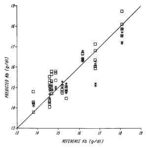
FIG. 4 shows a graph of predicted hemoglobin concentration from a blood sample
versus measured hemoglobin using the apparatus of FIG. 1.
DETAILED DESCRIPTION OF THE INVENTION
Human blood includes a plurality of test constituents present in unknown
concentrations together with at least one reference constituent present in a
known
concentration. Because the concentration of water in blood is both known and
nearly
invariant across the human population and time, it is convenient to use water
as the
reference constituent. However, the methods described below can be applied to
any
constituent which is present at a known concentration.
The method has the steps of providing an illumination source that generates
illumination radiation to a measurement site across a portion of the spectrum
that
contains absorbance bands of said substance of interest. The measurement site
is
illuminated with said illuminating radiation and radiation transmitted,
transflected, or
reflected from said measurement site is detected by a detector array. In the
preferred
embodiment, the detector array has a plurality of detectors having distinct
maximum
spectral response characteristics in different regions of said portion of the
spectrum used
for illumination. In one embodiment, the plurality of detectors includes at
least a first
detector and a second detector having spectral characteristics with greater
responsiveness to the absorbance bands of water than to the other constituents
in the
blood. Each of the detectors in said detector array provides an output signal
indicative
of the amount of radiation it receives in a selected time period. The method
also
contains the step of determining a differential value of the output signals
for periods of
arterial pulsation by comparing values obtained during systolic portions of
the arterial
pulse with values obtained during a diastolic portions of said pulse.
CA 02369793 2001-10-19
WO 00/62661 PCT/US00/10806
-12-
The method of comparing predominantly water-responsive channels to
predominantly non-water-responsive channels is effective if good wavelength
separation
can be achieved. For example, oxyhemoglobin (and deoxyhemoglobin) are dominate
absorbers in the 600-1100 nm range, while water is the dominant absorber in
the
wavelength range greater than 1100 nm. Accordingly, the ratio method may be
used if
hemoglobin concentration is sought. However, this is not effective for glucose
which
has its primary absorbance in the greater than 1 I 00 nm range. Accordingly,
another
method of using water as a universal standard is needed to eliminate the
pathlength
variability and other interference problems.
In the case where water is a dominant absorbing constituent in all the
detection
channels required for measurement of a constituent of interest, then a method
is required
that represents the constancy of the water concentration of the arterial blood
over sets of
detection channels employed in the measurement. This representation can be
achieved
by the consideration of a set of the detection channels as the elements of a
vector. The
output of N detectors can be used to generate a vector in an N-dimensional
space. For
example,a vector B can be measured in vivo which represents the set of
fractional
modulations in each detection channel by the cardiac pulse caused by all the
absorbing
constituents in the blood. Similarly, a vector G, denoting absorptivity of
glucose or any
other constituent of interest, may be generated from the in vitro
absorptivities of said
constituent as measured in each of the N detection channels; each additional
absorbing
constituent has its own, unique vector. The underlying assumptions in the
disclosed
method are that the relative magnitude and direction of the vector of the
analyte of
interest and the vector of water from the detectors in the N-dimensional space
will
maintain substantially the same relative directions and magnitudes as they
have in vitro.
Based on these assumptions, an in vitro measurement of water can be taken
using the
same detector configuration employed for in vivo measurements, and a water
vector, w,
representing the in vitro water results in the same N-dimensional space, can
be
determined. By definition, the vector w is deemed to be a unit vector. The dot
product
of B and w, B~w, is a scalar which can then be used to normalize the vector B.
Normalization is achieved by the formula B/ B~w yielding a vector B which has
the
same direction as B but with two desirable properties. First, if the only
reason for
CA 02369793 2001-10-19
WO 00/62661 PCT/US00/10806
-13-
changes in B is variations in pathlength due to changes in blood pressure or
other
changes in pulse magnitude, the magnitude and direction of B, computed by this
method, will not vary. Second, the component of B in the water direction will
be the unit
vector w; the component of B in the water direction is B~w, resulting in B~w/
B~w = 1,
i.e., the unit vector in the water direction, w. The first property achieves
the goal of
eliminating dependency on pathlength changes, while the second provides
universality
of calibration related to the constancy of water content in blood. This self
normalization allows information from all the detection channels to be used in
the
measurement of B, unlike the prior art reference measurements. One may also
use
another constituent, G, and perform similar operations as with B to express
the response
to all the constituents of interest on the same water-normalized scale. Once
this has
been done, changes in the direction of B toward the vector G representing any
constituent of interest may be clearly seen to be indicative of increases in
the
concentration of that constituent. Furthermore, the normalization of all the
vectors to
the approximately known concentration of water, in the context of the
assumption of
constant relative directions and magnitudes of all the vectors so normalized,
permits the
calculation of the magnitude of the concentration change causing such a shift
in vector
B.
FIG. 1 shows an optical system useful for practicing the present method. This
optical system employs collimating optics for both illumination and detection,
with the
detector having a plurality of detector units placed such that they achieve
congruent
sampling. Radiation source 10 is selected so that it provides broad spectrum
illumination, e.g., 700-2500 nm illumination. Radiation from radiation source
10 passes
through collimating lens 12 before striking tissue 20. Optional aperture 14 is
shown
which helps define the collimation optics in conjunction with collimating lens
12.
Once the radiation has traversed tissue sample 20 and exits the tissue through
area defining aperture 25, it passes through detector collimating optics 30
formed of
converging lens 32, aperture 34 and recollimating lens 36: This type of
collimating
optics is conventionally used in telescopes and other devices where
collimation of light
is desired. The collimated beam exiting collimation optics 30, specifically
recollimating
lens 36, then goes through a series of beam splitters 42A, 42B and 42C and
onto four
CA 02369793 2001-10-19
WO 00/62661 PCT/US00/10806
- 14-
detector units 44A, 44B, 44C and 44D. The beam splitters and detector units
are
arranged such that the entire detection unit 40 provides congruent sampling of
the beam.
More particularly, the beam sputters and detector units are arranged such that
the
pathlength and angles from recollimating lens 36 to any of detector units 44
are equal
and each of detector units 44 are optically superimposable upon the other. If
the analyte
of interest has spectral characteristics which can be differentiated from
those of water, at
least two of detector units 44 should have spectral characteristics with
greater
responsiveness to the absorbance bands of water than to the other constituents
of the
blood. Although four detector units are shown, the exact number may be varied.
In operation, radiation Ion from the broadband source 10 penetrates the finger
20
where it interacts with the various constituents in the blood and the tissue.
As a result of
the heartbeat and the resulting pulsatile blood flow, this interaction is a
time-varying
phenomenon. During the diastolic phase of the heartbeat, as shown in FIG. 2A,
the
incident radiation interacts with the tissue 201d, the venous blood 202d, and
the arterial
blood 203d of the finger 20d. During the systolic phase, shown in FIG. 2B, the
amount
of tissue 201s and venous blood 202s in the finger 20s is approximately the
same as that
which was present in the diastolic phase. However, the amount of arterial
blood 203s
has increased. As a result, the output radiation Ig~ during the systolic phase
differs from
the output radiation IDS during the diastolic phase to the extent that the
extra blood
volume results in additional absorption.
FIG.3 shows a different apparatus for use in practicing the present invention.
FIG. 3 shows a system using the beam splitter array of Fig. 1 reversed for
congruent
illumination rather than congruent sampling. Four radiation sources 310A,
310B, 310C
and 310D, are used to illuminate the tissue sample. The radiation issuing from
each of
the radiation sources goes through a collimating lens (312A, 312B, 312C and
312D,
respectively) and then is redirected by one of the beam splitters 316A, 316B
or 316C to
illuminate tissue 320. The radiation transmitted by tissue 320 passes through
converging lens 332 and aperture 334 before striking detector 344. Optionally,
an
additional lens 336 (not shown) could be used to recollimate the transmitted
radiation
before it strikes detector 344. The radiation sources, collimating lenses and
beam
CA 02369793 2001-10-19
WO 00/62661 PCT/US00/10806
-15-
splitters are arranged to provide congruent illumination and each separate
radiation
source may have an associated modulator to provide a different modulation to
the
radiation issuing from that radiation source. This type of modulation
apparatus, and its
advantages, is described in more detail in United States Patent No. 5,424,545.
Briefly,
using a plurality of modulators, each providing a different modulation to the
associated
radiation issuing therefrom, and using a form of modulation differentiation at
the
detector (such as electronically separating the signals based on modulation
frequency)
provides a method which allows differentiation at the detector of the source
of the
illuminating radiation, and accordingly allows additional information to be
generated
from a single detector. For example, if the radiation sources cover different
wavelengths, a single detector can differentiate the intensity of the
transmitted radiation
at each wavelength range by using the modulation to determine the wavelength
range.
This can eliminate the requirement of the system illustrated above which
requires a
plurality of detector units. For improved results, both the congruent
illumination shown
in Fig. 3 and the congruent sampling shown in Fig. 1 may be used in the same
device.
Similarly, a filter wheel that provides different wavelength transmission can
be used on
either the illumination or detection side of the device. It is also possible
to use fiber
optics to transmit light on either the illumination or detector side of the
apparatus.
FIG. 4 shows a plot of predicted hemoglobin concentration, made using the
first
embodiment of the in vivo, non-invasive methods and apparatus described
herein, with
actual hemoglobin as measured using a blood sample. The actual hemoglobin
reading is
determined using capillary blood sample on a Hemocue B-hemoglobin analyzer
(Mission Viejo, CA) for 10 samples. The predicted values are determined using
an
apparatus such as is shown in FIG. 1 in a transmission mode with a fiber optic
input
placed next to a fingernail and the detector on the opposite side of the
finger. Four
congruent InGaAs detectors are used, with filters whose maximal transmission
is located
near 1064nm, 1200nm, 1300nm, and 1625nm before the individual detectors. The
1200nm and 1300nm filters are the water channels. The illumination source is a
2.7 W
halogen light source with the fiber optic output focused on elevated
fingernails. The
elevation of the finger above the heart also appears to improve accuracy.
Optical
transmission data from each detector channel is digitized by HP3458A
multimeters and
CA 02369793 2001-10-19
WO 00/62661 PCT/US00/10806
-16-
transmitted to a Pentium PC via Labview software for post-processing to derive
pulsatile
modulation values by Matlab software.
Each data run consists of thirty seconds of raw data, or about 30 cardiac
pulses at
normal cardiac rates. The data from each run was used in a ratio calculation,
with all
four possible hemoglobin to water detection channel ratios being used to
generate the
results. The linear regression analysis is shown in FIG. 4. The standard error
of
calibration was 0.24 g Hb/dl, on values ranging from 13-19 g Hb/dl.
The advantages of the present invention apply to spectrophotometric systems
such as those employed in pulse oximetry. While the shot-noise constraints on
the
detected intensity are lower because the absorption of the hemoglobins are so
much
larger the acceptance angle restrictions provide greater linearity and
improved
calibratability, as well as reduction in the severity of motion and breathing
artifacts, and
other limitations on universality of calibration.
For the analysis of trace constituents where the high photon flux requirement
is
critical, the present invention is particularly advantageous when combined
with the use
of broadband and broadband overlapping detectors, as taught in the Block '265
Patent
and the parent applications.
The foregoing description is meant to be explanatory only and is not intended
to
be limiting as to the scope of the invention. The invention is defined by the
following
claims. What is claimed is: