Note: Descriptions are shown in the official language in which they were submitted.
CA 02369868 2002-07-16
WO 00163694 PCTIUS00109714
Proteome Mining
US Government Rights
This invention was made with United States Government support
under Grant No. DK52378A, awarded by the National Institutes of Health. 1'he
United States Government has certain rights in the invention.
Field of the Invention
The present invention is directed to high throughput screening of
proteomics for identifying bioactive compounds. More particularly the
invention is
directed to the isolation of novel herbicides, antibiotics, antifungals,
antivirals,
insecticide or pharmaceuticals based on their interactions with target
molecules.
Backeround of the Invention
I 5 The animal, plant, prokaryotic and viral kingdoms contain within them
a vast array of genes that express 100,000's of distinct proteins whose
biological
function is essential life. 'The number of genes contained with in a
particular
organism varies greatly. Generally, the simpler the organism the fewer the
total
number genes. For example, completion of the yeast genome shows that these
organisms have about 8300 genes, the complete (.'. elegans genome contains
about
18,000 genes, and the human genome is estimated to contain about 100,000
distinct
genes. Each gene encodes a specific protein which has a predetermined
essential
function for the over all survival of the organism. Collectively, given the
biodiversity
that exists on earth, the numbers of distinct genes that exist in nature is
likely to
number in the billions.
Obviously not all of the proteins expressed by the genes of an organism
are likely to be of importance to man. Indeed the number of genes that are
likely to
express proteins of commercial or medical value is a tiny fraction of this
vast
biodiverse gene pool. Methods therefore that allow one to rapidly and
effectively
screen large numbers of proteins within this pool for valuable proteins are of
great
importance. In the case of the human genome it has been estimated that
approximately 4000 of its genes are responsible for the causes of non-pathogen
CA 02369868 2002-07-16
WO 00163694 PCT/US00109714
-2-
induced human disease. In short this means that human tissue contain 4000
proteins
of potential medical and commercial value. 'fhe same analogies can be made to
other
species. For example if one was screening for a new antibacterial agent, one
would be
looking for bacterial protein targets that were peculiar to the particular
bacterial strain
of interest. In the case of bacteria this has traditional been enzymes
involved in the
synthesis of the bacterial cell wall. The classic example of a drug that is
selective for
bacteria is penicillin which inhibits an essential enzyme required for
synthesis of the
bacterial cell wall. Humans do not possess any of the enzymes that make
bacterial
cell walls. One cannot simply target any bacterial protein when searching for
new
antibiotics, this is because even though there are many differences between
humans
and bacteria, a significant portion of the bacterial genome encodes proteins
of similar
structure or function as found in humans. Drugs that inhibited proteins with a
common function in both organisms are unlikely to discriminate between the two
species.
To identify new drugs or commercially important bio-active molecules
one needs methods that have the ability to encompass entire species genomes
and
immediately identify candidate proteins of importance. The present invention
is
directed to a method of identifying compounds that selectively interact with
important
biological components. 'this selective interaction is an essential element
that makes a
particular chemical have medical or commercial value. Without selectivity a
compound has no bio-active value; selectivity is the single most important
factor in all
drug, antibiotic, antifungal, antiviral, insecticide and herbicide action.
1'he selectivity of a valuable bio-active compound in 99% of all cases
is based on its interactions with one or more specific proteins contained
within the
target cell or organism of interest. One or two percent of valuable bio-active
molecules maybe directed towards non-protein targets such as DNA, RNA, lipids
or
sugars. Without exception a valuable bio-active chemical interacts with its
protein
target in a highly specific manner. The target protein will contain on its
surface a
domain or pocket that binds the chemical with high affinity. This domain or
pocket is
unique to the target and not the several 1000 proteins that may also be
contained with
in the cell expressing the target protein.
In most cases the binding site for the chemical on the target protein is
CA 02369868 2002-07-16
WO 00/63694 PCT/US00109714
-3-
important to the biological activity of the protein, The binding site may be
required for
enzymatic activity, or be site of hormone interaction (e.g. a receptor) or a
binding site
for an all osteric regulator. When a chemical binds to one of these sites and
affects
the biological activity of the target protein it invariably contains
structural components
that resemble the natural biological ligand that normally occupies the site.
The
affinity of the interaction of the chemical for the target protein generally
increases as
the overall structural components of the chemical mimic the natural ligand. in
some
cases bio-active molecules iit better into the natural ligand-binding site
than the
natural ligand itself. These types of molecules are likely to have extremely
potent bio-
1 b activity. If these molecules possess structural features that prevent
their metabolism
then this increases their bio-activity even greater.
One of the primary' mechanisms for identifying bio-active chemicals of
medical or commercial value is to screen large combinatorial libraries with
some form
of an enzymatic or biological assay. Combinatorial libraries can be extremely
diverse
I ~ and contain many hundred thousands of distinct molecules of known or
unknown
structure. They can be derived from very diverse sources, including plant
extracts,
animal extracts, soil samples, bacteria, fungi, chemical industry byproducts
etc.
Theoretically, these libraries contain within them molecules of every
conceivable
shape and form. However, like the proteins to be targeted, only a small
percentage of
20 these libraries contain molecules that have important bio-activity.
Prior high through put screens for drug discovery begin with a disease,
the choice of which is invariably determined by potential market size. The
etiology of
the disease is first defined by basic research to determine likely underlying
cause.
This research identifies potential protein targets that may be useful drug
targets; e.g.
25 receptors or enzymes. The purified receptors or enzymes are then used to
screen
chemical libraries for agents either bind, inhibit or activate. Similar
approaches are
also used to screen for anticancer drugs. Transformed cell lines are used to
screen
large chemical libraries that rnay contain compounds that revert them to their
normal
phenotype or kill the cancer. Further investigation is then used to identify
the
30 molecular mechanisms by which active compounds from these screens bring
about
their cellular effect.
Therefore in the traditional search for a new bio-active compounds one
CA 02369868 2002-07-16
WO 00163694 PCTIUS00/09714
-4-
begins with a specific biological problem in mind. For example a particular
pharmaceutical house maybe focused on discovering new antihypertensive agents.
Its
decision to enter into the screening process is always based on the disease
market size.
Thus the search for drugs that would treat small populations of afflicted
individuals is
S unlikely to happen in the private sector. In the case of a new
antihypertensive agent
for example, a drug screen will generally begin with an assay that includes a
specific
receptor or enzyme that has important functions in the regulation of blood
pressure.
One then has to hope that the libraries one screens contain bio-active
molecules that
selectively effect the proteins selected in the assay. Once candidate
chemicals are
identified one then has to demonstrate that these compound act selectively and
predictably for the targeted protein in the assay and not others. Thus in the
initial
stages one ends up with many false positives which must be eliminated in a
second
round of screening because the entire expressed genome was not taken into
account in
the first instance. The invention described herein eliminates this problem at
the start
because it encompasses both the diversity contained within the chemical
library to be
screened, with the diversity of the expressed genorne itself in one step.
The selectivity is achieved in the analysis following sequencing of the
targeted proteins. A decision as to whether a particular protein/chemical
interaction is
likely to have commercial and medical value is made during the last stages of
analysis. Therefore, in addition to identifying bio-active agents that have
commercial
value the screen of the present invention does not exclude compounds that may
have
humanitarian value. This is because we could conceivably identify agents that
bind to
proteins important in the pathology of obscure diseases with small patient
populations.
Finally, the present invention can readily cross platforms with no
change in protocol or equipment. There is no difference in screening
procedures for
herbicides, antibiotics, antifungals, antivirals, insecticide or
pharmaceuticals. All one
changes is the expressed genome (proteorne) that is to be screened. One can
even use
the same libraries for each screen; i.e. a library that did not yield any
useful
pharmaceutical agents may contain a useful herbicide.
CA 02369868 2002-07-16
WO 00/63694 PCTJUS00/09714
-5-
Summary of the Invention
The present invention is directed to compositions and methods for
identifying bioactive compounds. Advantageously, the present method of
identifying
bioactive compounds utilizes both the diversity contained within a chemical
library to
be screened, with the diversity of the expressed genome itself in one step to
maximize
the efficacy of the screening procedure. The method comprises the steps of
contacting
an immobilized combinatorial library with the protein members of a proteome,
characterizing the proteins that interact with members of the library to
identify those
proteins having important biological value, and isolating the corresponding
compound
from the library that interacts with a protein having important biological
value.
Brief Description of the Drawings
Fig. 1 is a diagramatic representation of the steps used in accordance
with one embodiment to isolate bioactive compounds from a complex mixture of
proteins (proteome) using an immobilized combinatorial library.
Fig. 2 is a diagramatic representation of the steps used in accordance
with one embodiment to isolate bioactive compounds from a complex mixture of
proteins (proteome) using an immobilized combinatorial library.
Fig. 3 is a diagramatic representation of the steps used in accordance
with one embodiment to isolate bioactive compounds from a complex mixture of
proteins (proteome) using an immobilized combinatorial library.
Fig. 4 is a diagramatic representation of the steps used in accordance
with one embodiment to identify cell surface receptors and their peptide
ligands en
masse from a predetermined cell type.
Fig. 5 represents the stained SDSPAGE results from characterization
of proteins isolated from rabbit skeletal muscle through the use of
gammaphosphate
linked ATP-Sepharose. Rabbit skeletal muscle extract was prepared from 350 g
of
tissue (wlw) and passed over 50 mls of gamma phosphate linked ATP-Sepharose
containing 10 umols/ml of linked ATP. Following washing, the column was eluted
sequentially with NADH, AMP, ADP and ATP and fractions collected (lOmls).
Column fractions were separated by SDSPAGE then transfered to PVM and stained
with amido black. Proteins 1-17 were identified b}' mixed peptide sequencing
(see
CA 02369868 2002-07-16
WO 00/63694 PCTIUS00109714
-6-
Table 1 ).
Fig. 6 represents the stained SDSPAGE results from geldanarnycin
released muscle extract proteins. ATP-Sepharose was loaded with skeletal
muscle
extract and eluted successively with the indicated concentrations of
geldanamycin,
followed by 10 mM ATP. Peak fractions (20u1 of I .0) were analyzed by SDSPAGE
and silver staining or transferred to PVM for identification by peptide
sequencing.
Numbers indicate that proteins that wee identified on the PVM membrane: I.
HSP90
and proteolytic fragments of HSP90; 2. purine synthetase (ADE2); 3. myosin
light
chain kinase; 4. phosphorylase kinase; S. p98 glucose indued kinase; 6. HSP70;
arginine succinate synthetase; 7. glutamate dehydrogenase; 8. glutamate
ammonium
ligase; 9. glutathione sythetase; 10. aldehydc dehydrogenase; 11. MAPK;
t 2. GAPDH; 13. PKA
Detailed Description of the Invention
In describing and claiming the invention, the following terminology
will be used in accordance with the definitions set forth below.
As used herein, "nucleic acid," "DNA," and similar terms also include
nucleic acid analogs, i.e. analogs having other than a phosphodiester
backbone. For
example, the so-called "peptide nucleic acids," which are known in the art and
have
peptide bonds instead of phosphodiester bonds in the backbone, are considered
within
the scope of the present invention.
As used herein, bioactive compounds include any compound that is
capable of inducing an effect on a living cell or organism. Bioactive
compounds
include bui are not limited to pharmaceuticals, hormones, chemotherapeutics,
nucleic
acids and the like.
As used herein the term "proteome" relates to a complex mixture of
proteins that are derived from a common source, such as an extract isolated
from a
particular cell or tissue. For example a human proteome represents a mixture
of
proteins isolated from human cells. The category can be further defined by
specifying
a particular cellltissue source for the proteome (i.e. a human myocardial
tissue
proteome represents all the proteins isolated from human myocardial tissue).
As used herein the term "combinatorial library" relates to a collection
CA 02369868 2002-07-16
WO 00/63694 PCT/US00/09714
_'j_
of compounds. The combinatorial library can be a biological synthesized
library that
comprises nucleic acid sequences that include a common vector sequence
(allowing
for replication of the library in a host species) and a protein encoding
region. The
biological synthesized library can be further provided with regulatory
elements that
allow for the expression of the encoded proteins (i.e. an expression library).
Chemical
libraries are collections of compounds that were isolated from a natural
source or were
synthesized in a laboratory using chemical or biological processes. A
"combinatorial
chemical library" is a collection of compounds created by a combinatorial
chemical
process, wherein the compounds of the combinatorial chemical library have a
common scaffold with one or more variable substituents.
As used herein the term "solid support" relates to a solvent insoluble
substrate that is capable of forming linkages preferably covalent bonds) with
soluble
molecules. The support can be either biological in nature, such as, without
limitation,
a cell or bacteriophage particle, or synthetic, such as, without limitation,
an
acrylamide derivative, agarose, cellulose, nylon, silica, or magnetized
particles.
As used herein the term "naturally-occurnng" relates compounds
normally found in nature. Although a chemical entity may be naturally
occurring in
general, it need not be made or derived from natural sources in any specific
instance.
As used herein the term "non naturally-occurring" relates to
compounds rarely or never found in nature and/or made using organic synthetic
methods.
As used herein the term "functional analog" of a library
compound/ligand relates to a compound that has a binding affinity for the same
ligand
as one of the members of the library, such that the functional analog will
compete
with the library component for binding to that ligand.
The present invention is directed to a novel method for the rapid
identification of bioactive compounds, including but not limited to novel
drugs,
antibiotics, antifungals, antiviral, insecticide or herbicides. The overall
strategy
behind the invention is to screen complex protein mixtures with an immobilized
library of compounds for proteins that bind specifically to components in the
library.
The bound proteins are then identified by protein microsequcncing to determine
if the
CA 02369868 2002-07-16
WO 00/63694 PCT/US00/09714
_g_
identified protein is therapeutically relevant. 'The protein has therapeutic
relevance if
the protein is known to be central to the development of a disease, is a
metabolic
enzyme unique to a particular microorganism, yeast, virus, or fungi, or is an
enzyme
peculiar to a type of insect, or an enzyme required for photosynthesis in a
particular
weed.
T he advantage of the present screening methodology derives from the
initial assumption that the entire genome is a potential drug target. The only
decision
prior to screening is to decide what proteome should be utilized; i.e. for
drugs
important in human disease, human tissue is the choice, for herbicide a plant
species,
for antibiotic, a bacterial strain.
In one aspect of the invention, systems and methods are provided for
rapidly screening a combinatorial library for bioactive agents. The method is
based on
the identification of those library components that interact with proteins of
a
preselected proteome, wherein the proteome protein is a potential target for
therapeutics. The method of identifying bioactive compounds comprises the
steps of
contacting a combinatorial library with the protein members of a proteome
under
conditions that allow for specific interactions between proteins of the
proteome and
the bound library. Proteins that interact with the immobilized library
components are
then isolated and analyzed to determine if the protein is interesting from a
therapeutic
standpoint. Those proteins that have therapeutic relevance are then used to
identify
the component of the immobilized library that interacts with the protein.
In one embodiment, the method of identifying bioactive compounds
present in a compound library comprises the steps of contacting an immobilized
compound library with the protein members of a preselected proteome, and
washing
the immobilized compound library with a buffered solution. In one embodiment
the
immobilized compound library comprises a column of particulate solid support,
such
as sepharose or agarose beads, that has the individual components of the
compound
library bound to the support, and the wash comprises the use of a low ionic or
high
ionic buffer. In one embodiment the column is washed with both a high ionic
buffer
and a low ionic buffer. After the solid support has been washed with buffer,
the
remaining bound proteins are released from the solid support by contacting the
bound
proteins with one or more individual members of the compound library or with
CA 02369868 2002-07-16
WO 00/63694 PCTlUS00/09714
-9-
functional analogs of the library components. Alternatively, the bound
proteins can be
released from the immobilized library through the use of a chaotropic agent,
including
but not limited to detergents such as SDS, TritonX, sarkosyl, denaturants such
as urea,
or chelators such as EGTA or EDTA. The released proteins are then identified
by
protein sequencing or mass spectrometry; and the identity of the specific
compounds
of the compound library that bind to the released proteins is determined.
The libraries
Combinatorial libraries can be constructed using techniques known to
the skilled practitioner to provides researchers vast number of chemical
candidates to
screen for potential bioactivity. In accordance with the present invention the
library
comprises a collection of compounds that are capable of specific binding to
their
target. For example, suitable library components include, but are not limited
to
peptides, proteins, carbohydrates, lipids, glycoproteins or nucleic acids.
Biologically synthesized combinatorial libraries have been constructed
using techniques of molecular biology. These library components are expressed
using
bacteria or bacteriophage particles. For example, U.S. Pat. No. 5,270,170 and
5,338,665 to Schatz describe the construction of a recombinant plasmid
encoding a
fusion protein created through the use of random oligonucleotides inserted
into a
cloning site of the plasmid. In other biological systems, for example as
described in
Goedell et al., U.S. Pat. No. 5,223,408, nucleic acid vectors are used wherein
a
random oligonucleotide is fused to a portion of a gene encoding the
transmembrane
portion of an integral protein. Upon expression of the fusion protein it is
embedded in
the outer cell membrane with the random polypeptide portion of the protein
facing
outward. Thus, in this sort of combinatorial library the compound to be tested
is
linked to a solid support, i.e., the cell itself and the cell itself adheres
to the cell
culture substrate. The Goedell patent is incorporated herein by reference.
Similarly, bacteriophage display libraries have been constructed
through cloning random oligonucleotides within a porkian of a gene encoding
one or
more of the phage coat proteins. Upon assembly of the phage particles, the
random
polypeptides also face outward for screening. Such phage expression libraries
arc
described in, for example, Sawyer et al., 4 Protein Engineering 947-53 (1991
);
CA 02369868 2002-07-16
WO 00163694 PCT/US00/09714
-10-
Akamatsu et al., 151 J. Irnmunol. 4651-59 (1993), and Dower et al., U.S. Pat.
No.
5,427,908. These patents and publications are incorporated herein by
reference.
While synthesis of combinatorial libraries in living cells has distinct
advantages, including the linkage of the compound to be tested with its
nucleic acid,
there are clear disadvantages to using such systems as well. The diversity of
a
combinatorial library is limited by the number and nature of the building
blocks used
to construct it; thus modified or R-amino acids or atypical nucleotides may
not be able
to be used by living cells (or by bactetiophage or virus particles) to
synthesize novel
peptides and oligonucleotides. There is also a limiting selective process at
play in
such systems, since compounds having lethal or deleterious activities on the
host cell
or on bacteriophage infectivity or assembly processes will not be present or
may be
negatively selected for in the library.
Another approach to creating molecularly diverse combinatorial
libraries employs chemical synthetic methods to make use of atypical or non-
I 5 biological building blocks in the assembly of the compounds to.be tested.
Thus,
Zucketmann et al., 37 J. Med. Chem. 2678-85 ( 1994), describe the construction
of a
library using a variety of N-(substituted) glycines for the synthesis of
peptide-like
compounds termed "peptoids". The substitutions were chosen to provide a series
of
aromatic substitutions, a series of hydroxylated side substitutions, and a
diverse set of
substitutions including branched, amino, and heterocyclic structures. This
publication
is incorporated by reference herein.
Alternatively, chemical synthetic methodologies can be used to create
large diverse libraries of potentially useful compounds and permits the
synthesis of
compounds joined to a solid support of some kind or joined to an identifiable
marker
such as a flourescent tag. In accordance with one embodiment, the
combinatorial
library is chemically synthesized on solid supports in a methodical and
predetermined
fashion, so that the placement of each library member gives information
concerning
the synthetic structure of that compound. Examples of such methods are
described,
for example, in Geysen, U.S. Pat. No. 4,833,092, in which compounds are
synthesized
on functionalized polyethylene pins designed to fit a 96 well microtiter dish
so that the
position of the pin gives the researcher information as to the compound's
structure.
Similarly Hudson et al., PCT Publication No. W094105394, desct7be methods for
the
CA 02369868 2002-07-16
WO OOI63694 PCT/US00/09714
-11-
construction of combinatorial libraries of biopolymers, such as polypeptides,
oligonucleotides and oligosaccharides, on a spatially addressable solid phase
plate
coated with a funetionalized polymer film. In this system the compounds are
synthesized and screened directly on the plate. Knowledge of the position of a
given
S compound on the plate yields information concerning the nature and order of
building
blocks comprising the compound.
Another approach has been the use of large numbers of very small
derivatized beads, which are divided into as many equal portions as there are
different
building blocks. In the first step of the synthesis, each of these portions is
reacted
with a different building block. The beads are then thoroughly mixed and again
divided into the same number of equal portions. In the second step of the
synthesis
each portion, now theoretically containing equal amounts of each building
block
linked to a bead, is reacted with a different building block. The beads are
again mixed
and separated, and the process is repeated as desired to yield a large number
of
1~ different compounds, with each bead containing only one type of compound.
This
methodology, termed the "one-bead one-compound" method, yields a mixture of
beads with each bead potentially bearing a different compound. The compounds
displayed in the surface of each bead can be tested for the ability to bind
with a
specific compound (i.e. a protein member of a proteomel.
The libraries used in the present invention can be well defined,
containing known mixtures of molecules, or the library can be one in which the
chemical content is poorly defined.
In accordance with one embodiment the libraries are immobilized on a
solid support. Biological material, including but not limited to proteins,
carbohydrates, nucleic acids, lipids, glyeoproteins can be bound to a solid
surface
using standard techniques known to those skilled in the art. In preferred
embodiments
the library compounds are linked through covalent bonds. The solid surface can
be
selected from any surface that has been used to immobilize biological
compounds and
includes but is not limited to polystyrene, agarose, silica or nitrocellulose.
In one
embodiment the solid surface comprises functionalized silica or agarose beads.
In accordance with one embodiment the components of a sample are
bound to silica or agarose beads in separate reactions using different
reaction reagents
CA 02369868 2002-07-16
WO 00/63694 PCTfUS00109714
-12-
and conditions to ensure that a diverse array of compounds are bound to the
solid
surface. Fractions of these separate reactions can then be combined to form a
single
affinity chromatography column. For example, a portion of the library can be
reacted
with an inert solid support (e.g. agarose, Sepharose, polystyrene or other
chromatography beads) using standard protocols for linking primary amines to
NHS,
cyanogen bromide activated or maleimide activated resins. Many of these resins
are
commercially available, for example, Pharmacia CH-activated Sepharose. Another
portion of the library is then reacted to an inert support that would select
any
molecules containing carboxylic acid residues. A commercially available resin
in this
case would be Pharmacia EAH-activated Sepharose. A third and fourth portion of
the
library could be linked through thiol (SH2), phosphate or aldehyde (CHO)
containing
residues. The goal is to link as many components as possible with in the
library and
in as many orientations as possible. The orientation of molecule when it is
tethered is
critical to its ability to interact with potential target proteins. Thus for
some
1 S molecules reaction through primary amines may cause binding of a target
protein to be
sterically hindered because of the thether. However, tethering of the same
molecule at
a carboxyl residue may not hinder interaction with a target protein.
The entire library can be linked to the resin or portions of the library
linked can be linked separately. One should aim to achieve as reasonably high
a
ligand concentration as possible per immobilized molecule. Ideally this should
be
between 10 nmol to 1 qmollml. In the case of libraries in which the chemical
content
is poorly defined two mixtures from the library are prepared, a mixture that
is soluble
in organic phase and a mixture that is soluble in aqueous phase. The same
linking
strategy is then employed for the preparation of the resins from the organic
and
aqueous soluble library members. Each resin is then placed into a
chromatography
column and equilibrated into the protein extraction buffer.
In another embodiment (shown in Fig 2), individual components in a
library, or mixtures containing between 1-10 chemicals, are attached to beads
separately in mierotitre plate wells. Several 100 beads can be reacted in each
well at
the same time. One bead is then selected from each well and placed in a
chromatography column. Again each ligand should be attached in multiple
orientations. In cases where a target compound is identified that interacts
with a large
CA 02369868 2002-07-16
WO 00/63694 PCTlUS00/09714
-13-
number of proteins (e.g. ATP, see Fig 3) only the target compound is
immobilized on
the solid support. The bound proteins can be released by the addition of an
excess of
free target compound, a derivative of the target compound or a functional
analog of
the target compound (see Example 2).
The proteomes
The immobilized libraries are contacted with a proteome under
conditions conducive to the formation of specific interactions between
components of
the proteome and components of the immobilized library. Choice of the proteome
used depends on the problem being addressed. If one is looking to isolate a
new drug
for the treatment of a human disease the human protcome (i.e. human tissue) is
the
best choice. If one wishes to discover a new antibiotic then the target
pathogen (e.g. a
gram negative bacterium) of interest is the obvious choice. For an
insecticide, the
targeted insect and so on.
In accordance with one embodiment the proteome comprises a natural
products library which represents a collection of natural products that have
been
recovered from biological material and have been determined to have biological
activity. For example the natural products library may include a mixture of
natural
products wherein the mixture is known to induce a phenotypic change in a
population
of cultured cells.
The amount of starting tissue used to isolated the proteome proteins is
critical and should be based on theoretical recovery of target proteins. For
example, if
one is interested in identifying drugs that may bind to signal transduction
molecules
one should take into account the amount of these proteins (or copy number)
that may
be in the cell. Many of these types of molecules may be expressed as low as
200
copies per cell. A quick calculation predicts that if one wanted to recover as
much as
10 pmol of a particular protein that was expressed at 200 copies per cell one
would
require 12 grams of wet weight tissue. For high copy number proteins, such as
metabolic enzymes, less tissue mass would be required to achieve 10 pmol of
protein.
One should also factor in potential losses due to inefficiency of extraction
or
proteolysis. Thus although 12 g of tissue may contain a total of 10 pmol of a
target
protein of interest one may only recover a small percentage of this in the
initial screen.
CA 02369868 2002-07-16
WO 00/63694 PCT/US00/09714
-14-
Fortunately, modern protein detection and sequencing methods enable one to
identify
proteins in the femto molar range. However, increasing the starting tissue
mass as
much as possible will improve the odds of recovering sufficient protein for
later
identification.
Tissue from the chosen target proteome is ground and homogenized in
buffers appropriate for solubilizing proteins and retaining their native
conformations.
This involves standard procedures utilized in most biochemical laboratories.
Following clarification by centrifugation the portions of the extraction
mixture are
passed over the immobilized chemical library resins.
When interactions are sought between library compounds and cell
surface proteins, it may be advantageous to investigate such interactions with
the
surface proteins in their natural state (i.e. embedded in the cell membrane).
In
accordance with one embodiment, the "proteome" represents the set of surface
molecules displayed by cells cultured on a cell culhmc substrate, and may
include
proteins, glycoproteins, carbohydrates and lipids.
In accordance with one embodiment certain components of the
proteome can be removed prior to contacting the immobilized compound library
with
the proteome. Components can be removed, for example, by fractionating the
components based on molecular weight, electric charge and/or hydrophobicity.
Alternatively, specific components can also be removed by ligand or antibody
binding. Such methods allow the removal of protein components that do not
warrant
further investigation but are know to bind to certain components of the target
compound library. In addition such prescreening allows for the removal or
reduction
of proteins that are expressed at high levels in the tissue used to generate
the
proteome.
The compounds of the proteorne are placed in contact with the library
component under conditions favorable for specific interactions between members
of
the two groups. The interaction may result in the alteration of a physical
characteristic
such as fluorescence, absorption, enzymatic activity, but typically the
specific
interaction involves binding of the two components to one another. In
accordance
with one embodiment the library components will be immobilized on a solid
support
and the proteome components will be solubilized or suspended in a .solvent.
The
CA 02369868 2002-07-16
WO 00163694 PCTlUS00109714
-15-
solvent will then be incubated with the immobilied library for a time
sufficient for
specific interaction between the library components and the proteome
components.
In accordance with one embodiment the library components are
covalently linked to a particulate solid support and the particulate is used
to form a
column. In this embodiment the proteome solution/suspension is passed through
the
column to provide contact between the library components and the proteome
components. Some of the proteome components will bind to the immobilized
library
components. The immobilized library is then washed with a buffered solution to
remove any non-specifically bound proteins. 1n accordance with one embodiment
the
step of washing the library comprises the steps of washing with a high ionic
strength
buffer and a low ionic strength buffer to remove proteins that may be
associated
because of non-specific ionic or hydrophobic interactions..
Isolation and identification of target proteins with a defined immobilized
library
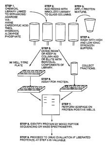
In accordance with one embodiment the library comprises a defined set
of compounds that have been covalently linked to sepharose/agarose beads
(resin) via
amine, carboxylic acid, thiol, hydroxyl, aldehyde or phosphate linkages and
the beads
are combined to form a column (see Fig 1 ). The protein mixture (proteome) is
then
applied to the column followed by washes of high and low strength buffers. In
accordance with one embodiment the solutionslsuspensions are allowed to
percolate
through the column based on gravity. Alternatively, additional force can be
applied to
speed the flow of the eluate through the column; for example the column can be
spun
in a centrifuge to enhance flow through the column.
After the column has been washed writh the buffered solutions, the
beads (resins) are either kept in the columns or removed and placed in equal
amounts
into 96 well microtitre plates (see step 5, Fig. 1 ). Maintaining the resins
in a column
has the advantage of potentially recovering more protein per library ligand,
however,
it has the disadvantage of being much slower procedure overall.
When using the microtitre plate approach, the number of plates and
wells used depends upon the number of components that are in the defined
chemical
library. This could be a few 100 to several 1000 to 10,000's. Individual
components
from the library are added at high concentration (milimolar at least) 1.o each
well.
CA 02369868 2002-07-16
WO 00/63694 PCT/US00109714
-16-
Application of resin and subsequent addition of the library component can be
automated using commercially available robots. Following addition of the
individual
components to each well the plates are agitated for a pre-determined length
and
temperature (e.g. 30 minutes at 30°C). .
In the case of resins packed in chromatography columns, each
component in the library (free of the solid support) is passed separately over
the
immabilized library and any eluted material collected. It is anticipated that
a certain
portion of the library components will be able to compete with the bound
ligand and
selectively liberate one or more proteins from the resins in the plates or
columns. A
portion of each eluate is then placed into a micratitre plate for protein
analysis.
Advantageously, using this method identifies the specific library component
that binds
to the released protein. Further analysis of the protein will determine if
this binding
interaction has potential therapeutic value.
If the column is broken down and distributed into the individual wells
of a microtitre plate prior to releasing the bound proteome proteins, an
additional step
must be taken to isolate the released proteins. Library components are added
to each
well and incubated to release the bound proteins. Following incubation with
each
library component, the resin is allowed to settle or the resin suspension is
centrifuged
briefly (300 x g) to pellet the beeds. In accordance with one embodiment the
resin
beads can be magnetized beads, and a magnetic field is applied to the bottom
of the
plate to hold the resin at the bottom. Afrer the resin has been separated from
the
supernatant, a portion of the supernatant from each well is removed and placed
in a
second well containing a high sensitivity protein staining reagent. This last
step can
be automated using standard robots familiar to those skilled in the ari.
The protein detection reagent used to detect the presence of released
protein can be any of those known to the skilled practitioner. In one
preferred
embodiment the detection reagent is one that changes color in the presence of
protein.
Radioactive isotopes that bind proteins (I'zs), fluorophors (e.g. FITC) or
gold stains
may also be used to increase sensitivity. Specialized detection systems
capable of
detecting these markers are known to those skilled in the art.
Wells/column fractions that are positive for protein are selected for
further analysis using standard techniques. It is anticipated that the number
of
CA 02369868 2002-07-16
WO 00!63694 PCTlUS00/09714
_ 17-
positive wells would be a few percent of the total number of components that
are in
the library screened - although this may be several 100 if the total number of
components in the initial library numbers in the 10,000's. The proteins will
be
subjected to gel electrophoresis analysis, typically using SDSPAGE, to measure
the
purity of the protein and quantitate the amount of recovered protein. The
supernatant
from each well that contains liberated proteins) is mixed with SDSPAGE sample
buffer and characterized by SDS gel electrophoresis. Many hundreds of
supernatants
can be easily characterized using this method. The gel is stained with silver,
Commassie or colloidal gold (for sequencing by mass spectrometry) or
transferred to
polyvinyl membrane (for mixed peptide sequencing). At this point the molecular
weights and amount of protein recovered are determined.
For mixed peptide sequencing, the proteins on the polyvinyl membrane
(PVM) are stained then excised (See Damer et al., (1998) J. Biol. C'hem 273:
24396-
24405). The membrane pieces are digested briefly with CnBr, washed and placed
directly into an automated Edman sequenator. Mass spectrometry can also be
used
but may be less desirable because of the amount of labor that is required and
its
inability to handle many protein samples at one time. In the case of mixed
peptide
sequencing between 6 and 12 rounds of Edman sequencing are carried out and the
mixed peptide sequences generated sorted and matched against the databases
with the
2U FASTF (protein databases) and TFASTF algorithms (DNA data bases). This
process
identifies the liberated proteins in each well.
At this point a determination is made as to whether or not the liberated
protein is interesting (i.e. is the protein involved in a human disease, is it
an important
enzyme to bacterial metabolism.. etc). If the protein is determined to have
therapeutic
value, then the chemical that liberated the protein from the immobilized
library is
chosen for further characterization using conventional approaches. For
example, an
affinity assay will typically be conducted to determine if the protein has
sufficient
affinity for the target library compound (i.e. binding at nanomolar
concentrations) to
be useful as therapeutic agent. In addition, analysis will be conducted to
determine
what is the biological impact of the interaction and whether the affinity of
interaction
with the targeted protein can be imprnved by modification.
This screen may yield several candidate proteins that are valuable in
CA 02369868 2002-07-16
WO 00!63694 PCT/USOOI09714
-18-
the initial round and not confine one to a single field or market or interest.
Importantly, a single round of screening will not only identify a potentially
useful
bioactive agent, but will also provide valuable information about its targets,
what
groups can be modified without affecting function and where the agents should
be
applied.
In another embodiment of the present invention, library components
are fractionated and linked to a solid support in separate vessels (see Fig.
2). The
various immobilized fractions of the library are then combined and packed into
a
column. The proteome of interest is then place in contact with the library
under
conditions suitable to allow for specif c interaction between the proteome and
library
components. The resin is then washed to remove non-specifically bound
proteins,
typically using both a high ionic and a low ionic strength buffer. The bound
proteins
are then labeled with a detectable maker, for example either iodinating with
I'-'S, or
reacting with fluorescent marker or dye (e.g. iodofluorescein). The excess
probe is
washed away and the beads removed and individual beads placed into 96 well
microtitre plates, The plates are then scanned for protein either by detecting
radioactivity, fluorescence or color.
Beads that are positive for protein are treated with SDSPAGE sample
buffer and their protein content determined by gel electrophoresis as
described
previously. If a protein is deemed to be of value, the bead that contained the
identified protein is treated to liberate the ligand (library component) for
chemical
identification.
In some instances a library component may be identified that binds to
many protein targets. As outlined in Fig. 3 a ligand that interacts with many
protein
targets can also be used to identify important drugs. In accordance with this
embodiment a proteome is passed over a resin containing a single ligand (e.g.
gamma-
phosphate linked ATP Sepharose). Following washing to remove non-specifically
bound proteins, either the bound proteins arc labeled as described in Fig. 2
or the
column is successively washed with components in a chemical library and
fractions
collected as described in Fig. 1. If the proteins are labeled then the beads
are removed
from the column and placed into microtitre plate wells (1-10 beads/well to
give ~20
nmols total protein). Individual components in the library are then applied to
each
CA 02369868 2002-07-16
WO 00!63694 PCT/US00/09714
-19-
well and the supernatants sampled for protein release. Preferably, each
library
component should be added at increasing doses in orders of magnitude ranging
from 1
nM to 1 mM. Proteins that are selectively liberated in the nm-1tM range are
analyzed
by SDSPAGE and mixed peptide sequencing is conducted as described above.
In accordance with one embodiment the method of identifying
bioactive compounds from a complex mixture of proteins comprises contacting a
combinatorial library with the complex mixture of proteins under conditions
that
allow for specific binding of the proteins to library components. Preferably
the library
components are immobilized on a solid support via a covalent linkage, and
numerous
compounds of the library are present in multiple copies that are bound to the
solid
support in multiple orientations. The immobilized library is then washed with
a
buffered solution, preferably with a high ionic strength buffer and a low
ionic strength
buffer, to remove non-specifically bound proteins. In accordance with one
embodiment the solid support is in particulate form, and the method further
comprises
1 S the step of distributing equal portions of the support particles into a
plurality of wells
of a microtitre plate after the step of washing the immobilized compound
library.
The proteins bound to library components by specific interactions are
then released, preferably by a competition reaction using one or more of the
components of the library (in an unbound state). Vv~herein the library has
been
fractionated and equal portions of the support particles have been distributed
into a
plurality of wells of a microtitre plate the step of releasing the bound
proteins
comprises adding to each microtitre plate well one or more compouzids of the
library.
The released proteins are characterized using standard techniques and the
compounds
of the library that specifically bind to the released proteins are identified.
The release proteins will be identified primarily based on
microsequence analysis and comparison to existing protein databases. This has
been
made feasible because of the near completion of the nucleotide sequencing of
several
genomes, including human, mammalian, C.elegans, bacteria, yeast, viral, rice,
corn.
The invention will have increasing relevance as more species specific genomes
become complete. The proteins remaining bound to the immobilized library after
the
washing steps can also be labeled to assist the detection of the proteins.
CA 02369868 2002-07-16
WO 00!63694 PCT/USOOI09714
-20-
Isolation and identification of target proteins with an undefined
immobilizedchemical library.
All of the initial steps are the same as described above. Following
preparation of the resins and application of protein mixture, the resins are
again
aliquoted into titre plate wells. However, since the components in the library
are
poorly defined and of unknown number some fractionation of the library using
standard methods is required. Useful steps for fractionation include organic
and
aqueous extraction, HPLC or ionic-exchange fractionation. The fracaionated
library is
then applied to each well containing resin and incubated as described. In one
embodiment these steps are carried out robotically. The column chromatography
approach out-lined above is also applicable with an undefined library.
Following incubation with each fraction of the library the resin is
pelleted as described above and a sample of the supernatant taken for protein
analysis_
Fractions that contain liberated protein are selected for characterization by
SDSPAGE.
At this point it is likely that some fractions of the library will liberate
many proteins,
some only a few. In either case, mixed peptide sequencing as mass spectrometry
can
be used to identify all these components in a short space of time. With mixed
peptide
sequencing a standard Edman sequences containing 4 reaction chambers can
identify
20-30 proteins per week. Mass spectrometry will be somewhat slower if a
species-
specific database is not available. The list of proteins that are identified
for each well
is then surveyed for the criteria stated earlier.
Proteins that are deemed valuable are expressed as recombinant
proteins and immobilized on a second resin (Sepharose or agarose beads). The
fraction of interest or entire chemical library is then passed over the
protein affinity
column to selectively recover the chemical compounds with in the library that
bind the
protein of interest. Mass spectrometry or NMR techniques can then be used to
identify or determine the structure of the bioactive compound. One then
proceeds
with the standard methods to characterize the bioactivity of the isolated
chemical. All
three strategies as outlined in Figs. 1-3 can be applied to an undefined
library.
In accordance with an alternative embodiment bioactive compounds
are isolated through the use of intact cells. This method is particularly
useful for
identifying bioactive agent that interact with cell surface molecules such as
receptors.
CA 02369868 2002-07-16
WO 00/63694 PC'f/US00/09714
-21-
The cells are grown on a cell culture substrate suitable for the type of cell
grown. A
complex mixture of labeled proteins or peptides is then added to the cells
under
conditions that allow for specific binding of the labeled proteins to the cell
surface
proteins. In one embodiment the cells are culture in multiple plates and the
complex
mixture of labeled proteins/peptides is divide equally among the multiple
plate of
cells. The cells are then washed under conditions that do not dislodge the
cells from
the substrate. In accordance with one embodiment the cells can be fixed to the
cell
culture substrate prior to incubation with the labeled proteins.
After the plates have been washed to remove non-specifically bound
1 U proteins, the plates are screened for the presence of labeled proteins.
The labeled
proteins/peptides arc released using the same procedures as described above
and
analyzed by gel electrophoresis and microsequencing.
In accordance with one embodiment, the complex mixture of labeled
proteins comprises randomly generated peptide sequences that have been labeled
with
a fluorescent entity. The binding of such labeled proteins to the cell surface
molecules
effectively concentrates the label at the bottom of the culture plate and thus
a positive
reaction can be detected even in the absence of washing the cells to remove
unbound
labeled protein. For example, an excitation light source can be provided
wherein the
beam of light is parallel to the cell surface and the detector is similarly
position so that
only signal generated from the cell surface will be detected. The bound
protein can
then be released using any of the techniques described previously herein, and
the
protein can be analyzed as describe above.
Fig. 4 exemplifies one approach used in accordance writh the present
invention for identifying cell surface receptors and their peptide ligands en
masse.
The overall scheme outlined in Fig. 4 is a variation of that disclosed in Fig.
2.
Bioactive peptides are of pharmaceutical value because they mimic naturally
occurring proteins or larger peptides that bind to important cell surface
receptors e.g.
interferon's, cytokines, growth factors, endorphins. Bioactive peptides can be
generated randomly in large libraries using combinatorial approaches. Peptides
in
these libraries are general range from 4 to 20 amino acids in length. 'These
libraries
can be generated synthetically or recombinantly as fusion proteins. In the
case of
fusion proteins, random peptide sequences are displayed at the N or ~ termini
of a
CA 02369868 2002-07-16
WO OOi63694 PCT/US00109714
-22-
known recombinant protein expressed in yeast or bacteria (Blum et al. 2000
PNAS 97,
2241; Geyer et al. 1999 PIfAS 96: 8567). The fusion protein displaying the
peptide
are recovered by affinity chromatography through an affinity tag that is
present at the
opposite end of the molecule (C or N termini) from the peptide.
There are 24 naturally occurring amino acids in nature which can be
used to construct a random combinatorial library. A peptide library consisting
of
peptides of 20 amino acids in length can therefore have 20z° possible:
combinations.
'this gives an extremely large number of possible variations of peptide and
theoretically covers all possible peptide sequences that could occur in
nature.
Typically these types of peptide libraries can be used to search for cell
surface
receptors or protein partners that would selectively bind one or more peptide
sequences contained within them. As shown in Fig. 4, a recombinant peptide
library
cultured in bacteria or yeast, or a synthetic combinatorial peptide library
tagged with a
floor (e.g. fluorescein), is mixed (at 1nM - 1 ~M concentration) with a
designated
target cell line (e.g. cancer cell line, B or T cell) that is present in the
wells of a multi
chamber titer plate. The plate is placed in an instrument capable of detecting
fluorescent labeled probes on cell surfaces at 100 -5000 molecules per cell.
In our
laboratory we use the PE-Biosystems FMAT robot (Swartzman et al. 1999 Anal.
Biochern. 271: 143). In the case of synthetic peptides the cells are screened
for
specific binding of floor tagged peptides on the cell surface. In the case of
recombinant fusion proteins displaying the random peptides a floor tagged
antibody
that recognizes the fusion protein is added.
The peptides that produce positive resutts are sequenced. In the case of
the synthetic peptides this can be done directly without further purification
in an
Edman sequences or mass spectrometer. In the case of the bacterially or yeast
expressed fusion protein two methods can be used to sequence the peptide.
Positive
clones can be cultured and the expression vector encoding the fusion protein
can be
sequenced across the region encoding the random peptide by DNA sequencing. Or
alternatively, the fusion protein can be isolated from a culiure of bacteria
or yeast and
the random peptide sequence determined by mass spectrometry or Edman
sequencing.
Once the peptide sequence is identified it is produced synthetically and
tagged with a
fluorophor. The affinity of the peptide for the cell surface receptor is
determined. If
CA 02369868 2002-07-16
wo ao~63694 PC."TIUSOal097i4
-23-
the affinity is determined to be sub ~M, an affinity column is constructed
from the
peptide for purification of the receptor target. Typically this would involve
linking the
peptide via its C or N terminus to a C8 spacer attached to a Sepharose bead.
Cell
extract prepared from the designated cell target would then be passed over the
resin to
recover the receptor target for identification by protein sequencing. Bound
proteins
would be recovered by either eluting the resin with free peptide or washing
with SDS.
SDSPAGE and protein staining would be used to quantitate and
evaluate purity. Mixed peptide sequencing or mass spectrometry would be used
to
identify the protein. If the protein is found to be an important cell surface
receptor of
commercial or medical value the peptide and protein target would be fully
characterized. This screen is anticipated to identify many cell surface
receptors and
their bioactive peptide ligands. Some receptors will be well characterized,
many
others are anticipated to be novel. Significantly, in addition to identifying
new
peptide ligands and their physiological targets, our assay, like the other
methods, also
gives a measure of selectivity and potential toxicity. 'this is because the
identified
bioactive ligands, in addition to their true physiological target, had an
equal
opportunity to interact with all other cell surface receptors that happen to
be expressed
on the designated cell target.
Examale 1
Isolation of Adenine Nucleotide Binding Protein from a Proteome
As a proof of principle and to evaluate the types of proteins that bind to
gamma-phosphate linked ATP-Sepharose, tissue extracts prepared from rabbit
skeletal
muscle, liver, kidney, brain or bladder were passed over a gamma-phosphate
linked
ATP-Sepharose affinity resin. Following extensive washing to remove non-
specifically associated proteins, the resin was washed sequential with NADH,
AMP,
ADP and A'hP. Fig. 5 shows the results from characterization of proteins
isolated
from skeletal muscle. Similar results were obtained with other tissues,
although the
pattern, abundance and complexity between tissues varied considerably due to
varied
ievels of expression of individual proteins (See Fig. 5 and Table 1).
Gamma phosphate linked ATP -Sepharose was washed with extracts
prepared from rabbit, skeletal muscle, kidney, liver, brain or bladder.
Following
CA 02369868 2002-07-16
WO 00!63694 PCT/US00I09714
-24-
washing the resin was eluted successively with the indicated nucleotides as
described
in Fig. 5.
Proteins 1-17 (see Fig. 5) were identified by mixed peptide sequencing
(Table 1). Eluted proteins were characterized by SDSPAGE, transferred to PVM
and
treated with CnBr or Skatol prior to mixed peptide sequencing, Mixed peptide
sequencing was carried out on average for 6-12 Edman cycles. The mixed
sequences
were sorted and matched against the entire published protein or DNA data bases
with
the FASTF or TFASF algorithms respectively (Damer et al. 1998., Alms et al.
1999).
Expectation scores for the identified proteins ranged from 2.6 e-' for PKA to
1.2 a -5~
for GAPDH. Expectation scores after each search for the next highest scoring
non-
related protein were generally < 2.3 c ~~ The experiment shown was repeated on
several occasions, and on several different tissue including liver (120g) ,
kidney (60g),
brain (60g) and bladder (20g) with similar results (Table 1).
CA 02369868 2002-07-16
WO 00163694 PCT/US00/09?14
-25-
Table 1
NADH pmol AMP pmol
GAPDH (M) 1 * 25,000.0Purine synthetase 5.0
(M,L) 2*
malate dehydrogenase 0.5 Phosphorylase (M) 10.0
(M) 8* 3*
glutamate dehydrogenase1.5 AMP activated protein2.0
(L) 9* kinase (L) #
aldehyde dehydrogenase2.0 ES'T AA254816 (L) 4.0
(M) 7*
lactate dehydrogenaseX0.0 ESTAA571903 (L) 5.0
(M,L) #
6-phosphogluconate 5.0 phosphatidylinositol-4-2.U
dehydrogenase (M,L) phosphate S-kinase
(L)
I isocitrate dehydrogenaseI .0 protein kinase DI3N1.0
0 (L) I
(related) (L)
3-hydroxyacyl-CoA 1.0
dehydrogenase(L)
sorbitol dehydrogenase2.0
(L)
alcohol dehydrogenase20.0
(M,L)
glucose-6-phosphate 17.0
dehydrogenase (M,L)
CA 02369868 2002-07-16
WO 00/63694 PCT/IJS00/09714
-26-
Table 1 (cont.)
ADP pmol ATP pmol
Heat shock 15,000.0MAPK (M) 5*# 5.0
protein
90 (MIL) 4*#
Purine synthetase14.0 MEK1 (M)# 6.0
(MlL) 2*
Pyridoxal kinase (M, L) 10.0
12*
Arginnosuecinate synthetase1.0
(M) 1 I *
Glutamate ammonium ligasc2.0
(M,L) 6*
Adenosine kinase (M,L) 5.0
15*
CSK (M) 16*# 4.0
HSPAS (M) 17* 4.0
P90 S6 kinase (M, L) 14* 0.5
P70 S6 kinase (L) 0.2
Pyridoxal kinase (L) 5.0
P98 glucose induced kinase10.0
(M,L,SM,Ii,K) 1 U*
Heat shock protein 70 55.0
(L,M,B,SM,K)
PKA (L,M,B,SM) 6.0
Glutamine synthetase (L) 9.0
ZIP Kinase (SM)# 0.2
Phosphofructokinase(L) 6.0
Heat shock protein 60 10.0
(M,L,B,SM,K)
RNA helicase (L) 1.0
Protein kinase PC-1 (L) 0.6
Protein kinase C epis(on 0.2
(L)#
Beta tubulin (B)# 50
CA 02369868 2002-07-16
wu umbsoya PCT/USOOI09714
-27-
P6 electron transport 1.0
flavoprotein a
subunit (L)
serine/theonine-protein 0.2
kinase ip 11
(related) (L)
protein kinase C beta-II 0.2
(L)
protein kinase kem (L) 0.3
cyclic G kinase (SM)# S.U
lupus nephritis protein I.0
LN1 (SM)
casein kinase 1 (M) 5.0
casein kinase 11 (M) 6.0
GSKIII (M) 4.0
I lim domain kinase 1 (SM) 0.2
0
protein kinase pkxl (SM) 0.3
pp60c-src (M) 1.0
MLCK (M,SM}# 6.0
Phosphorylase kinase (M) 20.0
1 Arginine deimidase (M) S.U
S 18*
CAM kinase II (B,M)# 1.0
fructose-I-G-bisphosphatase2.0
(M,L)
'The pmol amount of protein recovered was determined from PTH amino acid
recovery during mixed sequencing multiplied by the volume applied to the gel
and
20 fraction volume (mls). *Indicates proteins identified in Fig. 5; #
indicates proteins
tested for binding efficiency by Western analysis of the column flow through.
To unambiguously identify the eluted proteins in each case, peak
column fractions were transferred to PVM following SDSPAGE and subjected to
25 mixed peptide sequencing (Table 1 ). Table 1 shows the identification of
over 70
proteins that were eluted from ATP Sepharose loaded with skeletal muscle,
liver,
brain, kidney or bladder. Without exception, all of the proteins identified in
the
protein data bases belonged either to the protein kinase, dehydrogenase or ATP-
grasp
CA 02369868 2002-07-16
WO 00163694 PCTJUS00f09714
-28-
classes of purine binding proteins. Analysis of PTH amino acid recovery during
mixed peptide sequencing reveals that the affinity resin recovered proteins of
both
high and low cell copy number (Table 1). Western blotting of the column flow
through with antibodies to several of the identified proteins demonstrated
that the
resin absorbed the tested proteins with >85 % efficiency from the initial
extract (Table
1 ). This finding indicated that the differences in recovery of individual
proteins in the
nucleotide washes was a reflection of cell copy number rather than because of
affinity
differences for the immobilized ATP.
Several of the identified proteins have been crystallized with NADH,
AMP, ADP or ATP bound and these published structures explain selective
recovery
of each classification of protein from the affinity resin. Inspection of the
three
dimensional structure of 10 of the dehydrogenases identified in the NAD wash
shows
in each case the adenine portion of these nucleotides is buried within a cleft
containing the conserved Gly-X-Gly-X-X-Gly loop. The diphosphate portion of
the
bound nucleotides spans an open region on the surface connecting to the
nicotinamide
moiety accommodated within a closely situated second binding site.
The finding that 0.5 mM NADH/NAD exclusively eluted
dehydrogenases over other purine binding proteins is consistent with the well
established preferences of these types of enzymes for nicotinamide containing
purines.
Although it should be noted that in separate experiments increasing the
concentration
of NADH/NAD to >10 mM did begin to elute many of the proteins found in the
subsequent AMP wash. Characterization by microsequencing of the AMP cluate
identified two proteins that are allosterically regulated by the nucleotide,
glycogen
phosphorylase and the AMP activated protein kinase. In the case of
phosphorylase,
elution with AMP is consistent with the crystal structure of enzyme in its 'T'
state,
which is favored by the presence of glucose, ADP and ATP competitors, low
concentrations of substrate (Sprang et al. 1991 ). Purification of the AMP
activated
protein kinase over gamma phosphate linked A'fF-Sepharose has been reported
previously (Davis et al 1996). The enzyme is known to contain both an ATP and
AMP binding pocket and is activated by AMP in vitro. Recovery of the kinase
with
AMP therefore is most likely because of interaction with the immobilized ATP
with
its AMP binding pocket. Elution of multifunctional protein ADE2 with AMP (and
CA 02369868 2002-07-16
wU uU163694 PCT/US00109714
-29--
also ADP) is consistent with involvement of this protein in catalyzing the
conversion
of A1R to LAIR (steps 6 and 7) in purine biosynthesis. Although the three
dimensional structure of mammalian ADE2 has not been solved, the related
E.coli
enzyme, NS-carboxyaminoimidazole ribonucleotide synthetase (PurK) with ADP
bound was recently reported (Thoden et al. 1999). PurK belongs to the ATP
grasp
superfamily of purine binding proteins, and in prokaryotes, plants and yeast,
catalyzes
the conversion of AIR to CAIR in a two step process (steps 6 and 7 of 10)
involving a
second distinct gene product PurE. Recovery of mammalian ADE2 from the
affinity
resin in this present study suggests it also binds purine nucleotides in a
similar
orientatian to that found in PurK. Phosphatidylinositol-4-phosphate 5-kinase,
protein
kinase DUN1 (related) and the two EST AA254816, ESTAA571903 all contain
nucleotide binding motifs in their primary sequence and elution of these
proteins with
AMP suggests that they also bind the purine orientating the phosphate such
that it is
solvent accessible.
Elution of the resin with ADP following AMP eluted two proteins,
HSP90 and ADE2 in all tissues tested. The recovery of additional ADE2 with ADP
suggests that this enzyme may either have two separate adenine binding pockets
or
exist in two conformational states that discriminate the presence of _ and r
phosphates
on the two types of purine. Recovery of HSP90 with ADP is consistent with
recent
reports by Toft and co-workers identifying the N terminal domain of HSP 90 as
a Mg
2-+ ATP/ADP binding domain and the crystal structure of this domain with ADP
or
ATP bound (Prodromou et al. 1997, Stebbins et al. 1997 35-38). Interestingly,
the
purine binding pocket on HSP90 was not readily predicted to exist based on
primary
sequence alignments alone. Recovery of HSP90 suggests that other non-
conventional
purine binding proteins presenting adenine containing nucleotides in the
"protein
kinase" orientation are likely to be recovered from the affinity resin.
Examples of
other proteins that been crystallized with purine bound and classified as
having non-
conventional binding domains are the adenine binding domain of DNA gyrase B
(Wigleyet al. 1991 ), the AMP binding sites on glycogen phosphorylase and
adenylate
kinase, the ADP binding sites on fructose-1-6-bisphosphatase and
phosphofructokinase, the cyclic AMP binding sites on catabolite activator
protein and
ATP binding site in DD-ligase. Notably four of these proteins were
subsequently
CA 02369868 2002-07-16
WO 00/63694 PCT/US00/09714
-30-
recovered in the ATP wash (Table 1 ).
Final elution of the affinity resin with ATP eluted a diverse range of
proteins in all tissues tested, from heat shock proteins and metabolic enzymes
with
non-conventional nucleotide binding folds to a variety of protein kinase
family
members (Table 1 ). The majority of the proteins recovered are known to
utilize Mg
2+ ATP and show a high degree of specificity towards the nucleotide.
Consistent
with this observation, we have found that inclusion of mM NADII, AMP and ADP
in
the extraction buffer completely abolishes binding of all of the proteins
shown in
Table 1 that would normally be recovered from the resin in their absence (data
not
shown). In contrast, proteins identified in the ATP elution are retained on
the resin
under these conditions. Amongst the most frequent of all the adenine binding
proteins
sequenced in Table 1 are protein kinases. It has been estimated that up to 2%
of the
entire human genome may encode a protein kinase with a highly conserved ATP
binding cassette. When the amino acid sequences of over 400 protein kinases
are
aligned with that of cyclic AMP dependent protein kinase, 1 S amino acids
residues
within 11 conserved subdomains are nearly invariant. In addition, there are 19
hydrophobic amino acids of similar structure that are also conserved within
the
protein kinase family. In the activated state, the conserved and invariant
amino acids
of the ATP binding cassette make intimate contact with MgA'I'P and orientate
the
molecule such that its gamma phosphate is exposed at the lips of the catalytic
cleft.
The recovery of several distinct tyrosine and serine/threonine protein kinases
as
reported herein, and reports by others utilizing gamma phosphate linked ATP-
Sepharose in purification schemes for specific kinases, demonstrates that this
affinity
resin is likely to bind all protein kinase family members. Furthermore, this
finding,
combined with the frequency of occurrence of protein kinases, dehydrogenases
and
some of the other proteins identified in Table 1 in the current protein and
DNA data
bases suggests that the ATP resin may catch 3-5% of all proteins present in
most
eukaryotic genomes.
CA 02369868 2002-07-16
WO 00/63694 PCTIUS00/09714
-31-
Example 2
Screening for Selective Inhibitors for Purine Binding Proteins
To test the concept of proteome mining of a combinitorial and natural
products small molecule libraries for selective inhibitors of purine binding
proteins,
geldanamycin (GA) and 74 structural analogs were passed over gamma-phosphate
linked ATP Sepharose that had previously been loaded with whole skeletal
muscle
extract. To ensure that all proteins that bind adenosine in the "protein
kinase
orientation" the resin contained an ATP concentration between 10-15 ItMollml.
Initial ligand screens were performed at 10 pM which would enable only
pharmacologically relevant competitive inhibitors to be identified in the
small library.
This is because any protein that was selectively eluted from the ATP resin, by
a
particular GA analog, in order to have pharmacological relevance in subsequent
cell
based assays would have to be able to compete with a physiological ATP
concentration of ~10 mM. As discussed earlier, the high ligand concentration
also
ensured that proteins of both high and low affinity, and copy number would be
equally
and maximally recovered by the resin. Fig. 6 shows a silver stain of peak
column
fractions after eluting the affinity resin with increasing concentrations of
GA. A
similar gel was transferred to PVM and the most abundant proteins identified
by
mixed peptide sequencing.
Washing the amity column with 10 nM GA was found to almost
exclusively elute a single protein at 45kDa. The fraction also contained a
minor
amount of a 90 kDa protein. Mixed peptide sequencing identified the 45kDa
protein
as ADE2 and the 90kDa protein as HSP90. In particular, mixed peptide
sequencing
identified peptide sequences Met Phe Phe Lys Asp Asp Ala Asn Asn Asp Pro Gln
~l'rp
(SEQ ID NO: 1 ) and Met Lys Ile Glu Phe Gly Val Asp Val Thr Thr Lys Glu (SEQ
ID
NO: 2) which were 100% identical to peptide sequences of purine
multifunctional
enzyme (ADE2). Mixed peptide sequencing also identified peptide sequences Met
Thr Lys Ala Asp Leu Ile Asn Asn (SEQ ID NO: 3) and Met Ile Gly Ciln Phe Gly
Val
Gly Phe Tyr (SEQ ID NO: 4) which were 100% identical to peptide sequences of
heat
shock protein 90 beta (I-ISP90).
These findings identify ADE2 as a new target for GA in vitro. PTH
analysis of the sequenced proteins indicated that there were 2 pmols of ADE2
in the
CA 02369868 2002-07-16
WU UU163694 PCT/USOOf09714
-32-
gel compared with <150 fmol of HSP90. Increasing the concentration of GA to
100
nM eluted 2 proteins of 70kDa and 65 kDa respectively that were identified by
mixed
peptide sequencing as proteolytic fragments of HSP90. No other proteins were
eluted
from the resin until the concentration of GA reached 10 pM. Approximately
lpmol of
HSP90 was recovered in the gel at this step. Increasing the concentration of
GA to
100 pM eluted a large amount of HSP90 and several N terminal proteolytic
fragments
of the protein. Approximately 11 pmols of the proteins was sequenced from the
gel.
At 1mM GA 3 pmols of IISP90 was sequenced from the gel.
Significantly no other proteins were eluted at this concentration. Subsequent,
elution
of the column with 10 mM ATP liberated a complex mixture of proteins of
varying
abundance and molecular weights. Consistent with previous results, mixed
peptide
sequencing of a selection of these proteins identified them as adenine
nucleotide
binding proteins.
The finding that the GA concentration could be raised by 3 orders
magnitude over the concentration required to elute ADE2 before significant
HSP90
was recovered demonstrates that in solution GA prefers the former enzyme over
the
later. Although, increasing the concentration to 1 mM did not elute any other
proteins
and is a testament to the selective of GA towards HSP90 and ADE2, these
findings
have implications for the actions of GA in vivo. In particular, the elution of
ADE2 by
GA illustrates a potential unforeseen toxicity of GA. The enzyme ADE2
catalyzes
two essential steps that are required for the synthesis of purine nucleotides.
Inhibition
of ADE2 activity would therefore be toxic to all cell types.
To discriminate functional regions on GA that may discriminate sites
of interaction with HSP90 from ADE2, 74 structural analags (Table 2) of the
molecule were passed over gamma-phosphate linked ATP Sepharose that had been
charged with skeletal muscle extract.
Each analog of GA was washed over the gamma-phosphate linked
ATP Sepharose that had been charged with skeletal muscle extract at 10 wM and
the
elutes analyzed by fluorography and SDSPAGE following tagging of the eluted
proteins with iodofluorescein. The eluted proteins were visualized by laser
induced
fluorescence using a molecular dynamics flat bed imager. Using this method
proteins
that contained at least one reduced cysteine residue could be detected in the
eluate at
CA 02369868 2002-07-16
WO 00/63694 PCT/US00109714
-33-
<0.1 finol. Following fluorpohor labeling the eluted proteins were
characterized by
SDSPAGE and fluorography. Mixed peptide sequencing identified the eluted
proteins
as either HSP90 or ADE2. Several compounds within the small GA analog library
have selectivity for HSP 90, while others are more selective for ADE2:
Assay of purified rabbit and human ADE2 confirmed that all
compounds that selectively eluted ADE2 from the affinity also inhibited the
enzyme
in vitro (Table 3). All assays were performed against purified human ADE2.
Results
shown are from three separate experiments. Compounds that eluted HSP90
selectively
had no effect of ADE2 activity in vitro at ~M concentrations. Significantly,
compounds that selectively eluted HSP90 showed low biological activity in cell
based
growth inhibition assays. In contrast, compounds that showed selectivity for
ADE2
were potent inhibitors of cell growth. These findings demonstrate that in
vivo, the
biological effects of geldanamycin are because of its ability to inhibit ADE2
activity
rather than through any actions on HSP90.
CA 02369868 2002-07-16
WO 00/63694 PCTIUS00I09714
-34-
Table 3. Determination of apparent Ki for ADE2 against GA structural analogs
Inhibitor K.'( ~ M) Inhibitor I~-'( yvt)
210760 6.803 683663 37.037
189794 3.546 330506 45.455
182857 5.525 255109 38.462
189795 0.688 320877 30.303
255111 5.051 665479 2.037
189793 3.497 265482 10.417
604169 38.462 320947 21.277
697886 15.152 182858 58.824
683662 16.667 I 56219 I 000.000
672165 ! .250 683660 90.909
330500 500.000 320946 33.333
661581 6.250 330509 21.277
255107 62.500 I 56217 22.222
683201 11.905 169627 7.634
655480 7.143 156218 58.824
255104 23.810 359658 ' .815
682299 13.514 658515 0.484
655746 0.688 236651 ,4.483
682300 I 8.868 f 5193 7 0.185
330510 12.658 330499 0.615
683666 90.909 683664 1.692
661580 14.493 707545 62.500
662199 58.824 210753 0.362
690214 17.544 662199 50.000
607306 0.792 320944 20.000
607307 1.629 236652 I 0.526
255110 22.222 48810 14.706
321593 20.833 156216 26.316
674124 13.699 I 9990 20.408
156215 45.455 210761 27.778
CA 02369868 2002-07-16
WO 00/63694 PCT/US00/09714
-1-
SEQUENCE LISTING
<110> Haystead, Timothy A
University of Virginia Patent Foundation
<120> Proteome Mining
<130> 00164-02
<140>
<141>
<160> 4
<170> PatentIn Ver. 2.0
<210> 1
<211> 13
<212> PRT
<213> Oryctolagus cuniculus
<400> 1
Met Phe Phe Lys Asp Asp Ala Asn Asn Asp Pro Gln Trp
1 5 10
<210> 2.
<211> 13
<212> PRT
<2.13> Oryctolagus cuniculus
<400> 2
Met Lys Ile Glu Phe Gly Val Asp Val Thr Thr Ly~s Glu
1 5 10
<210> 3
<211> 9
<212> PRT
<213> Oryctolagus cuniculus
<400> 3
Met Thr Lys Ala Asp Leu Ile Asn Asn
1 5
CA 02369868 2002-07-16
WO 00/63694 PCT/US00I09714
<210> 4 u2__
<211> 10
<212> PRT
<213> Oryctolagus runiculus
<400> 9
Met Ile Gly Gln Phe Gly Val Gly Phe Tyr
1 5 10
CA 02369868 2002-07-16
SEQUENCE LISTING
<110> University of Virginia Patent Foundation
<120> Proteome Mining
<130> 7854-148 LAH
<150> 60/129,917
<151> 1999-04-15
<150> 60/132,595
<151> 1999-05-05
<160> 4
<170> PatentIn Ver. 2.0
<210> 1
<211> 13
<212> PRT
<213> Oryctolagus cuniculus
<40U> 1
Met Phe Phe Lys Asp Asp Ala Aan Asri Aap Pro Gln Trp
1 5 10
<210> 2
<211> 13
<212> PRT
<213> Oryctolagus cuniculus
<400> 2
Met Lys Ile Glu Phe Gly Val Asp Val Thr Thr Lys Glu
1 5 10
CA 02369868 2002-07-16
<210> 3
<211> 9
<212> PRT
<213> Oryctolagus cuniculus
<400> 3
Met Thr Lys Ala Asp Leu lle Asn Asn
1 5
<210> 4
<211> 10
<212> PRT
<213> Oryctolagus cuniculus
<400> 4
Met Ile Gly Gln Phe Gly Val Gly Phe Tyr
1 5 l0