Note: Descriptions are shown in the official language in which they were submitted.
CA 02370222 2001-10-15
WO 00/62672 PCT/US00/10070
UTILITY PATENT APPLICATION
TITLE:
to
METHODS FOR IN VIVO MAGNETIC RESONANCE IMAGING
20
WO 00/62672 cA o23~0222 2ooi-io-is PCT/US00/10070
METHODS FOR IN VIVO MAGNETIC RESONANCE IMAGING
REFERENCE TO PRIOR APPLICATIONS
This application claims the benefit of U.S. Provisional Patent Application No.
60/129,368 filed April 15, 1999, U.S. Provisional Patent Application No.
60/129,364,
filed April 15, 1999, U.S. Provisional Patent Application No. 60/192,133 filed
March
24, 2000, and is a continuation-in-part of U.S. Patent Application serial
number to be
to assigned entitled "Magnetic Resonance Imaging Antenna and Guidewire," to
Albert C.
Lardo et al., filed March 24, 2000. The disclosures of these applications are
incorporated herein by reference.
BACKGROUND OF THE INVENTION
1. Field of the Invention
The invention relates in general to magnetic resonance imaging, and in
particular
to methods for interventional in vivo magnetic resonance imaging.
2. Related Art
Magnetic resonance imaging (MRI) is a well known, highly useful technique for
imaging matter. It has particular use with imaging the human body or other
biological
tissue without invasive procedures or exposure to the harmful radiation or
chemicals
present with x-rays or CT scans. MRI uses changes in the angular momentum or
"spin"
of atomic nuclei of certain elements to show locations of those elements
within matter.
In an MRI procedure, a subject is usually inserted into an imaging machine
that contains
a large static magnetic field generally on the order of 0.2 to 4 Tesla
although machines
3o with higher and lower strength fields are being developed and used. This
static magnetic
field tends to cause the vector of the magnetization of the atomic nuclei
placed therein to
2
WO 00/62672 CA 02370222 2001-10-15 PCT/115~~/1~~~~
align with the magnetic field. The subject is then exposed to pulses of radio
frequency
(RF) energy in the form of a second, oscillating, RF magnetic field having a
particular
frequency referred to in the art as a resonant or Larmor frequency. This
frequency is
equal to the rate that the spins rotate or precess.
This second field is generally oriented so that its magnetic field is oriented
in the
transverse plane to that of the static magnetic field and is generally
significantly smaller.
The second field pulls the net magnetism of the atomic nuclei off the axis of
the original
magnetic field. As the second magnetic field pulses, it pulls the spins off
axis. When it
1o is turned off, the spins "relax" back to their position relative to the
initial magnetic field.
The rate at which the spins relax is dependent on the molecular level
environment.
During the relaxation step, the precessing magnetization at the Larmor
frequency
induces a signal voltage that can be detected by antennas tuned to that
frequency. The
magnetic resonance signal persists for the time it takes for the spins to
relax. Since
15 different tissues have different molecular level environments, the
differences in
relaxation times provides a mechanism for tissue contrast in MRI. The magnetic
resonance signal is detected in the form of a voltage that the precessing
magnetization
induces in an antenna placed nearby.
2o In order to image the magnetic resonance signal it is necessary to encode
the
locations of the resonant spins. This is performed by applying pulses of
gradient
magnetic fields to the main magnetic field in each of the three dimensions. By
creating
these fields, the location of resonant nuclei can be determined because the
nuclei will
resonate at different Larmor frequencies since the magnetic field they
experience differs
25 from their neighbors. The magnetic resonance (MR) image is a representation
of the
magnetic resonance signal on a display in two or three dimensions. This
display usually
comprises slices taken on an axis of interest in the subject, or slices in any
dimension or
combination of dimensions, three-dimensional renderings including computer
generated
three-dimensional "blow-ups" of two-dimensional slices, or any combination of
the
30 previous, but can comprise any display known to the art.
WO 00/62672 CA 02370222 2001-10-15 pCT/US00/10070
MR signals are very weak and therefore the antenna's ability to detect them
depends on both its size and its proximity to the source of those signals. In
order to
improve the signal of an MRI, the antenna may be placed near or inside the
subject to be
imaged. Such improvements can enable valuable increases in resolution
sensitivity and
reduction of scan time.
Interventional magnetic resonance antennas and coils have been known and used
for in vivo examination of organs, tissue, and other biological structures.
See, e.g., U.S.
Patent No. 5,699,801 to Atalar et al. However, such devices are not optimized
for
clinical utility in transesophageal, transtracheal or transbronchial,
transurethral,
transrectal, transvaginal, intravascular, and other interventional
applications because the
probes have undesirable mechanical properties, are of incorrect dimension to
be useful in
these areas, or have not been specifically designed for use in procedures
associated with
the areas.
SUMMARY OF THE INVENTION
It is desired in the art to produce systems and methods for evaluation of
anatomic
areas. Evaluation of an anatomic area may pertain to normal or abnormal
features of the
2o anatomic area. Evaluation of an anatomic area may be undertaken
simultaneously with
other diagnostic procedures, including those interventional procedures that
require
insertion of a diagnostic tool within the human body, through a naturally
occurring or
iatrogenically produced orifice. Evaluation of an anatomic area may be
undertaken
simultaneously with therapeutic interventions, using techniques for
therapeutic
interventions well-recognized in the art such as biopsies, excisions,
ablations, drug
deliveries or other types of local or systemically directed treatments.
It is further desired in the art to produce systems and methods for performing
medical interventions, where guidance for the interventions can be
anatomically detailed
and can further include the entire region of anatomic interest. A medical
intervention
may be a diagnostic or a therapeutic procedure or some combination thereof. As
4
WO 00/62672 CA 02370222 2001-l0-15 PCT/US00/10070
understood herein, any person who views images produced that represent an
anatomic
area in order to understand that anatomic area may be termed a
"diagnostician," even
though that person is viewing the images for therapeutic as well as diagnostic
purposes,
and even if that person is viewing the images only to understand the anatomy
and not to
diagnose an abnormality.
In certain embodiments, the present invention provides systems and methods for
the evaluation of anatomy of the mediastinum, and for diagnosis and treatment
of
abnormalities therein.
In certain embodiments, the present invention provides systems and methods for
the evaluation of the pancreaticohepaticobiliary anatomy, and for diagnosis
and
treatment of abnormalities therein.
In certain embodiments, the present invention provides systems and methods for
the evaluation of the tracheobronchopulmonary anatomy, and for diagnosis and
treatment of abnormalities therein.
In certain embodiments, the present invention provides systems and methods for
the evaluation of the head and neck anatomy, and for diagnosis and treatment
of
abnormalities therein.
In certain embodiments, the present invention provides systems and methods for
the evaluation of the genitourinary anatomy, and for diagnosis and treatment
of
abnormalities therein.
In certain embodiments, the present invention provides systems and methods for
the evaluation of the vascular anatomy, and for diagnosis and treatment of
abnormalities
therein.
S
W~ 00/62672 CA 02370222 2001-10-15 PCT/US00/10070
In certain embodiments, the present invention provides systems and methods for
the evaluation of the gastrointestinal system, and for diagnosis and treatment
of
abnormalities therein.
In certain embodiments, the present invention provides methods for evaluating
internal fluid collections, for diagnosing and for treating them.
Other features and advantages of the present invention will become more
apparent from the following detailed description, taken in conjunction with
the
to accompanying drawings which illustrate, by way of example, the principles
of the
invention.
BRIEF DESCRIPTION OF THE DRAWINGS
i5 Objects, features, and advantages of the invention will be apparent from
the
following more particular description of preferred embodiments as illustrated
in the
accompanying drawings, in which reference characters refer to the same parts
throughout the various views. The drawings are not necessarily to scale,
emphasis
instead being placed upon illustrating principles of the invention.
FIG. 1 shows a detailed cross-sectional side view illustrating one embodiment
of
a probe according to the invention.
FIG. 2 shows a high-level diagrammatic cross-sectional side view of the probe
of
the invention according to an embodiment of the present invention.
FIG. 3 shows a cross-sectional side view of the coaxial cable assembly of the
invention.
FIG. 4 shows a cross-sectional side view illustrating details of the coil loop
assembly of the invention prior to its attachment to the coax cable.
6
WO 00/62672 CA 02370222 2001-10-15 PCT/US00/10070
FIG. 5 shows a cross-sectional side view illustrating the typical shape and
dimensions of a nasogastic tube used in the invention according to an
embodiment of the
present invention.
FIG. 6 shows a schematic diagram illustrating details of the interface circuit
21
for loopless antenna circuits of the invention.
FIG. 7 shows a schematic side view illustrating an embodiment of the invention
l0 employing a loop antenna imaging coil. This particular probe is one
embodiment of a
probe for use in the urethra.
FIG. 8 shows a cross-sectional side view of a probe of the invention within an
access structure such a catheter.
FIG. 9 shows a probe designed for rectal use, for example, as to examine the
prostrate. This particular embodiment has an elliptical shape.
FIG. 10 shows a flexible printed circuit board with a loop etched on the
surface
2o which could comprise a loop antenna design for use with the instant
invention.
FIG. 11 shows another embodiment of a probe designed for use in the urethra.
DETAILED DESCRIPTION
The invention will now be described with reference to certain illustrated
embodiments and certain exemplary practices. Specifically, the invention will
be
described hereinafter in connection with evaluating an anatomic area, with
diagnosing an
abnormality of the anatomic area and with treating a diagnosed abnormality
thereof.
3o However, it should be understood that the following description is only
meant to be
illustrative of the invention and is not meant to limit the scope of the
invention which is
7
WO 00/62672 CA 02370222 2001-10-15 PCT/US00/10070
applicable to other forms of anatomic evaluation, diagnosis and treatment, as
will be
evident to practitioners in the art.
With reference to FIG. 1, a probe of the invention according to one embodiment
comprises an insulating tube 8 (such as, but not limited to, a catheter or a
nasogastric
tube) encasing a magnetic resonance antenna whip 13 which can comprise a wire
2
coiled around a mandrel 3. The insulating tube 8 shown here is significantly
larger than
the probe shaft 1 but could be a thin sleeve or coating contoured to follow
the shape of
the probe. In one embodiment, the insulating tube 8 would have a thickness of
.005
l0 inches or less. In another embodiment, the probe does not contain the
insulating tube.
The wire 2 preferably comprises a copper wire having a diameter of 32 AWG, and
the
mandrel 3 preferably comprises PVDF or Nitinol tubing having a 0.016 inch
outer
diameter and 0.008 inch inner diameter, +/- 0.002 inches. A probe shaft (which
can
comprise any type of coaxial cable) 1 is operatively connected to the wire 2
for
conducting a received signal to a magnetic resonance scanner via a connector 7
and an
interface 21 (FIG. 2). The probe shaft 1 preferably has a 50-ohm impedance
Teflon
dielectric with silver-plated copper as shielding, but such design is not
required. The
core 4 of the probe shaft 1 preferably extends beyond the sheath 14 and is
connected to
the coiled wire 2 via a soldering joint 6, whereby the wire 2 acts as a
continuation of the
2o core 4. The coiled wire 2 and probe shaft 1 are completely sealed inside
the insulating
tube 8, thereby isolating them from direct contact with any physiological
membrane or
fluids. A hub and strain relief can be provided at a proximal end of the
insulating tube 8.
FIG. 1 shows only one possible design of probe antenna which could be used
with the current inventions. One of skill in the art would recognize that many
different
antenna designs could be used. In particular, the antenna could be a loop
design such as
the one depicted in FIG. 7 or could alternatively be any type of relatively
linear or
slightly bent design of antenna including, but not limited to, any of the
designs described
or depicted in U.S. Patent Application serial number to be assigned entitled
"Magnetic
Resonance Imaging Antenna and Guidewire," to Albert C. Lardo et al. filed
March 24,
WO 00/62672 CA 02370222 2001-l0-15 PCT/US00/10070
2000. Alternatively, the antenna may be of loop design where the shape of the
antenna
comprises a loop of wire (or a similar substance) including, but not limited
to, a
solenoid. The loop design can be a collapsible loop design such as the type
described in
U.S. Provisional Patent Application No. 60/192133. Additionally, the probes of
the
current invention can be inserted through an access structure 81 such as would
be known
to the art to get the probe inside the body. In FIG. 8 one possible access
structure 81 is
depicted showing a probe placed inside an access structure 81 such as, but not
limited to,
a catheter. FIG. 8 further shows a mark 83 which could optionally be placed on
the
probe to show the proper insertion distance of the probe.
In a first embodiment, the invention provides an endo-esophageal magnetic
resonance imaging probe for use in magnetic resonance imaging and analysis of
the
esophagus and anatomy near the esophagus, e.g., the aorta, coronary arteries,
mediastinum, etc., with minimal intervention and with a high level of
accuracy.
Positioning the probe within the esophagus can delineate the adjacent anatomy
while a
biopsy or ablation procedure is simultaneously caxried out on a designated
anatomic
structure. For example, a MRI probe positioned within the esophagus may allow
the
anatomy of the mediastinum to be delineated during the performance of a
mediastinoscopy. In this example, the signals produced by the endo-esophageal
probe
may be used to guide biopsy of mediastinal lymph nodes safely, without
impinging upon
the major blood vessels in the region. Using the methods of the present
invention, endo-
esophageal lesions may also be evaluated, diagnosed and treated. Neoplasms,
for
example, may be diagnosed using MRI signals, so that malignancies are
identified at
early stages and are distinguished from benign and pre-malignant lesions.
Esophageal
varices, for example, may be diagnosed, assessed and treated using MRI images
obtained according to the present invention.
In certain embodiments of the present invention, anatomy of the mediastinum
may be delineated by these systems and methods. Structures within the
mediastinum are
considered to include those anatomic structures within the anterior,
posterior, middle and
superior mediastina, as those areas are understood within the medical arts.
For example,
9
WO 00/62672 CA 02370222 2001-10-15 PCT/US00/10070
the middle mediastinum is understood to contain the pericardium and its
contents, as
well as certain of the great vessels, the tracheal bifurcation, the two
mainstem bronchi,
and certain bronchial lymph glands. The posterior mediastinum is understood to
contain
the esophagus, the thoracic duct, the azygos veins and the descending thoracic
aorta.
The anterior mediastinum contains loose areolar tissue and certain lymphatic
tissue. The
superior mediastinum, lying above the upper level of the pericardium and
behind the
manubrium, contains certain great vessels, some lymphatic glands, the trachea,
the
esophagus, and several nerves. The superior mediastinum may be reached via
mediastinoscopy, wherein a small incision is made in the sternal notch and
dissection is
to carried into the tissues behind the manubrium and the sternum to gain
access to lymph
nodes that may be abnormal and to permit their biopsy. Positioning a MRI probe
in the
esophagus may yield anatomic information to permit a biopsy of a mediastinal
lymph
node. Positioning a MRI probe in the superior mediastinum, as through a
mediastinoscopy incision or similar incisional access, may permit information
about
adjacent structures to be obtained. This information may permit, through the
same or a
different access route, the sampling or the biopsy or the excision of
designated structures
whose MRI images identify them as abnormal. Placing a MRI probe according to
the
present invention in areas of the mediastinum, including inside the esophagus,
within the
pericardium or within the areolar tissue of the anterior or posterior
mediastinum, may
2o provide images that can guide other diagnostic or therapeutic procedures,
including
those performed via other access routes, including endovascular access routes.
For
example, a procedure involving the coronary arteries may be conducted based on
images
generated from a MRI probe according to the present invention, wherein the MRI
probe
is placed within a mediastinal structure. A MRI probe according to the present
invention
could also guide interventional cardiology procedures from an intraluminal
position.
In one embodiment, referencing FIG. 1, the insulator tube 8 may comprise a non-
magnetic nasogastric tube which isolates the coil from any direct contact with
physiological membranes or fluids. The nasogastric tube used as an insulator
tube in
3o this embodiment is preferably a vinyl polymer tubing, e.g., medical-grade
PVC, having a
Shore D hardness of approximately 60. The nasogastric tube may have an outer
W~ 00/62672 CA 02370222 2001-10-15 pCT/US00/10070
diameter of 9 French. The nasogastic tube may be similar to a standard feeding
tube
which is inserted in the nasal cavity and advanced through the esophagus into
the
stomach, or a pediatric feeding tube. It is known in the art that standard
feeding tubes
have apertures at their distal end for fluid exchange. To ensure complete
isolation of the
imaging cable and coil from fluids and other body materials, the apertures in
the
standard feeding tube may be sealed off, using a medical grade silicone
adhesive, such as
those manufactured by Dow Corning or could be reformed (such as by using heat
on an
appropriately shaped mold) to seal and reshape the tube. This shape is likely
to be
rounded and atraumatic, but such design is not required. Those of ordinary
skill in the
to art will recognize other particular properties, characteristics and
dimensions associated
with nasogastric tubes. Other types of nasogastric tubes may be used for the
purposes of
the present invention, or a nasogastric tube may be expressly produced for
these
purposes. The length of the probe shaft 1 may be selected so as to be
locatable within
the espohagus, and in one embodiment is approximately 61 cm.
Insertion of a MRI probe may be performed in a wide range of anatomic areas.
For example, in another embodiment, a MRI probe may be inserted into the
hepaticobiliary system or the pancreas in order to yield anatomic information
about the
structures in those areas. Collectively the structures of the liver,
gallbladder, bile ducts
and pancreas may be referred to as "pancreaticohepaticobiliary" structures. To
access
these structures, a catheter of appropriate dimensions may be delivered into
the region of
the stomach or duodenum in closest proximity to the pancreas, for evaluation
of the
pancreas. Endoscopic retrograde cannulation of the pancreas and other accesses
to the
pancreaticohepaticobiliary ductal system are well-known in the art; these
access routes
may be advantageously employed for presenting a MRI probe into the proper
anatomic
region so that it can be imaged. Images obtained using these techniques may be
able to
delineate the entire pancreas, or may be able to identify sources of external
compression
impinging upon the ductal structures with anatomic clarity and high
resolution.
3o In another embodiment, the MRI probe of the present invention may be
dimensionally adapted for insertion into an endotracheal or a tracheostomy
tube. The
11
W~ 00/6262 CA 02370222 2001-10-15 pCT/US00/10070
MRI probe may be inserted through a non-magnetic catheter directed through the
endotracheal or tracheostomy access route, to be directed into the more distal
airway for
evaluation of lesions therein. The MRI probe may be used in concert with
biopsy or
ablation tools that are addressing the same lesion. Alternatively, a probe
positioned
within the airway may be used to delineate the anatomy of adjacent structures
to identify
abnormalities or to provide guidance for procedures in the adjacent areas. For
example,
a probe positioned at the carina could provide imaging for biopsies of lymph
nodes in
the subcarinal area or in the mediastinum more generally. Without the need for
imaging
optics, a catheter directing a MRI probe according to the present invention
could be of
1o smaller diameter to permit less irntating access to the patient's airways
and to permit
entry into the smaller caliber parts of the bronchial tree. An ultrathin MRI
receiver
according to these systems and methods may be inserted into the airway to
illuminate the
surrounding region of the lung, imaging the tissues with near-microscopic
resolution,
thereby permitting characterization of the tumor type and its response to
therapy without
the need to damage the tissue with a biopsy and potentially spread a
malignancy. A
probe according to these systems and methods could also be positioned in the
intrapleural space using a standard chest tube that had been inserted for
diagnostic or
therapeutic purposes, with the probe optionally being protected within a
sealed catheter
to prevent its contact with body fluids, said sealed catheter being
dimensionally adapted
2o for insertion into a standard chest tube system. Those structures including
the lungs and
the tracheobronchial tree may be referred to herein as tracheobronchopulmonary
structures.
Catheters of smaller dimensions can be readily envisioned by practitioners of
ordinary skill in the art to permit access into the more remote regions of the
nasopharynx
system. A nasopharyngeal catheter, for example, can be positioned in the
posterior nasal
passages or the pharynx to allow anatomic evaluation of adjacent structures
using a MRI
probe according to the present invention. Positioning the probe sufficiently
posteriorly
within the nasal cavity could permit assessment of adjacent intracranial
lesions,
3o including lesions of the pituitary or the sella. In other embodiments, a
probe positioned
12
WO 00/62672 CA 02370222 2001-10-15 pCT/US00/10070
transnasally may be adapted for evaluating the arterial circle of Willis and
related
vascular structures for abnormalities, for example congenital or other
aneurysms.
Further, the MRI probe of the present invention, appropriately positioned,
could
be used to evaluate lesions of the proximal aerodigestive system or the
thyroid. As an
example, these systems and methods may be advantageously used in conjunction
with or
as a substitute for the panendoscopic evaluation of the nasopharynx performed
as part of
the diagnostic work-up for an isolated neck nodule. More distal positioning of
a catheter
in the upper airway could transport a MRI probe according to the present
invention to
l0 the upper esophagus or to the larynx, for evaluation of lesions therein.
Such anatomic
diagnosis could be readily combined with biopsy or local ablation of lesions,
using
techniques familiar to practitioners in the otolaryngological art. Similarly,
catheters
dimensionally adapted for positioning within the ear canal or the Eustachian
tube,
permitting anatomic assessment of abnormalities of the middle or inner ear,
and further
permitting evaluation of adjacent intracranial structures and lesions. These
systems and
methods may be used advantageously to delineate minute anatomic abnormalities
of the
ossicles, or anatomic abnormalities along the facial nerve. An MRI probe may
be
combined with traditional surgical techniques in otolaryngology, such as
middle ear
reconstruction or facial nerve decompression, to provide finely detailed real
time images
that can guide surgical interventions.
The anatomic imaging presented by these systems and methods may be
combined with a variety of diagnostic and therapeutic interventions, as will
be
recognized by practitioners of ordinary skill in the arts. Therapeutic
interventions may
include those procedures performed in internal areas of the head and neck,
using
instruments such as scopes and probes. In addition, however, these systems and
methods may provide information about the extensiveness of various tumors
requiring
resection and the adequacy of that resection, all in real time. Procedures may
be
performed using MRI guidance where the MRI probe in the head and neck area may
provide real time information about where the tumor is anatomically located
and how
much of it, within which structures, remains to be removed. The systems and
methods
13
WO 00/62672 CA 02370222 2001-10-15 PCT/US~O/1~~70
of the present invention may be particularly useful in those lesions whose
extent is not
readily diagnosed, such as basal cell carcinomas. These lesions may follow
nerves into
the orbit or into the intracranial area, extensions not evident with
traditional imaging
modalities to the surgeon undertaking the resection. Using these systems and
methods,
by contrast, a surgeon may be able to determine where the tumor is going, what
it
involves and how much needs to be resected to obtain clean margins. Other
tumors
where this information may be useful will be readily apparent to head and neck
surgeons. These systems and methods may also be advantageously employed to
provide
real time information to the resecting surgeon or the surgeon performing a
biopsy as to
l0 the likely areas of lymph node invasion.
As understood herein, the term "head and neck" will be used to refer
collectively
to those structures of the ear, nose and throat and proximal aerodigestive
system as
described above, traditionally falling within the province of
otorhinolaryngology. The
term "head and neck," as used herein, will further include those structures of
the neck
such as the thyroid, the parathyroid, the parotid and the cervical lymph
nodes, and will
include also the extracranial portions of the cranial nerves, including but
not limited to
the facial nerve, this latter nerve being included from its entry into the
internal auditory
meatus outward. The term "head and neck, as used herein, will also include
those
2o structures of the orbit or of the globe, including the oculomotor muscles
and nerves,
lacrimal glands and adnexal structures. As used herein, the term "head and
neck" will
further include those intracranial structures in proximity to the aforesaid
head and neck
structures. These intracranial structures may include, as examples, the
pituitary gland,
the pineal gland, the nuclei of various cranial nerves, the intracranial
extensions of the
cranial nerves, the cerebellopontine angle, the arterial circle of Willis and
associated
vascular structures, the dura, and the meninges.
In yet another embodiment, the invention provides a transurethral magnetic
resonance imaging probe for use in magnetic resonance imaging and analysis of
the
3o urethra, prostate, bladder, and anatomies in proximity thereto. In this
embodiment, the
insulating tube 8 preferably comprises a non-magnetic Foley catheter. Those of
ordinary
14
WO 00/62672 CA 02370222 2001-l0-15 pCT/USOU/10070
skill in the art will recognize the particular properties, characteristics and
dimensions
associated with Foley catheters. Positioning the Foley catheter in the bladder
will permit
insertion of the probe to reach the designated anatomic targets. Using a probe
in this
manner, for example, the anatomy of the prostate can be delineated and areas
of
abnormality may be defined. This use of the probe may be combined with biopsy
techniques well known in the art to permit sampling of lesions identified
thereby. The
combination of biopsy techniques with anatomic mapping using the MRI probe
according to these systems and methods may facilitate diagnosis or extirpation
of lesions
when they are at an early stage, possibly at an earlier stage than other
diagnostic
l0 modalities now extant in the art. It is understood in the art that a
critical sign of prostate
malignancy is the observation of capsular invasion, which is well shown with
MRI. The
application of radiotherapy via seed implantation could be guided with MRI,
and the
response to therapy can be monitored. A transurethral MR coil can be combined
with a
transrectal MR coil to provide a larger field-of view image of the prostate
than is
available with a single coil. Furthermore, using a transurethral catheter to
access the
bladder may permit insertion of MRI probes according to these systems and
methods to
diagnose bladder lesions, ideally and possibly at an earlier stage than
current techniques,
and may furthermore be used to guide biopsies and to direct endovesical
therapies.
2o Transurethral placement of MRI probes according to these systems and
methods
offers a novel modality for evaluation and treatment of female urinary
incontinence. In
diagnosing this condition, identifying its cause and guiding anatomically
precise
treatment, high resolution images of the different layers of the paraurethral
tissues would
be extremely valuable. It is understood, for example, that a clearly
identified disruption
in the muscle layers surrounding the urethra may be repaired surgically, but
also must be
guided by detailed anatomic information about the site of the abnormality. MRI
probes
provided according to these systems and methods may produce the images that
would be
useful for planning this therapy and monitoring its success.
3o Other non-magnetic catheters adapted for placement in the genitourinary
system
may in like manner be utilized as conduits for positioning the MRI probe
according to
WO 00/62672 CA 02370222 2001-10-15 PCT/US00/10070
the systems and methods of the present invention. For example, a ureterostomy
catheter
placed according to standard urological techniques may permit the introduction
of a
probe into the ureter or renal pelvis. The probe may provide information about
lesions
in the surrounding anatomic region to target for biopsy. In another
embodiment,
positioning a MRI probe within a non-magnetic ureteral catheter can be an
ongoing
source of anatomic guidance for surgeons performing procedures in the vicinity
of the
ureter, where damage to the ureter is a constant danger. In alternate
embodiments, MRI
probes according to the present invention may be positioned within the urinary
tract
using the variety of percutaneously placed drainage devices, whether
catheters, drainage
1o tubes or other means of endourinary access presently known in the art or
yet to be
devised.
In an additional embodiment, the invention provides a transvaginal magnetic
resonance imaging probe for use in magnetic resonance imaging and analysis of
the
vagina and anatomies in proximity thereto. In this embodiment, the insulator
tube 8
preferably comprises a non-magnetic uterine manipulator, e.g., a Homie or
Zoomie
catheter. Such catheters are described in U.S. patent No. 4,430,076, the
entire disclosure
of which is incorporated herein by reference. Those of ordinary skill in the
art will
recognize the particular properties, characteristics and dimensions associated
with such
2o uterine manipulators. The probe may also have a "C' shape to further aid in
navigation.
Transvaginal or transcervical endouterine placement may be useful in the
diagnosis of
neoplasia, in the diagnosis and treatment of endometriosis and in the
evaluation of
infertility. As an example, these systems and methods may be advantageously
applied to
the diagnosis and treatment of pelvic disorders resulting in pelvic pain
syndromes.
Current optical techniques permit imaging of the surface of pelvic structures,
while a
MRI probe would permit transmural evaluation of the affected organs. Further,
a MRI
probe according to these systems and methods may be used to direct the
ablation of
hypertrophied tissues and to monitor local tissue responses to treatment. As a
further
example, MRI probes according to these systems and methods may be employed to
diagnose cervical and uterine malignancies and to determine their stages. MRI
images
can identify the extent of tumor invasion so that appropriate therapy can be
selected.
16
WO 00/62672 CA 02370222 2001-10-15 PCT/US00/10070
Implantation of radiation seeds may be performed to treat certain tumors;
these may be
positioned within a lesion using the images produced by the MR coils of the
present
invention. As another example, the systems and methods of the present
invention may
permit more detailed diagnosis of anatomic abnormalities contributing to
infertility such
as inflammation-induced scarring or obstruction of the fallopian tubes; these
systems
and methods may further be combined with therapeutic interventions intended to
correct
the identified abnormalities. Furthermore, the transvaginal or transcervical
placement of
MRI probes may be advantageously combined with other techniques useful in
treatment
of infertility, such as ovum harvest or embryo placement or manipulation.
Other uses
1o for this embodiment will be apparent to practitioners of ordinary skill in
the art. Uses
may be envisioned, for example, in diagnosis of various obstetric conditions
where the
competence of the cervix needs to be determined or the position of the
placenta needs to
be identified. With appropriate dimensional modifications, a system according
to the
present invention may be adapted for other obstetric needs, permitting
anatomic
evaluation of mother and fetus using transvaginal probes as described herein.
As used herein, the term "genitourinary" shall include those structures of the
urinary tract, the male genital system and the female genital system. The
urinary tract
structures include the urethra, the bladder, the ureters, the kidney and
related neural,
vascular, lymphatic and adnexal structures. The male genital tract includes
the prostate,
the seminal vesicles, the testicles, the epididymus and related neural,
vascular,
lymphatic, ductal and adnexal structures. The female genital tract includes
the vagina,
the cervix, the non-gravid and gravid uterus, the fallopian tubes, the
ovaries, the ova, the
fertilized egg, the embryo and the fetus. The term "genitourinary" further
refers to those
pelvic structures that surround or support the abovementioned structures, such
as the
paraurethral tissues, the urogenital diaphragm or the musculature of the
pelvic floor.
In another embodiment, a MRI probe according to these systems and methods
can be positioned within the rectum or colon by the transrectal route. A
catheter of
3o appropriate dimensions can be inserted through the anus to a level within
the rectum,
sigmoid or descending colon where the designated anatomy can be visualized.
For
17
w0 00/62672 CA 02370222 2001-10-15 PCT/US00/10070
example, this approach may be used to delineate the anatomy of the prostate
gland, and
may further guide the biopsy or the extirpation of lesions undertaken
transrectally or
transurethrally. As another example, a diagnostic probe using a MRI probe
within a fine
caliber non-magnetic catheter may be advanced to the level of a known neoplasm
to
permit determination of the extent of the lesion; such information can be used
for
staging and may provide the indication for preoperative chemotherapy or
radiation.
In one embodiment, the systems and methods of the present invention may be
used for the evaluation, diagnosis or treatment of a structure in the
gastrointestinal
1o system, or for the evaluation, diagnosis or treatment of a region of the
gastrointestinal
anatomy. As used herein, the term "gastrointestinal" shall include structures
of the
digestive system including the esophagus, the stomach, the duodenum, jejunum
and
ileum (small intestine), the appendix and the colon. The term
"gastrointestinal anatomy"
shall refer to the structures of the gastrointestinal system as well as the
surrounding
15 supporting structures such as the mesentery and the enclosing structures
such as the
peritoneum, the diaphragm and the retroperitoneum. Disorders of the
gastrointestinal
system are well-known in the medical arts, as are disorders of the
gastrointestinal
anatomy. Diagnostic or therapeutic interventions into these areas using the
systems and
methods of the present invention may take place using access structures
familiar to
20 skilled artisans, such as endoscopes, laparoscopes, tubes, catheters,
needles, cannulae, or
any other access structure, whether used for other purposes or whether
specifically
fabricated to provide access to the structures of the gastrointestinal system
or to regions
of the gastrointestinal anatomy. In an exemplary embodiment, a MRI probe
according to
these systems and methods may be passed into the stomach using a conventional
25 endoscope, using a conventional nasogastric tube as an access structure or
using a
modified nasogastric tube as an insulating tube, as previously described.
According to
these systems and methods, a MRI probe may be passed into the gastrointestinal
system
or into any other system through an access structure to gain access thereto,
or the MRI
probe may be insulated from body contact within an insulating tube, said
insulating tube
3o to be passed into the target structure either by passage through an access
structure or by
passage into the target structure without an access structure. These
principles, well-
18
WO 00/62672 CA 02370222 2001-10-15 PCT/US00/10070
exemplified by embodiments directed to the gastrointestinal system, apply for
using
these systems and methods in any anatomic region of the body or in any
anatomic
structure.
In one embodiment, the systems and methods of the present invention may be
used for the evaluation, diagnosis and treatment of the vascular system. The
vascular
system is understood to include the blood vessels of the body, both arterial
and venous.
The vascular system includes both normal and abnormal blood vessels, named and
unnamed vessels, and neovascularization. Access to the vascular system takes
place
io using techniques familiar to practitioners of ordinary skill in the art.
Positioning a MRI
probe in the vascular system may be used in combination with other techniques
for
vascular evaluation, diagnosis and therapy, as would be well-known to skilled
artisans.
The present invention may be used in blood vessels of all sizes, limited only
by the
dimensional specifications required in order to fabricate the MRI probe, as
disclosed
herein. Hence, with appropriate miniaturization as would be familiar to one of
ordinary
skill in the art, using for example, miniaturization techniques such as
printed flexible
circuits, a MRI probe according to the present invention may be dimensionally
adapted
to enter smaller caliber vessels, such as those comprising the distal coronary
circulation,
the intracranial circulation, the circulation of the distal extremities or the
distal
2o circulation of the abdominal viscera. As techniques for miniaturization
evolve in the art,
it is understood that these techniques may be readily applied to the systems
and methods
as disclosed herein, without departing from the scope of the present
invention.
According to these systems and methods, furthermore, positioning a MRI probe
within
the vascular system may be useful for evaluating, diagnosing and treating
conditions in
structures adjacent to or in proximity to the particular vessel within which
the probe is
situated. Such structures are termed "perivascular structures." As an example,
a probe
placed within a vessel feeding a neoplasm may provide information about the
vasculature specific to the neoplasm and may further provide information about
the
neoplasm itself. The probe may then be used to guide other therapeutic
modalities
3o directed to the neoplasm itself, with those modalities approaching the
neoplasm either
via an intravascular approach or via an extravascular approach. As another
example, a
19
WO 00/62672 CA 02370222 2001-10-15 PCT/US00/10070
MRI probe placed within a coronary artery may provide information about the
vessel
itself and about the myocardium that is perfused by the vessel or that is
adjacent to the
vessel. A probe thus positioned may be able to guide therapeutic interventions
directed
to the myocardial tissue, and may also be able to guide endovascular or
extravascular
manipulations directed to the vessel itself. It will be readily appreciated by
those of
ordinary skill in the art that a number of other applications exist or may be
discovered
with no more than routine experimentation using the systems and methods of the
present
invention within the vascular system.
l0 It is understood that access to anatomic structures using the systems and
methods
of the present invention may be provided via naturally occurnng anatomic
orifices, as
indicated in the examples above. It is further understood, however, that
access to
anatomic structures using these systems and methods may be additionally
provided
using temporary or permanent orifices that have been created medically. For
example, a
15 non-magnetic t-tube or other endobiliary tube put in place with surgical
methods or
during interventional radiology may provide an access route for a catheter
bearing a MRI
probe according to the present invention to be inserted for evaluation of the
relevant
anatomy. As another example, a drainage catheter placed surgically or
radiologically to
drain a cyst, a pseudocyst or a fluid collection may provide an access route
for a catheter
2o bearing a MRI probe to be inserted to evaluate the relevant anatomy, a
feature that is
particularly advantageous for the diagnosis and treatment of unusual anatomic
arrangements that may be giving rise to the cyst, pseudocyst or fluid
collection. In
certain embodiments, a drainage catheter or other access structure may be used
for
draining the cyst, pseudocyst or fluid collection or for injecting an agent
into the cyst,
25 pseudocyst or fluid collection, thereby to treat it or thereby to delineate
its anatomy
better, e.g., by using a contrast agent or a vital dye like Methylene Blue.
Other examples
where an iatrogenic orifice may be employed to provide access for a MRI probe
according to the present invention will be readily apparent to those of
ordinary skill in
the arts. As used herein, the term "access structure" may be applied to any
tube, conduit,
3o catheter, stoma, cannula or other medical device suitable for allowing a
MRI probe to be
inserted into a subject's body, thereby to gain access to a body area or a
body tissue of
WO 00/62672 CA 02370222 2001-10-15 PCT/US00/10070
interest. An access structure may be left in place while a MRI probe is used
to evaluate
an anatomic area, or an access structure may be removed, so that the MRI probe
is left in
situ without an access structure in place. As an example of this latter
situation, an access
structure such as a non-magnetic needle or a cannula may be used to gain entry
into an
anatomic area such as an internal cyst, fluid collection or pseudocyst. The
MRI probe
may be placed through the needle to be positioned within the targeted area.
The needle
or cannula may be used to withdraw fluid from the targeted area for diagnosis
or for
treatment; after fluid removal, the needle or cannula may be withdrawn,
leaving the
MRI probe in its preselected position, from which signals may be obtained to
delineate
l0 any anatomic abnormalities in the area after fluid removal. This technique
may be used,
with appropriate dimensional modifications, for example, to diagnose the
presence of a
breast cancer in juxtaposition to a breast cyst.
Probe systems according to the present invention may further be employed in
conjunction with traditional endoscopic procedures or as a replacement for
optical
endoscopies. An MRI probe may be inserted in combination with a standard (non-
magnetic) cystoscope, for example, or as a substitute for optical cystoscopy.
Alternatively, an optical cystoscopy could be performed, followed or preceded
by a MRI
evaluation using the present invention. These systems and methods may also be
adapted
2o for use in conjunction with endoscopic surgical procedures such as
laparoscopies, where
the MRI image would be used as a substitute for or an adjunct to optical
methods. The
image produced using MRI is also available for digital enhancement and
manipulation,
so that image quality can be improved and more precise anatomic data can be
obtained.
Further, MRI data can be processed to produce digital coordinates that may be
used to
guide robotic or other telesurgical interventions. In certain contexts, MRI
guidance may
prove superior to conventional optical technologies. For example, the biopsy
of
retroperitoneal lymph nodes may be advantageously guided using MRI probes
according
to the present invention, where the MRI images can identify pathological lymph
nodes
and their extent, and can further readily distinguish between lymph nodes and
surrounding anatomic structures. Positioning a MRI probe according to the
present
invention can take place using a catheter that may be placed in a relatively
inaccessible
21
WO 00/62672 CA 02370222 2001-10-15 PCT/US00/10070
anatomic space such as the axilla or inguinal area, to permit identification
of abnormal
lymph nodes therein and further to permit their diagnosis using imaging, image-
guided
biopsy or both. Techniques to open up a space surrounding a catheter can be
directed by
images produced by the MR probe to direct the probe and any associated biopsy
device
efficiently towards the target lesion.
With appropriate dimensional modification of the MRI system, probes according
to the present invention may be utilized during conventional surgical
procedures to
provide information about adequacy of extirpation or about the surgeon's
proximity to
various structures rendered inaccessible to direct vision. For example, during
complex
pelvic procedures, it is imperative to protect the ureters from damage.
Positioning a
MRI probe within the ureter may provide the surgeon with important information
about
the location of the structure and about the proximity of dissection thereto.
As another
example, in the extirpation of an extensive head and neck tumor, an
appropriately
positioned MR probe can yield anatomic information about the extent of
extirpation and
the impingement of the lesion upon adjacent structures. Other examples may be
readily
envisioned by those of ordinary skill in the arts.
The aforesaid embodiments are understood to be exemplary only. Other
2o embodiments wherein MRI probes may be used within body areas such as body
canals,
cavities, lumens, passageways, actual or potential spaces will be apparent to
practitioners
of ordinary skill in the relevant arts. As exemplified herein, a variety of
access
structures may be used to permit the insertion of MRI probes into the body;
while
access structures such as catheters, endoscopes, anuscopes, chest tubes,
drainage tubes,
tracheostomy tubes, introducers and cannulae have been described, other access
structures are familiar to practitioners in these arts. Probes sealed within
sterile catheters
may be used to penetrate and evaluate areas where asepsis is essential, such
as the
various body interiors.
With appropriate dimensional modifications, probes according to these systems
3o and methods may be adapted for insertion into any body area. Use in the
central nervous
system, for example, may require fabrication of probes that can be inserted
within a
22
WO 00/62672 CA 02370222 2001-10-15 PCT/US00/10070
sheath through a burr hole or other cranial aperture, or that can be inserted
and directed
over long distances after intrathecal insertion. Routine experimentation,
familiar to
practitioners in the art, will permit adaptation of these systems and methods
to a range of
anatomic locations. Use of these systems and methods in this plurality of
anatomic
locations, therefore, is understood to fall within the scope of the present
invention.
Further, practitioners will be able to envision situations where more than one
probe
assembly according to these systems and methods may be advantageously
employed. A
plurality of probe assemblies, for example a loopless and a looped antenna,
may be
combined in a single probe to insert in a single anatomic area. In addition,
separate
l0 probe assemblies may be used simultaneously, each to be inserted into a
particular
anatomic area, so that a combined signal is obtained, better to delineate
features of the
anatomic area. As an example, input from a transurethral and a transrectal
probe may be
combined to provide more extensive anatomic information about the prostate and
surrounding structures. Or, as another example, an endovesical and an
endovaginal
probe may together provide useful anatomic information about a set of
endopelvic
structures, or about a structural abnormality leading to incontinence. Other
probe
combinations can be arranged by practitioners using no more than routine
experimentation. As understood herein, an area of anatomic interest may
include any
part of a subject's body or any body tissue. The examples of areas of anatomic
interest
that have been provided, therefore, are intended to be illustrative only, with
other areas
of anatomic interest being readily identifiable by practitioners of ordinary
skill in the art.
In particular, it will be understood that the aforesaid probe systems and
methods for
specific preferred embodiments of anatomical applications may incorporate
either or
both loop antenna or loopless antenna configurations with specific probe
geometrics and
properties for the procedures described. These antenna configurations can be
of any type
known to the art including, but not limited to, those described in U.S. Patent
No.
5,699,801 to Atalaar et al.; U.S. Patent Application serial number to be
assigned entitled
"Magnetic Resonance Imaging Antenna and Guidewire" to Albert C. Lardo, filed
March
24, 2000; Atalar E, Bottomley, PA, Ocalio, Correia LL, Kelemen MD, Lima JA,
Zerhouri EA "High resolution intravascular MRI and MRS by using a catheter
receiver
coil" Magn Reson Med 1996 Oct; 36(4): 596-605; and Ocalio, Atalar E.
"Intravascular
23
WO 00/62672 CA 02370222 2001-10-15 PCT/US00/10070
magnetic resonance imaging using a loopless catheter antenna" Mag~t Reson Med
1997;
37:112-118 all of which are herein incorporated by reference.
Probe dimensions suitable for various anatomic locations based on some of the
uses as described above are provided in Table 1. The antenna lengths given in
this table
are valid for its use in about 1.5T magnets or larmor frequencies of about
64MHz and
generally scale inversely with increasing magnetic field strength as would be
understood
by one of skill in the art. This table lists types of the applications as
discussed above
along with some preferred designs for the style of antenna used on the probe.
It then
i o lists a recommended internal diameter range and preferred internal
diameter. This
internal diameter refers to the diameter of the probe within the insulator
tube as depicted
in FIG 1. There is also a recommended outer diameter range and preferred outer
diameter. This outer diameter refers to the diameter of the insulator tube as
depicted in
FIG 1. There are recommended probe length ranges and preferred probe lengths
relating
to the size of the probe depicted in FIG 1, and finally recommended and
preferred
antenna lengths for the different types of applications. In the table, antenna
length refers
to the length of the antenna 13 whether the whip of loopless antennas, or the
length of
the coil of loop antennas such as the one depicted in FIG 7. This table is by
no means
exhaustive, and other lengths could potentially be used. These dimensions have
been
2o chosen as generally more desirable for the performance of more common
procedures in
these areas, not because they are the only dimensions available.
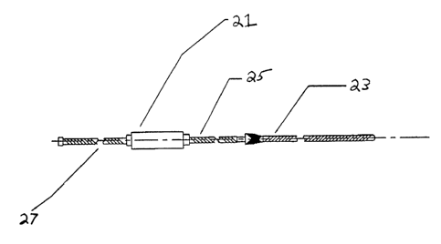
FIG. 2 shows a high-level diagrammatic cross-sectional side view of the probe
assembly of the invention according to a preferred embodiment, including an
antenna
that can be encased in a catheter 23, a connecting coaxial cable 25, an
interface 21, and a
third coaxial cable 27 connecting the probe to the surface coil port of a
magnetic
resonance scanner. The interface 21 includes, e.g., a balun and decoupling and
tuning/matching components. Further details of the interface 21 are shown in
FIG. 6 and
discussed below with reference thereto.
24
WO 00/62672 CA 02370222 2001-10-15 PCT/US00/10070
FIG. 3 shows a cross-sectional side view of one embodiment of the probe shaft
1.
The probe shaft can 1 comprise a conductor core 4 sandwiched concentrically in
layers
of insulation/dielectric, conductive shielding, and dielectric/insulation,
respectively. The
preferred embodiment is of the "loopless" antenna type wherein such probe
shaft 1 has
its top layers of insulation and shielding removed at a distal end 33 and its
central
conductor core 4 exposed. The exposed central conductor 4 is then insulated
with an
ultra-thin layer of insulation and may be fabricated from gold-plated Nitinol.
The
central conductor 4 then acts as an imaging pole or imaging coil of the
antenna. The
central conductor 4 is optimized by varying its length according to the
wavelength and
to frequency of the signals of interest. The length of the central conductor 4
is
approximately .25 times this wavelength, but this relationship is more
accurately
described by a complex function of several parameters, including dielectric
constant,
wavelength and frequency as described in U.S. Patent Application serial number
to be
assigned entitled "Magnetic Resonance Imaging Antenna and Guidewire" to Albert
C.
Lardo et al. Filed March 24, 2000. The central conductor 4 may be coiled or
may be
straight, depending on the application. A connector 31 (such as a BNC
connector) can
be attached to the proximal end of the coaxial cable for connection to a
preamplifier.
The function of the loopless antenna can be described as follows. The coil and
2o the shielding of the coaxial cable form a closed loop and RD signals
generated are
collected by the loop. These signals are transmitted to the scanner via the
coaxial cable.
For optimal performance, the input impedance of the coils should be matched
with the
characteristic impedance of the coaxial cable. The noise resistance of the
coil is
approximately 20-100 ohms.
FIG. 4 shows a cross-sectional side view illustrating details of one
embodiment
of the coil loopless assembly of the invention prior to its attachment to the
coax cable.
The assembly includes the wire 2 coiled around the mandrel 3 in the
configuration and
dimensions as shown.
25
WO 00/62672 CA 02370222 2001-10-15 PCT/US00/10070
FIG. 5 shows the typical shape and dimensions of a nasogastic tube 51 used in
one embodiment of the invention, as discussed above in detail.
FIG. 6 shows details of the interface circuit 21 for an antenna circuit of the
invention. The interface circuit 21 comprises a balun circuit, a decoupling
circuit, and a
tuning/matching circuit. The balun circuit is provided for preventing
unbalanced current
from being induced in the scanner, and includes a rigid coaxial cable inductor
coil 63
and a capacitor 62 connecting the ground to a housing 69. The decoupling
circuit is
provided for selecting the antenna of the invention for MRI detection and for
limiting
to current flow in the antenna when magnetic resonance is not being detected,
and includes
a decoupling capacitor 64 and a diode 66. A DC current signal generated by the
scanner
activates the diode, which in turn grounds the DC current, thereby preventing
current
flow in the coil. However, for normal magnetic resonance detection, the diode
is not
active and the circuit functions normally. The tuning/matching circuit is
provided for
matching the impedance of the antenna to the cable and preamplifier input, and
includes
a tuning/matching capacitor 67 and an inductor 65 in parallel to match the
output
impedance of the circuit to approximately 50 ohms, which is the input
impedance
typically required by the preamplifier of a scanner for optimum performance.
The
specific values of the capacitors 61, 62, 64 and the inductor 65 may be
determined in
2o accordance with the specific application by those skilled in the art. A
copper plate 610 is
provided for isolating the balun from the tuning and matching circuits. A
ground-
isolated connector 61 and a connector 68 (which can be BNC connectors) can be
provided for connecting the interface to coaxial cables 25 and 27 shown in
FIG. 2.
The coil antenna of the invention is typically connected to the surface coil
or
auxiliary coil port of the MRI scanner, and can be used in conjunction with
other
external receiver coils, e.g., surface coils, etc. In use, MRI signals are
excited by the
scanner's transmitter coil. The decoupling circuit in FIG 6 substantially
eliminates
induced current in the coil of the invention during excitation. In embodiments
wherein
3o the balun, tuning/matching and decoupling circuit are proximally located on
the probe,
these elements preferably remain outside the body during the examination. The
signals
26
WO 00/62672 CA 02370222 2001-10-15 pCT/IJS00/10070
received by the coil are then transmitted through interface circuitry to a
magnetic
resonance scanner or other magnetic resonance signal processing device. For
example,
the signals received by the coil may be transmitted to a GE Signa scanner via
that
scanner's surface coil or auxiliary coil port.
FIG. 7 shows a schematic side view illustrating an embodiment of the invention
employing a loop antenna imaging coil. This design also provides a reference
point 71
where the type of shielding can change a coaxial cable 73 a triaxial balun 75
and a
meeting point 77 between the coaxial cable 73 and the triaxial balun 77.
The elongated loop 13 shown in FIG 7 can comprise two parallel wires shortened
at one end. The wires can be made flexible while keeping the separation of the
wires
constant. Two or more capacitors can be used for tuning and matching as is
known in
the art. The number of capacitors increases the quality factor (Q) (increases
and
therefore the signal-to-noise ratio performance of the coil). A shunt diode of
the coaxial
cable 73 can be used as a decoupling circuit. The circuit eliminates the
induced currents
on the wire which results in a uniform flip angle and RF excitation of the
object of
interest. A balun circuit can be used to reduce/eliminate the unbalanced
currents on the
shield or on the coil. This can result in an increase in signal-to-noise ratio
performance
of the coil and can also decrease the risk of excessive heating.
FIG. 9 shows one embodiment of a probe designed for use in the rectum. The
probe design is similar to other probe designs herein disclosed. The antenna
13 here
depicted is a flexible circuit antenna such as the one depicted in FIG. 10.
The probe also
comprises a coaxial cable 95 and interior shielding 93 and a balun shielding
91. The
probe is elliptical in shape having a width 97 greater than its height 99.
FIG. 10 shows one embodiment of a flex circuit that can comprise the antenna
13
in the invention. Here the loop 1013 is etched onto a circuit board 1015 or
similar
substance. The circuit board 1015 can comprise holes 1009 for flexibility and
can
further incorporate circuitry such as parallel capacitors 1002, series
capacitors 1001, and
27
W~ 00/62672 CA 02370222 2001-10-15 PCT/US00/10070
a diode 1005. The flex circuit is also likely to have a connective structure
1007 enabling
connection to a coaxial cable (such as 95 in Figure 9) or another type of
connection as is
known to the art.
FIG. 11 shows a further embodiment of the probe of the instant invention
designed for use in the urethra. This probe comprises a braiding or balun
sleeve 1101 as
shielding.
Although certain embodiments of these systems and methods are disclosed
herein, it should be understood that other embodiments are envisioned as would
be
understood by one of ordinary skill in the art. Although the invention has
been
described by reference to specific embodiments thereof, it is not intended
that the
invention be limited to those illustrative embodiments. Rather, it is intended
that all
variations and modifications as fall within the spirit of the invention be
included within
the scope of the following claims. Accordingly, no limitation of the invention
is
intended by the foregoing description and accompanying drawings, except as is
set forth
in the appended claims.
28