Note: Descriptions are shown in the official language in which they were submitted.
CA 02371025 2001-10-18
WO 00/69355 PCTIUSOO/12936
BIOENGINEERED ANTERIOR CRUCIATE LIGAMENT
Background of the Invention
Every year more than 135,000 Americans tear or
rupture their anterior cruciate ligament (ACL) (Chen et
al., J. Biomed. Mat. Res. 14: 567-586 (1980); Butler, D.
L., J. Orthop. Res. 7: 910-921 (1989); Langer et al.,
Science 260: 920-926 (1993)). The ACL serves as a
primary stabilizer of anterior tibial translation and as
a secondary stabilizer of valgus-varus knee angulation,
and is often susceptible to rupture or tear resulting
from a flexion-rotation-valgus force associated with
sports injuries and traffic accidents. Ruptures or tears
often result in severe limitations in mobility, pain and
discomfort, and the loss of an ability to participate in
sports and exercise. Failures of the ACL are classified
in three categories: (1) ligamentous (ligament fibers
pull apart due to tensile stress), (2) failure at the
bone-ligament interface without bone fracture, and (3)
failure at the bone-ligament interface with bone fracture
at the attachment site of bone and ligament. The most
common type of ACL failure is the first category,
ligamentous.
Total surgical replacement and reconstruction are
required when injury to the ACL involves significant tear
or rupture. Four options have been utilized for repair
or replacement of a damaged ACL: (1) autografts, (2)
allografts, (3) xenografts, and (4) synthetic prostheses
(degradable and non-degradable). To date, no surgical
repair procedure has been shown to restore knee function
completely, and novel treatment options would likely
benefit a large number of patients.
The problems associated with the use of synthetic
ACL replacements, along with the limited availability of
the donor tissue, have motivated research towards the
development of functional and biocompatible equivalents
of native tissues. This shift from synthetic to
biologically-based ACL replacements first applied in
early studies in which collagenous ACL prostheses were
CA 02371025 2001-10-18
WO 00/69355 PCT/US00/12936
-2-
prepared as composite structures consisting of
reconstituted type I collagen fibers in a collagen I
matrix with polymethylmethacrylate bone fixation plugs,
and used as anterior cruciate ligament replacement
tissues in rabbits (Dunn et al., Am. J. Sports Medicine
20: 507-515 (1992)). Subsequent studies incorporated
active biological components into the process, such as
ligament fibroblasts seeded on cross-linked collagen
fiber scaffolds that were used as ligament analogs (Dunn
et al., J. Biomedical Materials Res. 29: 1363-1371
(1995); Dunn, M. G., Materials Res. Soc. Bulletin, Nov:
43-46 (1996)), and suggested that structures
approximating native ligaments can be generated. A
tendon gap model, based on pre-stressed collagen sutures
seeded with mesenchymal stem cells provided improved
repair of large tendon defects (Young et al., 1998).
Goulet et al. modified the collagen-fibroblast system by
using ligament fibroblasts in non-cross-linked collagen,
with bone anchors to pre-stress the tissue and facilitate
surgical implantation (Goulet et al., Tendons and
Ligaments. In Principles of Tissue Engineering, Ed. R.
Lanza, R. Langer, W. Chick. R. G. Landes Co. pp 633-643,
R. G. Lanz Co. and Academic Press, Inc., San Diego, CA
(1997)). Passive tension produced by growing the new
ligament in a vertical position induced fibroblast
elongation and the alignment of the cells and surrounding
extracellular matrix.
However, to date, no human clinical trials have been
reported with tissue culture bioengineered anterior
cruciate ligaments. This is due to the fact that each
approach has failed to address one or more of the
following issues: (1) the lack of a readily available
cell or tissue source, (2) the unique structure (e.g.
crimp pattern, peripheral helical pattern and isometric
fiber organization) of an ACL, and (3) the necessary
remodeling time in vivo for progenitor cells to
differentiate and/or autologous cells to infiltrate the
graft, thus extending the time a patient must incur a
CA 02371025 2001-10-18
WO 00/69355 PCTIUSOO/12936
-3-
destabilized knee and rehabilitation. The development of
methods for generating more fully functional
bioengineered anterior cruciate ligaments would greatly
benefit the specific field of knee reconstructive
surgery, and would also provide wider benefits to the
overall field of in vitro tissue generation and
replacement surgery.
Summary of the Invention
The present invention provides a method for
producing an anterior cruciate ligament ex vivo. The
method comprises seeding pluripotent stem cells in a
three dimensional matrix, anchoring the seeded matrix by
attachment to two anchors, and culturing the cells within
the matrix under conditions appropriate for cell growth
and regeneration, while subjecting the matrix to one or
more mechanical forces via movement of one or both of the
attached anchors. In a preferred embodiment, the
pluripotent cells are bone marrow stromal cells.
Suitable matrix materials are materials to which cells
can adhere. A preferred matrix material is collagen type
I gel. Suitable anchor materials are materials to which
the matrix can attach. Preferred anchor material
includes Goinopra coral which has been treated to convert
the calcium carbonate to calcium phosphate, and also
demineralized bone. In a preferred embodiment, the
mechanical forces to which the matrix is subjected mimic
mechanical stimuli experienced by an anterior cruciate
ligament in vivo. This is accomplished by delivering the
appropriate combination of tension, compression, torsion,
and sheer, to the matrix.
Another aspect of the present invention is the
bioengineered ligament which is produced by the above
method. The ligament is characterized by a cellular
orientation and/or matrix crimp pattern in the direction
of the applied mechanical forces, and also by the
production of collagen type I, collagen type III, and
fibronectin proteins along the axis of mechanical load
CA 02371025 2001-10-18
WO 00/69355 PCT/US00/12936
-4-
produced by the mechanical forces. In a preferred
embodiment, the ligament is characterized by the presence
of fiber bundles which are arranged into a helical
organization.
Another aspect of the present invention is a method
for producing a wide range of ligament types ex vivo
using an adaptation of the method for producing an
anterior cruciate ligament by adapting the anchor size to
reflect the size of the specific type of ligament to be
produced, and also adapting the specific combination of
forces applied, to mimic the mechanical stimuli
experienced in vivo by the specific type of ligament to
be produced. Similar adaptations of the method can be
made to produce other tissues ex vivo from pluripotent
stem cells, by adapting the mechanical forces applied
during cell culture to mimic stresses experienced in vivo
by the specific tissue type to be produced. The methods
of the present invention can be further modified to
incorporate other stimuli experienced in vivo by the
particular developing tissue, some examples of the
stimuli being chemical stimuli, and electro-magnetic
stimuli.
Another aspect of the present invention relates to
the specific tissues which are produced by the methods of
the present invention. Some examples of tissue which can
be produced include other ligaments in the body (hand,
wrist, elbow, knee), cartilage, bone, tendon, muscle, and
blood vessels.
Brief Description of the Figures
Figure 1 is a picture of bioreactor tubes containing
growing ligaments.
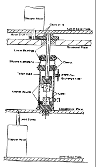
Figure 2 is a schematic of the mechanical apparatus
with an attached bioreactor tube.
Figure 3 is a picture of an actual mechanical
apparatus with attached reactor tubes containing growing
ligaments.
CA 02371025 2001-10-18
WO 00/69355 PCTIUSOO/12936
-5-
Figure 4 is a diagrammatic representation of the
rotational strain rates and the translational strain
rates experienced by the growing ligaments in Experiment
2.
Figure 5 contains diagrammatic representations of
the mechanical regime applied to the growing ligaments in
Experiment 3. a) Indicates the rotational and
translational strain rates as a % of strain over time
(min). b) Indicates rotational and linear deformation
experienced over the course of time.
Figure 6 graphically illustrates the cell density
data obtained from Experiment 3. a) Legend; b) Schematic
diagram representing the cross sectional areas from which
cell density measurements were taken; c) Data graphically
representing the average of 8 individual cell density
measurements taken from peripheral and central regions of
a cross sectional area of a mechanically stimulated and a
statically grown ligament.
Detailed Description of the Invention
The present invention is based on the finding that
the histomorphological properties of an in vitro produced
bioengineered tissue generated from pluripotent cells
within a matrix are affected by the direct application of
mechanical force to the matrix during tissue generation.
This discovery provides important new insights into the
relationship between mechanical stress, biochemical and
cell immobilization methods, and cell differentiation,
and has applications in producing a wide variety of
tissues in vitro from pluripotent cells.
One aspect of the present invention relates to a
method for producing an anterior cruciate ligament (ACL)
ex vivo. Cells capable of differentiating into ligament
cells are grown under conditions which simulate the
movements and forces experienced by an ACL in vivo
through the course of embryonic development into mature
ligament function. This is accomplished by the following
steps: Under sterile conditions, pluripotent cells are
CA 02371025 2001-10-18
WO 00/69355 PCT/US00/12936
-6-
seeded within a three dimensional matrix, of cylindrical
shape, which is comprised of a material to which the
cells can adhere (e.g. collagen gel). The faces of the
matrix cylinder are each attached to respective anchors,
through which a range of forces are to be applied to the
matrix. To facilitate force delivery to the matrix, it
is preferable that the entire surface of each respective
face of the matrix contact the face of the respective
anchors. Anchors with a shape which reflects the site of
attachment (e.g. cylindrical) are best suited for use in
this method. Once assembled, the cells in the anchored
matrix are cultured under conditions appropriate for cell
growth and regeneration. The matrix is subjected to one
or more mechanical forces applied through the attached
anchors (e.g. via movement of one or both of the attached
anchors) during the course of culture.
In the experiments described in the Exemplification
section below, the applied mechanical stimulation was
shown to dramatically influence the morphology, and
cellular organization of the progenitor cells within the
resulting tissue. The extracellular matrix components
secreted by the cells and organization of the extra
cellular matrix throughout the tissue was also
significantly influenced by the forces applied to the
matrix during tissue generation. During in vitro tissue
generation the cells and extra cellular matrix aligned
along the axis of load, reflecting the in vivo
organization of a native ACL which is also along the
various load axes produced from natural knee joint
movement and function. These results suggest that the
physical stimuli experienced in nature by cells of
developing tissue, such as the ACL, play a significant
role in progenitor cell differentiation and tissue
formation. They further indicate that this role can be
effectively duplicated in vitro by mechanical
manipulation to produce a similar tissue. The more
closely the forces produced by mechanical manipulation
resemble the forces experienced by an ACL in vivo, the
CA 02371025 2001-10-18
WO 00/69355 PCT/US00/12936
-7-
more closely the resultant tissue will resemble a native
ACL.
One or more types of pluripotent cells are used in
the method. Such cells have the ability to differentiate
into a wide variety of cell types in response to the
proper differentiation signals. More specifically, the
method requires cells which have the ability to
differentiate into cells of ligament tissue. In a
preferred embodiment, bone marrow stromal cells, also
known as mesenchymal cells are used. If the resulting
bioengineered ligament is to be transplanted into a
patient, the cells should be derived from a source which
is compatible with the intended recipient. The recipient
will generally be a human, although applications in
veterinary medicine also exist. In one embodiment, the
cells are obtained from the recipient, although
compatible donor cells may also be used. The
determination of compatibility is within the means of the
skilled practitioner.
The three dimensional matrix used in the method is
potentially comprised of any material to which the cells
can adhere. This matrix serves as a preliminary matrix,
which is supplemented and possibly even replaced by
extracellular matrix components produced by the
differentiating cells. Use of a more specialized matrix
may enhance or accelerate the development of the ACL.
For instance, a matrix which has specific mechanical
properties (e.g. increased tensile strength) can
withstand strong forces prior to reinforcement from
cellular extracellular matrix components. Other
properties which may also be useful in a preliminary
matrix include, without limitation, biocompatibility and
susceptibility to biodegradation.
The matrix used in the examples disclosed herein was
a collagen gel. One of skill in the art will recognize
that the properties of the preliminary matrix can be
modulated and enhanced by modifying the matrix
components, and that use of an enhanced matrix is likely
CA 02371025 2001-10-18
WO 00/69355 PCTIUSOO/12936
-8-
to increase the efficiency of production of a
bioengineered ACL. Such modifications include, without
limitation, modifications aimed at modulating the
mechanical and mass transport properties of the matrix.
In particular, the concentration of collagen and the
degree of crosslinking of collagen in the matrix can
significantly influence the mechanical properties of the
matrix, as well as the diffusional transport rates of
nutrients and large molecules. Since the ACL is made
primarily of collagen type I, it is particularly well
suited for use as a preliminary matrix component. The
concentration of collagen type I in the matrix should be
sufficient to support cell adhesion, proliferation and
differentiation. In one embodiment, collagen type I is
used at a final concentration from about 2 mg/ml to about
6 mg/ml. In another embodiment the final concentration of
collagen type I in the matrix is about 2 mg/ml. In
another embodiment, the collagen in the preliminary
matrix is crosslinked. Suitable processes for cross
linking collagen include without limitation,
dehydrothermal crosslinking and ultraviolet irradiation
crosslinking. Other suitable matrix materials include,
without limitation polysaccharides, alginates, other
proteins such as silk and elastin, synthetic polymers
such as polyglycolic acid and polylactic acid and
copolymers of the two, and demineralized bone.
The cells are seeded within the preliminary matrix
either pre - or post-matrix formation, depending upon the
particular matrix used and the method of matrix
formation. Uniform seeding is preferable. In theory,
the number of cells seeded does not limit the final
ligament produced, however optimal seeding may increase
the rate of generation. Optimal seeding amounts will
depend on the specific culture conditions. In one
embodiment, the matrix is seeded with from about 0.05 to
5 times the physiological cell density of a native
ligament.
CA 02371025 2001-10-18
WO 00/69355 PCT/US00/12936
-9-
The seeded matrix is subjected to mechanical forces
which are applied through a set of attached anchors.
Anchors are defined herein as comprising a solid surface
to which force can be applied and transmitted to an
attached matrix. The anchors must be made of a material
suitable for matrix attachment, and the resulting
attachment should be strong enough to endure the stress
of the mechanical forces applied. The preliminary matrix
must be able to attach to the anchors. In addition, it
is preferable that the anchors be of a material which is
suitable for the attachment of extracellular matrix which
is produced by the differentiating cells. Some examples
of suitable anchor material include, without limitation,
Goinopra coral and demineralized bone. In a preferred
embodiment, the anchors are Goinopra coral which has a
pore size of 500 AM, and the coral is treated by means to
convert the calcium carbonate of the coral to calcium
phosphate, prior to use.
Alternatively, anchor material may be created or
further enhanced by infusing a selected material with a
factor which promotes matrix binding. The term infuse is
considered to include any method of application which
appropriately distributes the factor onto the anchor
(e.g. coating, permeating, contacting). Examples of such
factors include without limitation, laminin, fibronectin,
any extracellular matrix protein that promotes adhesion,
silk, factors which contain arginine-glycine-aspartate
peptide binding regions. Growth factors or bone
morphogenic protein can also be used to enhance anchor
attachment. In addition, anchors may be pre-seeded with
cells (e.g. stem cells, ligament cells, osteoblasts).
which adhere to the anchors and bind the matrix, to
produce enhanced matrix attachment.
The matrix is attached to the anchors via contact to
the anchor face or alternatively by actual penetration of
the matrix material through the anchor material. Because
the force applied dictates the final ligament produced
and the force is applied through the anchors, the size of
CA 02371025 2001-10-18
WO 00/69355 PCT/US00/12936
-10-
the final ligament produced is in part dictated by the
size of the attachment site of the anchor. One of skill
in the art will appreciate that an anchor of appropriate
size to the desired final ligament should be used. A
preferred anchor shape for the formation of an ACL is a
cylinder, however, one of skill in the art will
appreciate that other anchor shapes and sizes will also
function adequately. In a preferred embodiment, anchors
have an appropriate size and composition for direct
insertion into bone tunnels in the femur and tibia of a
recipient.
The cells are cultured within the matrix under
conditions appropriate for cell growth and
differentiation. During the culture process, the matrix
is subjected to one or more mechanical forces via
movement of one or both of the attached anchors. The
mechanical forces of tension, compression, torsion and
shear, and combinations thereof, are applied in the
appropriate combinations, magnitudes, and frequencies to
mimic the mechanical stimuli experienced by an ACL in
vivo.
Various factors will influence the amount of force
which can be tolerated by the matrix (e.g. matrix
composition, cell density). Matrix strength is expected
to change through the course of tissue development.
Therefore, mechanical forces applied will increase or
decrease in magnitude, duration, and variety over the
period of ligament generation, to appropriately
correspond to matrix strength at the time of application.
The more accurate the intensity and combination of
stimuli applied to the matrix during tissue development,
the more the resulting ligament will resemble a native
ACL. Two issues must be considered regarding the natural
function of the ACL when devising the in vitro mechanical
force regimen that closely mimics the in vivo
environment, (1) the different types of motion
experienced by the ACL and the responses of the ACL to
knee joint movements and (2) the extent of the mechanical
CA 02371025 2001-10-18
WO 00/69355 PCTIUSOO/12936
-11-
stresses experienced by the ligament. Specific
combinations of mechanical stimuli are generated from the
natural motions of the knee structure and transmitted to
the native ACL. To briefly describe the motions of the
knee, the connection of the tibia and femur by the ACL
between provides six degrees of freedom when considering
the motions of the two bones relative to each other: the
tibia can move in the three directions and can rotate
relative to the axes for each of these three directions.
The knee is restricted from achieving the full ranges of
these six degrees of freedom due to the presence of
ligaments and capular fibers and the knee surfaces
themselves (Eiden et al., Experimental methods used to
evaluate knee ligament function. In Knee Ligaments:
Structure, Function, Injury and Repair, Ed. D. Daniel et
al. Raven Press, pp.135-151 (1990)). Small translational
movements are also possible. The attachment sites of the
ACL are responsible for its stabilizing roles in the knee
joint. The ACL functions as a primary stabilizer of
anterior-tibial translation, and as a secondary
stabilizer of valgus-varus angulation, and tibial
rotation (Shoemaker et al., The limits of knee motion. In
Knee Ligaments: Structure, Function, Injury and Repair,
Ed. D. Daniel et al. Raven Press, pp.1534-161 (1990)).
Therefore, the ACL is responsible for stabilizing the
knee in three of the six possible degrees of freedom. As
a result, the ACL has developed a specific fiber
organization and overall structure to perform these
stabilizing functions. The present invention simulates
these conditions in vitro to produce a tissue with
similar structure and fiber organization.
The extent of mechanical stresses experienced by the
ACL can be similarly summarized. The ACL undergoes
cyclic loads of about 300 N between one and two million
cycles per year. It is also critical to consider linear
stiffness (-182 N/mm), ultimate deformation (100% of ACL)
and energy absorbed at failure (12.8 N-m) (Woo et al.,
The tensile properties of human anterior cruciate
CA 02371025 2001-10-18
WO 00/69355 PCT/US00/12936
-12-
ligament (ACL) and ACL graft tissues. In Knee Ligaments:
Structure, Function, Injury and Repair, Ed. D. Daniel et
al. Raven Press, pp.279-289 (1990)) when developing an
ACL surgical replacement.
The Exemplification section below details the
production of a prototype bioengineered anterior cruciate
ligament (ACL) ex vivo. Mechanical forces mimicking a
subset of the mechanical stimuli experienced by a native
ACL in vivo (rotational deformation and linear
deformation) were applied in combination, and the
resulting ligament which was formed was studied to
determine the effects of the applied forces on tissue
development. Exposure of the developing ligament to
physiological loading during in vitro formation induced
the cells to adopt a defined orientation along the axes
of load, and to generate extracellular matrices along the
axes as well. These results indicate that the
incorporation of additional mechanical forces into the
regime to produce a more complex network of load axes
that more accurately mimics the environment of the native
ACL, will produce a bioengineered ligament which more
closely resembles a native ACL. The different mechanical
forces to be applied include, without limitation,
tension, compression, torsion, and shear. These forces
are applied in combinations which simulate forces
experienced by an ACL in the course of natural knee joint
movements and function. These movements include, without
limitation, knee joint extension and flexion as defined
in the coronal and sagittal planes, and knee joint
flexion. Optimally, the combination of forces applied
mimics the mechanical stimuli experienced by an anterior
cruciate ligament in vivo as accurately as is
experimentally possible. Varying the specific regimen of
force application through the course of ligament
generation is expected to influence the rate and outcome
of tissue development, with optimal conditions to be
determined empirically. Potential variables in the
regimen include, without limitation: (1) strain rate,
CA 02371025 2001-10-18
WO 00/69355 PCTIUSOO/12936
-13-
(2) repetition number, (3) duration at extreme points of
ligament deformation, (4) force levels, and (5) different
force combinations. It will be recognized by one of
skill in the art that a potentially unlimited number of
variations exist. In a preferred embodiment the regimen
of mechanical forces applied produces helically organized
fibers similar to those of the native ligament, described
below.
The fiber bundles of a native ligament are arranged
into a helical organization. The mode of attachment and
the need for the knee joint to rotate -140 of flexion
has resulted in the native ACL inheriting a 900 twist and
with the peripheral fiber bundles developing a helical
organization. This unique biomechanical feature allows
the ACL to sustain extremely high loading. In the
functional ACL, this helical organization of fibers
allows anterior-posterior and posterior-anterior fibers
to remain relatively isometric in respect to one another
for all degrees of flexion, thus stabilizing the knee
throughout all ranges of joint motion. In a preferred
embodiment of the invention, mechanical forces which
simulate a combination of knee joint flexion and knee
joint extension are applied to the developing ligament to
produce an engineered ACL which possesses this same
helical organization. The mechanical apparatus used in
the experiments presented in the Exemplification below
provides control over strain and strain rates (both
translational and rotational). An improved mechanical
apparatus will monitor the actual load experienced by the
growing ligaments, serving to `teach' the ligaments over
time through monitoring and increasing the loading
regimes. Such a reactor can be designed by starting from
the features of the first generation bioreactor used in
the Experiments described in the Exemplification section
below. To these features (e.g., ports for medium and gas
exchange, sterilizable) will be added features, including
e.g. a flexibility to run multiple mechanical deformation
CA 02371025 2001-10-18
WO 00/69355 PCT/US00/12936
-14-
programs concurrently. Such a system should have a
precise (strain-gauge) control of the applied forces, and
an on-line monitoring and control of mechanical loading
parameters.
Another aspect of the present invention relates to
the bioengineered anterior cruciate ligament produced by
the above described methods. The bioengineered ligament
produced by these methods is characterized by cellular
orientation and/or a matrix crimp pattern in the
direction of the mechanical forces applied during
generation. The ligament is also characterized by the
production/presence of extra cellular matrix components
(e.g. collagen type I, and type III, elastin, and
fibronectin proteins) along the axis of mechanical load
experienced during culture. In a preferred embodiment,
the ligament fiber bundles are arranged into a helical
organization, as discussed above.
The above methods are not limited to the production
of an ACL, but can also be used to produce other
ligaments found in the knee (e.g. posterior cruciate
ligament) or other parts of the body (e.g. hand, wrist,
ankle, elbow and shoulder). All moveable joints in a
human body have specialized ligaments which connect the
articular extremities of the bones in the joint. Each
ligament in the body has a specific structure and
organization which is dictated by its function and
environment. The various ligaments of the body, their
locations and functions are listed in Anatomy,
Descriptive and Surgical (Gray, H. , Eds. Pick, T. P.,
Howden, R., Bounty Books, New York (1977)) the pertinent
contents of which are incorporated herein by reference.
By determining the physical stimuli experienced by a
given ligament, and incorporating forces which mimic
these stimuli, the above described method for producing
an ACL ex vivo can be adapted to produce a bioengineered
ligament ex vivo which simulates any ligament in the
body.
CA 02371025 2001-10-18
WO 00/69355 PCT/USOO/12936
-15-
The specific type of ligament to be produced is
predetermined prior to ligament generation since several
aspects of the method vary with the specific conditions
experienced in vivo by the native ligament. The
mechanical forces to which the developing ligament is
subjected during cell culture are determined for the
particular ligament type being cultivated. The specific
conditions can be determined by those skilled in the art
by studying the native ligament and its environment and
function. One or more mechanical forces experienced by
the ligament in vivo are applied to the matrix during
culture of the cells in the matrix. The skilled
practitioner will recognize that a ligament which is
superior to those currently available can be produced by
the application of a subset of forces experienced by the
native ligament. However, optimally, the full range of
in vivo forces will be applied to the matrix in the
appropriate magnitudes and combinations to produce a
final product which most closely resembles the native
ligament. These forces include, without limitation, the
forces described above for the production of an ACL.
Because the mechanical forces applied vary with ligament
type, and the final size of the ligament will be
influenced by the anchors used, optimal anchor
composition, size and matrix attachment sites are to be
determined for each type of ligament by the skilled
practitioner.
Another aspect of the present invention relates to
the production of other tissue types ex vivo using
methods similar to those described above for the
generation of ligaments ex vivo. The above described
methods can also be applied to produce a range of
engineered tissue products which involve mechanical..
deformation as a major part of their function, such as
tendon, muscle (e.g. smooth muscle, skeletal muscle,
cardiac muscle), bone, cartilage, vertebral discs, and
some types of blood vessels. Bone marrow stomal cells
possess the ability to differentiate into these as well
CA 02371025 2001-10-18
WO 00/69355 PCT/US00/12936
-16-
as other tissues. The results present in the
Exemplification section below indicate that growth in an
environment which mimics the specific mechanical
environment of a given tissue type will induce the
appropriate cell differentiation to produce a
bioengineered tissue which significantly resembles native
tissue. The ranges and types of mechanical deformation
of the matrix can be extended to produce a wide range of
tissue structural organization. Preferably, the cell
culture environment reflects the in vivo environment
experienced by the native tissue and the cells it
contains, throughout the course of embryonic development
to mature function of the cells within the native tissue,
as accurately as possible. Factors to consider when
designing specific culture conditions to produce a given
tissue include, without limitation, the matrix
composition, the method of cell immobilization, the
anchoring method, the specific forces applied, and the
cell culture medium. The specific regimen of mechanical
stimulation depends upon the tissue type to be produced,
and is established by varying the application of
mechanical forces (e.g. tension only, torsion only,
combination of tension and torsion, with and without
shear, etc.), the force amplitude (e.g. angle or
elongation), the frequency and duration of the
application, and the duration of the periods of
stimulation and rest.
The method for producing the specific tissue type ex
vivo is an adaptation of the above described method for
producing an ACL. Components involved include
pluripotent cells, a three dimensional matrix to which
cells can adhere, and a plurality of anchors which have a
face suitable for matrix attachment. The pluripotent
cells (preferably bone marrow stroma cells) are seeded in
the three dimensional matrix by means to uniformly
immobilize the cells within the matrix. As discussed
above, the only requirement for the matrix is that it be
of a substance, or contain a substance, to which the
CA 02371025 2001-10-18
WO 00/69355 PCT/US00/12936
-17-
cells can adhere, although certain matrix compositions
will most likely prove optimal for specific tissues.
Matrix shape is not viewed as a limiting factor to the
method, however, a specific shape which resembles the
final desired product may facilitate generation of the
tissue. The number of cells seeded is also not viewed as
limiting, however, seeding the matrix with a high density
of cells may accelerate tissue generation.
Once seeded, the matrix is attached to a plurality
of anchors. The number of anchors, as well as their
shape, and the shape and size of their sites of
attachment to the matrix, depends upon the particular
tissue being produced, and will reflect the nature of the
forces applied to the matrix. For some tissues (e.g.
cartilage, bone, vertebral discs), use of a solid matrix
(e.g. demineralized bone or Goinopra coral) will be
optimal. Because mechanical forces can be applied
directly to a solid matrix, solid matrices may be
considered herein to possess inherent anchors. If deemed
necessary, the location and size of these inherent
anchors is determined by the position and area of the
solid matrix to which the mechanical force is applied.
The specific forces applied are to be determined for
each tissue type produced through examination of native
tissue and the mechanical stimuli experienced in vivo. A
given tissue type experiences characteristic forces which
are dictated by location and function of the tissue
within the body. For instance, cartilage is known to
experience a combination of shear and compression/tension
in vivo, bone experiences compression. Determination of
the specific mechanical stimuli experienced in vivo by a
given tissue is within the means of one of skill in the
art.
Additional stimuli (e.g. chemical stimuli, electro-
magnetic stimuli) can also be incorporated into the above
described methods for producing bioengineered ligaments
and other tissues. Cell differentiation is known to be
influenced by chemical stimuli from the environment,
CA 02371025 2001-10-18
WO 00/69355 PCTIUSOO/12936
-18-
often produced by surrounding cells, such as secreted
factors, cell-cell contact, chemical gradients, and
specific pH levels, to name a few. Other more unique
stimuli are experienced by more specialized types of
tissues (e.g. the electrical stimulation of cardiac
muscle). The application of such tissue specific stimuli
in concert with the appropriate mechanical forces is
expected to facilitate differentiation of the cells into
a tissue which more closely approximates the specific
natural tissue.
Tissues produced by the above described methods
provide an unlimited pool of tissue equivalents for
surgical implantation into a compatible recipient.
Engineered tissues may also be utilized for in vitro
studies of normal or pathological tissue function, e.g.
for in vitro testing of cell- and tissue-level responses
to molecular, mechanical, or genetic manipulations. For
example, tissues based on normal or transfected cells can
be used to assess tissue responses to biochemical or
mechanical stimuli, identify the functions of specific
genes or gene products that can be either over-expressed
or knocked-out, or to study the effects of
pharmacological agents. Such studies will likely provide
more insight into ligament development, normal and
pathological function, and eventually lead toward fully
functional tissue engineered replacements, based in part
on already established tissue engineering approaches, new
insights into cell differentiation and tissue
development, and the use of mechanical regulatory signals
in conjunction with cell-derived and exogenous
biochemical factors to improve structural and functional
tissue properties.
The production of engineered tissues such as
ligaments has the potential for applications such as
harvesting bone marrow stoma cells from individuals at
high risk for tissue injury (e.g. ACL rupture) prior to
injury. These cells could be either stored until needed
or seeded into the appropriate matrix and cultured and
CA 02371025 2001-10-18
WO 00/69355 PCTIUSOO/12936
-19-
differentiated in vitro under mechanical stimuli to
produce a variety of bioengineered prosthetic tissues to
be held in reserve until needed by the donor. The use of
bioengineered living tissue prosthetics that better match
the biological environment in vivo, provide the required
physiological loading to sustain for example, the dynamic
equilibrium of a normal, fully functional ligament,
should reduce rehabilitation time for a recipient of a
prosthesis from months to weeks, particularly if the
tissue is pre-grown and stored. Benefits include a more
rapid regain of functional activity, shorter hospital
stays, and fewer problems with tissue rejections and
failures.
Exemplification
The feasibility of using directly applied forces
during tissue cultivation to promote in vitro formation
of ACL-like structures was tested. A three-dimensional
tissue culture system was developed utilizing precursor
cells, obtained from bone marrow stroma, immobilized in a
collagen gel matrix. The matrix was positioned within a
bioreactor that subjected the matrix to defined types,
magnitudes and frequencies of mechanical forces,
corresponding in part to those experienced by an ACL
during physiological loading in vivo. Cells within the
matrix were cultured under conditions appropriate for
proliferation during exposure of the matrix to the
various mechanical forces, to produce a bioengineered
anterior cruciate ligament ex vivo. Control tissues were
cultured with no mechanical stimulation of the matrix
under otherwise identical conditions.
A bioreactor that would provide a reasonable range
of mechanical options for deformation of the growing
ligaments was constructed. The reactor provided
tensile/compressive and torsional loads along the
longitudinal axis and could accommodate up to 12
individual reactor tubes for the growth of ligaments.
CA 02371025 2001-10-18
WO 00/69355 PCT/US00/12936
-20-
Although this reactor did not subject the growing
ligament to the full range of deformations experienced in
vivo, the information obtained from these preliminary
experiments can be used to design a more advanced
reactor.
The two major parts of the device included: (1) the
bioreactor tubes which provide the growth environment and
attachment sites for the growing ligaments (shown in
Figure 1), and (2) the mechanical apparatus to provide
mechanical loading regimes to the bioreactor tubes.
Figure 2 presents a schematic of the mechanical apparatus
and Figure 3 is a picture of the working device.
Experiment 1
The first experiment was performed to better
characterize adhesion of the collagen matrix to the coral
anchors. This experiment ran for a total of 12 days and
encompassed rotational deformation of 100 initially,
increasing to 65 by the end of the experiment. The
linear deformation was 0.5 mm along the longitudinal axis
of the ligament for the majority of the 12 days. No loss
of adhesion between the collagen matrix and the coral
anchors was observed, indicating that at least a 65
rotational deformation could be tolerated by present
system.
Experiment 2
The next set of experiments were conducted to study
the effects of increased mechanical stresses on the
growing ligaments. Linear deformation was kept constant
at 1 mm (double that of Experiment 1) throughout the 13
day experiment while rotational deformation was increased
progressively from 100 to 65 by the end of the
experiment. The time to progress from one rotational
extreme to the other extreme was maximally 30 minutes,
with rests at the extreme points varying from 0 to 3
hours over the course of the experiment. Thus, complete
cycles of mechanical deformation ranged from 0.33 hr to 4
CA 02371025 2001-10-18
WO 00/69355 PCTIUSOO/12936
-21-
hr. A detailed description of the mechanical processes
employed in this experiment is shown below to illustrate
the range of control over the bioreactor that we can
achieve with the apparatus (Table 1). Figure 4
summarizes the rotational and translational strain rates
used in this experiment.
Histology was performed on the resulting tissue to
examine cell morphology. Ligament tissue samples were
stained with hemotoxylin and eosin and visualized by
light microscopy at 400 X. Results indicated that
approximately 50% of the cells from the mechanically
stimulated ligaments exhibited ovid morphology and
alignment along the longitudinal axis of the ligament.
Immunohistochemistry was not performed.
CA 02371025 2001-10-18
WO 00/69355 -2111- PCT/US00/12936 Experiment 3
This set of studies was conducted to provide insight
into the influence of frequency and cycling on ligament
formation in the bioreactors (Figure 5 a&b). After a 48
hr rest period (represented as cycle 1), the rotational
and translation strain rates and linear and rotational
deformation were kept constant for 18.5 days. As
illustrated in Figure 5a, the ligaments were exposed to a
constant rotational and translational strain rate of 0.83
min-1 and 0.33 min-1, respectively, for 18.5 days.
Rotational deformation (0 degrees) and linear deformation
(Omm) were kept constant at 900 and 2 mm respectively.
Figure 5b shows the deformation pattern. Slopes of the
plotted lines indicate strain rate.
Following the culture period, ligament samples, both
the mechanically challenged as well as the controls
SUBSTITUTE SHEET (RULE 26)
CA 02371025 2001-10-18
WO 00/69355 PCTIUSOO/12936
-22-
rn
1
zs
m
0
U
N .+
E ~.E -~noo~noVno~no0ooo0
A ~I c~ (~ N N M M V V' kn to ~o 06 O\
U) a X
1J 4J
d d
V A N to 'n 'n ' - "n in sn *n
''"'( CDC) 00 O CDC) O O O
F U F q
co
U O
N -- N N N N N N N N O 00 N
-H (0 ai
ri -H
41
Q) -H o C a ,O
' +^+ M O r- en J) r( ca O LL i d M ~n ~p CO -. -. N N W) kr)
M
0 a C C N O O O O N M
4-) (0 A~"UC4
'CS TS
4) N C
c a~ i OoCD CD CD 0CD 0OCD OO0ooCD 0
a O a a
(0 S4
a c, c
E
O O O O O o 0 0 0 0 0 --"- N?
U) N X =O C4 r N
4) r-1 W a
.0 >4
(0 0 O O
O O kn O O O O O O O O O O O O
r='I G\ w 'y -+ ^~ N N en cn m M M M en m M Cl m M
(0 Q) a O X 0
U L1F~p'
-H 0
a o----------------
0
a) N
ra E
-H
N }-1
:tt [Yv)~or-mcx,
A a Qr4
(o X
H W
SUBSTITUTE SHEET (RULE 26)
CA 02371025 2001-10-18
WO 00/69355 PCT/US00/12936
-23-
(static) were characterized for: (1) general
morphological appearance (by visual inspection); (2) cell
distribution (image processing of histological sections);
(3) cell orientation (histological analysis); and (4)
tissue specific markers (immunostaining).
Mechanical stimulation markedly affected the
morphology of the engineered ACL, the distribution of
cells along the matrix, and the extracellular matrix
which was generated by the cells.
The mechanical stimulation markedly affected the
dimensions and overall appearance of the engineered
ligaments. As compared to static controls, mechanically
stimulated ligaments contracted laterally to a diameter
of 5.1 mm after 21 days in culture, as compared to 6.4 mm
diameter for static controls (n = 3 for each group) in
Experiment 3.
The mechanical stimulation also had a dramatic
effect on cell density in the engineered ACL. Cells
counts (N=8 fields, one ligament from each group,) were
taken from cross sections of the control and mechanically
challenged ligaments from Preliminary Experiment 3
(Figure 6). Cell density of the ligament was
approximately 3-fold higher in the center and -2-fold
higher in the periphery in the mechanically deformed
ligaments in comparison to the static controls. These
data indicate that mechanical stimulation provides
suitable signals to the BMSCs to promote proliferation in
the bioreactor environment.
The mechanical stimulation also had a dramatic
effect on cell orientation. Ligament tissue samples from
Experiment 3 were stained with hemotoxylin and eosin and
visualized by light microscopy at 400 X. Significant
alignment in the BMSCs from the mechanically stimulated
ligament was clearly seen, in comparison to the control
(static) sample. Furthermore, this alignment had a
lengthwise orientation along the longitudinal axis of the
bioreactor tube, thus in the direction of the applied
tension. The longitudinal orientation was similar to
CA 02371025 2001-10-18
WO 00/69355 PCT/US00/12936
-24-
ligament fibroblasts found within an ACL in vivo (Woods
et al. Amer. J. Sports Med. 19: 48-55 (1991)).
The mechanical stimulation also had a dramatic
effect on the development of tissue specific markers.
Collagen I accounts for -88% of total collagen in the
ACL. Collagen III accounts for -12% of total collagen
and fibronectin accounts for 2 pg/mg dry tissue weight of
an ACL (Amiel et al., Ligament structure, chemistry, and
physiology. In Knee Ligaments: Structure, Function,
Injury, and Repair. Eds. Daniel, D.; Akeson, W.; O'Connor.
J. Raven Press (1990)). Collagen I, collagen III and
fibronectin (as indicators of new ligament tissue
formation and organization) were identified by
immunostaining mechanically stimulated and control
(static) ligament tissue samples from Experiment 3.
Mechanically stimulated ligaments expressed ligament-
specific molecular markers (collagen III and
fibronectin), in contrast to static controls in which the
expression was either low or not detectable. The
diameter of the collagen I structures observed in the
mechanically challenged ligaments approached that of
similar structures seen in naturally formed ACL collagen
bundles, -20 m. The morphology of these markers
suggested the beginning of differentiation of BMSCs into
ligament cells and similar structural features to an ACL
in terms of fiber bundle orientation and diameter.
The above results indicate that the mechanical
apparatus and bioreactor system can provide a suitable
environment for in vitro formation of tissue engineered
ligaments starting from bone marrow stromal cells
immobilized in a collagen gel matrix.
The culture conditions used in these preliminary
experiments can be further expanded to more accurately
reflect the physiological environment of a ligament (e.g.
increasing the different types of mechanical forces) for
the in vitro creation of functional equivalents of native
ACL for potential clinical use. These methods are not
CA 02371025 2001-10-18
WO 00/69355 PCTIUSOO/12936
-25-
limited to the generation of a bioengineered ACL.
Indeed, by applying the appropriate magnitude and variety
of forces experienced in vivo, any type of ligament in
the body can be produced ex vivo by the methods of the
present invention.
The above results gathered from these controlled in
vitro studies of the roles of mechanical regulatory
signals on precursor cell differentiation into ligament
cells and in vitro development of an engineered ACL,
further the understanding of the roles of mechanical
regulatory signals in cell differentiation and tissue
development.
Methods of the Invention
Cell Isolation and Culture.
Bone Marrow Stromal Cells (BMSC), pluripotent cells
capable of differentiating into osteogenic, chondrogenic,
tendonogenic, adipogenic and myogenic lineages, were
chosen since the formation of the appropriate conditions
can direct their differentiation into the desired
ligament fibroblast cell line (Markolf et al., J. Bone
Joint Surg. 71A: 887-893 (1989); Caplan et al.,
Mesenchymal stem cells and tissue repair. In The
Anterior Cruciate Ligament: Current and Future Concepts,
Ed. D. W. Jackson et al., Raven Press, Ltd, New York
(1993); Young et al., J. Orthopaedic Res. 16: 406-413
(1998)). Bone marrow cultures were established from the
tibias and femurs of 2-3 week old bovine calves. The
contents of the bone marrow cavity were aseptically
harvested in a 50 ml centrifuge tube containing 15 ml
phosphate buffered solution (PBS) with 0.05 mM ethylene
diamine tetraacetic acid (EDTA). Single cell suspensions
were made by repeatedly passing the marrow through
needles of different gauges (16 to 20), and resuspended
in Dulbecco's Modified Eagle Medium (DMEM) supplemented
with 10% fetal bovine serum (FBS), 0.1 mM nonessential
amino acids (NEAA), 100 U/ml penicillin and 100 mg/L
CA 02371025 2001-10-18
WO 00/69355 PCT/US00/12936
-26-
streptomycin (P/S). White blood cells were counted using
a hemocytometer, plated in 100 mm Petri dishes at 2 x 10⁶
cells per dish (approximately 25 x 103 cells/cm2) in 10 ml
of medium supplemented with 1 ng/ml fibroblast growth
factor-2 (FGF-2) and cultured in a humidified 37 C/5% CO2
incubator.
BMSCs were selected pre-plating, based on their
ability to adhere to the Petri dish; non-adherent
hematopoietic cells were removed with the culture medium
during medium replacement. The medium was changed twice
per week. When BMSC became near confluent, after
approximately 2-3 weeks, upon which they were detached
using 0.25% trypsin/1 mM EDTA and replated in 100 mm
dishes at 3 x 10⁵ cells per dish. After 1 more week, when
dishes again became confluent, 1" passage (P1) cells were
trypsinized and 1 ml aliquots containing 20 x 106 cells
(based on Goulet et al., 1997) in 1X DMEM were either
seeded directly into the collagen gels or spun down and
frozen in 8% DMSO 10% FBS 1X DMEM solution for future
use.
The final medium in the 20 ml total volume of the
reactor vessel consisted of: 5.6 ml of 3.6X DMEM at pH
8.0, 3.7 ml heat inactivated FBS (30 min at 56 C), 9.5 ml
of 4.22 mg/ml collagen [acid soluble collagen type I
(Sigma type III)], 0.2 ml of 0.7 N NaOH and 1.0 ml of the
cell preparation containing the 20 X 106 cells. The 3.6X
DMEM consisted of 36 ml lOX DMEM containing 4500 mg
glucose/L, 0.4 mg powered folic acid, 2 ml of 200 mM L-
glutamine, 0.37 g sodium bicarbonate (NaHCO3), 200 U/ml
penicillin, 200 mg/L streptomycin, 0.5 g/ml Fungizone
(P/S and Fungizone were purchased from Life Technologies)
and ddH2O to bring the volume to 90 ml; the pH was
adjusted to 8.0 with 2N NaOH, and enough ddH2O to bring
the final volume to 100 ml. The 3.6X DMEM solution was
well mixed and filtered through a 0.2 m filter unit and
stored at 4 C.
CA 02371025 2001-10-18
WO 00/69355 PCT/US00/12936
-27-
Cell immobilization in Collagen Matrix.
To prepare an individual bioreactor tube for a
ligament growth experiment 20 x 106 P1 BMSCs were
resuspended in 1 ml 1X DMEM, 9.5 ml of 4.22 mg/ml bovine
collagen type I, 5.6 ml 3.6X DMEM, 3.7 ml heat
inactivated FBS, and 0.2 ml 0.7 N NaOH. The final
concentration of collagen type I in solution was 2 mg/ml.
These reagents were first added to a 50 ml centrifuge
tube on ice, then quickly transferred to the bioreactor
tube. The bioreactor tube was fitted with a PTFE gas
filter, loaded into the mechanical device, and placed in
a humidified 37 C/5% CO2 incubator. The collagen was
allowed to gel for 24 hours. During a 24 or 48 hr
initial growth period, the ligaments were not exposed to
any mechanical stimulation except for gravity to allow
for sufficient adhesion to develop between the collagen
matrix and the coral anchors. Fifty percent of the
medium was exchanged with 10% FBS in 1X DMEM containing
200 U/ml penicillin, 200 mg/L streptomycin, 0.5 pg/ml
Fungizone, after 24 hrs and two times a week thereafter.
Anchors for Ligament Matrix.
Cylindrical pieces of Goinopra coral, 12 mm in
diameter and 20 mm in length with a pore size of 500 m
(supplied by Interpore-Cross International) were used as
the anchors. The coral was treated by a hydrothermal
process to convert the calcium carbonate to calcium
phosphate (hydroxyapatite). This mineral content and
pore size is similar to some types of human cancellous
bone and this material has been approved by the FDA for
bone grafts.
Bioreactor Design.
The bioreactor tube design provided an environment
for the growth of a 4 cm long ligament when considering
the anchors, and approximately 2 cm long extending
between the anchors. The terminology used in this
document will be defined as follows: (a) translation
CA 02371025 2009-07-22
-28=
load along the longitudinal axis of the ligament
tension; (b) rotational load about the longitudinal axis
of the ligament - torsion; (c) change in length (ALt)
along the: longitudinal axis of the ligament linear
deformation; (d)" change in rotational degree (ALr) about
the longitudinal axis of the ligament - rotational
deformation; (e) strain (4Lt/L t, where L'ot= 20 mm initial
length. of. ligament) along the longitudinal axis of the,
ligament - translational strain; (f) strain (ALr/Lor,
where Lob 3 60 initial non-strained position of
ligament') about the longitudinal axis. of the ligament
rotational strain, (g) strain rate . (4Lt/L t/time) along
the longitudinal axis of the ligament - translat.iorial
strain rate; (h) strain rate (AL,./Lor/time) about the
longitudinal axis of the ligament.- rotational strain
rate, Note: strain is reported as a percentage of AL/Lo"
The reactor tubes and the apparatus were placed in
an incubator at 37 C with 5o CO The readtor tubes. are
2.54 cm in diameter and 10 cm long. The tubes were cut
from Teflon stock tubing (McMaster-Carr Supply_Co.).
Each reactor tube was fitted with two nylon bulkhead-
mounted luers which serve as ports for. medium and gas
exchange. The luers were fit within tapped holes to
avoid protrusion into the inner area of_,the tube: The
anchor mounts were machined from Teflon rod stock and. a..
12: mm diameter by 10 mm length hole was machined in the
center of each anchor mount- to allow for co-axial
alignment of the coral anchors. The coral anchors were
held in place with set=screws spaced 90 apart.. The
bottom section of the lower anchor mount and the.lower
translational plate, 'respectively, were machined with a
square shape to prevent rotation of the reactor. tube with
re'spec.t to the translational plate The cylindrical
sect.ion of the lower' anchor mount is inserted into .the
3"5 bottom of, the teflon reactor tube and attached with a
hose.clamp: A stepper motor (Servo Systems; 400
steps:/360 ) coupled to a high precision lead screw: (lead
CA 02371025 2009-07-22
-29-
0.. and low drag torque.anti.- backlash nut
mounted into the translational plate provide
translational tolerances: precise to 1.6 m.
The upper anchor mount was attached to a rotational
shaft with set screws: The shaft extended into the.
reactor tube through two teflon bearings. The lower of
the two.bearing was inserted into the top of the Teflon
reactor tube and attached.via a worm-drive clamp. The
lower bearing did not move while allowing for the free
rotation of the shaft. Super stretch silicone rubber
thick yeas used to extend between the upper and lower
teflon bearings in order to.enclose the top of the
reactor tube and provide. a barrier against contamination.
The system used allowed for the application of a
variety of loading regimes based on a combination of
linear deformation (up to 2 mm and a 10%., translational
strain) and rotational strain (up to 25% and 90 degrees),
with a collagen matrix which remained adherent to the
coral anchors-
2 0 Bioreactor Operating.Conditions.
The coral anchors were fastened into the anchor
mounts .using the set screws The upper and lower mounts,.
linear bearings, rotational shaft, and. silicone membrane
are assembled with the teflon tube. Two. caps were placed
on the luer ports and the reactor tube is-,autoclaved for
20 minutes.. All -materials were selected to be.stable'in.
..the autoclave. After `autoelav.i q, the. upper l.uer cap is
replaced with a Gelman Acrodisc CR PTFE 1.0 pm filter for
gas exchange. The matrix and tissue, culture medium
containing the :cells were injectedthrou.gh the-lower port
of the reactor tube using a 20 ml syringe... Following
injection, the lower cap was replaced and the reactor
tube inserted into the translational plate at a lowered
position in the: mechanical device. The translational
plate was then raised so that the end of the rotational
shaft extending from the reactor tube inserted, into a
linear bearing press fit into the rotational plate and a
CA 02371025 2009-07-22
-30-
pin hub spur gear (120 teeth, 1.666 inch pitch diameter,
Nordex)Tsitting above the plate. Once inserted into the
gear, the rotational shaft was. fastened with a set-screw.
A second stepper motor (400 steps/3600) coupled to =a
smaller pin .hub spur gear (30 teeth, .4166 inch pitch
diameter) was used to rotate the rotational -shaft and
hence the top coral.anchor. Since the two gears (motor
gear/rotation gear) are in a 4:1 ratio, tolerances
precise to 0.225 degrees can be achieved with this
device-
Controls.
In all experiments, control tubes consisted of
ident.ical. components and conditions (cells, media,
matrix, anchors.) to those described for the bioreactor
tube experimental set up.with the. exception that these
tubes were not mechanically deformed (static) in. the
apparatus.
Software.
Software used to control the mechanical device was.
written using C programming language. and Borland C++
Compiler Version 5Ø The mechanical device was designed
specifically for periodic torsional and tensile:loads
along the :longitudinal axis of the growing ligament. The
software-provided precise independent control over the
rotational and linear. movement and the rates of these
movements. Rates for linear and rotational movement.
range from 1 mm/day and 1 ./day, respectively, to a.
maximum of 0.32 mm/sec and 45 /sec. The software allowed
the user to input.the forward and return rota:tional. and'
3:0 -linear rates, the duration.to reach and return` from the
extreme points (e.g., maximum angle and distance), an
intermediate period of rest or static mode at the extreme
point., a rest or mode at the home point, and the
number of repetitions for: the cycle. Several different
cycles with varying loading regimes can be programmed and
run for the duration of the experiment.'
CA 02371025 2009-07-22
-31-
Initial Experimental Runs.
In preliminary studies, up to six reactor-tubes have been
run concurrently for up to'21 days. A variety of. loading
regimes were studied to evaluate device performance, to
determine. ranges of conditions suitable for ligament
formation, and to define limits of mechanical stress
which can be applied while maintaining sufficient.
adhesion of the matrix to the anchors during ligament
growth.
.10 Histology and.Immunohistochemistry:
Samples for bistoloica1 analysis were fixed in
neutral buffered. forma1in (496) for 24 h at 4-8 ,C,
embedded in paraffin; and sectioned (5 pm thick) both
along the longitudinal axis and in cross section through
the center of the ligament. Sections were stained with
hematoxylin and eosin (for cells) and trichrome (for
cross-linked collagen). Polyclonal antibodies (for type
I and III collagen) and monoclonal antibodies (-for
elastin and fibronectin) were used to determine the
presence and distribution of secreted matrix components
by immunofluorescence.
Cell Density, Distribution.: and Morphology-
Spatial distributions of cells within constructs
were assessed by image analysis of-the hematoxylin and
25, eosin stained cross-sections. Black and white images
were acquired using an inverted microscope (Nikon
TM
Diapholt) video camera (HltachiHV-C2.0),..a frame
grabber card (LG-3, Scion, Frederick, MD), and NIH-Image
version 1:.61 software. Fields measuring 0.30 mm2 were
randomly chosen and.classified as either central (C) or
peripheral (P), depending if the region was more or less
than 0. 63. mm from the outer surface of the ligament,.
respectiyel y. In each field, the number of cells was
determined by automated counting. For each tissue sample
CA 02371025 2001-10-18
WO 00/69355 PCTIUSOO/12936
-32-
and time point, the average cell density was calculated
from at least eight central or peripheral fields.
Cell specific markers.
Immunofluorescence was used to determine BMSC
differentiation into ligament cells, by assessing the
production of specific proteins known to be necessary to
maintain the overall integrity of an ACL: fibronectin,
collagens I & III. Monoclonal mouse anti-bovine elastin,
goat serum, and anti-mouse IgG FITC conjugate developed
in goat was obtained from Sigma. Polyclonal rabbit anti-
bovine type I collagen and anti-bovine collagen type III,
and anti-rabbit IgG FITC developed in goat was obtained
from Chemicon; polyclonal rabbit anti-human fibronectin
was obtained from DAKO.