Note: Descriptions are shown in the official language in which they were submitted.
CA 02374208 2001-11-27
WO 00/72766 PCT/US00/14658
ULTRASOUND TRANSMISSION APPARATUS HAVING A TIP
CROSS REFERENCE TO RELATED APPLICATIONS
This application is a continuation-in-part of copending U.S. Application
Serial
No. 08/700,064, filed on August 19, 1996, and a continuation-in-part of
copending U.S.
Application Serial No. 08/858,247, filed on May 19, 1997, which is a
continuation-in-part of
provisional U.S. Application Serial No. 60/038,180, filed February 13, 1997,
all of which are
hereby incorported by reference.
BACKGROUND OF THE INVENTION
The invention relates generally to medical devices and, more particularly, to
an improved
ultrasound probe and a method of using the improved ultrasound probe for
treating conditions
to such as stenotic or occlusive vascular disorders.
Stenotic or occluded arteries are commonly treated by using one or several
methods,
which include, balloon or laser angioplasty, atherectomy and bypass surgery.
While these types
of treatments have had some success, they each have undesirable side effects.
For example,
following a balloon angioplasty procedure, the stenosis often rebounds to
again obstruct the
vessel; laser angioplasty and atherectomy procedures carry the risk of
damaging the arterial
structure; and bypass surgery is traumatic and requires a prolonged recovery
period.
In recent years, a number of devices that use ultrasonic energy to ablate
obstructive
material from blood vessels have been described in U.S. patents, such as
Patent Nos. 4,870,953
(Don Michael), 4,920,954 (Alliger et al.), and 5,269,287 (Weng et al.), the
contents of which are
2o incorporated herein by reference. In general, ultrasound transmitting
devices have been
reasonably successful when used to ablate obstructions located in peripheral
blood vessels, such
as the femoral artery. However, conventional ultrasound devices have been
shown not to be fully
satisfactory. For example, in applications within smaller blood vessels, such
as the distal sections
of coronary arteries, successful applications have been harder to achieve in
practice due in part to
the more tortuous paths and smaller vessel diameters involved.
While a number of devices that use ultrasonic energy to ablate obstructive
material from
blood vessels have been described in recent years, very little has been
written about methods of
using such devices. One method of ablating material from blood vessels by
using heat is
CA 02374208 2001-11-27
WO 00/72766 PCT/US00/14658
disclosed in U.S. Patent No. 4.773.413 (Hussein et al.). which is herehy
incorporated by
reference. A second U.S. patent, Patent No. ~,324,25~ (Passafaro et al.),
describes a method of
using ultrasound to treat vasospasm, the content of which is incorporated
herein by reference.
However, little has been written on a method of using ultrasound devices,
apparently due to a
general lack of success in providing a safe, effective ultrasonic device
capable of ablating clots.
Accordingly, it is desirable to provide an improved device and method for the
treatment
of stenotic or occluded arteries and the like which overcomes shortcomings of
the prior art.
SUMMARY OF THE INVENTION
Generally speaking, in accordance with the invention, an ultrasonic treatment
system and
t o method for utilizing ultrasound to treat stenotic and occluded regions of
blood vessels and
artificial vessels, such as grafts or shunts used by dialysis patients, are
provided. The ultrasonic
treatment system includes an ultrasonic probe, having a proximal and distal
end, and an
ultrasonic energy source. The distal end vibrates at ultrasonic frequencies at
the treatment site
when the energy source is applied to the proximal end. The amplitude of
vibration is herein also
~ 5 referred to as displacement. A guide catheter may be provided, and the
probe may be slidably
disposed within the guide catheter. A guidewire may be provided and the probe
may be slidably
disposed over the guidewire. The probe may include a horn at the proximal end.
a transmission
member with a proximal and a distal end connected to the horn at the
transmission member's
proximal end, and a distal tip at the transmission member's distal end. The
transmission member
20 may include one or more co-axial transmission wires, having proximal and
distal ends, connected
serially.
One section of the transmission member may be formed with multiple
transmission wires
arranged in parallel. The diameter or cross-sectional area of the proximal end
of each successive
transmission wire, moving toward the distal end of the probe, whether the
transmission wires are
25 in series or in parallel. may be less than the cross-sectional area of the
distal end of the preceding
transmission wire.
The diameter or cross-sectional area of the proximal end of the initial
transmission wire
may be less than the diameter or cross-sectional area of the distal end of the
horn. In accordance
with the foregoing elements, a step-down in cross-sectional area can occur at
the transition
CA 02374208 2001-11-27
WO 00/72766 PCT/US00/14658
between the horn and the tirst transmission ~~ire. successive transmission
wires and elsewhere in
the probe. Some or all of the step transitions should be located at or near
displacement nodes
{minima) in order to effect maximum displacement amplification and maximize
the delivery of
ultrasound energy to the distal working end of the device. Each step
transition that is located at
or near a displacement node is consequently located at or near a stress
maximum. Therefore, this
invention reaps the benefit gained from high displacement amplification at
each step transition
located at or near a displacement node because the design withstands the high
stresses at these
transitions.
It will be understood by those experienced in the art that both the frequency
and
to wavelengths of resonance (or anti-resonance) of the probe and the
associated standing wave
developed along the probe may shift depending upon the tortuosity of the blood
vessels to be
treated. Thus, the various nodal and anti-nodal portions of the standing wave
may shift as a
probe is advanced, retracted, and manipulated in a blood vessel. It will be
understood that the
probe is designed in accordance with an average frequency and an average
standing wave. More
particularly, the placement of the standing wave nodes and anti-nodes relative
to the structural
elements of the probe are referenced herein to the average geometric
conditions or tortuosity of
the treatment application.
It is an advantage of this invention that the placement of one or more step
transitions at or
near displacement nodes will tend to reduce the probe's sensitivity to
tortuosity, especially when
2o the step transitions are located proximal of the most tortuous sections.
Therefore, in accordance
with another aspect of this invention, those step transitions which are placed
at or near
displacement nodes can be used to effectively reduce the probe's sensitivity
to tortuosity.
It will be understood that the techniques for assembling the sections of this
invention are
equally applicable to systems that promote or focus ultrasound energy to
enhance the absorption
of drugs, induce apoptosis in cells, and/or treat tissue, tumors,
obstructions, and the like, within
and without the body, and in systems to be utilized for laproscopic surgery,
for ultrasonic
scalpels, and to induce tissue hyperthermia for cancer radiation therapy, for
example.
Furthermore, it will be understood that while several examples given herein
refer to
intravascular applications of the invention employing guide catheters,
introducer sheaths,
-,
J
CA 02374208 2001-11-27
WO 00/72766 PCT/US00/14658
~~uide~~ires. and the like. the insention is equally applicable to topical or
superficial treatments.
therapies administered in cavities of the body, infra-muscular and infra-
tissue treatments,
including the application of ultrasound to fatty deposits to assist in their
removal, the use of
ultrasound to enhance healing, or to stimulate or suppress the functioning of
bodily organs.
In accordance with another aspect of this invention, some or all of the step
transitions are
fashioned as joints wherein the same or different materials, selected for
their particular
advantageous properties, are joined to form the step transition. For example,
an aluminum wire
of a thick diameter may be joined to a higher strength titanium wire of a
smaller diameter.
In accordance with another aspect of this invention, each step transition,
which is
to fashioned as a joint, is designed as a high strength coupling. For example,
a crimp joint may be
used with enhanced strength by roughening the surface of one or all of the
joining members.
In accordance with another aspect of this invention, there is provided a tip
having a distal
section, a proximal section and an intermediate section connecting the distal
section and the
proximal section. The proximal section can have a first diameter that is
larger than the diameter
of the transmission wire. The intermediate section can include a portion
having steps of
decreasing diameter, a narrowed portion and a portion having steps of
increasing diameter. The
distal section can have a second diameter, the second diameter being narrower
than said first
diameter. Preferably the distal section is formed generally as a hemispheroid,
having a diameter
larger than the intermediate section diameter.
In accordance with yet another aspect of this invention, there is provided an
ultrasound
transmission member having a moisture-blocking coating material. The
transmission member
coating serves to reduce or eliminate stress corrosion and may be formed of a
variety of thin film
coating materials, including hydrocarbon material, such as parylene. Parylene
may be vacuum
deposited so as to provide complete, microscopic coverage of the component,
even as a thin film.
In accordance with another embodiment of the invention, a low friction
sheathing
material for the transmission member is provided that improves the
transmissive qualities of the
transmission member. The sheathing material is selected to minimize friction
with the
transmission member and may be formed of a flexible polymer material such as
polyimide.
4
CA 02374208 2001-11-27
WO 00/72766 PCT/US00/14658
Polyimide is a low friction. high temperature polymer that can be Formed into
tubes with
extremely thin walls.
In accordance with another embodiment of the invention, the distal working
end, or tip, of
the device may be fashioned with an axial through-hole. A tubular assembly may
be affixed
within that hole and may be configured to be slidably disposed within a second
tubular assembly
positioned proximal of the first tubular assembly, so that a tubular piston-
cylinder arrangement
may be effected. Alternatively, the second tubular assembly positioned
proximal of the first
tubular assembly may be configured to be slidably disposed within the first
tubular assembly.
This piston-cylinder arrangement may then be utilized as a guidewire pathway
with unique
abrasion-resistant properties.
Accordingly, it is an object of the invention to provide an improved device
for treating
thrombosis, stenosis and the like.
Another object of the invention is to provide an improved ultrasound probe.
Still another object of the invention is to provide an ultrasound probe having
improved
flexibility, guidability and reduced diameter.
Yet another object of the invention is to provide an apparatus that is
designed to
maximize transmission of ultrasonic energy for a given application.
Still a further object of the invention is to provide an ultrasonic tip that
prevents abrasion
by a guidewire which is fed through a bore in the tip.
Still other objects and advantages of the invention will in part be obvious
and will in part
be apparent from the specification and drawings.
The invention accordingly comprises the several steps and the relation of one
or more of
such steps with respect to each of the others, and the apparatus embodying
features of
construction, combinations of elements and arrangement of parts which are
adapted to effect
such steps. all as exemplified in the following detailed disclosure. The scope
of the invention will
be indicated in the claims.
s
CA 02374208 2001-11-27
WO 00/72766 PCT/US00/14658
BRIEF DESCRIPTION OF THE DRAWINGS
For a fuller understanding of the invention, reference is had to the following
description,
taken in connection with the accompanying drawings, in which:
FIG. 1 is a side view of an ultrasound transmission device constructed in
accordance with
an embodiment of the present invention;
FIG. 2 is a side view of an ultrasound transmission device constructed in
accordance with
another embodiment of the invention;
FIG. 3 is a side view of an ultrasound transmission device constructed in
accordance with
another embodiment of the invention, having a straight transmission member,
integral with the
t o horn;
FIG. 4A is a side view of a horn of an ultrasound transmission device in
accordance with
an embodiment of the invention, having a straight transmission member,
integral with the horn, a
way of attaching a second transmission member at its distal tip, a way of
attaching sheathing via
a keyed o-ring groove section, and a way of attaching a transducer at its
proximal end;
FIG. 4B is an enlarged view of the distal tip of the transmission member of
FIG. 4A;
FIG. 4C is an end view of the distal tip of the transmission member of FIG.
4B.
FIG. 5 is a cross-sectional view taken along line 5-5 of FIG. 4A;
FIGS. 6A and 6B are side and end views, respectively, of a keyed O-ring groove
section
of the horn of FIG. 4A;
FIG. 7A is a side view of an additional embodiment of an ultrasound
transmission
member in accordance with an embodiment of the invention;
FIG. 7B is an enlarged view of the distal tip of the transmission member of
FIG. 7A;
FIG. 7C is an end view of the distal tip of FIG. 7B;
FIG. 7D is a cross-sectional view taken along line 7D-7D of FIG. 7A;
FIGS. 7E and 7F are side and end views, respectively, of a keyed O-ring groove
section of
the horn of FIG. 7A;
FIGS. 8-13, and 13A are side views of ultrasound transmission devices
constructed in
accordance with embodiments of the invention;
G
CA 02374208 2001-11-27
WO 00/72766 PCT/US00/14658
FIG. l4 is an enlarged side view of a section of an ultrasound transmlsslon
device with a
multiwire construction constructed in accordance with an embodiment of the
invention;
FIGS. 15-27, and 29 are side views of variations of the distal tip section of
ultrasound
transmission devices constructed in accordance with embodiments of the
invention with the
energy transmission wires not shown;
FIGS. 28 and 30 are side views of variations of the distal tip section of
ultrasound
transmission devices constructed in accordance with embodiments of the
invention;
FIG. 31 is a cross-sectional view taken along line 31-31 of FIG. 30;
FIGS. 32-39 are side views of additional embodiments of the distal tip section
of an
to ultrasound transmission device constructed in accordance with embodiments
of the invention;
FIGS. 40A and 40B are schematic side views showing the relationship between
wavelength and first transmission member length;
FIGS. 41 A and 41 B are schematic side views showing the relationship between
wavelength and first transmission member length;
FIGS. 42A-SOA are side views of the distal tips of ultrasound transmission
devices
constructed in accordance with embodiments of the invention;
FIGS. 42B-44B are rear views of the distal tips of FIGS. 42A-SOA;
FIG. 42C is a side view of the distal tip of FIG. 42A rotated 90 degrees about
a
longitudinal axis;
2o FIGS. 45B-SOB are front views of the distal tips of FIGS. 42A-SOA;
FIG. S 1 is a perspective view of a distal tip of the ultrasound transmission
device
constructed in accordance with embodiments of the invention; and
FIG. 52 is a side view of a distal tip section of the ultrasound transmission
device
constructed in accordance with embodiments of the invention.
DETAILED DESCRIPTION OF THE PREFERRED EMBODIMENTS
It has been determined that an effective way of ablating thrombus, occlusions
and the
like, is to use an ultrasound probe to deliver ultrasound energy to a selected
area within a
patient's vasculature. However, in order to reach relatively inaccessible
areas of the vasculature,
it is necessary to provide an extremely flexible device which is of adequate
length and
7
CA 02374208 2001-11-27
WO 00/72766 PCT/US00/14658
sufficiently ~~uideable. (n order to transmit sufficient power. it is
desirahlc to receive ultrasound
energy from an energy source with a probe having a relatively large diameter
proximal end.
However, large diameters lead to undesirable stiffness and insertion problems.
Accordingly, to
accomplish the foregoing objectives, an ultrasound probe is provided, which
makes a rapid
transition from the large diameter "horn" section that receives the ultrasound
energy from an
ultrasound source, to relatively thin and flexible transmission media, while
minimizing the loss
of transmission power, strength or guidability.
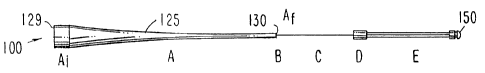
An improved ultrasound probe in accordance with an embodiment of the invention
for
accomplishing the foregoing is illustrated generally as probe 100 in FIG. 1.
Probe 100 is formed
1o with a tapered member horn section 125, formed with a proximal end 129 of
diameter A;
constructed to be coupled to a source of ultrasound energy (not shown). When
coupled to a
source of ultrasound energy, proximal end 129 is preferably located at a
displacement maximum
relative to the standing ultrasound wave supported by the overall device.
Proximal end 129 may
be coupled directly to a transducer or other energy source or to an
intermediate member located
between proximal end 129 and the energy source. From proximal end 129, tapered
member 125
tapers, in section A thereof, to a reduced diameter distal end 130, of
diameter Af at a transition
zone B. Proximal end 129 must be large enough to receive sufficient energy to
treat the
thrombus, occlusions and the like. However, in order to provide optimal
flexibility, it is desirable
to reduce the diameter of distal portions of probe 100 as much as possible,
while minimizing loss
of energy, strength or guidability. Furthermore, the reduction in diameter
must be accomplished
in such a manner as to amplify, i.e. increase the amplitude of, the ultrasound
vibrations.
Following tapered section A of distal diameter Af (or one or more tapered
sections A), is
a constant diameter section C, of diameter C;, where C; < Af. In the event
additional reductions
in diameter are desired, a second transition zone D may be provided, for
coupling section C to a
section E of one or more lengths of transmission media, each of diameter E;,
where E; < C;.
Section C may be composed of a different material than section A. For example,
section
A may be composed of aluminum, which has superior ultrasound transmission
properties, is
easily machined and is inexpensive, while section C may be composed of
titanium, titanium
alloys or other materials (including other metals, glass, ceramics. cermets,
polymers and
8
CA 02374208 2001-11-27
WO 00/72766 PCT/US00/14658
composites) that have adequate ultrasound transmission properties. but
~~reater tensile stren~,th
for the smaller diameters required.
FIGS. 41A and 41B provide a simplified representation of a portion of a
longitudinal
standing wave that could be induced in section A of FIG. 1. Longitudinal
displacement is plotted
using the vertical axis of the figure and longitudinal position in section A
is plotted along the
horizontal axis. FIG. 41 A represents section A of FIG. 1 as composed of a
first straight section, a
second tapered section, which terminates at 351, and a final straight section,
which terminates at
355. This arrangement is also shown in FIG. 3. Similarly, where section A of
FIG. 1 is of a
constant diameter, FIGS. 40A and 40B provide a simplified representation of a
portion of a
l0 longitudinal standing wave that could be induced in that section. This
arrangement is also shown
in FIGS. 8 and 9.
It is advantageous for a step down in cross-sectional diameter to be located
at a
displacement minimum in order to obtain maximum amplification of displacement.
Therefore,
refernng to FIGS. 40A and 40B, given that a proximal end 340 of a first medium
341 of wide
diameter is at a displacement maximum 342, step downs in transmission media
diameter, such as
step down 343, are preferably located at displacement minimums 444, which will
be located at
odd multiples of ~./4. For illustrative purposes, in FIG. 40B, step down 343
is shown located at 9
~,/4. Similarly, as shown in FIGS. 41A and 41B, given that a proximal end 353
of a first medium
350 of wide diameter is at displacement maximum 417, step down 355 is
preferably located at
2o displacement minimum 356, at odd multiples of ~,/4. For illustrative
purposes, in FIG. 41B, step
down 355 is shown located at 11,/4.
It is to be understood that FIGS. 40B and 41B are simplifications of a portion
of the
standing wave pattern. In actuality, the ultrasound wavelength is a function
of the shape,
dimensions and material of the horn and probe. Therefore, the wavelength is
not necessarily
constant, as shown in FIGS. 40B and 41 B. but instead will vary in conformance
with the shape of
the device and the geometry of the vasculature during use. Furthermore, it is
understood that the
ultrasound wavelength may also be a function of the transmission wire
diameter, such that even
for constant diameter sections, the wavelength of the standing wave may vary
if the cross-
sectional shape of one section is different from that of a second section. For
example, the
9
CA 02374208 2001-11-27
WO 00/72766 PCT/US00/14658
transmission wire can be a substantially evp~nential. catenary. stl'al'~hi.
quadratically. or
hyperbolically tapered cross-sectional dimension, or a uniform cross-sectional
dimension, or
combinations thereof
With further reference to FIGS. 40B and 41B, it will be understood that the
means of
coupling the proximal end of the horn to an ultrasound energy source, the
operating mode (i.e.
resonance or anti-resonance), as well as the transmission characteristics of
the ultrasound source
itself (i.e. the structure of the transducer assembly) will all determine the
exact location of the
leading displacement maximum 342. Thus, it is important to note that this
invention should not
be construed to be limited by deviations of the leading displacement maximum
342 from the
to location shown in FIGS. 40B and 41B. That is, displacement maximum 342 is
shown in FIGS.
40B and 41 B for illustrative purposes only. The exact location of
displacement maximum 342
has no bearing on the positioning of displacement nodes and anti-nodes of the
standing wave
pattern relative to the step-transitions and other structures taught in this
invention.
It is to be further understood that the standing wave pattern that develops
within the
probe, and which is partially depicted in FIGS. 40B and 41 B, is a function of
the tortuosity of the
probe during use. That is, the geometric configuration of the blood vessels
within which the
probe is inserted, will determine to a greater or lesser extent, the exact
operating frequency and
exact location of the nodes and anti-nodes of the standing wave at any given
moment as the
probe is advanced or retracted within the vessels. In practice, the dimensions
of the probe and
the operating frequency of the probe are selected so that the desired nodal
positions of the
standing wave, as taught herein, are achieved for a selected range of
geometries. In this way, the
nodal positions in the probe will be ideal at certain locations within the
target vessel and will
deviate minimally from ideal at other locations. Thus, it is to be understood
that references
herein to the positioning of the displacement nodes and anti-nodes of the
standing wave pattern
2S relative to the step transitions and other structures taught herein relate
to the preferred or ideal or
average positions, around which some variation will naturally occur as the
probe is manipulated
through a given vessel tortuosity. For this reason, references herein to
standing wave positions
are designated "approximate," or "average."
CA 02374208 2001-11-27
WO 00/72766 PCT/US00/14658
(n the event the transmission member tapers. such as medium ,~0 of FIG. 41A,
then a
distal end 3~ 1 of the tapered portion preferably is located at a displacement
maximum 352. This
tapered section then functions as a half wavelength horn, the amplification
properties of which
are well understood. Thus, if a proximal end 353 is at a displacement maximum
417, the distal
terminus of the taper 351 should be located at a distance equal to an integral
multiple of ~./2. For
illustrative purposes, in FIG. 41B, terminus 351 is shown located at 3~,/2.
The tapered section
may be followed by a constant diameter section with a distal step down 355
which should be at a
displacement minimum 356.
Referring again to FIG. 1, in accordance with preferred embodiments of the
invention,
1o section A, if it includes a taper, preferably has a tapered length equal to
an integral multiple of
half wavelengths of the intended frequency of operation. At the terminus of
section A, there may
be a transition zone B, which is a step transition to section C, wherein
section C has diameter C;<
Af. To effect maximum displacement amplification, step-transition zone B is
preferably placed
at or near a displacement node (i.e., a displacement minimum). Thus, if
section A includes a
tapered section which is an integral multiple of half wavelengths, it should
be followed by a
straight section of a length equal to an odd multiple (i.e. 1, 3, 5. . . ) of
quarter-wavelengths. In
this way, section A begins at the proximal end 129 at a displacement maximum,
and ends at its
distal end 130 at a displacement minimum (displacement node). If section A is
straight (i.e., has
a constant diameter as is shown in FIG. 40A), then it should begin at a
displacement maximum
2o and terminate at a displacement node.
Device 100 also includes a mass 150 at the distal tip thereof. Mass 150 is
designed and
shaped to distribute ultrasound energy and/or perform work in accordance with
the application of
interest.
Ultrasound device 100 (as well as other probes discussed herein) is understood
to operate
in a resonant (or anti-resonant) mode; i.e., it supports a standing wave
(preferably a longitudinal
wave) when energized by ultrasonic stimulation at proximal end 129.
Consequently, it is
preferred that mass 150 is located at a displacement maximum (anti-node).
Transition zone D
may be located at a displacement node or anti-node. For example, transition
zone D may involve
a joint that couples several parallel lengths of transmission media, of
diameter E;, to section C. In
CA 02374208 2001-11-27
WO 00/72766 PCT/iJS00/14658
that case. it may be determined that the mechanical strength ot~ transition
zone D is insufticient to
support maximum stress. For such a case, transition zone D may be located at
or near a
displacement maximum, as the displacement maximum corresponds with a location
having
minimal stress (stress node).
It is understood that the techniques for assembling the sections of this
invention are
equally applicable to systems that promote or focus ultrasound energy to
enhance the absorption
of drugs, induce apoptosis in cells, and/or treat tissue. tumors,
obstructions, and the like, within
and without the body, and for systems to be utilized in laproscopic surgery,
for ultrasonic
scalpels, and to induce tissue hyperthermia for cancer radiation therapy, for
example.
to Furthermore, it will be understood that while several examples given herein
refer to
intravascular applications of the invention employing guide catheters,
introduces sheaths,
guidewires, and the like, the invention is equally applicable to topical or
superficial treatments,
therapies administered in cavities of the body, intra-muscular and intra-
tissue treatments,
including the application of ultrasound to fatty deposits to assist in their
removal, the use of
I5 ultrasound to enhance healing, or to stimulate or suppress the functioning
of bodily organs.
An ultrasound probe constructed in accordance with a second embodiment of the
invention is shown generally as ultrasound probe 200 in FIG. 2. Probe 200 is
similar in
construction to probe 100, except that sections B, C, and D of probe 200 are
further subdivided to
provide for additional step downs in cross-sectional area. Thus, tapered
section A, which is
2o preferably machined from a single piece of metal, such as aluminum, can be
reduced in length.
This can significantly reduce the cost of probe 200, compared to probe 100.
Tapered section A
of probe 100 or 200 may be formed with any combinations of constant diameter
and reducing
sections, or a single section of diameter A;.
Probe 200 includes n sections (C, to C") each of constant diameter, separated
by n
25 transition zones B, to B", where preferably diameter C,<A,~ and C;+~< C;
for i = 1 to n. Each of
transition zones B or B, to B~ may be abrupt or tapered, and sections A and C,
or any of C, to C
may be formed from one material or from a multitude of materials, such as
aluminum or
titanium. Thus, constant diameter sections C or C; may be formed separately
(as, for example,
from drawn wire) and then joined at zones B or B; to sections A, D, E and F.
Alternatively,
12
CA 02374208 2001-11-27
WO 00/72766 PCT/US00/14658
constant diameter sections C or C; may be formed as an inte~~ral unit such as
from a sin~~le wire
which may be ground to conform to the aforementioned criteria. Accordingly,
sections A, B and
C or sections A, B, C and D .may be formed from an integral unit as from a
single rod, for
example, which may be machined to conform to the aforementioned design
criteria. In the event
probes 100 or 200 are formed from multiple sub-components which are joined at
zone B (or B;),
D and F, the connections should be free of voids and provide for the intimate
contact of the
joined members. The materials for each subsection may be carefully selected to
maximize
performance of the device by satisfying the specific requirements of the
device along its length.
Specifically, the physical requirements of the device may change along its
length as for example,
certain subsections may require greater flexibility and other subsections
greater strength, while
others may require greater erosion resistance and so on.
In the event probes 100 or 200 are formed from multiple sub-components which
are
joined at zone B (or B;), D and F, the connections may be achieved by a
variety of methods,
including, but not limited to, welding, adhesive bonding, swaging, crimping,
clamping, screwing,
or pinning. In addition, one or several of those connections may be fashioned
to be releasable,
thus permitting the interchange of components during or between procedures.
For example,
section A of probes 100 or 200 may be rendered reusable, resterilizable or
modifiable by the
interchange or addition of alternative sections C (or C;). Similarly, section
25 of FIG. 3 may be
rendered reusable, resterilizable, or modifiable by the interchange or
addition of alternative
2o transmission members 40 and tip 50.
It is also noted that any or all of the surfaces may be coated with a moisture
barrier or
hermetic coating to extend longevity by reducing stress corrosion.
Mass 150 can be in the shape of a sphere, a cylinder or a grooved cylinder. It
can be
ground or otherwise patterned, textured with holes or notches, and so forth to
promote or focus
ultrasound emissions, promote surface cavitation or promote selected flow
patterns. The shapes
disclosed in U.S. Patent No. 5,269,297, the content of which is incorporated
by reference, are
suitable.
Mass 1 ~0 may be formed directly as an integral portion of section E or mass
150 may be
formed separately and then attached to section E. For example, mass 150 may be
formed as a
I;
CA 02374208 2001-11-27
WO 00/72766 PCT/US00/14658
welded or brazed mass at the distal end of section E and then further
machined. if desired. to
impart additional surface texture or structure to mass 1 ~0. Alternatively,
mass 1 ~0 may be
formed or machined separately. and then attached to section E by a variety of
methods including
but not limited to welding, adhesive bonding, swaging, crimping, clamping,
screwing or pinning.
Mass 150 may be formed from a wide variety of materials which may be selected
based
upon the requirements of the specific application. For example, mass 150 may
be constructed
from any one or several of metals, ceramics, cermets, glass or polymers. Mass
150 may be
molded or otherwise formed directly onto section E of FIG. I or FIG 2.
To dissipate energy lost as heat and/or to damp unwanted vibrational modes, a
probe in
1 o accordance with the invention may be bathed with a coolant. The coolant
may be directed over
and around the probe, for example, by incorporating a sheath around some or
all sections of the
probe. The sheathing can be affixed to the probe at one or more of the
displacement nodes of the
standing wave, but preferably at any of the displacement nodes of section A,
which are proximal
of transition B. Additional sheathing may be incorporated for providing a
passageway for a
guidewire or other auxiliary tool which may serve to steer the device to, or
position the device at,
its intended location.
The coolant pathway may be used additionally or alternatively as a conduit for
the
delivery or withdrawal of other fluids, or bodily tissue matter, or gels or
suspensions or the like.
For example, the sheathing may serve as a pathway to administer therapeutic
drugs, or the
sheathing may serve as a conduit for the withdrawal of ablated material.
Furthermore, drugs, such
as streptokinase, urokinase, and platelet inhibitors, and contrast media, and
other fluids whose
function or efficacy would be enhanced by ultrasound or that would enhance the
application of
ultrasound at the treatment site, may be infused within the coolant fluid for
cooling the
ultrasound probe or delivered through a separate passageway within or without
the ultrasound
probe to the treatment site.
Referring to FIG. 3, a probe with a constant diameter section as part of the
horn section is
shown generally as probe 20. A horn 25, having a tapered section T and a first
constant diameter
section S is constructed to be coupled to an ultrasound energy source. Probe
20 also includes a
transmission member 40 coupled to horn 25 at transition zone B', and a tip 50
coupled to the
14
CA 02374208 2001-11-27
WO 00/72766 PCT/US00/14658
distal end of transmission memher X10. Ultrasound ener~~y sources disclosed in
II.S. Patent No.
~,269.2~)7, the content of which is incorporated by reference, are suitable.
Horn 25 includes a proximal end 29, a distal end 30, a tapered section 26 of
reducing
diameter from proximal end 29 to a transition point 28 and a straight section
27 with a constant
diameter from transition point 28 to distal end 30. Horn 25 is preferably
machined or turned
down from a single piece of metal, preferably aluminum 707. Horn 25
transitions from tapered
section 26 to straight section 27 at transition point 28, which should be
located approximately at
a displacement anti-node. The length of section 26 is approximately a multiple
of ~,/2, where ~./2
is the half wavelength of the standing wave, measured from anti-node to anti-
node. The
frequency of the ultrasonic energy generated by the ultrasonic energy source
used to excite the
device into resonance is designated f. In a preferred embodiment of the
invention, f ranges from
to 100 kHz, more preferably about 42 kHz. It is to be understood that the
selected frequency
of operation of the device may be an overtone, i.e., the operating frequency
is not necessary the
fundamental resonant (or anti-resonant) frequency of the device. Horn 25 is
preferably 7075
aluminum and the length of tapered section T is 144 mm long. In said preferred
embodiment, the
diameter of the proximal end of horn 25 is 12.7 mm, which tapers to a 1.0 mm
diameter at horn
transition point 28. While horn 2~ is preferably tapered, in alternate
embodiments, it may have a
constant diameter.
In a preferred embodiment of the invention, the diameter of straight section
27 remains a
2o constant 1.0 mm from transition point 28 to horn distal end 30. Distal end
30 is connected to
transmission member 40 at transition zone B' and includes at least one
transmission wire 45,
having a wire proximal end 46 and a wire distal end 47. Horn distal end 30 may
be connected to
transmission wire proximal end 46 by a number of coupling devices and
techniques which are
known in the art and otherwise, such as welding, including laser, diffusion,
and thermal welding,
adhesive bonding, swaging, crimping, clamping, screwing, pinning, or with a
mechanical
connector. The joint should be free of voids and provide for the intimate
contact of the joined
members.
Transmission member 40 also includes a highly flexible Section E', which is
shown in
FIG. 3 as being formed with three wires 60 of fine diameter. coupled to wire
45 at a 1-to-3
CA 02374208 2001-11-27
WO 00/72766 PCT/US00/14658
COLIpIIII~.T, joint JJ at transltloll lone D'. While Section E' prefei-ablv
consists of three wires. in this
embodiment, at least two wires are advantageous to give the device extra
flexibility and hi~~h
power transmission. Coupling 55 includes one opening at its proximal end for
insertion of distal
end 47 of wire 45 and three openings in its distal end for the proximal ends
of the three fine wires
60. At least ends of wires 60 are advantageously knurled prior to inserting
wires 60 within
openings of coupling 55. Wires 60 may be glued or otherwise coupled to
coupling 55 using
techniques known in the art, such as welding, adhesive bonding, swaging,
crimping, clamping,
screwing, pinning or with a mechanical connector.
Transition zone D' can also be designed as a single step amplification wherein
Section E'
t o consists of a single wire having a diameter less than wire 45. In a
preferred embodiment of the
invention, wires 45 and 60 are composed of high strength titanium wire.
A bullet-shaped tip 50 is coupled to the three fine wires 60 by means of three
openings in
the proximal end of tip 50. In a preferred embodiment, the three openings in
coupling 55 and in
tip 50 are spaced so as to form an equilateral triangle, concentric about the
central longitudinal
t 5 axis of coupling 55 and tip 50, as is shown in FIG. 31.
Tip 50 is provided with a notch 51 to improve cavitation as is shown in FIG.
14. It will
be understood that displacement amplitudes that exceed a threshold level
particular to a given
fluid type may be used to induce cavitation in that fluid. Cavitation bubbles
in a sound field may
be used advantageously to concentrate energy and enhance ablation or other
desired effects. Tip
20 50 may also be provided with proximal chamfers 52, as shown in FIG. 14. Tip
50 may also be
provided with proximal chamfers 52, as shown in FIG. 14 to aid in the
retraction of the probe
following a procedure. A radiopaque marker may be affixed to tip 50. The
radiopaque band may
be affixed to the proximal or distal end of tip 50, and may be contained
within a recess or affixed
to the outside of the tip. Alternatively, tip 50 may be fashioned from a
radiopaque material or it
25 may be coated with a radiopaque film. In a preferred embodiment, a pocket
or recess 53 in the
distal end of tip 50 is fashioned, as shown in FIG. 14, wherein a radiopaque
marker band is
affixed with adhesive.
Tip 50 may also be provided with an opening for a guidewire, and a guidewire
sheath
may be installed in the opening and extend proximally Ii~om the distal end. In
a preferred
16
CA 02374208 2001-11-27
WO 00/72766 PCT/US00/14658
embodiment. the guidewire opening is centrally located in tip ~0. passing
along its longitudinal
axis. Fine wires 60 may be separately sheathed, and said sheathing may extend
bemveen tip ~0
and coupling joint 55. Wire 45 may also be sheathed and said sheathing may be
connected to the
separate sheathing of wires 60 and may extend proximally to a coolant port to
allow coolant to be
injected to bathe all or part of sections 26, 27 and 40.
In another embodiment of the invention, tip 550 is shown generally in FIGS.
42A-42C.
Tip 550 includes a distal section 560, an intermediate section 570, and a
proximal section 580.
As with tip 50 depicted in FIG. 3, proximal section 580 of tip 550 is shaped
to accept three wires
of a mufti-wire section. As is shown in FIG. 42C, proximal section 580
includes bores 581 sized
1 o and shaped to accept wires 60. In a preferred embodiment, at least the
ends of wires 60 are
knurled prior to inserting wires 60 into bores 581 and proximal section 580 is
crimped to secure
wires 60 within bores 581. Wires 60 may also be glued or otherwise coupled to
tip 550 using
other techniques known in the art, such as welding, adhesive bonding, swaging,
crimping,
clamping, screwing, pinning or with a mechanical connector.
Tip 550 includes a central bore 590, which extends through proximal section
580,
intermediate section 570 and distal section 560, and is sized to accommodate a
guide wire (not
shown). In a preferred embodiment, that portion of central bore 590 contained
within distal
section 560 may include a counterbore 591 to provide a space for a radiopaque
marker (not
shown), which may be affixed within counter bore 591 with an adhesive or any
other affixation
means known in the art, including, but not limited to, those described above
in connection with
affixing wires 60 to tip 50.
As is shown best in FIGS. 42A and 42C, proximal section 580 includes a
substantially
cylindrical body 581 having a rear face 587, a surface 588a and a distal wall
583. Proximal
section 580 includes a first ring 584 and a second ring 585 spaced apart from
first ring 584 in a
longitudinal direction depicted in FIG. 42A as arrow X. First ring 584
preferably includes a
flattened surface 584, however, first ring 584 may have any cross-sectional
shape including a
rectangular, a square or an arcuate shape, for example. The walls of first and
second rings 584
and 585 are preferably substantially perpendicular to surface 588a of body 581
to facilitate the
17
CA 02374208 2001-11-27
WO 00/72766 PCT/US00/14658
creation of low pressure zones. when activated within a vessel. as is
described in more detail
below.
First ring 584 and second ring 585 extend from body 581 and can optionally be
formed,
as is shown in FIGS. 42A and 42B, with discontinuities 584a and 585a,
respectively, that permit
movement of the fluid medium in which tip 550 operates or an occlusion along
surface 588a and
through discontinuities 584a and 585a. Optionally, discontinuities 584a and
585a are located on
the same radial plane thereby forming a channel or gap 589. First ring 584 and
second ring 585
advantageously have at least two sets of discontinuities 584a and 585a equally
spaced apart
radially.
1o Channel 589 is advantageously formed substantially perpendicular to first
ring 584 and
second ring 585. Channel 589, however, can have any orientation in relation to
the longitudinal
axis X, including, as is described below, a spiral or oblique orientation, and
can have a variety of
widths. The width of channel 589 may vary.
Proximal section 580 includes beveled surfaces or flats 586, which extend from
rear face
587 distally toward first ring 584, thereby creating a truncated rear face
587. Proximal section
580 also includes fillets 582, which are preferably located at the base of
first ring 584 and second
ring 585 where rings 584 and 585 meet surface 588a of body 581. As is
discussed in more detail
below, flats 586 and fillets 582 serve as surfaces at which cavitation bubbles
can form when tip
550 is activated to move in the direction indicated by arrow A in FIG. 42A.
2o Intermediate section 570 optionally includes proximal steps 572, which step
down from
second ring 585 to a step landing 574, and distal steps 576 which step up from
step landing 574
to distal section 560. Proximal steps 572 and distal steps 576 can include one
or more increases
in diameter as measured from step landing 574. Preferably, proximal steps 572
and distal steps
576 form substantially vertical surfaces where cavitation bubbles can form in
low pressure areas
during oscillation of tip 550. Steps 572 and 576 in connection with step
landing 574 form a
radial channel having a first width at the level of step landing 574 and at
least a second width
formed at a fixed distance measured radially from step landing 574.
Intermediate section 570
optionally also includes lands 578, which extend from the top-most step of
proximal steps 572 to
the top-most step of distal steps 576, and which serve to support intermediate
section 570. Lands
18
CA 02374208 2001-11-27
WO 00/72766 PCT/US00/14658
578 along with the surface of the top-most steps of steps 57? and 576 form an
intermediate upper
surface ~ 71.
Distal section 560 is preferably substantially hemispheroidal or paraboloidal
in shape
with the nose of distal section 560 truncated by a plane substantially
perpendicular to the
longitudinal axis X. Distal section 560 includes a distal face 562 and an
outer surface 564. As
with proximal section 580, distal section 560 may include channels (not
shown), which are
preferably substantially parallel with the longitudinal axis X, to promote the
movement of an
occlusion from face 562 of tip 550 to intermediate section 570, where the
occlusion can be lysed
by combination of cavitation and fluid agitation.
Thus, tip 550 preferably consists of a narrow intermediate section 570, which
is
sandwiched by sections with comparatively larger diameters, proximal section
580 and distal
section 560. In a preferred embodiment, distal section 560 has a maximum
diameter that is less
than the maximum diameter of proximal section 580. Most preferably, distal
section 560 is
approximately 1.6 mm in diameter at its maximum diameter, and proximal portion
580 is
i5 approximately 2.2 mm in diameter at its maximum diameter.
As described above, tip 550 is constructed to induce cavitation in the blood
contained
within a blood vessel, for example. Because probe 100 is constructed to move
the tip at high
speeds in a direction parallel to the longitudinal axis of probe 100, it can
be advantageous to form
surfaces substantially perpendicular to the direction of motion so as to
create or enhance
cavitation. In this way, it is believed that cavitation bubbles form in low
pressure areas where
such surfaces create low pressure areas during oscillation. Thus, when tip 550
moves in a
direction parallel to the longitudinal axis, depicted as arrow A in FIG. 42A,
low pressure areas
form that lead to the formation of cavitation bubbles near the substantially
perpendicular walls of
distal steps 576. Similarly, when tip 550 moves in a direction parallel to the
longitudinal axis,
depicted by arrow B, low pressure areas form that lead to the formation of
cavitation bubbles
near the substantially perpendicular walls of proximal steps 572.
Lysing fields are also believed to be formed at proximal section 580 and
distal section
560, where areas of low pressure are believed to be formed when tip 550
oscillates in a
longitudinal direction. Lysing fields of proximal section 580 are formed at
flats 586 and fillets
1 ~)
CA 02374208 2001-11-27
WO 00/72766 PCT/US00/14658
>g? v.v:llile lysing field of the distal section 560 is formed at distal tace
J62. Thus. when tip >>0 is
moved forward longitudinally in the direction shown by arrow A. cavitation is
believed to be
promoted in the areas near distal steps 576, flats 586 and fillets 582. And,
when tip 550
oscillates in the direction depicted by arrow B, cavitation is believed to be
promoted near
proximal steps 572 and distal surface 562. Such phenomena have been observed
in several in
vitro and in vivo experiments.
In a preferred embodiment. tip 550 is approximately .124 inches in length as
measured in
the longitudinal direction, and the proximal dimensioned as follows: distal
section 560 measures
approximately .033 inches in length and has a maximum diameter of .065 inches,
intermediate
to section 570 measures approximately .032 inches in length and has a minimum
diameter of .046
inches, and proximal section 580 measures approximately .059 inches in length
and has a
maximum diameter of .069 inches; steps 572 and 576 measure approximately .003
inches in
length and range from .046 to .065 in diameter; rings 584 and 585 measure
approximately .015
inches in length and have a maximum diameter of .087 inches; bore 590 is
approximately .033
inches in diameter and counterbore 591 is approximately .035 inches in
diameter.
Thus, when the occlusion is located forward of distal surface 562, and tip 550
is
energized and moved toward contact with the occlusion, the occlusion can be
channeled over
distal section 560 toward distal steps 576 and proximal steps 572 to what may
be termed "lysing
fields," where a combination of cavitation and fluid agitation pulls and
breaks the occlusion into
2o its constituent parts. The hemispheroidal shape of distal section 560
promotes the flow of
portions of the occlusion over outer surface 564 of distal section 560 and
toward distal wall 583
of proximal section 580. The differences in diameters between proximal section
580,
intermediate section 570 and distal section 560 can also help create vortices
that help pull the
occlusion apart and direct the occlusion into the lysing fields created near
distal steps 576 and
proximal steps 572.
To promote the formation of cavitation bubbles, preferably all or part of the
surface of tip
550, including outer surface 564. can be roughened to provide imperfections.
Also, those
surfaces designed to create lysing fields may be roughened or treated with a
coating to enhance
the formation of cavitation bubbles.
CA 02374208 2001-11-27
WO 00/72766 PCT/US00/14658
FIGS. 43-50 depict tips constructed in accordance with fin-ther embodiments of
the
invention. Such further embodiments include tips having different combinations
of elements
designed to, among other benefits, facilitate the formation of cavitation
bubbles, the agitation of
the fluid medium and thrombus contained therein, and the movement of the
occlusion proximally
from the distal most portion of the tip along the length of the tip. It is to
be understood by those
skilled in the art that the elements depicted herein can be combined
differently to achieve similar
or enhanced effects.
Turning to FIGS. 43A and 43B, a tip 650 formed in accordance with the
invention is
shown having a proximal section 680, an intermediate section 670, and a distal
section 660. Tip
650 differs from tip 550 in that rather than having channels 589 formed
substantially
perpendicular to first ring 584 and second ring 585, tip 650 includes lands
689 that bridge first
ring 684 and second ring 685 in the longitudinal direction to support proximal
section 680 and
promote agitation of the fluid medium and the occlusive material that moves
near distal section
680 when tip 650 is actuated. Lands 689 need not be oriented parallel to the
longitudinal axis of
I5 tip 650. For example, lands 689 may bridge first ring 684 and second ring
685 at any orientation,
including an oblique orientation.
Turning to FIGS. 44A and 44B, there is depicted a tip 750 constructed in
accordance with
another embodiment of the invention, having a proximal section 780, an
intermediate section
770, and a distal section 760. Tip 750 differs from tip 550 in that distal
section 760 is formed
with a more cylindrical shape, having a substantially flat nose 762 and sides
765 and 766. As a
result, rather than having a generally hemispheric or bullet shape as is
depicted in FIG. 42B,
distal section 760 has a constant outer diameter, except at the rounded edges
764. Therefore, tip
750 is depicted as a parallelogram when viewed from the side. It is
understood. however, that
sides 765 and 766 need not be parallel to the longitudinal axis of tip 750.
Sides 765 and 766 can
have an arcuate or an oblique orientation. When the orientation is oblique,
sides 765 and 766
would thereby create a trapezoidal cross section when viewed from the side.
Thus, the shape of
distal section 760 when viewed from the side may be any shape, including
rectangular,
thromboidal or trapezoidal, by way of non-limiting example.
21
CA 02374208 2001-11-27
WO 00/72766 PCT/US00/14658
FICrS. 4~A and 4~B depicts a tip 8>0 constructed in accordance with yet
another
embodiment of the invention. Tip 8~0 includes a distal section 860, an
intermediate section 870
and a proximal section 880. Proximal section 880 includes a rear face 887 and
flats 886, which
extend from rear surface 887 distally toward intermediate section 870. Distal
section 880 also
includes a body 881, a rear outer surface 889 and steps 885, which are formed
on body 881. In
this manner, the diameter of distal section 880 increases from rear outer
surface 889 to body 881.
Intermediate section 870 includes proximal steps 872, distal steps 876 and a
step landing
874 located intermediate proximal steps 872 and distal steps 876. From body
881, the outer
diameter of tip 850 decreases through a series of proximal steps 872 to step
landing 874.
to Thereafter, the outer diameter of tip 850 increases through a series of
distal steps 876 to a surface
864 of distal section 860. As is shown in FIG. 45A, proximal steps 872 may be
of greater
number than distal steps 876. Further, proximal steps 872 and distal steps 876
may be formed as
a spiral, as is shown in embodiments described below, such that the diameter
of tip 850 decreases
or increases at a defined rate when measured at a point within proximal steps
872 or distal steps
~5 876.
FIGS. 46A and 46B depict another embodiment of a distal tip 950 constructed in
accordance with the invention, having a proximal section 980, an intermediate
section 970, and a
distal section 960. Proximal section 980 includes a body 981 having channels
989 that spiral or
corkscrew obliquely through a portion of body 981. Further, intermediate
section 970 includes
2o distal steps 974a, 974b and 974c, each having different heights, and
proximal steps 972a, 972b
and 972c, each having different heights. Distal steps 974a and 974b and
proximal steps 972a
and 972b are substantially perpendicular to the longitudinal axis of tip 950,
while distal steps
974c and proximal steps 972c are preferably the same width as distal steps
974a and 974b, and
proximal steps 972a and 972b , respectively.
25 FIGS. 47A and 47B depict another embodiment of a distal tip 1050
constructed in
accordance with the invention, having a proximal section 1080, an intermediate
section 1070,
and a distal section 1060. Intermediate section 1070 is eccentric in its
construction in that on one
side of intermediate section 1070 includes proximal steps 1072 that step down
from proximal
section 1080, while on the opposite side. the diameter surface of intermediate
section 1070 is on
CA 02374208 2001-11-27
WO 00/72766 PCT/US00/14658
the same plane as the surface of distal section l ()60. In this way. when tip
100 oscillates in the
longitudinal direction, the tip vibrates eccentrically causing further
agitation to the fluid medium
and occlusion.
FIGS. 48A and 48B depict another embodiment of a distal tip 1150 constructed
in
accordance with the invention, having a proximal section 1180, an intermediate
section 1170,
and a distal section 1160. Proximal section 1180 includes a body 1181 having a
channel 1188
and a series of steps 1184 that step down in diameter from body 1181 to
channel I 188 and a
series of steps 1185 that step up from channel 1188 to body 1181. These series
of steps I 184 and
1185 and channel 1188 are formed as spirals in body 1181 and are oblique in
orientation as
compared with the longitudinal axis X. Steps 1184 and 1185 and channel 1188
may be a curved
or straight in orientation. Intermediate section 1170 includes steps 1172,
which are also curved
spiral steps whose orientation is preferably opposed to the orientation of
steps 1184 and 1185 of
the proximal section 1180. The walls of steps 1172, 1184 or 1185 need not be
perpendicular to
the surface from which they extend. In fact, in a preferred embodiment, the
walls may extend
obliquely from the surface as is clearly shown in FIG. 48B. Steps 1184, I 185
and channel 1188
are constructed to promote agitation and longitudinal twisting of the fluid
medium to facilitate
agitation and dissolution of the occlusion.
FIGS. 49A and 49B depict another embodiment of a distal tip 1250 constructed
in
accordance with the invention, having a proximal section 1280, an intermediate
section 1270,
and a distal section 1260. Proximal section 1280 includes a first ring 1282
and a second ring
1283 formed on body 1281. In this embodiment, intermediate section 1270 has a
diameter
greater than that of distal section 1260. but is less than the diameter of
ring 1283 of proximal
section 1280. Intermediate section 1270 includes a body 1271 having a first
channel 1272 and a
second channel 1273 formed substantially perpendicular to the longitudinal
axis of tip 1250, and
a third channel 1273 formed in body 1271 and having a spiral orientation.
Intermediate section
1270 also includes steps 1275, which step down from body 1271 to third channel
1274 and steps
1276 which step up from channel 1274 to body 1271. Intermediate section 1270
also includes
step 1277a and step 1277b, which respectfully step down from ring 1283 to
channel 1272 and
23
CA 02374208 2001-11-27
WO 00/72766 PCT/US00/14658
step up from channel 1272 to body 1271. and step 1278a and step 1278b which
respectfully step
down from body 1271 to channel 1273 and step up from channel 1273 to distal
section 1260.
FIGS. SOA and SOB depict another embodiment of a distal tip 1350 constructed
in
accordance with the invention, having a proximal section 1380, and an
intermediate section 1370
and a distal section 1360. In this embodiment, distal section 1360 and
proximal section 1380 are
simplified, and the diameter of intermediate section 1370 is greater than both
the diameters of
proximal section 1380 and distal section 1360. As with prior embodiments,
intermediate section
1370 includes a channel 1372 having steps 1373 and 1374
FIG. 51 depicts another embodiment of a distal tip 1390 constructed in
accordance with
to the invention, having a proximal section 1391, an intermediate section 1392
and a distal section
1393. The primary difference of this embodiment is that distal section 1392
has channels 1395
spaced apart radially that extend substantially parallel to the longitudinal
axis. Alternatively,
channels 1395 can be oblique in orientation as compared with the longitudinal
axis X.
Referring to FIGS. 4A, 4B, 4C and 5, another preferred embodiment of the
invention is
IS exemplified by horn 525, which includes a straight section 527 of constant
diameter and a
transition section in the form of a joint 535 in the distal end thereof. Joint
535 is bored to accept
a transmission wire. This embodiment may include a region of increasing
diameter 529 prior to
joint 535 (see FIG. 4B), so that the diameter of joint 535 is slightly greater
than the diameter of
straight section 527 to provide greater strength at joint X35 between a horn
distal end 530 and a
2o transmission wire (not shown). In one example of such an embodiment, a horn
525 has a straight
section 527 with a diameter of 1 mm which increases to 1.09 mm at the distal
end of the region
of increasing diameter 529.
In one preferred embodiment, joint 535 has a bore diameter of approximately
0.63 mm,
and a bore depth of approximately 5 mm, and is mechanically crimped onto a
transmission wire,
25 which is preferably formed of titanium and preferably has a diameter of
approximately 0.62 mm.
To further increase the strength of the crimp joint, in accordance with a
preferred embodiment of
the invention, the surface of the proximal 4 mm of the transmission wire may
be roughened prior
to crimping.
24
CA 02374208 2001-11-27
WO 00/72766 PCT/US00/14658
In an alternative embodiment joint ~;~ is replaced with joint 73~ shown in
FIG. 7B.
which does not include a region of increasing diameter.
It is important to note that the placement of a stepped-down reduction in
diameter from
horn 525 to a transmission wire at or near a displacement node offers the
maximum displacement
amplification. Despite this fact, the prior art generally teaches away from
using step downs in this
fashion because of the high level of stress associated with such a transition.
However, this
shortcoming of the prior art is overcome by introducing a high strength joint
and the ability to
combine different and appropriate materials at the joint. Also, by locating
the joint
approximately at a displacement node, energy transfer can be made more
efficient.
~ Because an ultrasound transmission device must be sized to accommodate
different
treatment sites that have varying distances between the point of entry of the
probe into the
patient's body and the point within the body to be treated, it is understood
that other
embodiments will require different lengths and diameters than the preferred
coronary
embodiments. Variations in length are still described by the general
formulation described in
FIGS. l, 2 and 3.
Referring to FIGS. 8 through 13, a variety of probe designs which satisfy the
principles of
construction taught herein are shown. These variations employ step transitions
with joints
constructed according to the advantages taught herein, though details of those
connections, such
as that of FIG. 4B or connecting member ~5 of FIG. 3, are not shown. It is to
be understood that
neither the diameters nor the lengths of the sub-components in these or any of
the other figures
contained herein are to scale, nor are any of the proportions to be construed
as representative or
limiting. In FIG. 8, three consecutive step transitions (801, 802 and 803) are
shown, each of
which may be located at a displacement node. The first step transition is
shown with a radiused
transition, which may be applied similarly to any of the step transitions
taught herein, to effect a
strain relief.
FIG. 9 is similar to FIG. 8 except that all transitions (901, 902 and 903) are
shown as
abrupt steps. FIG. 10 employs a proximal, tapered horn section 1001. FIG. 11
employs an
elongated straight section 1101 which is integral with a proximal horn section
1102. FIG. 12
shows the use of two parallel wires 1201 a and 1201 b in the distal-most
transmission wire section
CA 02374208 2001-11-27
WO 00/72766 PCT/LJS00/14658
for enhanced flexibility of this section. The use of two or more wires in the
distal section permits
the passage of a guidewire along the central longitudinal axis of the distal
tip. FIG. 13 is similar
to FIG. 12, except that the distal two-wire section is replaced with a three-
wire ( 1301 a; I 301 b
and I 301 c) section. In FIG. 13A, the proximal section is shown to consist of
two consecutive
half wavelength horns followed by an integral straight section which
terminates at a
displacement node at transition point B.
Referring again to FIG. 3, in one preferred embodiment of the invention
designed for
coronary blood vessels, the ultrasound horn, 526, includes a proximal tapered
section T, formed
with a length of 144 mm and an initial diameter of 12.7 mm that tapers to a
diameter of 1 mm at
t o transition point 28. The horn then extends distally, over section S for a
distance of 567 mm at
this constant diameter, and terminates at distal end 30. The horn is connected
by means of joint
735 of FIG. 7B to transmission wire 45 which has a length of 544 mm. The
distal end of
transmission wire 45 is connected to distal three-wire section E' via
connector 55. Section E' has
a wire length of 160 mm and is connected to tip 50. In another modification of
a preferred
coronary embodiment, joint 735 is replaced with joint 535 of FIG. 4B. In
another modification
of a preferred coronary embodiment, section S is extended to a total length of
847 mm and
transmission wire 45 has a length of 264 mm. In another modification of a
preferred coronary
embodiment, section T has a total length of 233 mm.
Referring again to FIG. 3, in one preferred embodiment designed for peripheral
vessels.
such as AV shunt vessels, ultrasound horn 525 includes a proximal tapered
section T formed
with a length of 144 mm and an initial diameter of 12.7 mm that tapers to a
diameter of 1 mm at
transition point 28. The horn then extends distally, over section S for a
distance of 173 mm at a
constant diameter, and terminates at distal end 30. The horn is connected by
means of joint 735
of FIG. 7B to transmission wire 45, which has a length of 30 mm. The distal
end of transmission
wire 45 is connected to distal single wire section E' via connector 55.
Section E' has a wire
length of 227 mm and is connected to tip 50. In another modification of the
above-preferred
peripheral embodiment, joint 735 is replaced with joint 535 of FIG. 4B. In
another modification
of the above-preferred peripheral embodiment, transmission wire 45 has a
length of 89 mm and
section E' is constructed as a two- or three-wire section with a length of 160
mm. In another
26
CA 02374208 2001-11-27
WO 00/72766 PCT/US00/14658
moditication of the above-preferred peripheral embodiment. transmission wire
4~ has a length of
X44 mm and Section E' is constructed as a two- or three-wire section with a
length of 160 mm.
As discussed above, transmission member 40 of FIG. 3 may include one or more
transmission wires, each having constant diameters, and each successive
transmission wire
having a smaller diameter. The successive wires may be formed as an integral
unit by machining
each diameter down from a single rod of material or they may also be formed
separately and then
joined.
Referring again to FIG. 3, in a preferred embodiment of the invention, distal
end 47 of
transmission wire 45 is joined with a mufti-wire section 60, which in a
preferred embodiment,
1o includes three titanium wires. The diameter of transmission wire 45 range
between 1.0 mm and
0.2 mm, while the diameter of fine wires 60 can range between .5 mm and .O1
mm. The length
of transmission wire 45 can range between 0 mm and 1000 mm, while the length
of fine wires 60
can range between 0 mm and 300 mm. In a preferred embodiment, transmission
wire 45 has a
diameter of approximately 0.62 mm, and a length of approximately 544 mm, and
the wires of
the mufti-wire section 60 have a constant diameter of approximately 0.29 mm,
and a length of
approximately 160 mm. While in this example, junction 55 of transmission wire
45 and fine
wires 60 is located near a displacement maximum, junction j5 may be located at
any position
along the standing wave.
In a preferred embodiment, coupling 55 is fabricated from high strength
aluminum
(preferably aluminum 6061 ) and includes a high strength crimp connection to
transmission wire
45 and aerospace-grade epoxy connections to fine wires 60. In this example,
the bore diameter
for the crimp connection is approximately 0.63 mm with a depth of 3 mm, and
the bore diameter
for the fine wire adhesive connections is approximately 0.31 mm with a depth
of approximately
I .5 mm.
In another preferred embodiment of the invention, the proximal end of horn 29
of FIG. 3.
and proximal end 29' of FIG. 4A can include a threaded bore having a diameter
of one-quarter
inch and 12 mm deep for receiving the distal tip of an ultrasound source. In
other embodiments,
connection between the ultrasound source and the horn can be made via bayonet-
type twist
connections, spring-loaded snap connections, and a variety of other quick-
connections. Referring
27
CA 02374208 2001-11-27
WO 00/72766 PCT/L1S00/14658
to FIGS. 6n and 6B, a keyed O-ring groove 600 is utilized as a means of both
establishin~~ a Iluid
seal between the proximal end of sheathing 1» of FIG. 1=~ and horn 525 and
preventing any
relative twisting of sheathing 1.55 and horn X25. O-ring groove 600 is
preferably located at a
displacement node (i.e. a displacement minimum) so as to avoid damping of the
transmitted
energy by the O-ring groove. In one embodiment, O-ring groove 600 is located
at a distance of 83
mm from the proximal end of the first transmission member. Preferably, ring
601 may extend
0.25 mm from the surface of the horn and have a thickness of 0.5 mm. A hex
ring 602 may
extend 0.5 mm from the surface of the horn, have a thickness of 0.8 mm, and
have a diameter
between flat surfaces of 3.7 mm, and a diameter between opposite apex points
of 4.2 mm. It will
be evident to those of ordinary skill in the art that an ultrasound
transmission device constructed
in accordance with the invention, including the foregoing examples, can
readily fit within and be
delivered to a thrombus in a coronary artery through a 7 French guide
catheter.
Refernng again to FIG. 3, tip 50 is connected to the distal end of at least
one transmission
wire 60. Preferably, tip 50 is shaped to accept three wires of a mufti-wire
section, and is
positioned at a displacement maximum such that it will oscillate maximally in
a longitudinal
direction. In a preferred embodiment, tip 50 is formed of aluminum, preferably
6061 aluminum
and is 1.65 mm in diameter. Alternatively, tip 50 may be formed of a titanium
alloy such as
Ti6Al/4V, which can serve to strengthen tip 50 and eliminates the need for a
distal marker band,
as the vanadium within the alloy makes the tip 50 visible under an angiogram.
2o To dissipate energy lost as heat and/or to dampen unwanted vibrational
modes, the device
may be bathed with a coolant. The coolant may be directed over and around the
device by
affixing a thin flexible sheathing, preferably formed of polyimide, or other
high strength, thin
walled, low friction material around some or all sections of the device. The
sheathing is
preferably affixed to the device at one or several displacement nodes.
Additional sheathing may
be applied to the device to provide a passageway for a guidewire or other
auxiliary tool that may
serve to steer the ultrasonic probe to its intended location.
In another preferred embodiment, all or some surfaces of the horn and/or
transmission
wires are coated with a moisture barner or hermetic coating, such as parylene,
to extend the
longevity of those sections by reducing or eliminating stress corrosion.
28
CA 02374208 2001-11-27
WO 00/72766 PCT/US00/14658
Referring to FIGS. 6A and 14. a sheathings 155 of one or more sections can be
disposed
around horn 525 and extend distally to just beyond a coupling member 1455. The
sheathings can
be reduced in diameter as the diameter of the transmission medium decreases.
An advantageous
sheathing material is polyimide, in view of its ability to be formed into
extremely thin diameters,
its strength and its low friction.
Referring to FIG. 14, a single transmission wire 1445 of relatively large
diameter is
joined to three wires 1403 of relatively small diameter, including wires 1401
and 1402, shown,
and a third wire, not shown, but positioned behind wires 1401 and 1402. Wire
coupling 55 is
constructed to have a proximal single hole to receive wire 1455, and three
distal holes to receive
to wires 1403. In a preferred embodiment, the three wires 1403 and their
receiving holes in
coupling 55 are spaced equally in a circular pattern to form an equilateral
triangle, concentric
with the central longitudinal axis of coupling 55.
In a preferred embodiment, coupling 55 is mechanically crimped onto single
transmission
wire 1445. To further increase the strength of the crimp joint, in accordance
with a preferred
embodiment of the invention, the distal surface of transmission wire 1445 may
be roughened
prior to crimping. In a preferred embodiment, coupling 55 is bonded to fine
wires 1403 using a
high-strength, aerospace-grade epoxy. Alternative means of attaching coupling
55 may also be
employed as previously taught herein.
Sheathing 155 may be disposed about horn 525, single transmission wire 1445,
wire
2o coupling 55, and extend distally to beyond the proximal end of coupling
member 1455.
Sheathings 1481 and 1482 are disposed around wires 1401 and 1402,
respectively, and the third
wire is sheathed similarly. A distal end 155a of sheathing 155 overlaps a
portion of coupling
1455, and is attached to coupling 1455 with adhesive. Sheathing coupling 1455
is constructed to
have three through-holes positioned and sized to receive the sheathings that
are disposed around
wires 1403. The sheathings disposed around wires 1403 are attached to coupling
1455 with
adhesive. Coupling 1455 and the sheathings disposed around wires 1403 may be
separate
members joined as described above, or they may be formed of a single member,
as a single
length of extruded mufti-lumen tubing. It is to be understood that other
embodiments of wire and
sheathing arrangements are encompassed by this invention including designs
that use more or
29
CA 02374208 2001-11-27
WO 00/72766 PCT/US00/14658
fewer than three wires 140 f. and their associated sheathings. In one
embodiment. fluid. such as
saline solution, flows through sheathing 1 >j, through coupling 14~~, and out
through the distal
ends of the wire sheathings disposed about wires 1403.
If the ultrasonic probe design includes a multi-wire distal section, as shown
for example
in FIG. 14, then it is important that sheathing 155 be prevented from rotating
relative to the
central ultrasound transmission member 1445 and coupling 55. In such a case,
if relative motion
were permitted, transmission wires 1403 could become twisted just proximal of
coupling 1455,
and failure of one or more of the transmission wires could be accelerated. To
prevent this, O-ring
groove 600, which is located at a displacement node, may be shaped or keyed
(see FIG. 6A). A
similarly shaped receiving pocket is then fashioned in the sheathing
structure, such that once
engaged with the horn, the sheathing cannot be rotated relative to the horn.
It is understood that
for those embodiments where the transmission members always lie on the central
axis of the
probe, for example, in FIG. 14, if wires 1403 are replaced with a single
centrally located wire or,
for example, in FIGS. 8-1 l, such "keying" is not necessary. However, the
location of an O-ring
groove or similar sealing mechanism to establish a fluid pathway between the
sheathing and the
horn (or transmission members) should be at a displacement node whether keying
is used or not.
In another preferred embodiment, as shown in FIG. 14, the sheathing of wires
1403
terminates proximal to a cavitation tip 1450, to create an expansion gap 1480
between the
sheathing and tip 1450. Expansion gap 1480 is typically a few millimeters in
length, and thus
2o provides adequate spacing to allow the sheathings disposed around wires
1403 and sheathing 155
to expand or contract during use, without effecting interference with
cavitation tip 140. The
expansion and contraction of such sheathing is a consequence of normal
elongation and
compression of the polymeric sheathing material when the probe is energized
and manipulated
during a procedure.
FIG. 14 also shows a guidewire tube 1430 to facilitate the positioning of
probe tip 1450 at
the desired location. In the construction shown in FIG. 14, guidewire tube
1430 is of a similar
diameter as the sheathing of wires 1403, and extends from a location 10 to 15
centimeters
proximal cavitation tip 1450 into and through tip 1450. Tube 1430 can include
a flared distal tip
to provide a safety retention feature, in the event that tip 1450 breaks when
the probe is
CA 02374208 2001-11-27
WO 00/72766 PCT/OS00/14658
energized. such as at a notch 14~ 1. Tuhe 1430 is held in dace by bands 1490.
which are wrapped
around the sheathing of wires 1403. Bands 1490 are shown in end cross-section
in F1G. 31 as
3190, and again in FIG. 30 as 3190.
In another preferred embodiment of the invention, a cavitation tip 1550 is
fitted with a
safety insert 1501, 1601, 1701, 1801, or 1901 as shown in FIGS. 15-19,
respectively. Guidewire
tube 1430 of FIG. 14 is denoted as tube 1510, 1610, 1710, 1810, and 1910 in
FIGS. 15-19,
respectively. Safety inserts 1501, 1601, 1701, 1801, and 1901 serve to retain
sections of tip
1550, such as the radiopaque marker or a fractured front end of tip 1550, in
the event that
dislocation occurs during use. The safety inserts also serve to separate the
cavitation tip 1550
t o from a guidewire which may be inserted inside the safety insert. The
safety insert is preferably
tightly connected to, or locked onto, tip 1550 in such a manner as to
eliminate or minimize
differential movement between the safety insert and tip 1550.
A variety of methods of attachment of the safety insert to tip 1550 may be
employed
depending upon the materials of construction selected for the particular
application. Attachment
methods include, but are not limited to, adhesive bonding, crimping, molding,
melt-bonding,
swaging, flaring, screwing, pinning or coating. In addition, the safety insert
and tip may be
fashioned as a single component using a single material, as for the case where
the selected
material serves the multiple functions of applying ultrasound to the treatment
site, resisting
abrasion by the guidewire, and providing safety retention of fractured
components.
2o It is advantageous to decouple the safety insert from the probe sheathing,
which. as noted
above, will tend to expand and contract during use as the probe is energized
and manipulated
during a procedure. Decoupling prevents chafing or abrasion of the sheathing
by the cavitation
tip. Preferred embodiments of the associated decoupling mechanisms are shown
in FIGS. 15 to
35. It is understood that while retaining flares are drawn in figures 15 to 19
as a way of affixing
the safety insert to the cavitation tip, other affixment arrangements as
depicted in FIGS. 20 to 28
will serve as well, including adhesive and melt bonding.
FIG. 15 shows a auidewire tube 1510 terminating just proximal tip 1550. Safety
insert
1501 is positioned within and in a slidable piston-in-cylinder fit with tube
1510. It is preferable
that insert 1501 fit inside tube 1510 and not vice versa to avoid creating a
point at which the
31
CA 02374208 2001-11-27
WO 00/72766 PCT/US00/14658
'~uide~~ire would be impeded when it is threaded from distal end 1501a of
insert I~01 towards
the proximal end of the probe.
FIGS. 16-29 and 52 show additional embodiments of the invention including
different
safety inserts. Safety inserts are preferably formed of abrasion-resistant
materials. An insert may
optionally be included in the cavitation tip. In still other embodiments, a
gap may exist proximal
of the safety tip and in still other embodiments, the guidewire tube can
terminate proximal of the
cavitation tip and the guidewire can pass through the cavitation tip without a
guidewire tube or
safety insert therebetween.
Referring to FIG. 16, a safety insert 1601 is shown slidably disposed over the
distal end
of a guidewire tube 1610. Though preferably, guidewire tube 1610 or the
proximal diameter of
insert 1601 can be sized so that guidewire tube 1610 fits over insert 1601, as
in FIG. 15.
Referring to FIG. 17, an insert 1701 is shown slidably disposed over the
distal end of a
guidewire tube 1710 similar to the construction of FIG. 16. However, a flare
1711 is formed in
the distal end of tube 1710 and a constriction 1702 is formed in the proximal
end of insert 1701.
In this manner, tube 1701 and tube 1710 are mutually captive.
Referring to FIG. 18, an insert 1801 is shown slidably disposed in a distal
end of a
guidewire tube 1810, similar to the construction of FIG. 15. However, a
constriction 1811 is
formed in the distal end of tube 1810, and a flare 1802 is formed in the
proximal end of insert
1801. Thus, tube 1801 and tube 1810 are mutually captive.
2o Referring to FIG. 19, an insert 1901 is shown slidably disposed over the
distal portion of
a guidewire tube 1910. The guidewire tube 1910 passes through insert 1901.
Tube 1910 may
project distally through 1901 and may be fitted with a safety flare 1911.
Safety flare 1911 serves
to capture tube 1901 or tip 1550 in the event of release.
Referring to FIG. 20, an insert 2001 is shown slidably disposed inside
guidewire tube
2010. Insert 2001 consists of two sections, tube 2002 and tube 2003, which are
joined together
and attached to tip 1550. Tube 2002 fits over tube 2003.
Referring to FIG. 21, an insert 2101 is shown slidably disposed inside
guidewire tube
2110. Insert 2101 consists of two sections. tube 2102 which fits over tube
2103, and which are
joined together and attached to tip 1550.
CA 02374208 2001-11-27
WO 00/72766 PCT/US00/14658
Referring to FIG. 2?. an insert 2201. is shown slidahly disposed inside
~~uidewire tuhe
2210. Insert 2201 consists of two sections, tube 2203 and tube 2202, which
fits over tube 2203,
which are joined together and attached to tip 1550.
Referring to FIG. 23, an insert 2301 is shown slidably disposed inside a
guidewire tube
2310. Insert 2301 consists of two sections, a tube 2302 which fits over a tube
2303, and which
are joined together and attached to tip 1550.
Referring to FIG. 24, an insert 2401 is shown slideably disposed over a
guidewire tube
2410. Insert 2401 consists of two sections, a tube 2402, which fits over a
tube 2403, which are
both joined together and attached to tip 1550.
Referring to FIG. 25, an insert 2501 is shown slidably disposed inside a
guidewire tube
2510. Insert 2501 consists of three sections, a tube 2502 which fits over a
tube 2503 and over a
proximal safety stop 2504. All three sections are joined together and attached
to tip 1550.
Referring to FIG. 26, a guidewire tube 2610 is shown in a piston arrangement
with an
insert 2601, which is disposed through tip 1550. Insert 2601 consists of three
sections: a
proximal safety stop 2604, a tube 2603, and a tube 2602, which fits over tube
2603, and inside
proximal safety stop 2604, all of which are joined together and attached to
tip 1550.
Referring to FIG. 27, an insert 2701 is shown slidably disposed inside a
guidewire tube
2710. Insert 2701 consists of two sections, a tube 2702 which fits over a tube
2703. Tube 2703
is shown with an enlargement which serves as the proximal safety stop. Tubes
2702 and 2703
2o are joined together and attached to tip 1550.
Referring to FIG. 28, an insert 2801, which is similar to the assembly of FIG.
25, is
shown incorporated in a multi-wire distal assembly, including three titanium
wires 2880 (two
shown), and their respective sheathings 2881. Insert 2801 is shown slidably
disposed inside a
guidewire tube 2810. Insert 2801 consists of three sections, a tube 2802 which
fits over a tube
2s 2803 and over a proximal safety stop 2804 all three sections are joined
together and attached to
tip 1550. Insert 2801 is shown with a distal safety flare 2805. It is to be
understood that distal
safety flares may be added to any of the insert designs disclosed herein.
FIG. 29 is a sectional view of an insert similar to that of FIG. 28. Insert
2901 consists of
three sections, a tube 2902 which fits over a tube 2903 and over a proximal
safety stop 2904. All
33
CA 02374208 2001-11-27
WO 00/72766 PCT/US00/14658
three sections are joined to~~ether and attached to tip 150. Insert 2901 is
shown with a distal
safety flare 290. FIG. 29 also shows a tip 1550 in which a radiopaque marker
2906 is affixed
therein.
FIG. 52 depicts a sectional view of a distal tip section 5200 like that of
FIG. 15. Distal tip
section 5200 includes of a guidewire 5270, a tip 5250 having a bore 5205 sized
to accommodate
guidewire 5270, a first tube 5201 having a first diameter sized to fit within
bore 5205 and a
flared portion 5201a having a second diameter that is greater than the
diameter of bore 5205. In
this way, first tube 5201 is prevented from moving in a proximal direction Z.
First tube 5201 is
preferably formed using Hytrel or another art-known material, such as a
plastic or polymer.
Distal tip section 5200 also includes a guidewire tube 5210 terminating just
proximal tip
1550, and a piston tube 5215 having a free end 5212 and a fixed end 5211
connected to first tube
5201. Guidewire tube 5210 is positioned within and in a slidable piston-in-
cylinder fit with
piston tube 5215. It is preferable that guidewire tube 5210 fit inside piston
tube 5215 to avoid
creating a point at which the guidewire would be impeded when it is threaded
from flared portion
5201 a of first tube 5201 toward the proximal end of the probe. In this way
the first tube 5201
can be decoupled from guidewire tube 5210, while guidewire 5270 is
advantageously shielded
from contacting wires 60 and causing damage thereto.
First tube 5201 is preferably glued or heat-bonded to free end 5212 of piston
tube 5215,
but can be connected using any techniques known in the art. Preferably, piston
tube 5215 is
formed of polyimide, has an inside diameter of .020 inches, and a length that
is approximately
.35 inches, but in any event is sized to permit guidewire tube 5210 to expand
during sonication.
Referring to FIG. 31, a partial view of a sheathing bundling arrangement is
shown. A
tube 3110 corresponds to tube 2810 of FIG. 28. A plurality of sheathings 3181
correspond to
sheathings 2881 of FIG. 28, and band 3190 corresponds to band 1490 of FIG. 14.
A
corresponding band may be applied to the assembly of FIG. 28. Tubes 3110 and
3181, and band
3190 are understood to be mutually attached. Sheathings 3181 are shown to fit
loosely about fine
wires 3103. The distal portion of guidewire tube 3110 is positioned between
sheathings 3181,
coaxial with the central axis of the device, and band 3190 is wrapped about
sheathing 3181 of
-1
CA 02374208 2001-11-27
WO 00/72766 PCT/US00/14658
tine wires 3103. Thus the distal portion of tube > 1 10 is positioned to
accept a slidably disposed
safety insert; for example, insert 2801.
Referring to FIG. 30, guidewire tube 3010 is shown to project beyond
sheathings 3081.
Such an arrangement may be employed to accept a slidably disposed safety
insert of the types
shown in FIGS. 16, 19 and 24.
Referring to FIG. 32, a distal tip assembly is shown in which an insert 3201
is shown
spaced apart from the distal end of a guidewire tube 3210. In FIG. 33, insert
3201 is replaced
with a polymeric coating 3301. In still another embodiment, polymeric coating
3301 and insert
3201 can be omitted.
Referring to FIG. 34, a construction similar to that of FIG. 19 is shown,
except that the
distal end of a guidewire tube 3410 which is slideably disposed relative to
insert 3401 terminates
inside insert 3401. In FIG. 35, a pair of insert retention sleeves 3511 are
shown attached to tube
3502. In FIG. 36, sleeves 3511 are replaced by insert retention bulbs 3611.
Referring to FIG. 37, a guidewire tube 3710 is shown slideably disposed
through tip
1550. In order to provide enhanced abrasion resistance, tube 3710 may be
formed of abrasion
resistant polymer such as polyethylene, nylon, polyester, polyurethane and
polypropylene.
Referring to FIG. 38, a construction similar to that of FIG. 37 is shown,
except that
wireguide tube 3710 is replaced with tube 3810 having a safety flare or bulb
3811 at its distal
end, to help retain a broken distal portion of tip I SSO, in the event tip
1550 fails.
FIG. 39 shows a tip 1550 in which a radiopaque marker or the cavitation tip
itself may be
flared to reduce the abrasive surface contact between tip 1550 and a guidewire
or safety insert
configurations shown herein.
The following example of a therapeutic ultrasound method for the ablation of
an
occluding thrombus in a human's coronary blood vessel is provided for purposes
of illustrating
features and aspects of the invention and is not intended to be construed in a
limiting sense.
Prior to undergoing the ablation procedure, a human patient is treated with
intracoronary
nitroglycerine (200mg), aspirin (250-325 mg chewable or intravenous) and
intravenous heparin
(15.000 units), to obtain an Activated Clotting Time of greater than 300
throughout the
procedure. First an introducer sheath is used to establish a point of entry
into the body. Through
3S
CA 02374208 2001-11-27
WO 00/72766 PCT/US00/14658
the introducer sheath. a relatively stiff wire is introduced. over which a
'~uidin~~ catheter is
advanced to the area proximal the lesion. In one embodiment of the invention.
the guide catheter
is advanced to the ostium of the coronary artery. Then a guidewire is advanced
through the guide
catheter and through the lesion. Next, an ultrasound transmission device in
accordance with the
invention is loaded onto the guidewire (not shown) and advanced through the
guide catheter until
the tip of the device is positioned in close proximity to the occlusion within
the blood vessel.
Alternatively, the ultrasound probe can be loaded onto the guidewire and both
advanced together
through the guide catheter. Preferably, the tip includes a radiopaque marker
that permits the
physician to locate the tip accurately, using fluoroscopy.
to The tip is then positioned in close contact with the occlusion, preferably
about I to 2 mm
beyond the proximal end of the occlusion. Sonication of the blockage is then
carried out for
approximately 60-second intervals by transmitting ultrasonic energy from the
energy source
through the ultrasound transmission device to the tip. During sonication, the
ultrasound
transmission device is preferably kept stationary for the first approximately
30 to 60 seconds, and
~ 5 then moved slowly back and forth over approxirr~ately 3 mm. The blockage
is thereby ablated by
the cavitation.
In an alternative method, after locating the ultrasound transmission device in
close
contact with the occlusion and sonicating the thrombus for approximately 30 to
60 seconds, the
user may attenuate the strands of the thrombus by modifying the handling of
the probe. One
2o means of attenuating the thrombus strands is to advance the tip in a
stepwise motion through the
thrombus while the tip is oscillating in a longitudinal direction. This action
effectively pulls
apart or mechanically disorganizes the blockage by utilizing a vortex to suck
the blockage toward
the tip as the tip is being withdrawn. Further, moving the tip toward and away
from the
obstruction can speed the ablation of the obstruction.
25 It is understood that the ultrasound transmission device and method for
using the device
shown and described herein is readily adapted to be guided in a human's blood
vessel for the
purpose of ablating unwanted material. The ultrasound transmission device may
be utilized in
different applications and therefore need not be limited solely to coronary
angioplasty, nor even
to medical applications.
36
CA 02374208 2001-11-27
WO 00/72766 PCT/US00/14658
It will thus be seen that the object set forth above. among those made
apparent from the
preceding description, are efficiently attained and, since certain changes may
be made in the
above article without departing- from the spirit and scope of the invention.
it is intended that all
matter contained in the above description and shown in the accompanying
drawings shall be
interpreted as illustrative and not in a limiting sense.
It is also to be understood that the following claims are intended to cover
all of the
generic and specific features of the invention herein described and all
statements of the scope of
the invention which, as a matter of language, might be said to fall
therebetween.
p7