Note: Descriptions are shown in the official language in which they were submitted.
CA 02374910 2001-11-21
WO 00/75709 PCT/EP00/04987
1
ROBUST AUTOFOCUS SYSTEM FOR A MICROSCOPE
The present invention relates to an autofocus system especially to an
autofocus system
suitable for a wide range of different microscope types, for example, but not
limited to, a
fluorescent microscope or a phase contrast microscope.
TECHNICAL BACKGROUND
Along with the introduction of high throughput screenings, quantitative
microscopy is gaining importance in pharmaceutical research. Fully automatic
acquisition
of microscope images is an unattended operation coupled to an automatic image
analysis
system allows for the investigation of morphological changes. Time lapse
experiments
reveal the effect of drug compounds on the dynamics of living cells.
Histochemical
assessment of fixed tissue sections is used to quantify pathological
modification.
A critical step in automatic screening is focusing. Fast and reliable
autofocus
methods for the acquisition of microscope images are indispensable for routine
use on a
large scale. Autofocus is also a requirement for any fully automated
microscope-based
image processing system that must scan areas larger than a single field. This
requirement
for autofocus may be generated by several factors including mechanical
instability of the
microscope and irregularity of the sample and/or its container, e.g. a glass
slide. For
example, thermal expansion could account for several microns of instability in
microscopes with lamps acting as unevenly distributed heat sources. Mechanical
instability may also arise from backlash between moving components in the
microscope
stage driving motor and gears. Preferably autofocus algorithms should be
generally
applicable on a large variety of microscopic modes and on a large variety of
preparation
techniques and specimen types. Although autofocusing is a long-standing topic
in
literature, no such generally applicable solution is available. Methods are
often designed
for one kind of imaging mode. Further the assumptions made for determining the
focal
plane in fluorescence microscopy are often not compatible with the same in
phase
contrast microscopy. There has been a long felt need for a method which is
generally
applicable in light microscopy.
From Fourier optics it has been deduced that well-focused images contain more
detail than images out of focus. Conventionally a focus score is used to
measure the
CA 02374910 2007-12-11
2
amount of detail. The focus curve can be estimated from sampling the focus
score for
different levels of focus, Best
focus is found by searching for the optimum in the focus curve. In a
conventional
approach the value of the focus score is estimated for a few focus positions.
Evaluation of
the scores indicates where on the focus curve to take the next sample.
Repeating the
process iteratively should ensure convergence to the focal plane. A major
drawback is
that such optimization procedure presupposes 1) a uni-modal focus function,
and 2) a
broad-tailed extremum to obtain a wide focus range. The example focus curves
in Figs.
I a to c show that this does not hold true in general. In reality, the focus
=curve depends on
the microscope set-up, imaging mode and preparation characteristics. When the
assumed
shape of the focus curve does not match the real focus curve, or when local
extrema
emerge, convergence to the focal plane is not guaranteed, see "A comparison of
different
focus functions for use in autofocus algorithms", Cytometry 1985; 6: pp 81-91,
Groen et
al.
Groen et al. suggest eight criteria for comparing the performance of autofocus
functions. These are: 1) unimodality, or the existence of a single maximum or
minimum;
2) accuracy, or coincidence of the extremum and best focus; 3)
reproducibility, or a sharp
extremum; 4) range, or the vertical distance over which the function will
unambiguously
determine the direction to best focus; 5) general applicability, or the
ability to work on
different classes of images; 6) insensitivity to other parameters, or
independence from
influences such as changes in mean intensity; 7) video signal compatibility,
or the ability
to use the same video signal as is utilized for image analysis; and 8)
implementation, that
is, it should be possible to calculate the function rapidly. Groen et al.
concluded that three
autofocus functions, i.e., two gradient functions and the intensity variance,
performed the
best. However, some autofocus functions that performed well on one specimen
did not
perform well on others and the authors cautioned against extrapolating the
results to
other imaging modes and specimens. Under insensitivity to other parameters is
considered
robustness against noise and optical artifacts common to microscopic image
acquisition.
Further, it is preferable to avoid that unimodality of the focus curve is an
absolute
necessary requirement because unimodality cannot be achieved in regularly
practice_ As a
consequence, the range of broadness of the extremum in the focus curve is of
less
relevance.
CA 02374910 2001-11-21
WO 00/75709 PCT/EP00/04987
3
Most autofocus methods fall into two categories: position sensing and image
content analysis. Position sensing methods, such as interferometry, require
independent
calibration of the best focus location and, more importantly, a single well-
defined surface
from which to reflect light or sound. In light microscopy there are often two
reflective
surfaces, the coverslip and slide. In addition, tissue specimens can have
significant depth
and best focus is not necessarily achieved at the surface of the glass. These
problems
make absolute position sensing methods impractical for use in light
nucroscopy. Image
content analysis functions depend only on characteristics measured directly
from the
image. Best focus is found by comparison of these characteristics in a series
of images
acquired at different vertical positions. This method of autofocus requires no
independent
reference and is not affected significantly by any additional reflective
surfaces. Its most
important linutation is speed, which is dependent on the video rate, the
vertical
repositioning time, function calculation time and search range.
The uncertainty in applying autofocus test results from one microscope method
to
another led to the present invention. The development of the present invention
included
exploring autofocus performance in microscopy of fluorescent stained
biological
specimens. The fluorescent signal can be used directly for autofocus. However,
problems
summarized by others, such as Chen (Chen L B: Fluorescent labeling of
mitochondria, in
Fluorescence Microscopy of Living Cells in Culture, Part A, Wang Y L and
Taylor D L,
eds. Academic Press, San Diego, 103-123, 1989), including photobleaching and
the
formation of free radicals, singlet oxygen, and heat, can create conditions
under which
minimizing fluorescent excitation becomes critical. The most critical
conditions probably
occur in analyzing live cells. If the signal is weak and antiphotobleaching
agents cannot be
used because of toxicity, the signal could easily be completely lost in the 5-
10 video
frames of exposure required for autofocus. In addition, the fluorescence by-
products
themselves are toxic, and excessive exposure could alter the results or damage
living
cells. Therefore, it is desirable to find a non-destructive imaging technique
for autofocus.
With brightfield microscopy, fluorescent stained cells appear unstained,
showing very
little contrast. Phase-contrast microscopy, on the other hand, gives high
contrast images
of unstained cells and is more useful for autofocus. It would be preferable if
a single
autofocus algorithm would provide good autofocus performance for both phase
contrast
and fluorescence microscopy.
CA 02374910 2001-11-21
WO 00/75709 PCT/EP00/04987
4
An object of the present invention is to provide an autofocus method which is
generally applicable in different microscopic modes.
A further object is to provide an autofocus method especially suited for an
unattended operational environment, such as high throughput screenings.
A further object of the present invention is to provide an autofocus method
especially suited for capturing and monitoring images which vary with time.
Still a further object of the present inventions is to provide an autofocus
method
which is robust against confounding factors common in microscopy, such as
noise,
optical artifacts and dust on the preparation surface.
SUMMARY OF THE INVENTION
The present invention includes a method of autofocus of an optical instrument
for
viewing an object and having an auto-focusing mechanism; comprising the steps
of:
step 1: acquiring a first digital image of the object through the optical
instrument,
the first digital image comprising a plurality of pixels having pixel values;
step 2: applying a digital filter to at least some of the pixel values of the
first
digital image to obtain a focus score for the image, the digital filter
settably selects
elements of the image depending on their size in the image for autofocusing.
The present invention includes a method of autofocus of an optical instrument
for
viewing an object and having an auto-focusing mechanism, comprising the steps
of:
step 1: acquiring a first digital image of the object through the optical
instrument,
the first digital image comprising a plurality of pixels having pixel values;
step 2: applying a digital gradient filter to at least some of the pixel
values of the
first digital image to obtain a focus score for the first digital image;
wherein the digital
gradient filtering step includes a smoothing operation having a settable
spatial extent.
The present invention also includes an optical instrument for viewing an
object
and having an auto-focusing mechanism, the optical instrument being adapted to
acquire
a first digital image of the object through the optical instrument, the first
digital image
comprising a plurality of pixels having pixel values; and the auto-focusing
mechanism
having a digital filter to filter at least some of the pixel values of the
first digital image and
to obtain a focus score for the first digital image, wherein the digital
filter is adapted to
settably select elements of the image depending on their size in the image for
CA 02374910 2001-11-21
WO 00/75709 PCT/EP00/04987
autofocusing.
The present invention also includes an optical instrument for viewing an
object
and having an auto-focusing mechanism, the optical instrument being adapted to
acquire
a first digital image of the object through the optical instrument, the first
digital image
5 comprising a plurality of pixels having pixel values; and the auto-focusing
mechanism
having a digital gradient filter to filter at least some of the pixel values
of the first digital
image and to obtain a focus score for the first digital image, wherein the
digital gradient
filter includes a smoothing function having a settable spatial extent.
The present invention also includes an auto-focusing mechanism for an optical
instrument, the optical instrument being provided for viewing an object and
for acquiring
a digital image of the object, the digital image comprising a plurality of
pixels having pixel
values; the mechanism comprising: a digital gradient filter to filter at least
some of the
pixel values of the digital image to obtain a focus score for the digital
image, wherein the
digital gradient filter includes a smoothing function having a settable
spatial extent.
The present invention also includes an auto-focusing mechanism for an optical
instrument, the optical instrument being provided for viewing an object and
for acquiring
a digital image of the object, the digital image comprising a plurality of
pixels having pixel
values; the mechanism comprising: a digital filter to filter at least some of
the pixel values
of the digital image to obtain a focus score for the digital image, wherein
the digital
gradient filter is adapted to settably select elements of the image depending
on their size
in the image for autofocusing.
In the above method, apparatus and mechanism the spatial extent of the
smoothing function may be manually or electronically settable or a combination
of both.
For instance, the spatial extent may be manually settable. The operator may
enter a
dimension of an object in the image to be captured which is to be used for
autofocusing
purposes. Alternatively, a default value may be selected by the apparatus and
a focusing
attempt made. If no suitable focus score is achieved, an alternative spatial
extent for the
smoothing function may be automatically selected by the apparatus.
Alternatively, the
operator may input a range, e.g. 1 to 5 microns and the apparatus selects the
spatial
extent of the smoothing function based on a value derived from the range, e.g.
the mid-
value or the lowest value derived from the range. This value may be used for a
first
attempt at autofocusing. If this first attempt is not successful, the
apparatus may select
CA 02374910 2001-11-21
WO 00/75709 PCT/EP00/04987
6
another value within the range specified by the operator. The larger the
spatial extent of
the smoothing function the less the noise in the image but also the greater is
the chance
that small objects in the image are not "seen" by the filter and therefore are
not used for
autofocusing. Where these small objects are dust particles, the failure of the
filter to see
these particles is an advantage. Hence, a larger value of the spatial extent
can eliminate
erroneous results caused by "noise", e.g. dust particles. The spatial extent
should not be
chosen too large otherwise the objects which are to viewed in the image may be
smaller
that the spatial extent of the smoothing function with the result that the
details of the
sought object no longer drive the convergence of the filter on the correct
focus position.
The ability to manually or electronically select the spatial extent of the
smoothing
function has the advantage that the optimum smoothing extent can be chosen
which
reduces noise to a minimum while still allowing the autofocusing system to
select the
focus position based on the object to be captured in the image.
The present invention also includes a method of autofocus of an optical
instrument for viewing an object and having an auto-focusing mechanism,
comprising the
steps of:
step 1: acquiring a first digital image of the object through the optical
instrument,
the first digital image comprising a plurality of pixels having pixel values;
step 2: applying a digital gradient filter to at least some of the pixel
values of the
first digital image to obtain a focus score for the first digital image;
wherein the digital
gradient filter includes a smoothing operation limited in spatial extent in
that it extends
over a distance smaller than or equal to the image size and extends at least
over three
pixels either side of a pixel whose value is being filtered. The larger
spatial extent of the
smoothing function of the present invention (7 pixels) compared with the
spatial extent of
known autofocus gradient filters provides a more robust autofocusing method
which is
able to resolve focus positions accurately and quickly with specimens which
are difficult
to focus with conventional filters.
The present invention also includes an optical instrument for viewing an
object
and having an auto-focusing mechanism, the optical instrument being adapted to
acquire
a first digital image of the object through the optical instrument, the first
digital image
comprising a plurality of pixels having pixel values; and the auto-focusing
mechanism
having a digital gradient filter to filter at least some of the pixel values
of the first digital
CA 02374910 2001-11-21
WO 00/75709 PCT/EPOO/04987
7
image to obtain a focus score for the first digital image, wherein the digital
gradient filter
includes a smoothing function limited in spatial extent in that it extends
over a distance
smaller than or equal to the image size and extends at least over three pixels
either side of
a pixel whose value is being filtered.
The present invention also includes an auto-focusing mechanism for an optical
instrument, the optical instrument being provided for viewing an object and
for acquiring
a digital image of the object, the digital image comprising a plurality of
pixels having pixel
values; the mechanism comprising: a digital gradient filter to filter at least
some of the
pixel values of the digital image to obtain a focus score for the digital
image, wherein the
digital gradient filter includes a mathematical smoothing function limited in
spatial extent
in that it extends over a distance smaller than or equal to the image size and
extends at
least over three pixels either side of a pixel whose value is being filtered.
The digital gradient filter of the method and system in accordance with the
present
invention may be defined by a mathematical function which includes both a
differential
operator and the smoothing operator. For example, a digital gradient filter in
accordance
with an embodiment of the present invention may be defined by a function
having a
negative and positive lobe around the spatial origin thereof, the mathematical
function
being limited in spatial extent over a distance smaller than or equal to the
image size and
extends at least over three pixels either side of a pixel whose value is being
filtered, and
having only one zero crossing within the spatial extent. The function is
preferably the first
spatial derivative of a Gaussian function but the present invention is not
limited thereto.
The present invention also includes higher order differential filters, e.g.
Laplacian
operators. The gradient filter in accordance with the present invention may be
unimodal
but the present invention is not limited thereto.
Generally, with all embodiments of the present invention, the spatial extent
of the
gradient filter in accordance with the present invention determines how noise
is excluded
from the autofocusing procedure using the captured and filtered image. A real
image may
include large objects of interest embedded in a matrix of small objects.
During filtering of
the image, the small objects will act like noise. If a fine grained
autofocusing technique is
used, the autofocus mechanism will always focus on the small objects.
Preferably, the
spatial extent of the gradient filter in accordance with the present invention
is settable by
the operator and/or is electronically settable. By altering the spatial extent
of the gradient
CA 02374910 2001-11-21
WO 00/75709 PCT/EPOO/04987
8
filter in accordance with the present invention, a range of objects in the
image from small
to large may be selected by the autofocus filter. That is the contribution of
small or large
details in the image to the focus score is more or less depending upon the
spatial extent of
the filter. For instance, the present invention includes changing the value of
the spatial
extent of the gradient filter if a suitable focus score is not obtained at a
first setting of the
spatial extent. Further the present invention includes filtering each image
with several
different spatial extents for the filter in parallel. Thus, for each image
captured during the
autofocus scan, several focus scores are obtained simultaneously by parallel
processing of
the image data with several different spatial extents. At the end of the
travel of the object
to be viewed a plurality of focus curves are obtained from which the best may
be selected.
The present invention also includes other methods of obtaining a suitable
focus score
when an initial value at initial settings is not obtained. For instance, if a
suitable focus
score is not obtained initially the method and system in accordance with the
present
invention includes one or more refocusing attempts in specimen fields adjacent
to the field
in which good focusing was not obtained. The present invention also includes
calculating
a plurality of focus scores for each image using a different spatial extent
for each
calculation. To save time this may be done in parallel. The present invention
also includes
selectively setting the range of the values of the spatial extent used for
each image. By
specifying a range, the autofocus system and method according to the present
invention
does not try to focus on items which are too large or too small.
It is not anticipated that specific implementation of the digital gradient
filter in
accordance with the present invention is limiting on the present invention.
For example,
the digital filter may be implemented as a convolutional filter "ter Haar
Romey BM,
"Geometry driven Diffusion in Computer Vision", Boston, Kluwer academic
Publishers,
1999, page 439", a recursive filter (van Vliet et. al. "Recursive Gaussian
derivative
filters", Proc. ICPR'98, IEEE Computer Soc. Press, 1998, pp. 509-514), a
morphological filter (van den Boomgaard et. al. "Quadratic structuring and
functions in
mathematical morphology", Mathematical morphology and its application to
signal
processing", vol. 3, 1996) or similar.
The auto-focusing system, mechanism and method in accordance with the present
invention may find advantageous use in a microscope.
The dependent claims define discrete embodiments of the present invention. The
CA 02374910 2001-11-21
WO 00/75709 PCT/EP00/04987
9
present invention will now be described with reference to the following
drawings.
Brief description of the drawings
Figs. 1 a to c are examples of measured focus curves for a) a bright-field
image of
stained neuronal tissue from mice, b) an image of fluorescent beads, and c) a
phase
contrast image of living PC 12 cells, a rat pheochromcytoma cell line.
Fig. 2 is a schematic representation of a microscope system to which the
present
invention may be applied.
Figs. 3a to e are schematic representations of mathematical functions which
may
be used to define the gradient filter in accordance with embodiments of the
present
invention.
Figs. 4a to f show the mean, minimum and maximum focus scores (arbitrary
units)
as function of the z position (optical axis of the microscope) measured in
accordance with
a method of the present invention, a) quantitative neuronal morphology, b)
cardiac
myocyte dedifferentiation, c) immunohistochemical label detection, d) C.
Elegans GFP-
VM screening, e) and f) immunocytochemical label detection, nuclei and immuno
signal,
respectively. The measured focus curves indicated by "max" and "min" represent
the
focus events resulting in the lowest and highest maximum score which indicates
the
variability and influence of noise on the estimate of the focus score.
Fig. 5 is a representation to show the effect of different spatial extents for
the
gradient filter in accordance with an embodiment of the present invention.
Description of the illustrative embodiments
The present invention will be described with reference to certain drawings and
embodiments but the invention is not linuted thereto but only by the claims.
The present
invention will also be described with respect to a microscope system but the
present
invention is not limited thereto but only by the claims. For instance, the
present invention
may find advantageous use in any optical instrument in which autofocus is of
importance.
Further, the present invention will be mainly described with reference to a
microscope
with a specimen stage moveable stage along the optical axis of the microscope
but the
present invention is not limited thereto but includes optical instruments with
which
focusing is obtained by adjustment of an objective of the optical instrument
and not by
CA 02374910 2007-12-11
movement of the specimen. The present invention also includes combinations of
the two,
e.g. adjustment of an objective within a first range and if a suitable focus
score is not
obtained, movement of the specimen stage along the optical axis followed by a
further
focusing attempt.
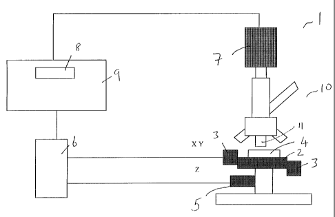
5 Fig. 2 illustrates an optical instrument, a microscope system 1, to which
the
autofocus system according to the present invention may be applied. The
hardware
components of the system I include a microscopelO,a motorized stage 2
controlled by a
pair of XY motors 3 and a Z motor 5, an XYZ stage controller 6, a video camera
7, an
image processor and host processor 9 with a video frame grabber 8. A separate
image
10 processor including video frame grabber may be provided but the present
invention is not
limited thereto. The XYZ stage controller controls the movements in the X, Y,
and Z
directions independently. The Z direction is along the optical axis (the
focusing axis) of
microscope 10. Typically, the microscope stage 2 will be moved laterally and
vertically
under computer control by stepper motors or DC servomotors. Suitable
components are
further described in detail below in the description of the examples. The
specimen 4 to be
viewed through the microscope 10 is located on the stage 2. Lamps, e.g.
fluorescent
lamps, and other optical accessories well known to the slalled person will not
be
described.
Although the present invention will mainly be described with reference to an
XYZ
stage 2 (movements in three orthogonal directions) the present invention is
not limited
thereto. To obtain focusing there is only a requirement for relative inovement
between the
objective 11 of the microscope 10 and the specimen 4. It is not anticipated
that the
method of achieving this is a limitation on the present invention.
The present invention also includes optical devices in which an objective 11
is
adjusted to determine a focus position rather than moving the specimen along
the optical
axis. In such an optical instrument the objective 11 may be adjusted by any
suitable
adjusting device which may be controIIed by the host processor such as a
piezoelectric
objective positioner. The positioner may be sandwiched between the objective
turret and
the objective 11. Such a positioner may be obtained from Polytech, PI, Costa
mesa,
California, USA, e.g. an E-810.10 closed loop controller. With such a system
the
objective 11 is adjusted instead of moving the specimen along the optical axis
of the
microscope. Combinations of objective and specimen movement are also included
within
CA 02374910 2001-11-21
WO 00/75709 PCTIEPOO/04987
11
the scope of the present invention.
In accordance with the present invention measurement of the focus score can
best
be based on the energy content of a linearly filtered image. Further, an
optimal focus
score is preferably output by a gradient filter.
In accordance with the present invention the gradient filter is limited in its
spatial
extent either side of the pixel of the captured image which is being
processed. In digital
filtering the pixel value to be filtered is combined in some way with pixel
values from
pixels around the pixel being filtered. The smoothing function has a spatial
extent, i.e. it is
a function of distance and determines whether or not the value of a pixel at a
certain
distance from the pixel being filtered should be included in the filtering for
that pixel and
if so with what weighting. Experiments have shown that this spatial extent
should not be
less than three pixels either side of the pixel being filtered. A reduced
spatial extent may
results in less accurate autofocusing or even in no ability to find a focus
position with
some types of image. The gradient filter may include one or more operators.
For instance,
the gradient filter may include at least one of the following:
1) a gradient operator to generate the first or higher order spatial
differential of the image
followed by a smoothing operator which determines and limits the spatial
extent of the
digital filtering either side of the pixel being filtered.
2) a combined gradient and smoothing operator which carries out both gradient
and
smoothing operations in one pass.
3) a smoothing operator applied to the image to limit the spatial extent of
the pixels
involved in the filtering around each pixel being filtered followed by a
gradient operator
to generate a first order or higher order spatial differentiation of the
smoothed image.
In accordance with embodiments of the present invention the digital gradient
filter
may include a mathematical function having a negative and positive lobe around
the
origin thereof, the mathematical function being limited in spatial extent in
that it extends
over a distance smaller than or equal to the image size and extends at least
over three
pixels either side of a pixel whose value is being filtered. Preferably, the
function has only
one zero crossing within the spatial extent.
Examples of the types of functions useful in embodiments of the gradient
filter in
accordance with the present invention are shown in Figs. 3A to E. Fig. 3A
shows a
function with its X axis as the spatial axis in pixel units and its Y axis
being the weighting
CA 02374910 2001-11-21
WO 00/75709 PCT/EP00/04987
12
factor used in the digital filtering. The spatial origin preferably coincides
with the pixel to
be filtered (as shown). The function is the spatial derivative of a normal
Gaussian curve.
It can be seen that the function has two lobes, one positive and one negative
either side of
the spatial zero. These lobes extend over a spatial distance of at least 3
pixels either side
of the spatial origin. In fact, in the example shown, the function has
appreciable values up
to 7 pixels each side of the origin. The effect of the difference in sign of
the lobes each
side of the spatial origin is to determine a gradient of the image when the
function is used
for digital filtering. Within the extent of the function which has appreciable
values there is
only one zero crossing. This is preferably at the spatial origin as shown in
Fig. 2A i.e. the
zero crossing coincides with the pixel to be filtered.
The present invention is not limited to a derivative of the Gaussian curve as
the
function defining the gradient filter. As shown in Fig. 3B an odd-order Bessel
function
may also be used (the figure shows a first order Bessel function) or any other
function
which has positive and negative lobes either side of the spatial origin. As
can be seen from
Fig. 3B, the first order Bessel function has two lobes, one positive and one
negative,
either side of the spatial origin. Due to the fact that a Bessel function has
more than one
zero crossing point, the spatial extent of the Bessel function is preferably
limited (e.g. by
truncation) to the distance either side of the origin which lies within the
first non-origin
zero crossing points, for example in Fig. 3B this would mean truncating the
Bessel
function at about the fourth pixel so that values of the function at higher
pixel values (or
lower negative values) are zero or negligible. This is shown by the dotted
line.
An alternative approach would be to damp the Bessel function so that the
number
of zero crossings is reduced. The function of Fig. 3C is a dampened version of
the first
order Bessel function of Fig. 3B using the damping function x2 + 1. It can be
seen that the
effect of the damping is to reduce the Y axis values effectively to zero
beyond the fourth
pixel. Accordingly, the damping function effectively truncates the Bessel
function so that
there is only one zero crossing within the range 3 pixels and values at
higher pixel
values are effectively zero.
The digital gradient filter according to the present invention is not limited
to one
dimensional filtering as would be indicated by the filtering functions of
Figs. 3A to C. A
three dimensional representation of a two-dimensional gradient filter function
in
accordance with an embodiment of the present invention is shown schematically
in Fig.
CA 02374910 2001-11-21
WO 00/75709 PCT/EP00/04987
13
3D. Fig. 3D shows a two-dimensional spatial differential of a two dimensional
normal
Gaussian curve. The X and Y-axes lie in the plane of the captured image and
have pixel
numbers as units. The Z-axis represents the weighting applied to the relevant
pixel
number as used in the filtering process. The origin 0,0,0 lies on the pixel to
be filtered.
The function has two three-dimensional lobes, one positive and one negative.
The origin
of the two lobes preferably coincides with the pixel to be filtered. Rotation
of the
orientation of the lobes as shown schematically in Fig. 3E is also included
within the
present invention and does not reduce the effectiveness of the filtering.
In accordance with one preferred embodiment of the image filter of the present
invention a two-dimensional Gaussian derivative as shown in Fig. 3D is used
within the
plane (x, y co-ordinates of an x,y,z co-ordinate system in which the direction
z is
perpendicular to the specimen to be imaged, i.e. the direction z is in the
focusing
direction) of the image to measure the focus score. The 6 of the Gauss filter
determines
the extent of the filtering and is related to the scale of prominent features.
A suitable focus function is:
F(a) ~ I [.f (x,.Y)* Gx(x,y, 6)]2 + [.f (x,y)* Gy(x,.Y> 6)12
x,y J
AM I fx fy (1)
x, y
where f(x,y) is the image gray value, Gx(x,y, 6) and Gy(x,y, 6) are the first
order Gaussian
derivatives in the x- and y-direction at scale 6, NM is the total number of
pixels in the
image, and fx, fy are the image derivatives at scale 6 in the x- and y-
direction,
respectively. Further, discussion of Gaussian gradient filters may be found in
the article by
ter Haar Romey BM, "Geometry driven Diffusion in Computer Vision", Boston,
Kluwer
academic Publishers, 1999, page 439; and in "Traitement de l'image sur micro-
ordinateur", Jean-Jacques Toumazet, Sybex, 1987, pages 156 to 158 which
describes the
method of J. F. Canny.
Often, a trade-off between noise sensitivity and detail sensitivity can be
observed
for a specific microscope set-up. For example, in fluorescence microscopy the
signal to
CA 02374910 2001-11-21
WO 00/75709 PCT/EP00/04987
14
noise ratio (SNR) is often low, and relatively smooth images are examined. For
phase
contrast microscopy, SNR is high, and small details (the phase transitions)
have to be
detected. Accuracy of autofocusing depends on the signal to noise ratio as
propagated
through the focus score filter. In accordance with an embodiment of the
present
invention, the 6 of the Gaussian filter may be freely chosen by the operator
so that it lies
within the range of 3 pixels to the size of the image. The value of 6 is
preferably chosen
such that noise is maximally suppressed, while the response to details of
interest in the
image is preserved. For bar-like structures, the value of 6 preferably
conforms to
d (2)
2~
where the thickness of the bar is given by d. Assuming that the smallest
detail to be
focused may be considered bar shaped, eq. 2 gives an indication for the value
of a.
The focal plane of the microscope is assumed to be within a pre-defined
interval dz
around the start z-position z. The scanning stage is moved down to the
position zmin = z -
'/z Az. Backlash correction is applied by always approaching a focusing
position from the
same direction, e.g. sending the stage further down than necessary, and
raising it again to
the given position. As a result, mechanical tolerance in cogwheels is
eliminated.
As t= 0 ms, the stage controller starts raising the stage to traverse the
complete
focus interval dz. During the continuous stage movement through focus,
successive
images of the preparation are captured at 40 ms intervals or at any other
standard video
rate. The focus score of each captured image is then calculated. In accordance
with a
preferred embodiment of the present invention the image buffer is re-used for
the next
video frame, necessitating only two image memory buffers to be active at any
time. One
of the buffers is used to provide the data necessary for the focus score
calculation of the
previously captured image, while the other is used for capturing the next
image. Hence,
calculation of the focus score is preferably performed within one video frame
time.
As soon as the stage has reached the end of the focus interval, timing is
stopped
at t= td ms. An estimation of the focus curve is obtained from the focus score
results for
the complete focus interval. The global optimum in the estimate for the focus
curve
represents the focal plane to be used as the final focusing position of the
specimen. Each
z-position is calculated from the time at which the corresponding image was
captured.
When linear movement of the stage is assumed, the position at which the image
at time ti
CA 02374910 2001-11-21
WO 00/75709 PCT/EP00/04987
is taken corresponds to
tl
zl = td az + zmin (3)
where td represents the travel duration, Az is the focus interval, and z,,,;,,
is the start
position (position at t= 0 ms).
5 It has been found that it is safe to assume that the focus curve is
parabolic around
the focal plane. From this a high focus precision can be achieved by quadratic
interpolation. When assuming linear stage movement, or z = vt + zm;n, the
focus curve
around the focal plane can be approximately by
s(t) = c+ bt + at 2 (4)
10 The exact focus position is obtained by fitting a parabola through the
detected optimum
and its neighboring measurements. Consider the detected optimum s(to) = so at
time t=
to. The time axis may be redefined such that the detected optimum is at time
t= 0. Then,
neighboring scores are given by (sn, t,) and (sp, tp), respectively. Solving
for a, b and c
gives
-sa(2 +sp12 +sotp-sntp s tn - sptn - sotp+sntp
15 c = s ,b = a = (5) 2
2 2 tntp - tntp tntp - tntp
The peak of the parabola, and thus the elapsed time to the focus position, is
given by
b S012 sPt2 - sotp+syltp
tf 2a + t 2(sotn - s ptn - s t p+ snt p) + to (6)
The focal plane is at a position given by:
tf
z f= td Az + zmin (7)
to which the specimen is moved, taking the backlash correction into account.
The depth of field of an optical system is defined as the axial distance form
the
focal plane over which details still can be observed with satisfactory
sharpness. The
thickness of the slice which can be considered in focus is then given by:
CA 02374910 2001-11-21
WO 00/75709 PCT/EP00/04987
16
n
zC11 = ~ (8)
NA
where n is the refractive index of the medium, A the wavelength of the used
light, and NA
the numerical aperture of the objective. The focus curve is sampled at Nyquist
rate when
measured at Zd intervals. Common video hardware captures frames at a fixed
rate. Thus
the sampling density of the focus curve can only be influenced by adjusting
the stage
velocity to travel Zd m per video frame time.
The present invention also includes additional method steps if the focus score
is
not good enough after filtering with one value of 6 or if the attempt to fit
the focus scores
to a polynomial, e.g. a parabola, is not successful, e.g. there is no
pronounced maximum
within the focus scores. In accordance with one embodiment of the present
invention the
filtering step may be repeated with another value of 6. This second value of 6
may be set
automatically by the host processor 9. For instance, if a suitable focus score
or maximum
thereof is not obtained, the host processor 9 may increase the value of 6 and
repeat the
focusing steps. Alternatively or additionally, if a suitable focus score or
maximum of the
focus score is not obtained, a field of the specimen adjacent the field used
for the focus
attempt may be chosen and the focusing steps in accordance with the present
invention.
This process may be extended by electronically setting different values of 6
at the new
field position and repeating the focusing steps again. After a predetermined
number of
failures, the autofocusing procedure may be terminated and the relevant field
or specimen
may be flagged in some way to indicate that focusing was not possible.
Preferably, the spatial extent 6 of the gradient filter in accordance with the
present
invention is settable in order to optimize noise rejection and object
location. In
accordance with preferred embodiments of the present invention the operator
may input a
spatial extent for the focusing process. For example, the operator may input a
value of 3
nucron to the host processor 9 of Fig. 2. The processor calculates the value
of 6 based on
equation 2 and uses this for the gradient filter. Alternatively, the operator
inputs a range
into the host processor 9, e.g. 2 to 5 micron. The host processor 9 is
progranuned to
select a value from this range, e.g. the mid-value of 3.5 or the lowest value
of 2 microns
and to determine the value of a from equation 2 using this value. If focusing
is not
successful, i.e. the focus score values are not adequate or a maximum is not
found, the
host processor 9 may sequentially and automatically select other extent values
within the
CA 02374910 2001-11-21
WO 00/75709 PCT/EP00/04987
17
range input by the operator. Alternatively, the processor may select values
for 6 without
operator input. For example, it may start with a default value and select
other values if
this proves unsuccessful. The exact value of a is not critical and the digital
filter in
accordance with the present invention is robust. As shown from Fig. 5 the same
focusing
position is identified over a range of 6 values.
A further embodiment of the present invention includes calculating the focus
score
for each image at each Z position with a plurality of 6 values e.g. 6, 62 ...
aN. This may
be done in parallel. The limit on the number of 6 values N may be limited only
by the
capability of the host processor 9 to calculate the focus scores within a
reasonable time.
After the focus scores 1-N have been calculated for all the images, each set
of scores
associated with one 6 value is fitted to a suitable curve, e.g. polynomial to
obtain the
focus position as described above. The result is a set of N focus positions or
less if some
of the 6 values did not result in a focus position. Various algorithms may be
applied to
obtain the best focus position from the obtained focus positions. For
instance, it is
anticipated that several of the focus positions will be the same or nearly the
same. Hence,
there may be a clustering of the focus positions. The cluster may be extracted
by digital
processing techniques, e.g. eliminating extreme focus positions more than 3
times the
standard deviation (least squares) from the average focus position calculated
from the set
of focus positions. Then an average best focus position is selected as the
focus position.
The number of different 6 values N does not generally have to be large. As the
value of 6 determines which size of element is selected by the filtering
operation for
autofocusing, small changes in a will not usually change the determined focus
point very
much (see Fig. 5). Hence, widely spaced 6 can be used. Secondly, it not
desirable for the
filter to select large or small elements of the image which are not related to
the desired
focused object. Hence, in accordance with an embodiment of the present
invention it is
preferred if the range of the values of 6 is settable y the operator. In this
way, the
operator has some control over what elements of the image will be selected for
autofocusing.
To calculate the focus score within the video frame time for current sensors
and
computer systems, a simplification of the focus function eq. 1 is preferably
considered
with present day computing speeds but the present invention is not limited
thereto. For
biological preparations, details may be distributed isotropically over the
image. In
CA 02374910 2001-11-21
WO 00/75709 PCT/EPOO/04987
18
accordance with an embodiment of the present invention the response of the
filter in one
direction is used for the determination of the focal plane. Further
computation time can be
saved by estimating the filter response from a fraction of the scan lines in
the image.
Then, the focus function may be given by
F(6) = AM I [f (x,Y)* G.X (x,Y, 6)]2 (9)
xIY
For example, each sixth row (L = 6) may be applied. A recursive implementation
of the
Gaussian derivative is used, for which the computation time is independent of
the value of
6. The calculation time could be kept under 40 ms for all computer systems
used, even
when the system was running other tasks simultaneously. Comparison between the
focus
curve in two dimensions for the whole image eq. 1 and the response of eq. 9
reveals only
by marginal differences for all experiments.
For the acquisition of multiple aligned images from large, flat preparations,
the
variation in focus position is assumed small but noticeable at high
magnification. Proper
acquisition of adjacent images can be obtained by focusing a few fields.
Within the
preparation, a procedure in accordance with an embodiment of the present
invention
starts by focusing the first field. Fields surrounding the focuses field are
captured, until
the next field to be captured is a given distance away from the initially
focused field.
Deviation from best focus is now corrected for by focusing over a small
interval. The
preparation is scanned, keeping track of focus position at fields further away
than a given
distance from the nearest of all the previously focused fields. The threshold
distance for
which focusing is skipped depends on the preparation flatness and
magnification, and has
to be empirically optimized for efficiency. Fields that have been skipped for
focusing are
positioned at the focus level of the nearest focused field. Small variations
in focus
position while scanning the preparation are corrected during acquisition.
The autofocus algorithm in accordance with the present invention has been
tested
in the following applications: a) quantitative neuronal morphology, b) time-
lapse
experiments of cardiac myocyte dedifferentiation, c) immunohistochemical label
detection
in fixed tissue d) C. Elegans GFP-VM screening, and e) immunocytochemical
label
detection in fixed cells. Each of these applications is described below. The
software
package SCIL-Image version 1.4 (16) (TNO-TPD, Delft, The Netherlands) is used
for
CA 02374910 2001-11-21
WO 00/75709 PCT/EP00/04987
19
image processing, extended with the autofocus algorithm and functions for
automatic
stage control and image capturing in accordance with the present invention.
Quantitative Neuronal Morphology in Bright-field Mode
Morphological changes of neurons were automatically quantified as described in
"Sodium butyrate induces aberrant tau phosphorylation and programmed cell
death in
human neuroblastoma cells", Brain Res. 1995, 688, pages 86-94. Briefly, PC 12
cells were
plated in poly-L-lysine (Sigma, St. Louis, MO) coated 12-well plates. In each
well 5 x 10⁴
cells were seeded. After 24 hours the cells were fixed with 1% glutaraldehyde
for 10
minutes. Then the cells were washed twice with distilled water. The plates
were dried in
an incubator.
The plates were examined with objective 5 x NA 0.15 Plan-Neofluar, in bright-
field illumination mode on an Axiovert 10 microscope (Carl Zeiss, Oberkochen,
Germany). A scanning stage (stage and MAC4000 controller, Marzhauser, Wetzlar,
Germany) was used for automatic position control. At power on, the stage was
calibrated
and an initial focus level was indicated manually. The camera used was an MX5
(Adaptec, Eindhoven, The Netherlands) 780 x 576 video frame transfer CCD with
pixel
size 8.2 x 16.07 m2, operating at room temperature with auto gain turned off.
Adjacent
images were captured by an Indy R4600 132 MHz workstation (Silicon Graphics,
Mountain View, CA). As a result, an 8 x 8 mosaic image covering an area of 6.7
x 6.7
mm2 was electronically stored on disk for each well. Prior to the acquisition
of the well,
autofocusing at the center of the scan area was performed. The smallest
details to focus
were the neurites, which were about 3 pixels thick, yielding 6= 1.0 (eq. 2).
Variability in
the z-position of the wells turned out to be within 500 m, which is taken as
the focus
interval. The wavelength of the illumination was about 530 nm, resulting in
23.6 m
depth of field (eq. 8). Therefore, stage velocity was reduced to 24.7 m per
video frame
(10,000 steps per second) during focusing. Due to the low magnification,
backlash
correction was not necessary.
Quantitative Neuronal Morphology - results
Fig. 4a shows the average focus curve for 180 wells, which were all accurately
focused according to an experienced observer. The measured focus curve with
the lowest
CA 02374910 2001-11-21
WO 00/75709 PCTIEPOO/04987
maximum score (peak at 0.004) is at a field containing only some dead cells.
The
variation in focus score is due to the different number of cells, and their
morphology. The
local maximum beneath focus is caused by a 180 phase shift in the point-
spread function
of the optical system.
5 The time needed for focusing was 1.7 seconds per field, consuming 7.5% of
the
time to acquire one 12-well plate (4.5 minutes). For these thoroughly stained
preparations, the autofocus method was able to focus all fields. Even for
fields containing
only a few dead cells, the focal plane was accurately determined.
10 Cardiac Myocyte Dedifferentiation in Phase Contrast Mode
Cardiac myocytes were isolated from adult rats (ca. 250 gram) heart by
collagenase as described in Donck LV, Pauwels, PJ, Vandeplassche G, Borgers M,
"Isolated rat cardiac myocytes as an experimental model to study calcium
overload: the
effect of calcium-entry blockers", Life Sci 1986;38:765-772. The cell
suspension
15 containing cardiomyocytes and fibroblasts was seeded on lazminin coated
plastic petri
dishes, supplied with M199 and incubated for one hour. Thereafter, unattached
and/or
dead cells were washed away by rinsing once with M199. The petri dishes were
filled
with M1999 + 20% fetal bovine serum and incubated at 37 C.
The petri dishes were examined with objective 32 x NA 0.4 Achrostigmat Phase
20 1, in phase contrast mode on an Axiovert 35 microscope (Carl Zeiss,
Oberkochen,
Germany). During the experiment, ambient temperature was maintained at 37 C.
Time-
lapse recordings (15 hours) were made in 6 manually selected fields, one in
each of the 6
petri dishes. A scanning stage (stage and MAC4000 controller, Marzhauser,
Wetzlar,
Germany) visited the selected fields at 120 second intervals. Fields were
captured using a
CCD camera (TM-765E, Pulnix, Alzenau, Germany). They were added to JPEG
compressed digital movies (Indy workstation with Cosmo compressor card, SGI,
Mountain View, CA), one for each selected field. Autofocusing was applied once
per
cycle, successively refocusing all the fields in 6 cycles. The smallest
details to focus were
the cell borders, which were less than 4 pixels thick, yielding 6= 1.0 (eq.
2). Variability
in the z-position between focus events was expected to be within 100 m, which
was
taken as the focus interval. The wavelength of the illumination was about 530
nm,
resulting in 3.3 m depth of field (eq. 8). Therefore, stage velocity was
reduced to 2.5
CA 02374910 2001-11-21
WO 00/75709 PCT/EP00/04987
21
m per video frame (1,000 steps per second) during focusing.
Cardiac Myocyte Dedifferentiation - results
Fig. 4b shows the average focus curve (75 events) for one field of the time
lapse.
All the fields were perfectly focused during the 15 hours of recording,
according to
experienced observers. The variation in focus score was due to the change in
image
content, caused by movement of fibroblasts and the dedifferentiation of the
myocytes.
The axial drift from the initial focal plane during the recording of the six
positions varied
from 3 m up to 27 m. The time needed for focusing was 2.8 seconds per field.
Rat cardiomyocytes are known to dedifferentiate spontaneously in culture,
i.e.,
they spread out, flatten and develop pseudopodia-like processes. Despite these
changes in
image content during the experiment, none of the time lapse movies was out of
focus any
time.
Immunohistochemical Label Detection in Bright-field Mode
Sections of the amygdala of mice injected with a toxic compound were cut at 15
m thickness through the injection site. They were subsequently immunostained
for the
presence of the antigen, using a polyclonal antibody (44-136, Quality Control
Biochemicals Inc., Hopkinton, MA) and visualized using the chromogen DAB.
Four microscope slides (40 brain slices) were mounted on the stage of an
Axioskop microscope (Carl Zeiss, Oberkochen, Germany) and examined with
objective
2.5 x NA 0.075 Plan-Neofluar, in bright-field illumination mode. A scanning
stage (stage
and MC2000 controller, Marzhauser, Wetzlar, Germany) was used for automatic
position
control. Adjacent images were captured (Meteor/RGB Framegrabber, Matrox,
Donval,
Quebec, Canada in an Optiplex GXi PC with Pentium 200 MHz MMX, Dell, Round
Rock, TX) by use of an MX5 CCD camera (Adaptec, Eindhoven, The Netherlands).
As a
result, mosaics of complete brain slices were stored electronically on disk.
Prior to
acquisition, autofocusing at approximately the center of the brain slice was
performed.
The smallest detail to focus was the tissue structure, which was about 3
pixels thick,
yielding 6= 1.0 (eq. 2). Variability in the z-position between the glass
slides turned out to
be within 1000 m, which was taken as the focus interval. The wavelength of
the emitted
light was about 530 nm, resulting in 94 m depth of field (eq. 8). Therefore,
stage
CA 02374910 2001-11-21
WO 00/75709 PCT/EP00/04987
22
velocity was reduced to 98.7 m per video frame (40,000 steps per second)
during
focusing. Due to the low magnification, backlash correction was not necessary.
Immunohistochemical Label Detection - results
Fig. 4c shows the average focus curve for the rat brain slices. From the 100
fields
examined, 2 fields contained not enough contrast for focusing. These are not
included in
Fig. 4c. The variation in focus score was caused by the differences in
contrast between
the slices.
The time needed for focusing was 1.5 seconds per field, consuming 7% of the
time to acquire one glass slide (3 minutes). The autofocus method was able to
focus 98%
of the fields. The contrast in the remaining fields was to low for accurate
focusing.
C Elegans GFP-VM Screening in Fluorescence Mode
Individual C. Elegans worms transgenic for GFP expressing vulval muscles (GFP-
VM) were selected from stock, and one young adult hermafrodite (Po) was placed
in each
of the 60 center wells of a 96-well plate (Costar, Acton, MA) filled with
natural growth
medium, and incubated for five days at 25 C to allow F, progeny to reach adult
stage.
Before image acquisition, fluorescent beads (F-8839, Molecular Probes, Eugene,
OR) were added to the wells as background markers for the focus algorithm. The
well
plate was examined with an objectives 40 x NA 0.6 Archoplan, in fluorescence
mode on
an Axiovert 135 microscope (Carl Zeiss, Oberkochen, Germany). A FITC filter
(B, Carl
Zeiss, Oberkochen, Germany) in combination with a 100W Xenbophot lamp was used
to
excite the green fluorescent protein (GFP). Images were captured (02 R5000 180
MHz
workstation, Silicon Graphics, Mountain View, CA) using an intensified CCD
camera
(IC-200, PTI, Monmouth Junction, NJ). The scanning stage (stage and MC2000
controller, Marzhauser, Wetzlar, Germany) was calibrated to capture adjacent
images.
Each of the selected wells was scanned and the adjacent images, completely
covering the
well, were stored electronically on disk. Variability in the z-position
between the center of
the wells turned out to be within 250 m, which was taken as the focus
interval. After
autofocusing on the well center, deviation from best focus while scanning the
well was
corrected. It turned out that autofocusing over one-fifth the focus interval
(50 m) for
fields further than 3 fields away from a focused field was sufficient. The
fluorescent
CA 02374910 2001-11-21
WO 00/75709 PCT/EP00/04987
23
spheres were 30 pixels in diameter, yielding 6= 8.5 (eq. 2). The wavelength of
the
emitted light was about 530 nm, resulting in 1.47 m depth of field (eq. 8).
The diameter
of the fluorescent spheres was 15 m, which is much larger than Zd. Since the
spheres
were homogeneously stained, the smallest detail to consider in the z-direction
was a
cylindrically shaped slice through the spheres, where the cylinder height was
determined
by the horizontal resolution. Therefore, the stage velocity was reduced to
approximately
one third of the sphere diameter during focusing, yielding 4.94 m per video
frame (2,000
steps per second). Backlash correction offset was determined to be 15 m.
C. Elegans GFP-VM Screening -results
Fig. 4d shows the average focus curve for 1786 fields of the C. Elegans
screening.
Fourteen of the 1800 fields did not contain fluorescent spheres, consequently
accurate
focusing was not possible. These are not included in Fig. 4d. The remaining
fields were
accurately focused according to an experience observer. The variation in focus
score was
caused by the different number of spheres and the presence or absence of worms
in the
focused fields. The time needed for focusing was 2.8 seconds for the first
field in a well,
and 1.1 seconds per field for keeping track of the focal plane within the
well.
Autofocusing consumed 12% of the time to acquire one 96-well plate (4.5 hours
for
28,000 images), which is reasonable given the time needed for the preparation.
The images were highly degraded by the presence of random noise (SNR ;zz~ 10
dB) due to fluorescent bacteria and structural noise caused by earth loops in
combination
with the extremely sensitive CCD camera. The focus algorithm was able to find
the
correct focus position for all but 14 out of 1800 fields examined. Failure was
caused by a
shortage of relevant image information content.
Immunocytochemical Label Detection in Fluorescence Mode
Human fibroblasts were seeded in a 96-well plate (Costar, Acton, MA) at 7000
cells per well, in 2% FBS/Optimem. The cells were fixed and permeabilized by
adding 2%
paraformaldehyde and 0.5% Triton-X-100 (Sigma, St. Louis, MO). The reaction
was
stopped after 30 minutes by washing with PBS, addition of 5% NGS during 60
minutes,
and washing with 0.2% B SAlPB S for 5 minutes. A dilution of 1/100 primary
antibody
rabbit anti human NF-KB (p65) (Santa Cruz Biotechnology, Santa Cruz, CA) in
0.2%
CA 02374910 2001-11-21
WO 00/75709 PCT/EP00/04987
24
BSA/PBS was added to each well for 2 hours at 37 C. After washing 3 times with
PBS
for 10 minutes, Cy3 labeled sheep anti rabbit (Jackson, Uvert-Grove, PA)
dilution (1/2
glycerol, 1/250 0.2% BSA/PBS) was added to each well for 60 minutes at 37 C.
After
washing with 0.2% BSA/PBS for 15 nunutes and repeated washing with PBS, and
nuclear counter staining with Hoechst 33342 (Molecular Probes, Eugene, OR),
the cells
were ready for quantitative evaluation.
Well plates were examined with an objective 40 x NA 0.6 Achroplan, in
fluorescence mode on an Axiovert 135 microscope (Carl Zeiss, Oberkochen,
Germany).
A DAPUFITC/TRITC filter (XF66, Omega Optical, Brattleboro, VT) in combination
with a 100W Xenophot lamp was used to excite the cells. A scanning stage
(stage and
MC2000 controller, Marzhauser, Wetzlar, Germany) was used for automatic
position
control. Adjacent images were captured (02 workstation R5000 180MHZ, Silicon
Graphics, Mountain View, CA) using an intensified CCD camera (IC-200, PTI,
Monmouth Junction, NJ). As a result, two 5 x 5 mosaic images, one for the
nuclei and
one for the immuno signal, was stored on disk for each well. The covered area
per well
was 1.2 x 1.2 mm2. Prior to the acquisition of each mosaic image, autofocusing
at
approximately the center of the scan area was performed. The smallest details
to focus
were the nuclei, which were minimally 30 pixels in diameter, yielding 6= 8.5
(eq. 2).
Variability in the z-position of the wells turned out to be within 250 m,
which was taken
as the focus interval. The wave length of the emitted light was about 450 nm
(nuclei) and
600 nm (immuno signal), resulting in 1.25 m and 1.67 m depth of field (eq.
8),
respectively. Cell thickness was about 5-15 m, much larger than Zd.
Therefore, the stage
velocity was reduced to 4.94 m per video frame (2,000 steps per second)
during
focusing. Backlash correction offset was determined to be 15 m.
Immunocytochemical Label detection - results
Fig. 4e shows the average focus curve for the nuclei in the immunocytochemical
label detection. All the 150 fields were accurately focused according to an
experienced
observer. The variation in focus score was due to the different number of
cells present in
each field. The average focus curve as measured for the immuno signal is shown
in Fig.
4f. From the 150 fields examined, 2 fields were completely saturated due to
preparation
artifacts, causing the focus algorithm to fail. These fields are not included
in Fig. 4f. the
CA 02374910 2001-11-21
WO 00/75709 PCTIEPOO/04987
remaining fields were accurately focused. The time needed for focusing was 2.8
seconds
per field, consunvng 14% of the time to acquire one 96-well plate (20
minutes).
The signal to noise ratio was estimated to be about 10 dB for the nuclei, and
4dB
for the immuno signal. Despite the noise, the autofocus method was able to
focus all but
5 two out of 300 fields. Failure was caused by a shortage of relevant image
information
content.
Comparison of Performance with Small Derivative Filters
In order to evaluate the effect of the scale 6 in the estimate for the focus
score,
10 experiments with a fixed (non-selectable) 6= 0.5 were performed.
For the quantitative neuronal morphology, accurate focusing with 6= 0.5 was
not
possible for 1 out of 24 fields. In this case, the algorithm focused on the
reversed contrast
image. Application of the small scale in focusing of the cardiac myocyte
dedifferentiation
failed whenever fungal contamination at the medium surface occurred, which was
taken
15 as the local plane. Taking 6= 1.0 solved this problem, that is by focusing
persistently on
the myocytes. Focusing with 6= 0.5 on the immunohistochemical label detection
resulted
in focusing on dust particles at the glass surface for 5 out of 24 fields.
For the fluorescence applications, accurate focusing was not possible with 6=
0.5, due to the small signal to noise ratio. Experiments taken with 6= 0.75
resulted in
20 inaccurate focusing for 18 out of 30 fields for the C. Elegans GFP-VM
screening.
Further, the algorithm was not able to focus accurately on 13 out of 30 fields
for the
nuclei in the immunocytochemical label detection, and failed for 17 out of 30
fields on the
immuno signal.
The effect of the scale 6 results in robustness against noise and artifacts. A
larger
25 scale results in robustness against phase reversion (quantitative neuronal
morphology),
fungal contamination at the medium surface (cardia myocyte dedifferentiation),
dust on
the glass surface (immunohistochemical label detection) and noise (the
fluorescence
applications). From these results it can be inferred that the performance of
small
differential filters, as used conventionally, is poor given the number of
inaccurately
focused images for 6= 0.5 or 6= 0.75. Further, it can be inferred that the
scale 6 should
preferably be at least 1. This is equivalent to 3 pixels. Hence, a preferred
range for the
scale 6 in accordance with the present invention is from at least 3 pixels up
to the size of
CA 02374910 2001-11-21
WO 00/75709 PCT/EP00/04987
26
the image itself.
For the different applications, the chosen focus interval was effectively used
for
about 30%. The focus interval is preferably not taken too narrowly to ensure
that the
focal plane is inside the interval, regardless of the manual placement of the
preparations.
The time needed for the autofocus algorithm varied from 1.5 up to 2.8 seconds
for current sensors and computer systems, which is in the same time range as
trained
observers. Focus time is determined by the depth of field and the video frame
time, both
of which can be considered as given quantities, and by the size of the focus
interval.
Further reduction of focus time can be achieved by a smaller focus interval,
on the
condition that the variability in preparation position is limited. When
positional variability
is low or well known, the focus interval Az can be reduced to exactly fit the
variability.
For the applications given, the focus time can be reduced up to a factor 3 in
this way.
Failure of the autofocus algorithm due to a shortage of image content can be
well
predicted. If the focal plane is inside the focus interval, there should be
global maximum
in the estimate of the focus curve. Comparing the maximum focus score so with
the
highest of the focus scores at the ends of the focus interval, se = max(s(0),
s(td)) which
are certainly not in focus, determines the signal content with respect to
noise. When the
maximum score does not exceed significantly the focus scores at the ends of
the interval,
or (so - se)/se < a, the found focus position should be rejected. In this
case, focusing can
better be based on a neighboring field. For the reported results, a threshold
of a= 10%
safely predicts all failures. Accordingly, in accordance with the present
invention a
specimen may be focused correctly, or if the specimen cannot be focused
correctly this
can be determined by the system from the focus scores and this sample and/or
its image
can be labeled/flagged that focusing was inadequate automatically. This
provides a
significant advantage as the operator may then identify these specimens and
carry out
other forms of focusing as required, e.g. manually without the time-consuming
necessity
to check every image to see if any are out of focus.